Review

Usage of Silver Nanoparticles in Orthodontic Bonding Reagents

1
School of Dental Medicine, University of Pennsylvania, Philadelphia, PA 19104, USA
2
Department of Preventive and Restorative Sciences, School of Dental Medicine, University of Pennsylvania, Philadelphia, PA 19104, USA
3
Department of Orthodontics, School of Dental Medicine, University of Pennsylvania, Philadelphia, PA 19104, USA
4
Division of Restorative Dentistry, School of Dental Medicine, University of Pennsylvania, Philadelphia, PA 19104, USA
5
Department of Periodontics, School of Dental Medicine, University of Pennsylvania, Philadelphia, PA 19104, USA
*
Authors to whom correspondence should be addressed.
These authors contributed equally to this work.
J. Funct. Biomater. 2025, 16(7), 244; https://doi.org/10.3390/jfb16070244
Submission received: 17 May 2025 / Revised: 13 June 2025 / Accepted: 29 June 2025 / Published: 3 July 2025
(This article belongs to the Special Issue Dental Biomaterials in Implantology and Orthodontics)

Abstract

Fixed orthodontic appliances, which are cemented to tooth surfaces, complicate the maintenance of oral hygiene and create a rough surface that is favorable for bacteria attachment. Additionally, the presence of orthodontic appliances may conceive a unique environment that interacts with cariogenic microorganisms, fostering a distinct microbial ecosystem compared to that of the patients without orthodontic appliances, thus increasing the vulnerability of tooth surfaces to demineralization and caries formation. Silver (Ag) has shown strong antimicrobial effects and has been extensively investigated in the medical field. Here, we aim to review the antibacterial properties and potential side effects of silver nanoparticles (AgNPs) when incorporated into orthodontic bonding reagents. This valuation could contribute to the development of novel bonding reagents designed to prevent the formation of white spot lesions and caries during orthodontic treatments.

1. Introduction

Orthodontics is a specialized branch of dentistry that focuses on correcting occlusal alignment and enhancing facial aesthetics, which can significantly improve patients’ quality of life in collaboration with other specialists [1]. To date, fixed orthodontic appliances, such as braces, remain the most commonly used option, especially for more complex cases, and are generally considered more effective than removable alternatives. However, because fixed orthodontic appliances are cemented on the tooth surface, they can complicate oral hygiene maintenance [2] and create a rough surface that promotes bacterial attachment [3]. As a result, fixed orthodontic appliances may not only trap food remnants and microbial secretions, such as toxins, tissue-damaging enzymes, and acidic byproducts, but also promote dental plaque accumulation. Consequently, it can create a favorable environment for pathogens to evade the host’s immune defenses and resist medications [4]; the use of fixed orthodontic appliances may contribute to oral-dental problems such as caries and periodontal inflammation [5,6]. Furthermore, regardless of whether they are fixed or removable, orthodontic appliances can foster a unique environment that interacts with cariogenic microorganisms, resulting in a distinctive microbial flora for patients wearing these devices compared to those who do not [7,8]. This altered microbial ecosystem increases the susceptibility of tooth surfaces to demineralization and caries development while also elevating the risk of periodontal damage, such as gingival inflammation and alveolar bone resorption [5,9]. To address these challenges, clinicians and researchers have dedicated significant efforts to developing innovative dental materials that offer antimicrobial benefits while remaining biocompatible [10].
Among the various materials and agents explored, silver has emerged as a leading option due to its strong antimicrobial properties [8,10,11,12,13,14]. Notably, both metallic and ionic forms of silver exhibit antimicrobial potency [8,10]. Silver ions, in particular, target various bacterial structures. For example, silver ions bind to bacterial cell walls and cytoplasmic membranes through electrostatic attraction and their affinity for sulfur proteins, increasing membrane permeability and disrupting its integrity [15,16,17]. As a result, the disruption leads to RNA and DNA degeneration. Additionally, silver ions can also interfere with amino acid function and impair cellular respiration [17]. Due to the complexity of silver’s anti-microbial mechanism, mutations in at least three key systems are necessary for microbes to develop silver resistance [12,13]. Therefore, silver is effective in treating and preventing multiple diseases caused by drug-resistant microorganisms [10]. Furthermore, silver is abundantly available in nature and relatively cost-effective, making it an appealing substance for scientists to harness its potential in dental treatment [8,18].
Meanwhile, the particle size of a given agent significantly influences its antimicrobial effectiveness [19,20,21]. Advances in nanotechnology have enabled the production of various materials at the nanoscale, with nanoparticles (NPs) typically measuring less than 100 nm [22]. Owing to their large surface area-to-volume ratios, NPs can effectively release ions from their constituent materials, which disturbs microorganisms’ cell function. Moreover, NPs can attach and penetrate the cell walls of Gram-positive and Gram-negative bacteria, and have thus been considered a more effective formulation of antibacterial agents [18].
In particular, silver nanoparticles (AgNPs) exhibit exceptionally high antimicrobial activity against a broad range of microorganisms, including bacteria, viruses, and fungi, outperforming many other antimicrobial agents [16]. Thus, AgNPs have been employed as the primary component in inorganic and polymeric-based antimicrobial (nano)composites to prevent biofilm formation, combat infection, and promote caries arrestment [16,23]. AgNPs are considered relatively safe bactericidal agents so far, and they can act synergistically with several antibiotics [24]. More promisingly, AgNPs have been found to induce osteogenesis [12,24,25]. As a result, AgNPs have been broadly incorporated into dental materials, such as dental implants and root canal irrigation [24,26].
To date, numerous attempts have been made to utilize AgNPs in orthodontic appliances [27]. However, a gap still exists between bench-top investigation findings and their clinical application. Furthermore, the impacts of AgNPs on the biomedical, physical, and mechanical properties of orthodontic materials require a thorough assessment. Since incorporating AgNPs in orthodontic bonding reagents has been investigated extensively in the past two decades, here, we aim to consolidate existing knowledge to facilitate further advancements in developing innovative, multipotent orthodontic bonding reagents.

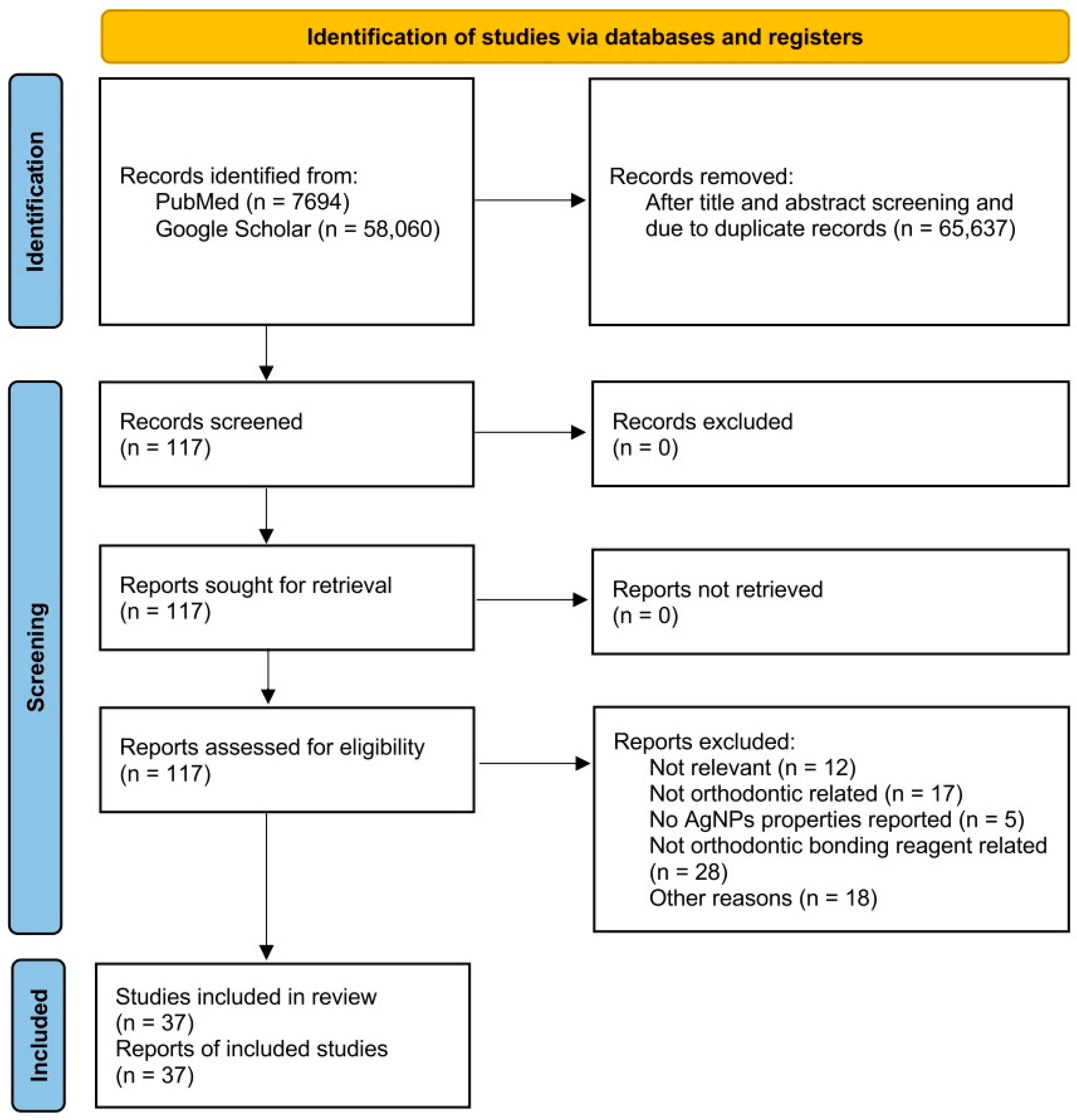
2. Materials and Methods

Original studies reporting the AgNPs in orthodontic bonding reagents were identified through comprehensive searches of PubMed and Google Scholars, using the following keywords: “silver nanoparticles”, and “orthodontics”, “orthodontic materials”, “orthodontic bonding”, “orthodontic composite”, “orthodontic cement”, “orthodontic adhesive”, “glass ionomer”, or “resin-modified glass ionomer cement”. The literature search, completed in March 2025, included only peer-reviewed, English-language original studies published since year 2010, and excluded conference abstracts, editorials, opinions, preprints, thesis, and literature reviews. To avoid missing any related articles, each database was searched independently by two authors. The data extraction from each original article was also performed by two authors separately. The information being extracted from each article includes the type of bonding reagent used, the type and concentrations of AgNPs, the type of study, the reported antibacterial effect, and any reported side effects. In cases of disagreement, a third author was involved to verify the accuracy of data extraction. This study is registered on Open Science Framework (osf.io/8nxp7), and the PRISMA Extension for Scoping Reviews (PRISMA-ScR) checklist was employed as the guideline. The PRISMA flow diagram is shown in Figure 1.

3. Nanosilver Particles in Orthodontic Primers

3.1. Antibacterial Effects

AgNPs have been incorporated into various bonding primers (Table 1 and Table 2). Notably, even at a low concentration of 0.11% (w/w), the inclusion of AgNPs in the TransbondTM XT primer (3M Unitek Corp., Monrovia, CA, USA) [28] and Universal bonding (Dentonics Inc., Monroe, LA, USA) [29] significantly inhibited the growth of Streptococcus mutans, an indicator cariogenic bacterial species often prevalent during orthodontic treatment [30] (Table 1).
Table 1. The antibacterial effects of the silver nanoparticle (AgNPs)-added bonding primers. Conc.: concentration; w/w: weight per weight; HA: hydroxyapatite; N/A: not applicable.
Bonding ReagentReferencesTested Conc. of AgNPs (w/w)Combinatory MaterialsType of StudyAntibacterial Effect
TransbondTM XT primer (3M, Monrovia, CA, USA)Degrazia et al., 2016 [28]0.11%, 0.18%, 0.33%N/AIn VitroAll Conc. inhibit S. mutans
TransbondTM XT primer (3M)Blocher et al., 2015 [31]0.11%, 0.18%, 0.33%N/AIn VitroN/A
Universal Bonding (Dentonics Inc., Monroe, LA, USA)Jenabi et al., 2023 [29]0.5%, 1%, 2.5%, 5%N/AIn VitroAll Conc. inhibit S. mutans
TransbondTM XT primer (3M)Akhavan et al., 2013 [32]1%, 5%, 10% Ag-HADoped HA with various concentration of Ag nanoparticleIn VitroN/A
Single Bond™ Universal Adhesive (3M)Gilani et al., 2020 [33]1%, 5%, 10% Ag-HAAg-HA nanoparticle powderIn VitroN/A

3.2. Side Effects

3.2.1. Influence on Shear Bond Strength (SBS)

Degrazia et al. reported that lower concentrations of AgNPs, ranging from 0.11 and 0.33% (w/w), also markedly reduced the SBS of the TransbondTM XT primer, although the resulting SBS values remained above the clinically accepted threshold of 6–8 MPa [28] (Table 2). In contrast, Blocher et al. found that, within the same concentration range, neither silver microparticles (3.5–18 μm) nor AgNPs (12.6–18.5 nm) considerably altered the SBS of the TransbondTM XT primer after the bonded brackets were stored in distilled water at 37 °C for 24 h [31]. Thus, whether the incorporation of AgNPs at these low concentrations genuinely impacts the clinical bonding performance of the TransbondTM XT primer warrants further validation.
Regarding Universal Bonding primer, Jenabi et al. noted that their SBS values decreased with the concentration of incorporated AgNPs [29]. However, this study was conducted using a combination of an AgNP-modified Universal Bonding primer and an AgNP-modified composite [29]. As a result, it remains unclear whether using AgNP-modified Universal Bonding primers with a standard, unmodified composite would yield SBS values that meet clinically acceptable levels.
The effects of modified AgNPs, specifically silver-hydroxyapatite (Ag-HA) nanoparticles, on SBS have also been investigated. Hydroxyapatite (HA), a calcium phosphate compound, prevents calcium loss from demineralized enamel, enhancing tooth resistance to cavities [33]. HA alone can also inhibit the growth of S. mutans without disturbing the SBS of orthodontic composites [34]. In previous studies, 1%, 5%, and 10% (w/w) Ag-HA nanoparticles were mixed with either TransbondTM XT primer [32] or Single Bond™ Universal Adhesive (3M) [33], respectively. Interestingly, the same concentration of Ag-HA nanoparticles exhibited contrasting effects depending on the primer type. For instance, 1% Ag-HA nanoparticles increased the SBS of TransbondTM XT primer but decreased that of Single Bond™ Universal Adhesive, while 10% Ag-HA nanoparticles reduced the SBS of TransbondTM XT primer but enhanced that of Single Bond™ Universal Adhesive. Unfortunately, neither study provided data on the antibacterial effects of these Ag-HA nanoparticle-modified primers. Therefore, the currently available evidence is short for defining the optimized concentration of Ag-HA nanoparticles that can provide promising antibacterial effects while maintaining or even improving the SBS.

3.2.2. Discoloration

One major disadvantage of Ag particles is their dark color, which can compromise esthetics [31], especially in dental applications. For example, Blocher et al. compared the impacts of incorporating silver microparticles and AgNPs into the TransbondTM XT primer. They found that the addition of AgNPs led to more pronounced discoloration after debonding compared to silver microparticles or no-silver controls [31], highlighting the aesthetic concerns that AgNP-modified adhesives may heighten the risk of enamel discoloration, which potentially hinders their suitability for dental applications where appearance is a priority.
Table 2. The side effects of the silver nanoparticle (AgNPs)-added bonding primers. Conc.: concentration; w/w: weight per weight; SBS: shear bonding strength; HA: hydroxyapatite; N/A: not applicable.
Bonding ReagentReferencesTested Conc. of AgNPs (w/w)Combinatory MaterialsType of StudySide Effects
SBSCytotoxicityDiscoloration
TransbondTM XT primer (3M)Degrazia et al., 2016 [28]0.11%, 0.18%, 0.33%N/AIn VitroDecreased with all Conc.N/AN/A
TransbondTM XT primer (3M)Blocher et al., 2015 [31]0.11%, 0.18%, 0.33%N/AIn VitroNo significant differenceN/AAll Conc. showed silver spots under 10× Magnification
Universal Bonding (Dentonics Inc.)Jenabi et al., 2023 [29]0.5%, 1%, 2.5%, 5%N/AIn VitroDose-dependently decreased *N/AN/A
TransbondTM XT primer (3M)Akhavan et al., 2013 [32]1%, 5%, 10% Ag-HADoped HA with various concentration of Ag nanoparticleIn Vitro1%: significantly increased;
5%: no significant difference;
10%: significantly reduced
N/AN/A
Single Bond™ Universal Adhesive (3M)Gilani et al., 2020 [33]1%, 5%, 10% Ag-HAAg-HA nanoparticle powderIn Vitro1% and 5%: significantly reduced;
10%: no significant difference
N/AN/A
*: the bonding agent was tested in combination with AgNPs-modified composite for SBS.

3.3. Summary

It is important to emphasize that all currently existing studies on AgNP-incorporated bonding primers have been conducted in vitro and focused on targeting one single bacteria strain. This limitation restricts the evaluation of their effects on the complex biofilm inhabited on the tooth surface of patients. If and how saliva affects the release and function of AgNPs are unclear yet. Therefore, further ex vivo or in vivo studies are needed to confirm the efficacy of incorporating AgNPs into primers for reducing bacterial colonization while maintaining the bonding strength and natural teeth color in human usage [31]. However, the effects of AgNPs on SBS vary across studies, making it unclear whether these variations stem from differences in AgNP concentration, the physical and chemical interactions between AgNPs and various materials, or differing testing conditions. Furthermore, it remains uncertain whether the influence of AgNPs on SBS is consistent in an intraoral environment. These questions highlight the need for further investigation.

4. Nanosilver Particles in Orthodontic Composites

4.1. Antibacterial and Anti-Demineralization Effects of AgNPs

Multiple studies have suggested a diversity of benefits associated with incorporating specific concentrations of silver nanoparticles into orthodontic bonding composites (Table 3). As the clinically commonly used orthodontic bonding composite, Transbond™ XT (3M Unitek, Monrovia, CA, USA) is the most tested orthodontic composite with a broad range of concentrations of AgNPs [0.05–1% (w/w) in vitro and 1–10% (w/w) in vivo, respectively]. For instance, Eslamian et al. incorporated 0.3% (w/w) AgNPs into Transbond™ XT and found that its antibacterial effect against S. mutans did not significantly decline between 24 h and 30-day test time points [35]. Marco Sánchez-Tito et al. mixed 0.05%, 0.1%, 0.5%, and 1% (w/w) AgNPs with Transbond™ XT and observed that the 0.5% and 1% AgNPs modified Transbond™ XT could inhibit the growth of S. mutans and Lactobacillus acidophilus in vitro [36,37]. In addition, Transbond™ XT with 1% AgNPs significantly decreased the area and depth of white spot lesions on the tooth surfaces in an in vitro microbiological caries induction experiment [37]. The anti-demineralization effects of AgNPs have been further tested and compared with titanium dioxide (TiO2) nanoparticles in an in vitro circulating microbial model inoculated with S. mutans and Lacticaseibacillus Casei [38]. Notably, although 0.5% (w/w) AgNPs and 1% (w/w) TiO2 nanoparticles exhibited comparable anti-demineralization effects at a distance of 25–30 μm from the brackets, AgNPs demonstrated sustained anti-demineralization potential at a distance of 1.5 mm from the brackets, where the efficacy of TiO2 nanoparticles diminished [38]. Thus, based on the currently available in vitro studies, when incorporated with Transbond™ XT, a higher concentration (1%) of AgNPs is expected to provide long-lasting antibacterial and anti-demineralization effects, outperforming TiO2 nanoparticles. The antibacterial efficacy of AgNPs-modified Transbond™ XT was further assessed in vivo using Wistar rats [24]. In this study, all three tested concentrations [1%, 5%, and 10% (w/w)] of AgNPs markedly reduced the colony count of S. mutans in a dose-dependent manner. However, only 5% and 10% (w/w) AgNPs were effective in significantly lowering the colony count of Streptococcus sanguinis and L. acidophilus [24].
The antibacterial properties of AgNPs have also been evaluated in other orthodontic composites, such as Enlight Light Cure Composite (Ormco, Orange, CA, USA) [39] and Light-Cured experimental composite adhesive [40]. Again, AgNPs demonstrated superior antibacterial effects against S. mutans and L. acidophilus compared to TiO2 nanoparticles, as demonstrated by the disk agar diffusion test over a 30-day period [39]. Moreover, when compared to conventional Transbond™ XT and RMGI (Fuji Ortho LC, GC Corporation, Tokyo, Japan), experimental composite adhesives containing silica nanofillers and AgNPs displayed AgNP dose-dependent anti-adhesion effects against S. mutans and Streptococcus sobrinus [40].
The application of AgNPs was further explored in flowable composites. For instance, the addition of 1%, 2%, or 5% (w/w) AgNPs in Flow Tain composite resin (Reliance, Scottsdale, AZ, USA) significantly inhibited biofilm formations by S. mutans, S. sanguinis, and L. acidophilus in vitro, with the inhibitory effect exhibiting an AgNP dose-dependent pattern [8]. Similarly, incorporation of AgNPs at 0.5%, 1%, 2.5%, and 5% (w/w) into Master-Dent® Flow Composite (Dentonics Inc.) also significantly reduced S. mutans colony counts, with the inhibition effect increasing proportionally with the AgNP concentration [29].
Table 3. The antibacterial properties of the silver nanoparticle (AgNPs)-added bonding composite. Conc.: concentration; w/w: weight per weight; WSL: white spot lesion; N/A: not applicable.
CompositeReferencesTested Conc. of AgNPs (w/w)Type of StudyAntibacterial Effect
TransbondTM XT (3M)Reddy et al., 2016 [41]1%In VitroN/A
TransbondTM XT (3M)Eslamian et al., 2020 [35]0.3%In VitroLong-lasting antibacterial effect on S. mutans at both 24 h and 30 days
TransbondTM XT (3M)Najafi et al., 2020 [38]0.5%In VitroInhibit demineralization caused by S. mutans and L. casei up to 1.5 mm away from the brackets
TransbondTM XT (3M)Sánchez-Tito et al., 2021 [36]0.05%, 0.1%, 0.5%, and 1%In VitroDose-dependently inhibit S. mutans and L. acidophilus and area of WSL
TransbondTM XT (3M)Sánchez-Tito et al., 2022 [42]0.05%, 0.1%, 0.5%, and 1%In VitroN/A
TransbondTM XT (3M)Sánchez-Tito et al., 2023 [37]0.05%, 0.1%, 0.5%, and 1%In VitroDoes-dependently decrease the depth of the demineralization zone and area of WSL
TransbondTM XT (3M))Tavakolinejad et al., 2023 [43]0.3%In VitroN/A
TransbondTM XT (3M)Sánchez-Tito et al., 2024 [44]0.05%, 0.1%, 0.5%, and 1%In VitroN/A
TransbondTM XT (3M)Bahador et al., 2020 [24]1%, 5%, 10%In Vivo
(Wistar Rats)
Dose-dependently inhibit S. mutans, S. sanguinis, and L. acidophilus at 24 h
Enlight Light Cure Composite (Ormco, Orange, CA, USA)Mahendra et al., 2022 [39]1%In VitroLong-lasting antibacterial effect on S. mutans and L. acidophilus up to 30 days
Light-cured experimental composite adhesiveAhn et al., 2009 [40]0 ppm, 250 ppm, 500 ppmIn VitroAntibacterial effect by decreased S. mutans and S. sobrinus adhesion, and prevention of WSL
Flow Tain (Reliance Orthodontic Products, Inc., Itasca, IL, USA)Mirhashemi et al., 2021 [8]1%, 2%, 5%In VitroDose-dependently inhibit S. mutans, S. sanguinis, and L. acidophilus
Flow-It™ ALC™ Flowable Dental Composite (Pentron Clinical Technologies LLC, Orange, CA, USA)Al-Thomali et al., 2022 [45]0.05%In VitroN/A
Master-Dent® Flow Composite (Dentonics Inc.)Jenabi et al., 2023 [29]0.5%, 1%, 2.5%, and 5%In VitroDose-dependently inhibit S. mutans

4.2. Antibacterial Effects of Modified AgNPs

Several studies have also explored the antibacterial effectiveness of the combination of AgNPs with various particles (Table 4), and combining AgNPs with other antibacterial/anti-demineralization particles is the most common strategy. For example, an orthodontic adhesive containing nanoparticles of amorphous calcium phosphate-polydopamine-Ag (NPA) was developed to integrate the calcium phosphate remineralization system with the antibacterial Ag particles [46]. Adding NPA at concentrations as low as 0.2% (wt/wt) to Transbond™ XT demonstrated effective antimicrobial activities against S. mutans in vitro [46]. In another study, Aguiar et al. investigated a novel composite material by incorporating silicon dioxide-coated silver nanoparticles (Ag@SiO2-NPs) into Transbond™ XT and found that Ag@SiO2-NPs enhanced antibacterial properties against S. mutans [19]. Moreover, incorporating silver-doped zirconium dioxide nanoparticles (ZrO2AgDNPs) to Transbond™ XT also led to a significant reduction in S. mutans colony counts compared to control groups [47].
Since AgNPs are prone to self-agglomerate [48], which can significantly reduce their antibacterial efficacy, another widely adopted modification strategy focuses on controlling AgNP agglomeration and optimizing Ag+ release kinetics. For instance, graphene nanoplatelets (GNPs)-combined AgNPs were incorporated with Transbond™ XT, significantly inhibiting biofilm formation by S. mutans [49]. In another study, Kamran et al. loaded AgNPs into poly-l-glycolic acid (PLGA) nanoparticles, which exhibited a pH-sensitive, slow-releasing profile and significantly diminished the viability of S. mutans on Transbond™ XT in the short term (24 h) and long term (30 days) [50]. Additionally, to achieve a controlled release of silver ions over time, nano-bioactive glass-silver (nBG@Ag) was used to modify GC Ortho Connect adhesive (GC Orthodontics, Tokyo, Japan), which exhibited effective antibacterial potency against S. mutans in vitro [51].
Table 4. The antibacterial properties of the bonding composite with modified forms of silver nanoparticles (AgNPs). Conc.: concentration; w/w: weight per weight; WSL: white spot lesion; NPA: nanoparticles of amorphous calcium phosphate-polydopamine-Ag; Nacp: nanoparticles of amorphous calcium phosphate; GNP: graphene nanoparticles; NSPs-loaded PLGA: nanosilver particles loaded with poly-L-glycolic acid; PVA: polyvinyl alcohol; TCM: chloroform; Ag-HA NPs: silver hydroxyapatite nanoparticles; v/v%: Volume/volume percentage; Ag@SiO2NPs: silicon dioxide-coated silver nanoparticles; ZrO2AgDNPs: Zirconium dioxide silver-doped nanoparticles; β-AgVO3: Nanostructured silver vanadate decorated with silver nanoparticles; nBG@Ag: nano-bioactive glass-silver.
Bonding ReagentReferencesTested Conc. of AgNPs (w/w)Combinatory MaterialsType of StudyAntibacterial Effect
TransbondTM XTJia et al., 2023 [46]NPA: 0.1%, 0.2%, 0.3%, and 0.5%50 mM AgNO3 mixed with 50 mg Nacp + 2 mg/mL dopamine hydrochloride In VitroInhibit S. mutans growth, prevent WSL (Only tested with 0.2%)
TransbondTM XTSawan et al., 2021 [49]GNP-Ag: 0.25%, 0.5%80 mg AgNO3 mixed with 50 mg GNP solutionIn VitroDose-dependently inhibit S. mutans at 24 h and 30 days
TransbondTM XTKamran et al., 2022 [50]NSPs-loaded PLGA: 2.5%, 5%0.2 mL of 10 mM AgNO3 mixed with 2 mg of 0.5% PVA + 1 mL of NaBH4 + 10 mg of PLGA in 1.5 mL of TCMIn VitroDose-dependently inhibit S. mutans at 24 h and 30 days
TransbondTM XTSodagar et al., 2016 [52]Ag-HA NPs: 1%, 5%, and 10%100 mg AgNO3 mixed with 1 g HA nano powderIn VitroDose-dependent (5% and 10% are similar) antibacterial effect on S. mutans, L. acidophilus, and S. sanguinis at 3, 15, 30 days; prevent WSL
TransbondTM XTRajan et al., 2024 [53]Ag-HA NPs: 2%, 4% (v/v%)1 g of nanosized HA powder + Ag in 100 mL of ethanol + NH4H2PO4 + Ammonium hydroxide + AgNO3_Ca(NO3)24H2OIn VitroDose-dependent antibacterial effect on S. aureus, S. mutans, and E. coli
TransbondTM XTAguiar et al., 2022 [19]Ag@SiO2NPs: 0.5%, 1%, 3%Ag@SiO2NPsIn VitroDose-dependent antibacterial effect on S. mutans
TransbondTM XTAlmoammar et al., 2024 [47]ZrO2AgDNP: 2.5%, and 5%ZrO2AgDNPIn VitroDose-dependent antibacterial effect on S. mutans
TransbondTM XTUehara et al., 2024 [54]β-AgVO3: 2.5%, 5%β-AgVO3 In VitroDose-dependent antibacterial effect on S. mutans and S. sanguinis
No-mix self-cure composite resin (Unite Bonding System; Reliance, USA)Kachoei et al., 2021 [55]Ag/ZnO: 5%, 10%, 15%, and 20%AZ: Ag + ZnO synthesized;
AZ: ZnO nanoparticle + AgNO3 solution
In VitroAll Conc. showed antibacterial activity against S. mutans, S. aureus, E. coli, and L. gasseri; All Conc. has no effect against Candida albicans
GC Ortho Connect (GC Orthodontics, Japan)Seifi et al., 2024 [51]nBG@Ag: 1%, 3%, 5%2000M 2% PEG + di-ammonium hydrogen orthophosphate + AgNO3In VitroDose-dependent antibacterial effect on S. mutans
Moreover, several studies have assessed the antibacterial effects of modified AgNP systems against the polymicrobial species involved in white spot lesion formation. For instance, Ag/HA nanoparticles in Transbond™ XT composite significantly reduced the growth of S. mutans, L. acidophilus, S. sanguinis, Staphylococcus aureus, and Escherichia coli, thereby exhibiting high efficiency against white spot formation [52,53]. Meanwhile, modifying Transbond™ XT with nanostructured silver vanadate decorated with silver nanoparticles (βAgVO3) resulted in a marked reduction in S. mutans and S. aureus colony-forming units (CFUs) [54]. Since AgNPs can increase the surface roughness of orthodontic adhesives, a key factor that promotes bacterial adhesion [56], nanofillers have been recruited in the experimental composite adhesives to mitigate this issue [40]. Ahn et al. found that orthodontic adhesives containing AgNP-impregnated nanofillers have potent inhibitory effects on the adhesion and proliferation of cariogenic streptococci S. mutans and S. sobrinus in both non-saliva-coating and saliva-coating environments [40]. However, these AgNP-impregnated nanofiller composites failed to form the inhibition zone in the disk diffusion test [40], indicating a relatively low antibacterial effect compared to other types of AgNP composites. In a study aimed at developing a novel bioactive composite resin, Ag and zinc oxide (ZnO) were combined and integrated into a no-mix self-cure composite resin (Unite Bonding System; Reliance, USA) to synergize the antibacterial effects while minimizing the aesthetic concerns of Ag since ZnO imparts a white color [55]. Among the composites [control (no ZnO or Ag) group, ZnO nanoparticle-only group, AZ group with ZnO nanoparticles and silver ions, and AZ group with Ag/ZnO nanoparticles], the AZ group demonstrated the highest antimicrobial activity against S. mutans, S. aureus, Lactobacillus gasseri, and E. coli, but not Candida albicans [55].

4.3. Side Effects

4.3.1. Influences on Shear Bond Strength (SBS)

Numerous studies have consistently demonstrated a dose-dependent incorporation of AgNPs, either alone (Table 5) or in modified forms (Table 6), to decrease the SBS of Transbond™ XT [19,35,41,43,44,46,49,50,54], except ZrO2AgDNP, which increases the micro SBS of Transbond™ XT with dose-dependency [47]. Reddy et al. noted that, compared to ZnO or TiO2 nanoparticles, AgNPs have less influence on the SBS of Transbond™ XT [41]. On the contrary, Mahendra et al. observed a greater reduction in the SBS of Enlight composite (Ormco Corp, Brea, CA, USA) resulting from AgNP incorporation than that from TiO2 incorporation [39]. Furthermore, nano-bioactive glass-silver (nBG@Ag) reduced the SBS of GC Ortho Connect (GC Orthodontics, Japan) [51]. However, AgNPs did not considerably alter the SBS of light-cured experimental composite adhesives [40], nor did Ag/ZnO nanoparticles on no-mix self-cure composite resin (Unite Bonding System; Reliance, USA) [55].
The varying effects of AgNPs on SBS also extended to flowable composites. Yousef Al-Thomali reported that AgNPs significantly increased the SBS of the Nano-Bond Flow-It™ ALC™ Flowable Dental Composite (Pentron Clinical Technologies LLC., Orange, CA, USA) under different cyclic loading and thermal loading consistently [45]. On the other hand, Jenabi et al. [29] found that the SBS of fiber-reinforced composite (Master-Dent® Flow Composite (Dentonics Inc.) decreased only when the concentration of the incorporated AgNPs reached 5% (w/w), with no significant reduction observed at lower concentrations [29].

4.3.2. Discoloration

Research on enamel discoloration caused by incorporating AgNPs into orthodontic composites remains limited to date. However, a recent study by Sanchez-Tito et al. found that higher concentrations of the incorporated AgNPs were associated with a greater darkening of the teeth [42] (Table 5 and Table 6).

4.3.3. Cytotoxicity

While AgNPs alone exhibit promising biocompatibility (Table 5), recent studies have investigated the cytotoxicity of modified AgNPs to evaluate the potential risks introduced by additional components in the composition (Table 6). For example, Jia et al. reported that NPA fillers supported the growth of L929 cells, a fibroblast cell line, with 70% cell viability in vitro [46]. Similarly, Transbond™ XT modified with 0.25% (w/w) GNP-Ag exhibited over 80% cell viability when tested with human gingival fibroblasts (HGFs); however, cell survival rates decreased as the GNP-Ag concentration increased [49]. Kamran et al. [50] reported that 2.5% (w/w) NSPs-loaded PLGA nanoparticles promoted the proliferation of HGF cells, while when the concentration increased to 5% (w/w), the viability of HGF cells decreased [50]. Other studies have shown that Ag/ZnO nanoparticles exhibited no adverse effects on HGF viability at concentrations up to 0.1 µg/mL [55]. Additionally, nBG@Ag incorporated into GC Ortho Connect (GC Orthodontics, Japan) adhesive composites at concentrations up to 5% (w/w) showed no cytotoxicity to cellular structures [51]. Overall, these modified AgNPs generally exhibit low cytotoxicity, thereby enhancing their potential for safe use in orthodontic practice.
Table 5. The side effects of the silver nanoparticle (AgNP)-added bonding composite. Conc.: concentration; w/w: weight per weight; SBS: shear bonding strength; N/A: not applicable.
CompositeReferencesTested Conc. of AgNPs (w/w)Type of StudySide Effects
SBSCytotoxicityDiscoloration
TransbondTM XT (3M)Reddy et al., 2016 [41]1%In VitroDecreasedN/AN/A
TransbondTM XT (3M)Eslamian et al., 2020 [35]0.3%In VitroDecreasedN/AN/A
TransbondTM XT (3M)Najafi et al., 2020 [38]0.5%In VitroN/AN/AN/A
TransbondTM XT (3M)Sánchez-Tito et al., 2021 [36]0.05%, 0.1%, 0.5%, and 1%In VitroN/AN/AN/A
TransbondTM XT (3M)Sánchez-Tito et al., 2022 [42]0.05%, 0.1%, 0.5%, and 1%In VitroN/AN/ADose-dependent enamel discoloration after 6 months
TransbondTM XT (3M)Sánchez-Tito et al., 2023 [37]0.05%, 0.1%, 0.5%, and 1%In VitroN/AN/AN/A
TransbondTM XT (3M))Tavakolinejad et al., 2023 [43]0.3%In VitroDecreasedN/AN/A
TransbondTM XT (3M)Sánchez-Tito et al., 2024 [44]0.05%, 0.1%, 0.5%, and 1%In VitroDecreasedN/AN/A
TransbondTM XT (3M)Bahador et al., 2020 [24]1%, 5%, 10%In Vivo
(Wistar Rats)
N/AN/AN/A
Enlight Light Cure Composite (Ormco)Mahendra et al., 2022 [39]1%In VitroDecreasedN/AN/A
Light-cured experimental composite adhesiveAhn et al., 2009 [40]0 ppm, 250 ppm, 500 ppmIn VitroNo significant difference N/AN/A
Flow Tain (Reliance Orthodontic Products, Inc.)Mirhashemi et al., 2021 [8]1%, 2%, 5%In VitroN/AN/AN/A
Flow-It™ ALC™ Flowable Dental Composite (Pentron Clinical Technologies LLC.)Al-Thomali et al., 2022 [45]0.05%In VitroIncreasedN/AN/A
Master-Dent® Flow Composite (Dentonics Inc.)Jenabi et al., 2023 [29]0.5%, 1%, 2.5%, and 5%In VitroOnly significant reduction in 5%N/AN/A
Table 6. The side effects of the bonding composite with modified forms of silver nanoparticles (AgNPs). Conc.: concentration; w/w: weight per weight; SBS: shear bonding strength; NPA: nanoparticles of amorphous calcium phosphate-polydopamine-Ag; Nacp: nanoparticles of amorphous calcium phosphate; GNPs: graphene nanoparticles; HGF: human gingival fibroblast; NSPs-loaded PLGA: nanosilver particles loaded with poly-L-glycolic acid; PVA: polyvinyl alcohol; TCM: chloroform; Ag-HA NPs: silver hydroxyapatite nanoparticles; v/v%: Volume/volume percentage; Ag@SiO2NPs: silicon dioxide-coated silver nanoparticles; ZrO2AgDNPs: Zirconium dioxide silver-doped nanoparticles; µTBS: micro tensile bond strength; β-AgVO3: Nanostructured silver vanadate decorated with silver nanoparticles; nBG@Ag: nano-bioactive glass-silver; N/A: not applicable.
Bonding ReagentReferencesTested Conc. of AgNPs (w/w)Combinatory MaterialsType of StudySide Effects
SBSCytotoxicity DiscolorationSurface Roughness
TransbondTM XTJia et al., 2023 [46]NPA: 0.1%, 0.2%, 0.3%, & 0.5%50 mM AgNO3 mixed with 50 mg Nacp + 2 mg/mL dopamine hydrochloride In Vitro0.1% and 0.2%: met the minimal standard SBS;
0.3% and 0.5%: significantly reduced
All conc. showed greater than 70% cell viability (L929 cells)N/AN/A
TransbondTM XTSawan et al., 2021 [49]GNP-Ag: 0.25%, 0.5%80 mg AgNO3 mixed with 50 mg GNP solutionIn Vitro0.25%: no significant effect;
0.5%: decreased
0.25%: >80% HGF survival;
0.5%: <80% HGF survival after 48 h;
N/AN/A
TransbondTM XTKamran et al., 2022 [50]NSPs-loaded PLGA: 2.5%, 5%0.2 mL of 10 mM AgNO3 mixed with 2 mg of 0.5% PVA + 1 mL of NaBH4 + 10 mg of PLGA in 1.5 mL of TCMIn Vitro2.5%: no significant effect;
5%: decreased
2.5%: increased HGF viability rate after 24, 48, and 72 h;
5%: decreased HGF viability in 48 h and 72 h
N/AN/A
TransbondTM XTSodagar et al., 2016 [52]Ag-HA NPs: 1%, 5%, and 10%100 mg AgNO3 mixed with 1 g HA nano powderIn VitroN/AN/AN/AN/A
TransbondTM XTRajan et al., 2024 [53]Ag-HA NPs: 2%, 4% (v/v%)1 g of nanosized HA powder + Ag in 100 mL of ethanol + NH4H2PO4 + Ammonium hydroxide + AgNO3_Ca(NO3)24H2OIn VitroN/AN/AN/AN/A
TransbondTM XTAguiar et al., 2022 [19]Ag@SiO2NPs: 0.5%, 1%, 3%Ag@SiO2NPsIn VitroNo significant difference to the control; but 3% had significant lower SBS than 1%N/AN/AN/A
TransbondTM XTAlmoammar et al., 2024 [47]ZrO2AgDNP: 2.5%, & 5%ZrO2AgDNPIn VitroDose-dependent increase in µTBS N/AN/AN/A
TransbondTM XTUehara et al., 2024 [54]β-AgVO3: 2.5%, 5%β-AgVO3 In VitroDecreased in both concentrations regardless of thermocyclingN/AN/ADose-dependently decreased
No-mix self-cure composite resin (Unite Bonding System; Reliance, USA)Kachoei et al., 2021 [55]Ag/ZnO: 5%, 10%, 15%, and 20%AZ: Ag + ZnO synthesized;
AZ: ZnO nanoparticle + AgNO3 solution
In VitroNo significant differenceNo effect on HGF viability up to 0.1 µg/mL for AZ group, AZ group had lowest viabilityN/AN/A
GC Ortho Connect (GC Orthodontics, Japan)Seifi et al., 2024 [51]nBG@Ag: 1%, 3%, 5%2000 M 2% PEG + di-ammonium hydrogen orthophosphate + AgNO3In VitroDecreased within clinically acceptable rangeNo significant effectN/AN/A

4.4. Summary

Numerous studies have conclusively demonstrated that incorporating AgNPs, either alone or in modified forms, into the composite resins yields significant short-term antibacterial effectiveness in a dose-dependent manner. However, it is noteworthy that many current studies have been limited to single-species bacterial tests, predominantly focusing on S. mutans. In addition, various intraoral factors, such as thermal fluctuation, saliva composition, saliva flow, and acid–base balance, as well as other individual patient characteristics, which were not accounted for in most in vitro studies, can influence the performance of AgNPs [8]. Therefore, further investigations, particularly well-designed animal studies and clinical trials, are warranted to evaluate the in vivo effects of AgNPs on oral microbiota and to determine whether the antimicrobial effects of AgNPs can be sustained in the intraoral environment over time. In addition, agglomeration poses a significant challenge that can undermine the efficacy of AgNPs [48]. Targeted research on stabilization techniques, such as polymer coatings and encapsulation methods, is crucial to improve the dispersion and controlled release of AgNPs. Moreover, there is a notable research gap concerning enamel discoloration associated with modified AgNPs. Comprehensive studies are needed to evaluate the extent and impact of discoloration caused by these nanoparticles. By addressing these critical challenges, modified AgNPs are poised to become integral components of next-generation orthodontic composites, providing sustained antibacterial activities while preserving optimal mechanical and aesthetic properties.

5. Nanosilver Particles in Glass Ionomer Cement

5.1. Antibacterial Effects

AgNPs have also been incorporated into glass ionomer cement (GIC) and resin-modified glass ionomer cement (RMGIC) to arrest caries and prevent bacterial biofilm formation on the surface of bonding reagents (Table 7). For instance, Paiva et al. reported that AgNPs-incorporated GIC inhibited the growth of E. coli and reduced the metabolic activity of S.mutans biofilms [57]. Notably, these effects were dose-dependent, with 0.50% (w/w) AgNPs achieving a remarkable 99% reduction in bacterial viability compared to the negative control group. Similarly, Wang et al. found that incorporating AgNPs into RMGIC lowered the metabolic activity and lactic acid production of S. mutans, decreased the total counts of streptococci and planktonic bacteria, and prohibited biofilm formation [58]. The antibacterial activities of the AgNPs-incorporated RMGIC also exhibit a dose-dependent pattern and can reduce white spot lesion formation around brackets in vitro [58].
Meanwhile, other combinatory materials were also used with AgNPs for RMGIC modification. For example, a nanosilver base inorganic antibacterial powder (AgNaZr2(PO4)3·H2O; AGP-ZP003) exhibited a dose-dependent antibacterial effect against S.mutans both in vitro [59] and in vivo [60]. However, long-term storage may reduce the antibacterial effectiveness of AGP-ZP003-modified RMGIC, requiring further investigation on how to improve the stability of this material [59]. Notably, Ding et al. incorporated N-acetylcysteine (NAC) alongside AgNPs into RMGIC, resulting in lower CFU counts of S.mutans compared to formulations that did not include NAC [61].
Table 7. The antibacterial properties of the glass ionomer cement with silver nanoparticles (AgNPs). Conc.: concentration; w/w: weight per weight; WSL: white spot lesion; GIC: glass ionomer cement; RMGIC: resin-modified glass ionomer cement; NAC: N-acetylcysteine; Si-HA-Ag: Silica-hydroxyapatite-silver; AgNaZr2(PO4)3·H2O, AGP-ZP003: nanosilver base inorganic antibacterial powder; N/A: not applicable.
Bonding ReagentReferencesTested Conc. of AgNPs (w/w)Combinatory MaterialsType of StudyAntibacterial Effect
GIC (Queen Mary University of London)Paiva et al., 2018 [57]0.05%, 0.10%, and 0.50%N/AIn Vitrodose-dependent antibacterial effect against S. mutans and E. coli
GIC (GC Fuji II)Jowkar et al., 2019 [62]0.1%, 0.2%N/AIn VitroN/A
RMGIC (GC LC Fuji)Wang et al., 2015 [58]0.05%, 0.1%N/AIn Vitrodose-dependent effects on against S. mutans, total streptococci, and planktonic bacteria; and on reduced WSL
RMGIC (GC LC Fuji)Ding et al., 2021 [61]0.15%AgNPs + 0%, 5%, 10%, 20%, 30% NACIn VitroAgNPs alone group inhibits S. mutans; 20% NAC increased the AgNPs’ effects against S. mutans
RMGIC (GC LC Fuji II)Raghimi et al., 2024 [63]0.1%, 0.5%, 1% and 2%Si-HA-Ag hybrid nanoparticlesIn VitroN/A
RMGIC (GC LC Fuji)Biglar et al., 2023 [64]2%, 5%, 10%Si-HA-Ag hybrid nanoparticlesIn VitroN/A
RMGIC (GC LC Fuji)Li et al., 2013 [59]1%, 3%, 5%, 10%, 15%AgNaZr2(PO4)3·H2O, AGP-ZP003In VitroDose-dependent antibacterial effect against S. mutans
RMGIC (GC LC Fuji)Li et al., 2015 [60]1%, 3%, 5%, 10%, 15%AgNaZr2(PO4)3·H2O, AGP-ZP003In Vivo
(SD rats)
Dose-dependent bactericidal effect against S. mutans

5.2. Side Effects

5.2.1. SBS

The impact of AgNPs on SBS values appears to vary among different types of cements (Table 8), as previous studies have indicated that incorporating AgNPs at concentrations of 0.1% and 0.2% (w/w) increased the SBS of GIC with dentin [62], where the same concentrations did not affect the SBS of RMGIC [58]. However, in the study conducted by Ding et al., when 30% NAC was incorporated alongside AgNPs at a concentration of 0.15% (w/w), the SBS of RMGIC was significantly reduced, although lower concentrations of NAC did not result in any noticeable changes [61]. Furthermore, a dose-dependent decrease in SBS was observed when AGP-ZP003 was incorporated into GMGIC [59]. Similarly, silica-hydroxyapatite-silver (Si-HA-Ag) hybrid nanoparticles, which were explored due to HA’s potential ability to enhance the biocompatibility and mechanical properties of glass ionomer materials, also showed a statistically significant decrease in SBS at the concentration of 10% (w/w) but not at concentrations of 2% and 5% (w/w) [64].

5.2.2. Cytotoxicity

Currently, the evaluation of cytotoxicity for AgNP-modified glass ionomers is also limited (Table 8). However, Ding et al. revealed that incorporating 0.15% (w/w) AgNPs into RMGIC significantly reduced the viability of HGFs in vitro. Notably, this inhibitory effect was partially rescued by co-incorporating 20% NAC, indicating that using combinatory materials has the potential to enhance the biocompatibility of AgNP-modified glass ionomer materials [61].

5.2.3. Discoloration

Research on the discoloration effects of AgNPs on glass ionomer materials is also scarce and can hardly be compared head-to-toe (Table 8). Wang et al. concluded that incorporating 0.05% and 0.1% (w/w) AgNPs into RMGIC caused no noticeable color change [58]. On the contrary, incorporating Si-HA-Ag hybrid nanoparticles or AGP-ZP003 into RMGIC led to observable discoloration, with varying degrees of intensity reported [59,63].
Table 8. The side effects of the glass ionomer cement with silver nanoparticles (AgNPs). Conc.: concentration; w/w: weight per weight; SBS: shear bonding strength; GIC: glass ionomer cement; RMGIC: resin-modified glass ionomer cement; NAC: N-acetylcysteine; Si-HA-Ag: Silica-hydroxyapatite-silver; AgNaZr2(PO4)3·H2O, AGP-ZP003: nanosilver base inorganic antibacterial powder; N/A: not applicable.
Bonding ReagentReferencesTested Conc. of AgNPs (w/w)Combinatory MaterialsType of StudySide Effects
SBSCytotoxicity Discoloration
GIC (Queen Mary University of London)Paiva et al., 2018 [57]0.05%, 0.10%, and 0.50%N/AIn VitroN/AN/AN/A
GIC (GC Fuji II)Jowkar et al., 2019 [62]0.1%, 0.2%N/AIn Vitrodose-dependent increase (to dentin)N/AN/A
RMGIC (GC LC Fuji)Wang et al., 2015 [58]0.05%, 0.1%N/AIn VitroNo significant effect N/ANo noticeable change
RMGIC (GC LC Fuji)Ding et al., 2021 [61]0.15%AgNPs + 0%, 5%, 10%, 20%, 30% NACIn Vitro0–20% NAC: no effect;
30% NAC: decreased
AgNPs: reduced cell viability;
AgNPs + 20%NAC: increased cell viability than AgNPs alone
N/A
RMGIC (GC LC Fuji II)Raghimi et al., 2024 [63]0.1%, 0.5%, 1% and 2%Si-HA-Ag hybrid nanoparticlesIn VitroN/AN/ADose-dependent increased yellowish-brown
RMGIC (GC LC Fuji)Biglar et al., 2023 [64]2%, 5%, 10%Si-HA-Ag hybrid nanoparticlesIn Vitro2%: Slightly increased, no significant effect;
5%: Slightly decreased, no significant effect;
10%: Significantly decreased
N/AN/A
RMGIC (GC LC Fuji)Li et al., 2013 [59]1%, 3%, 5%, 10%, 15%AgNaZr2(PO4)3·H2O, AGP-ZP003In VitroDose-dependent decrease and was significant in 15% nanosilver but within the clinical acceptable rangeN/Avery light grey color for all conc.
RMGIC (GC LC Fuji)Li et al., 2015 [60]1%, 3%, 5%, 10%, 15%AgNaZr2(PO4)3·H2O, AGP-ZP003In Vivo
(SD rats)
N/AN/AN/A

5.3. Summary

Overall, incorporating AgNPs into GICs for bonding applications shows considerable promise, particularly regarding antibacterial properties. The combination with other materials, such as NAC and silica-hydroxyapatite, further improves their efficacy and biocompatibility. However, several challenges persist in their clinical application, including concerns related to SBS, cytotoxicity, and discoloration. It is also worth noting that the mixing process of GIC often introduces air inclusion, which can compromise its mechanical integrity. Incorporating small-sized AgNPs into GIC could address this issue by filling the spaces between larger glass particles, improving the packing process, reducing air entrapment, and providing additional bonding sites for the polyacrylic polymer. This approach may enhance the physical properties of GIC, such as flexural strength, compressive strength, and surface microhardness [62]. Furthermore, future research should evaluate the chemical interactions between AgNPs and GIC components, the potential effects of nanoparticle incorporation on the setting time of GIC, and the patterns of fluoride ion release [62].

6. Future Directions

6.1. Limitations of Currently Available Investigations

Numerous studies have investigated the antibacterial effects of AgNPs, either alone or in modified forms, incorporated into orthodontic bonding reagents. However, most of these studies are limited to in vitro experiments and target single bacteria species. The influence of intraoral environments and the presence of metal orthodontic appliances on the property of the AgNPs-modified bonding reagents remained largely unknown. In addition, although AgNPs show promising antibacterial effects over a 30-day test period, the clinical significance of this duration is uncertain, given that orthodontic treatment typically lasts years and rebonding patients every 30 days is impractical. Nevertheless, with the broad use of clear aligners, it has been shown that beta diversities of oral microbial communities differ between patients with fixed appliances and those with clear aligners [65]. Thus, whether AgNPs-modified bonding reagents can hold their antibacterial effects on clear aligner-associated predominant pathogenic bacteria when used on limited tooth surfaces for attachments is an interesting question to be answered. It is also critical to assess whether AgNPs can cause damage to the aligners in ways that may compromise orthodontic treatment outcomes.

6.2. Future Directions

Despite these uncertainties, AgNPs hold strong potential for helping to prevent caries and WSL during orthodontic treatment. A worldwide collaboration is warranted to advance the development of AgNPs-based materials in orthodontics by expanding in vivo tests, as well as by understanding the interaction between AgNPs released from the bonding reagents and orthodontic appliances, especially in the intraoral environment.

Author Contributions

Conceptualization, Z.Z. and C.L.; writing—original draft preparation, J.J.L., M.N. and Z.S.; writing—review and editing, G.H., C.-H.C., M.S.W., Z.Z. and C.L.; supervision, C.L.; project administration, C.L.; funding acquisition, C.L. All authors have read and agreed to the published version of the manuscript.

Funding

This research was funded by the American Association of Orthodontists Foundation (AAOF) Biomedical Research Award, the International Orthodontic Foundation (IOF) Young Research Grant, the American Association for Dental, Oral, and Craniofacial Research Anne D. Haffajee Fellowship, American Academy of Periodontology Foundation (AAPF) Dr. Colin Richman And Family Perio.-Ortho. Interface Award, and the J. Henry O’Hern Jr. Pilot Grant from the Department of Orthodontics, University of Pennsylvania School of Dental Medicine. The content is solely the responsibility of the authors and does not necessarily represent the official views of the funding agencies.

Institutional Review Board Statement

Not applicable.

Informed Consent Statement

Not applicable.

Data Availability Statement

No new data were created or analyzed in this study. Data sharing is not applicable to this article.

Conflicts of Interest

The authors declare no conflicts of interest.

References

  1. Gkantidis, N.; Christou, P.; Topouzelis, N. The orthodontic-periodontic interrelationship in integrated treatment challenges: A systematic review. J. Oral. Rehabil. 2010, 37, 377–390. [Google Scholar] [CrossRef]
  2. Zakrzewski, W.; Dobrzynski, M.; Dobrzynski, W.; Zawadzka-Knefel, A.; Janecki, M.; Kurek, K.; Lubojanski, A.; Szymonowicz, M.; Rybak, Z.; Wiglusz, R.J. Nanomaterials Application in Orthodontics. Nanomaterials 2021, 11, 337. [Google Scholar] [CrossRef]
  3. Skilbeck, M.G.; Cannon, R.D.; Farella, M.; Mei, L. The effect of surface roughening of orthodontic elastomers on hydrophobicity and in vitro adherence of Streptococcus gordonii. J. Mech. Behav. Biomed. Mater. 2023, 143, 105881. [Google Scholar] [CrossRef] [PubMed]
  4. Bapat, R.A.; Chaubal, T.V.; Joshi, C.P.; Bapat, P.R.; Choudhury, H.; Pandey, M.; Gorain, B.; Kesharwani, P. An overview of application of silver nanoparticles for biomaterials in dentistry. Mater. Sci. Eng. C Mater. Biol. Appl. 2018, 91, 881–898. [Google Scholar] [CrossRef] [PubMed]
  5. Ghasemi, T.; Arash, V.; Rabiee, S.M.; Rajabnia, R.; Pourzare, A.; Rakhshan, V. Antimicrobial effect, frictional resistance, and surface roughness of stainless steel orthodontic brackets coated with nanofilms of silver and titanium oxide: A preliminary study. Microsc. Res. Tech. 2017, 80, 599–607. [Google Scholar] [CrossRef]
  6. Manuelli, M.; Marcolina, M.; Nardi, N.; Bertossi, D.; De Santis, D.; Ricciardi, G.; Luciano, U.; Nocini, R.; Mainardi, A.; Lissoni, A.; et al. Oral mucosal complications in orthodontic treatment. Minerva Stomatol. 2019, 68, 84–88. [Google Scholar] [CrossRef] [PubMed]
  7. Matasa, C.G. Microbial attack of orthodontic adhesives. Am. J. Orthod. Dentofac. Orthop. 1995, 108, 132–141. [Google Scholar] [CrossRef] [PubMed]
  8. Mirhashemi, A.; Bahador, A.; Sodagar, A.; Pourhajibagher, M.; Amiri, A.; Gholamrezayi, E. Evaluation of antimicrobial properties of nano-silver particles used in orthodontics fixed retainer composites: An experimental in-vitro study. J. Dent. Res. Dent. Clin. Dent. Prospect. 2021, 15, 87–93. [Google Scholar] [CrossRef]
  9. Ahn, S.J.; Lim, B.S.; Lee, S.J. Prevalence of cariogenic streptococci on incisor brackets detected by polymerase chain reaction. Am. J. Orthod. Dentofac. Orthop. 2007, 131, 736–741. [Google Scholar] [CrossRef]
  10. Song, W.; Ge, S. Application of Antimicrobial Nanoparticles in Dentistry. Molecules 2019, 24, 1033. [Google Scholar] [CrossRef]
  11. Rodrigues, M.C.; Rolim, W.R.; Viana, M.M.; Souza, T.R.; Goncalves, F.; Tanaka, C.J.; Bueno-Silva, B.; Seabra, A.B. Biogenic synthesis and antimicrobial activity of silica-coated silver nanoparticles for esthetic dental applications. J. Dent. 2020, 96, 103327. [Google Scholar] [CrossRef]
  12. Liu, Y.; Zheng, Z.; Zara, J.N.; Hsu, C.; Soofer, D.E.; Lee, K.S.; Siu, R.K.; Miller, L.S.; Zhang, X.; Carpenter, D.; et al. The antimicrobial and osteoinductive properties of silver nanoparticle/poly (DL-lactic-co-glycolic acid)-coated stainless steel. Biomaterials 2012, 33, 8745–8756. [Google Scholar] [CrossRef]
  13. Zheng, Z.; Yin, W.; Zara, J.N.; Li, W.; Kwak, J.; Mamidi, R.; Lee, M.; Siu, R.K.; Ngo, R.; Wang, J.; et al. The use of BMP-2 coupled—Nanosilver-PLGA composite grafts to induce bone repair in grossly infected segmental defects. Biomaterials 2010, 31, 9293–9300. [Google Scholar] [CrossRef]
  14. Murphy, M.; Ting, K.; Zhang, X.; Soo, C.; Zheng, Z. Current development of silver nanoparticle preparation, investigation, and application in the field of medicine. J. Nanomater. 2015, 2015, 696918. [Google Scholar] [CrossRef]
  15. Ali, A.; Ismail, H.; Amin, K. Effect of nanosilver mouthwash on prevention of white spot lesions in patients undergoing fixed orthodontic treatment—A randomized double-blind clinical trial. J. Dent. Sci. 2022, 17, 249–255. [Google Scholar] [CrossRef]
  16. Noronha, V.T.; Paula, A.J.; Duran, G.; Galembeck, A.; Cogo-Muller, K.; Franz-Montan, M.; Duran, N. Silver nanoparticles in dentistry. Dent. Mater. 2017, 33, 1110–1126. [Google Scholar] [CrossRef]
  17. Prabha, R.D.; Kandasamy, R.; Sivaraman, U.S.; Nandkumar, M.A.; Nair, P.D. Antibacterial nanosilver coated orthodontic bands with potential implications in dentistry. Indian J. Med. Res. 2016, 144, 580–586. [Google Scholar] [CrossRef]
  18. Mohamed Hamouda, I. Current perspectives of nanoparticles in medical and dental biomaterials. J. Biomed. Res. 2012, 26, 143–151. [Google Scholar] [CrossRef]
  19. Aguiar, R.C.O.; Nunes, L.P.; Batista, E.S.; Viana, M.M.; Rodrigues, M.C.; Bueno-Silva, B.; Roscoe, M.G. Experimental composite containing silicon dioxide-coated silver nanoparticles for orthodontic bonding: Antimicrobial activity and shear bond strength. Dent. Press. J. Orthod. 2022, 27, e222116. [Google Scholar] [CrossRef]
  20. Siddiqi, K.S.; Husen, A.; Rao, R.A. A review on biosynthesis of silver nanoparticles and their biocidal properties. J. Nanobiotechnology 2018, 16, 1–28. [Google Scholar] [CrossRef]
  21. Agnihotri, S.; Mukherji, S.; Mukherji, S. Size-controlled silver nanoparticles synthesized over the range 5–100 nm using the same protocol and their antibacterial efficacy. Rsc Adv. 2014, 4, 3974–3983. [Google Scholar] [CrossRef]
  22. Laurent, S.; Forge, D.; Port, M.; Roch, A.; Robic, C.; Vander Elst, L.; Muller, R.N. Magnetic iron oxide nanoparticles: Synthesis, stabilization, vectorization, physicochemical characterizations, and biological applications. Chem. Rev. 2008, 108, 2064–2110. [Google Scholar] [CrossRef]
  23. Bruna, T.; Maldonado-Bravo, F.; Jara, P.; Caro, N. Silver Nanoparticles and Their Antibacterial Applications. Int. J. Mol. Sci. 2021, 22, 7202. [Google Scholar] [CrossRef]
  24. Bahador, A.; Ayatollahi, B.; Akhavan, A.; Pourhajibagher, M.; Kharazifard, M.J.; Sodagar, A. Antimicrobial Efficacy of Silver Nanoparticles Incorporated in an Orthodontic Adhesive: An Animal Study. Front. Dent. 2020, 17, 1–8. [Google Scholar] [CrossRef]
  25. Zhang, Y.; Zheng, Z.; Yu, M.; Hsu, C.; Berthiaume, E.A.; Pan, H.; Zhang, X.; Stieg, A.Z.; Wu, B.; Wang, H.; et al. Using an Engineered Galvanic Redox System to Generate Positive Surface Potentials that Promote Osteogenic Functions. ACS Appl. Mater. Interfaces 2018, 10, 15449–15460. [Google Scholar] [CrossRef]
  26. Yin, W.; Zheng, Z.; Liu, Y.; Wang, L.; Shi, C.; Zhang, L.; Liu, S.; Niu, W.; Ting, K.; Bian, Z. Disinfection of infected root canals: Nanosilver has good potential. Small Methods 2019, 3, 1900378. [Google Scholar] [CrossRef]
  27. Retnaningrum, Y.; Alhasyimi, A.A. Effect of Silver Nanoparticles Synthesized Using Betel Leaf Extract Added into Orthodontic Adhesive on the Bracket’s Tensile Bond Strength. J. Int. Dent. Med. Res. 2021, 14, 474–480. [Google Scholar]
  28. Degrazia, F.W.; Leitune, V.C.; Garcia, I.M.; Arthur, R.A.; Samuel, S.M.; Collares, F.M. Effect of silver nanoparticles on the physicochemical and antimicrobial properties of an orthodontic adhesive. J. Appl. Oral. Sci. 2016, 24, 404–410. [Google Scholar] [CrossRef]
  29. Jenabi, N.; Sadeghian, S.; Karimzadeh, F.; Pour, M.S.; Rakhshan, V. Antibacterial activity and shear bond strength of fiber-reinforced composites and bonding agents containing 0.5%, 1%, 2.5%, and 5% silver nanoparticles. Dent. Res. J. 2023, 20, 23. [Google Scholar] [CrossRef]
  30. Lundstrom, F.; Krasse, B. Caries incidence in orthodontic patients with high levels of Streptococcus mutans. Eur. J. Orthod. 1987, 9, 117–121. [Google Scholar] [CrossRef]
  31. Blocher, S.; Frankenberger, R.; Hellak, A.; Schauseil, M.; Roggendorf, M.J.; Korbmacher-Steiner, H.M. Effect on enamel shear bond strength of adding microsilver and nanosilver particles to the primer of an orthodontic adhesive. BMC Oral. Health 2015, 15, 42. [Google Scholar] [CrossRef]
  32. Akhavan, A.; Sodagar, A.; Mojtahedzadeh, F.; Sodagar, K. Investigating the effect of incorporating nanosilver/nanohydroxyapatite particles on the shear bond strength of orthodontic adhesives. Acta Odontol. Scand. 2013, 71, 1038–1042. [Google Scholar] [CrossRef]
  33. Gilani, M.A.H.; Ameli, N.; Ghorbani, R.; Akhavan, A.; Rabiei, A.; Zeinabadi, M.S.; Kameli, S. Effect of Adding Nano Silver-Hydroxyapatite to the Orthodontic Primer on Bracket-Enamel Shear Bond Strength. J. Evol. Med. Dent. Sci. 2020, 9, 3457–3463. [Google Scholar] [CrossRef]
  34. Grenho, L.; Barros, J.; Ferreira, C.; Santos, V.R.; Monteiro, F.J.; Ferraz, M.P.; Cortes, M.E. In vitro antimicrobial activity and biocompatibility of propolis containing nanohydroxyapatite. Biomed. Mater. 2015, 10, 025004. [Google Scholar] [CrossRef]
  35. Eslamian, L.; Borzabadi-Farahani, A.; Karimi, S.; Saadat, S.; Badiee, M.R. Evaluation of the Shear Bond Strength and Antibacterial Activity of Orthodontic Adhesive Containing Silver Nanoparticle, an In-Vitro Study. Nanomaterials 2020, 10, 1466. [Google Scholar] [CrossRef]
  36. Sanchez-Tito, M.; Tay, L.Y. Antibacterial and white spot lesions preventive effect of an orthodontic resin modified with silver-nanoparticles. J. Clin. Exp. Dent. 2021, 13, e685–e691. [Google Scholar] [CrossRef]
  37. Sanchez-Tito, M.; Castaneda-Via, J.A.; Tay, L.Y. Raman microscopy evaluation of the preventive effect of a modified orthodontic adhesive with silver nanoparticles on the formation of white spot lesions. J. Clin. Exp. Dent. 2023, 15, e706–e713. [Google Scholar] [CrossRef]
  38. Najafi, H.Z.; Azadeh, N.; Motamedifar, M. Evaluation of the Preventive Effect of Composites Containing Silver and TiO(2) Nanoparticles on Demineralization around Orthodontic Brackets. J. Contemp. Dent. Pract. 2020, 21, 874–879. [Google Scholar] [CrossRef]
  39. Mahendra, T.V.D.; Muddada, V.; Gorantla, S.; Karri, T.; Mulakala, V.; Prasad, R.; Chintala, S.K.; Mounica, K. Evaluation of antibacterial properties and shear bond strength of orthodontic composites containing silver nanoparticles, titanium dioxide nanoparticles and fluoride: An in vitro study. Dental Press. J. Orthod. 2022, 27, e222067. [Google Scholar] [CrossRef]
  40. Ahn, S.J.; Lee, S.J.; Kook, J.K.; Lim, B.S. Experimental antimicrobial orthodontic adhesives using nanofillers and silver nanoparticles. Dent. Mater. 2009, 25, 206–213. [Google Scholar] [CrossRef]
  41. Reddy, A.K.; Kambalyal, P.B.; Patil, S.R.; Vankhre, M.; Khan, M.Y.; Kumar, T.R. Comparative evaluation and influence on shear bond strength of incorporating silver, zinc oxide, and titanium dioxide nanoparticles in orthodontic adhesive. J. Orthod. Sci. 2016, 5, 127–131. [Google Scholar] [CrossRef]
  42. Sanchez-Tito, M.; Tay, L.Y. Effect of an orthodontic resin modified with silver-nanoparticles on enamel color change. J. Clin. Exp. Dent. 2022, 14, e241–e246. [Google Scholar] [CrossRef]
  43. Tavakolinejad, Z.; Mohammadi Kamalabadi, Y.; Salehi, A. Comparison of the Shear Bond Strength of Orthodontic Composites Containing Silver and Amorphous Tricalcium Phosphate Nanoparticles: An ex vivo Study. J. Dent. 2023, 24, 285–292. [Google Scholar] [CrossRef]
  44. Sanchez-Tito, M.; Tay, L.Y. Effect of the addition of silver nanoparticles on the mechanical properties of an orthodontic adhesive. Saudi Dent. J. 2024, 36, 359–363. [Google Scholar] [CrossRef]
  45. Al-Thomali, Y. Shear bond strength of orthodontic brackets after adding silver nanoparticles to a nano-bond adhesive at different thermal cycles and cyclic loading-An in vitro study. J. Orthod. Sci. 2022, 11, 28. [Google Scholar] [CrossRef]
  46. Jia, A.; Wang, P.; Tong, F.; Chen, Z.; Deng, Y.; Yao, H.; Wang, L.; Liu, Y.; Ge, H. Developing a Novel Enamel Adhesive with Amorphous Calcium Phosphate and Silver Nanoparticles to Prevent Demineralization during Orthodontic Treatment. J. Funct. Biomater. 2023, 14, 77. [Google Scholar] [CrossRef]
  47. Almoammar, S.; Kamran, M.A.; Alnazeh, A.A.; Almagbol, M.; Al Jearah, M.M.; Mannakandath, M.L. Orthodontic adhesive loaded with different proportions of ZrO2 silver-doped nanoparticles: An in vitro muTBS, SEM, EDX, FTIR, and antimicrobial analysis. Microsc. Res. Tech. 2024, 87, 1146–1156. [Google Scholar] [CrossRef]
  48. Morones, J.R.; Elechiguerra, J.L.; Camacho, A.; Holt, K.; Kouri, J.B.; Ramirez, J.T.; Yacaman, M.J. The bactericidal effect of silver nanoparticles. Nanotechnology 2005, 16, 2346–2353. [Google Scholar] [CrossRef]
  49. Sawan, N.M.; AlSagob, E.I.; Ben Gassem, A.A.; Alshami, A.A. Graphene functionalized with nanosilver particle-modified methacrylate-based bonding agent improves antimicrobial capacity and mechanical strength at tooth orthodontic bracket interface. Polym. Compos. 2021, 42, 5850–5858. [Google Scholar] [CrossRef]
  50. Kamran, M.A.; Alnazeh, A.A.; Hameed, M.S.; Yassin, S.M.; Mannakandath, M.L.; Alshahrani, I. Formulation and clinical performance of nanosilver loaded poly-l-glycolic acid modified orthodontic adhesive for orthodontic bonding. J. Mol. Struct. 2022, 1249, 131490. [Google Scholar] [CrossRef]
  51. Seifi, M.; Eskandarloo, F.; Amdjadi, P.; Farmany, A. Investigation of mechanical properties, remineralization, antibacterial effect, and cellular toxicity of composite orthodontic adhesive combined with silver-containing nanostructured bioactive glass. BMC Oral. Health 2024, 24, 650. [Google Scholar] [CrossRef]
  52. Sodagar, A.; Akhavan, A.; Hashemi, E.; Arab, S.; Pourhajibagher, M.; Sodagar, K.; Kharrazifard, M.J.; Bahador, A. Evaluation of the antibacterial activity of a conventional orthodontic composite containing silver/hydroxyapatite nanoparticles. Prog. Orthod. 2016, 17, 40. [Google Scholar] [CrossRef]
  53. Rajan, K.R.; Nagesh, S.; SP, M. Preparation, Characterization, and Assessment of Antimicrobial Properties of Silver-doped Hydroxyapatite Nanoparticles in Orthodontic Composite. J. Adv. Oral. Res. 2024, 15, 179–185. [Google Scholar] [CrossRef]
  54. Uehara, L.M.; Teixeira, A.B.V.; Valente, M.; Reis, A.C.D. Mechanical and microbiological properties of orthodontic resin modified with nanostructured silver vanadate decorated with silver nanoparticles (betaAgVO3). J. Dent. 2024, 145, 104836. [Google Scholar] [CrossRef]
  55. Kachoei, M.; Divband, B.; Rahbar, M.; Esmaeilzadeh, M.; Ghanizadeh, M.; Alam, M. A Novel Developed Bioactive Composite Resin Containing Silver/Zinc Oxide (Ag/ZnO) Nanoparticles as an Antimicrobial Material against Streptococcus mutans, Lactobacillus, and Candida albicans. Evid. Based Complement. Altern. Med. 2021, 2021, 4743411. [Google Scholar] [CrossRef] [PubMed]
  56. Quirynen, M.; Bollen, C.M. The influence of surface roughness and surface-free energy on supra- and subgingival plaque formation in man. A review of the literature. J. Clin. Periodontol. 1995, 22, 1–14. [Google Scholar] [CrossRef] [PubMed]
  57. Paiva, L.; Fidalgo, T.K.S.; da Costa, L.P.; Maia, L.C.; Balan, L.; Anselme, K.; Ploux, L.; Thire, R. Antibacterial properties and compressive strength of new one-step preparation silver nanoparticles in glass ionomer cements (NanoAg-GIC). J. Dent. 2018, 69, 102–109. [Google Scholar] [CrossRef]
  58. Wang, X.; Wang, B.; Wang, Y. Antibacterial orthodontic cement to combat biofilm and white spot lesions. Am. J. Orthod. Dentofac. Orthop. 2015, 148, 974–981. [Google Scholar] [CrossRef]
  59. Li, F.; Li, Z.; Liu, G.; He, H. Long-term antibacterial properties and bond strength of experimental nano silver-containing orthodontic cements. J. Wuhan Univ. Technol. Mater. Sci. Ed. 2013, 28, 849–855. [Google Scholar] [CrossRef]
  60. Li, F.; Fang, M.; Peng, Y.; Zhang, J. Antibacterial properties of nano silver-containing orthodontic cements in the rat caries disease model. J. Wuhan Univ. Technol. Mater. Sci. Ed. 2015, 30, 1291–1296. [Google Scholar] [CrossRef]
  61. Ding, R.; Qian, Y.; Chen, M.; Yi, J.; Zhao, Z. The effect of N-acetylcysteine on the antibacterial capability and biocompatibility of nano silver-containing orthodontic cement. Angle Orthod. 2021, 91, 515–521. [Google Scholar] [CrossRef]
  62. Jowkar, Z.; Jowkar, M.; Shafiei, F. Mechanical and dentin bond strength properties of the nanosilver enriched glass ionomer cement. J. Clin. Exp. Dent. 2019, 11, e275–e281. [Google Scholar] [CrossRef]
  63. Raghimi, E.C.; Biglar, N.; Sadighian, S.; Karamitanha, F.; Nouri, A.; Nourian, A. Compressive strength and fluoride release profile of a glass ionomer cement reinforced with silver-hydroxyapatite-silica hybrid nanoparticles: An in vitro study. Int. Orthod. 2024, 22, 100871. [Google Scholar] [CrossRef]
  64. Biglar, N.; Chaychi Raghimi, E.; Sadighian, S.; Karamitanha, F.; Zajkani, E.; Nourian, A. Effect of incorporating silica-hydroxyapatite-silver hybrid nanoparticles into the resin-modified glass ionomer on the adhesive remnant index score and shear bond strength of orthodontic metal brackets: An in vitro study. Int. Orthod. 2023, 21, 100761. [Google Scholar] [CrossRef] [PubMed]
  65. Shokeen, B.; Viloria, E.; Duong, E.; Rizvi, M.; Murillo, G.; Mullen, J.; Shi, B.; Dinis, M.; Li, H.; Tran, N.C.; et al. The impact of fixed orthodontic appliances and clear aligners on the oral microbiome and the association with clinical parameters: A longitudinal comparative study. Am. J. Orthod. Dentofac. Orthop. 2022, 161, e475–e485. [Google Scholar] [CrossRef]
Figure 1. The PRISMA flow diagram of current review.
Jfb 16 00244 g001
Disclaimer/Publisher’s Note: The statements, opinions and data contained in all publications are solely those of the individual author(s) and contributor(s) and not of MDPI and/or the editor(s). MDPI and/or the editor(s) disclaim responsibility for any injury to people or property resulting from any ideas, methods, instructions or products referred to in the content.

