Evaluation of the Genotoxicity and Cytotoxicity of Bioceramic Endodontic Sealers in HepG2 and V79 Cell Lines: An In Vitro Study Using the Comet and Micronucleus Assays

 ,
,  , ,
, , 
Abstract
1. Introduction
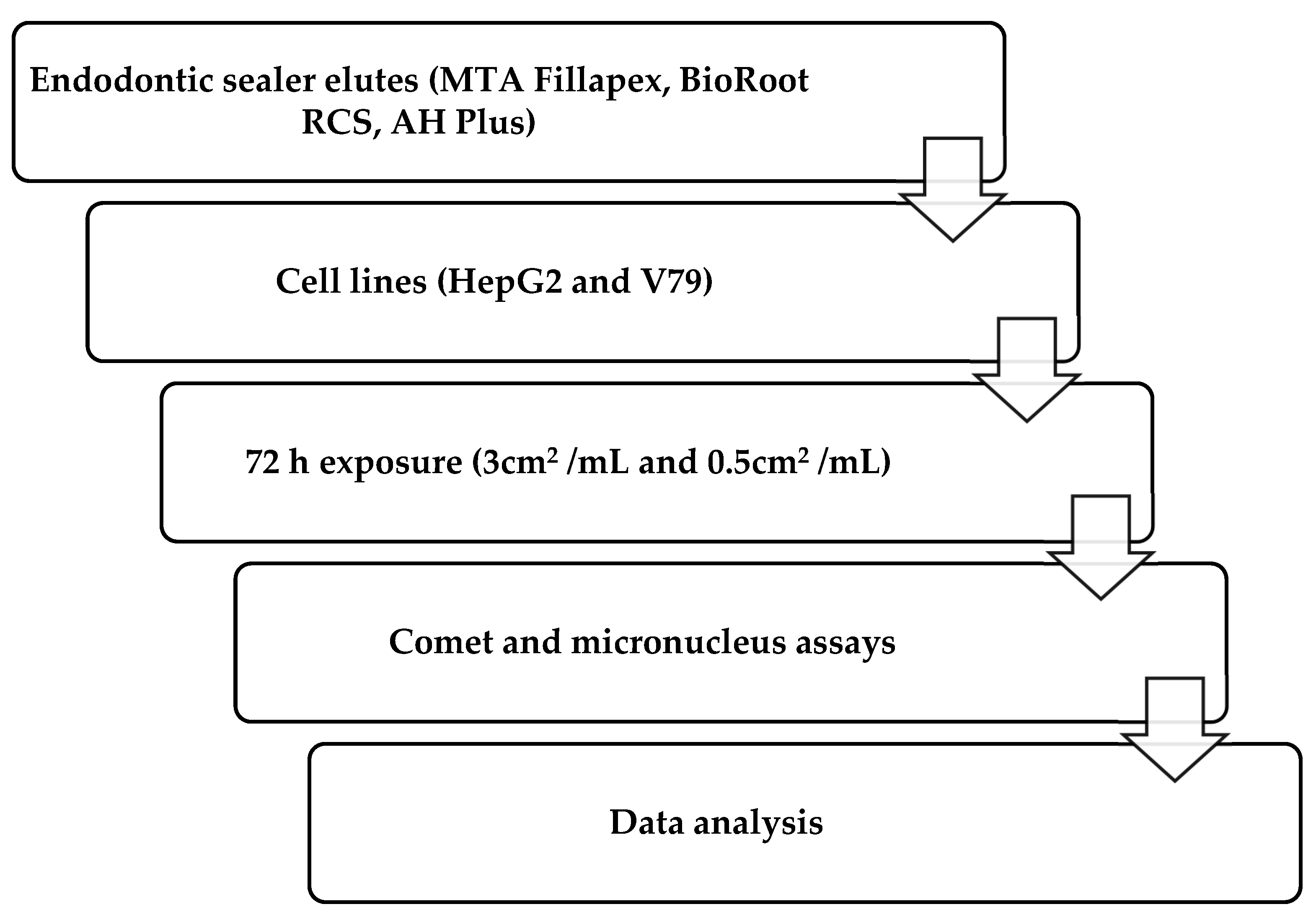
2. Materials and Methods
2.1. Preparation of Materials and Extraction of Non-Reacted Components
2.2. Cell Culture
2.3. Alkaline Comet Assay
2.4. Micronucleus Cytome Assay
2.5. Data Analysis
3. Results
4. Discussion
5. Conclusions
Author Contributions
Funding
Institutional Review Board Statement
Informed Consent Statement
Data Availability Statement
Conflicts of Interest
References
- Komabayashi, T.; Colmenar, D.; Cvach, N.; Bhat, A.; Primus, C.; Imai, Y. Comprehensive review of current endodontic sealers. Dent. Mater. J. 2020, 39, 703–720. [Google Scholar] [CrossRef]
- Reszka, P.; Nowicka, A.; Lipski, M.; Dura, W.; Droździk, A.; Woźniak, K. A comparative chemical study of calcium silicate-containing and epoxy resin-based root canal sealers. Biomed. Res. Int. 2016, 2016, 9808432. [Google Scholar] [CrossRef] [PubMed]
- Fonseca, D.A.; Paula, A.B.; Marto, C.M.; Coelho, A.; Paulo, S.; Martinho, J.P.; Carrilho, E.; Ferreira, M.M. Biocompatibility of root canal sealers: A systematic review of in vitro and in vivo studies. Materials 2019, 12, 4113. [Google Scholar] [CrossRef] [PubMed]
- Camilleri, J.; Atmeh, A.; Li, X.; Meschi, N. Present status and future directions: Hydraulic materials for endodontic use. Int. Endod. J. 2022, 55 (Suppl. S3), 710–777. [Google Scholar] [CrossRef]
- Kim, M.; Hayashi, M.; Yu, B.; Lee, T.K.; Kim, R.H.; Jo, D.W. Cytotoxicity and genotoxicity of epoxy resin-based root canal sealers before and after setting procedures. Life 2022, 12, 847. [Google Scholar] [CrossRef] [PubMed]
- Angulus Science and Technology. MTA-Fillapex Endodontic Sealer, Scientific Profile. 2011. Available online: http://www.angelusdental.com/img/arquivos/mta_fillapex_technical_profile_download.pdf (accessed on 7 October 2024).
- Lim, M.; Jung, C.; Shin, D.-H.; Cho, Y.-B.; Song, M. Calcium silicate-based root canal sealers: A literature review. Restor. Dent. Endod. 2020, 45, e35. [Google Scholar] [CrossRef]
- Borges, Á.H.; Orçati Dorileo, M.C.; Dalla Villa, R.; Borba, A.M.; Semenoff, T.A.; Guedes, O.A.; Estrela, C.R.; Bandeca, M.C. Physicochemical properties and surfaces morphologies evaluation of MTA FillApex and AH Plus. Sci. World J. 2014, 2014, 589732. [Google Scholar] [CrossRef]
- Silva, E.J.N.L.; Cardoso, M.L.; Rodrigues, J.P.; De-Deus, G.; Fidalgo, T.K.D.S. Solubility of bioceramic- and epoxy resin-based root canal sealers: A systematic review and meta-analysis. Aust. Endod. J. 2021, 47, 690–702. [Google Scholar] [CrossRef]
- Najafzadeh, R.; Fazlyab, M.; Esnaashari, E. Comparison of bioceramic and epoxy resin sealers in terms of marginal adaptation and tubular penetration depth with different obturation techniques in premolar teeth: A scanning electron microscope and confocal laser scanning microscopy study. J. Fam. Med. Prim. Care 2022, 11, 1794–1797. [Google Scholar] [CrossRef]
- Ashraf, H.; Najafi, F.; Heidari, S.; Yadegary, Z.; Zadsirjan, S. Cytotoxicity of Two Experimental Epoxy Resin-Based Sealers. Iran. Endod. J. 2018, 13, 257–262. [Google Scholar] [CrossRef]
- Huang, T.-H.; Yang, J.-J.; Li, H.; Kao, C.-T. The biocompatibility evaluation of epoxy resin-based root canal sealers in vitro. Biomaterials 2002, 23, 77–83. [Google Scholar] [CrossRef] [PubMed]
- Miletić, I.; Jukić, S.; Anić, I.; Željezić, D.; Garaj-Vrhovac, V.; Osmak, M. Examination of cytotoxicity and mutagenicity of AH 26 and AH Plus sealers. Int. Endod. J. 2003, 36, 330–335. [Google Scholar] [CrossRef]
- Gandolfi, M.G.; Parrilli, A.P.; Fini, M.; Prati, C.; Dummer, P.M. 3D micro-CT analysis of the interface voids associated with Thermafil root fillings used with AH Plus or a flowable MTA sealer. Int. Endod. J. 2013, 46, 253–263. [Google Scholar] [CrossRef] [PubMed]
- Donnermeyer, D.; Dornseifer, P.; Schäfer, E.; Dammaschke, T. The push-out bond strength of calcium silicate-based endodontic sealers. Head Face Med. 2018, 14, 13. [Google Scholar] [CrossRef]
- Chellapandian, K.; Reddy, T.V.K.; Venkatesh, V.; Annapurani, A. Bioceramic root canal sealers: A review. Int. J. Health Sci. 2022, 6, 5693–5706. [Google Scholar] [CrossRef]
- Kaul, S.; Kumar, A.; Badiyani, B.K.; Sukhtankar, L.; Madhumitha, M.; Kumar, A. Comparison of sealing ability of bioceramic sealer, AH Plus, and GuttaFlow in conservatively prepared curved root canals obturated with single-cone technique: An in vitro study. J. Pharm. Bioallied Sci. 2021, 13, S857–S860. [Google Scholar] [CrossRef]
- Colombo, M.; Poggio, C.; Dagna, A.; Meravini, M.V.; Riva, P.; Trovati, F.; Pietrocola, G. Biological and physico-chemical properties of new root canal sealers. J. Clin. Exp. Dent. 2018, 10, e120–e126. [Google Scholar] [CrossRef] [PubMed]
- Eldeniz, A.U.; Shehata, M.; Högg, C.; Reichl, F.X. DNA double-strand breaks caused by new and contemporary endodontic sealers. Int. Endod. J. 2016, 49, 1141–1151. [Google Scholar] [CrossRef]
- Jung, S.; Libricht, V.; Sielker, S.; Hanisch, M.R.; Schäfer, E.; Dammaschke, T. Evaluation of the biocompatibility of root canal sealers on human periodontal ligament cells ex vivo. Odontology 2019, 107, 54–63. [Google Scholar] [CrossRef]
- Taraslia, V.; Anastasiadou, E.; Lignou, C.; Keratiotis, G.; Agrafioti, A.; Kontakiotis, E.G. Assessment of Cell Viability in Four Novel Endodontic Sealers. Eur. J. Dent. 2018, 12, 287–291. [Google Scholar] [CrossRef]
- Collado-González, M.; García-Bernal, D.; Oñate-Sánchez, R.E.; Ortolani-Seltenerich, P.S.; Lozano, A.; Forner, L.; Llena, C.; Rodríguez-Lozano, F.J. Biocompatibility of three new calcium silicate-based endodontic sealers on human periodontal ligament stem cells. Int. Endod. J. 2017, 50, 875–884. [Google Scholar] [CrossRef] [PubMed]
- Kebudi Benezra, M.; Schembri Wismayer, P.; Camilleri, J. Interfacial characteristics and cytocompatibility of hydraulic sealer cements. J. Endod. 2018, 44, 1007–1017. [Google Scholar] [CrossRef]
- Zhou, H.M.; Du, T.F.; Shen, Y.; Wang, Z.J.; Zheng, Y.F.; Haapasalo, M. In vitro cytotoxicity of calcium silicate-containing endodontic sealers. J. Endod. 2015, 41, 56–61. [Google Scholar] [CrossRef]
- Portella, F.F.; Collares, F.M.; Dos Santos, L.A.; dos Santos, B.P.; Camassola, M.; Leitune, V.C.; Samuel, S.M. Glycerol salicylate-based containing α-tricalcium phosphate as a bioactive root canal sealer. J. Biomed. Mater. Res. Part B 2015, 103, 1663–1669. [Google Scholar] [CrossRef] [PubMed]
- Prüllage, R.K.; Urban, K.; Schäfer, E.; Dammaschke, T. Material properties of a tricalcium silicate-containing, a mineral trioxide aggregate-containing, and an epoxy resin-based root canal sealer. J. Endod. 2016, 42, 1784–1788. [Google Scholar] [CrossRef] [PubMed]
- Erdogan, H.; Yildirim, S.; Cobankara, F.K. Cytotoxicity and genotoxicity of salicylate- and calcium silicate-based root canal sealers on primer human periodontal ligament fibroblasts. Aust. Endod. J. 2021, 47, 645–653. [Google Scholar] [CrossRef]
- Oh, H.; Kim, E.; Lee, S.; Park, S.; Chen, D.; Shin, S.-J.; Kim, E.; Kim, S. Comparison of biocompatibility of calcium silicate-based sealers and epoxy resin-based sealer on human periodontal ligament stem cells. Materials 2020, 13, 5242. [Google Scholar] [CrossRef]
- Zheng, J.-J.; Kang, X.-Y.; Li, S.-M.; Liu, J.-C.; Sun, K.; Gao, L.; Hou, T.-Z. Effects of MTA, iRoot SP, and AH Plus on proliferation and differentiation of human periodontal ligament stem cells. Shanghai Kou Qiang Yi Xue 2020, 29, 449–455. [Google Scholar]
- Só, B.B.; Martins, M.D.; So, M.V.; Weissheimer, T.; Marques, M.M.; Moreira, M.S. Genotoxicity and Cytotoxicity Comparison of Calcium Silicate-Based and Resin-Based Sealers on Human Periodontal Ligament Stem Cells. Eur. Endod. J. 2022, 7, 129–134. [Google Scholar] [CrossRef]
- Jagtap, P.; Shetty, R.; Agarwalla, A.; Wani, P.; Bhargava, K.; Martande, S. Comparative evaluation of cytotoxicity of root canal sealers on cultured human periodontal fibroblasts: In vitro study. J. Contemp. Dent. Pract. 2018, 19, 847–852. [Google Scholar] [CrossRef]
- Zordan-Bronzel, C.L.; Tanomaru-Filho, M.; Rodrigues, E.M.; Chávez-Andrade, G.M.; Faria, G.; Guerreiro-Tanomaru, J.M. Cytocompatibility, bioactive potential and antimicrobial activity of an experimental calcium silicate-based endodontic sealer. Int. Endod. J. 2019, 52, 979–986. [Google Scholar] [CrossRef] [PubMed]
- Zordan-Bronzel, C.L.; Tanomaru-Filho, M.; Torres, F.F.E.; Chávez-Andrade, G.M.; Rodrigues, E.M.; Guerreiro-Tanomaru, J.M. Physicochemical properties, cytocompatibility and antibiofilm activity of a new calcium silicate sealer. Braz. Dent. J. 2021, 32, 8–18. [Google Scholar] [CrossRef] [PubMed]
- Janini, A.C.P.; da Silveira Bueno, C.E.; de De Martin, A.S.; Pelegrine, R.A.; Fontana, C.E.; Hussne, R.P.; Accorsi-Mendonça, T.; Pinheiro, S.L. In vitro evaluation of cytotoxicity in human osteoblastic cells and antimicrobial activity in the biofilm of different root canal sealers. Res. Soc. Dev. 2022, 11, e430111032842. [Google Scholar] [CrossRef]
- Sheela, S.; Nassar, M.; AlGhalban, F.M.; Gorduysus, M.O. In vitro cytotoxicity and mineralization potential of an endodontic bioceramic material. Eur. J. Dent. 2023, 17, 548–555. [Google Scholar] [CrossRef]
- Candeiro, G.T.M.; Moura-Netto, C.; D’Almeida-Couto, R.S.; Azambuja-Júnior, N.; Marques, M.M.; Cai, S.; Gavini, G. Cytotoxicity, genotoxicity and antibacterial effectiveness of a bioceramic endodontic sealer. Int. Endod. J. 2016, 49, 858–864. [Google Scholar] [CrossRef]
- Hoshyari, N.; Mesgarani, A.; Shokrzadeh Lamuki, M.; Motafeghi, F.; Mousavi, S.J.; Veghar Mousavi, M. Cytotoxicity of two calcium silicate-based sealers and AH Plus resin-based sealer for human gingival fibroblasts. J. Res. Dent. Maxillofac. Sci. 2023, 8, 203–209. [Google Scholar] [CrossRef]
- Malta, C.P.; Santi, S.S.; Barcelos, R.C.S.; Zanatta, F.B.; Bier, C.A.S.; Morgental, R.D. Premixed calcium silicate-based root canal sealers have better biological properties than AH Plus: A systematic review and meta-analysis of in vivo animal studies and in vitro laboratory studies. J. Conserv. Dent. Endod. 2024, 27, 345–359. [Google Scholar] [CrossRef]
- ISO 10993-12:2012; Biological Evaluation of Medical Devices—Part 12: Sample Preparation and Reference Materials. International Organization for Standardization (ISO): Geneva, Switzerland, 2012.
- Gerdemann, A.; Behrens, M.; Esselen, M.; Humpf, H.U. Metabolic Profiling as a Powerful Tool for the Analysis of Cellular Alterations Caused by 20 Mycotoxins in HepG2 Cells. Arch. Toxicol. 2022, 96, 2983–2998. [Google Scholar] [CrossRef]
- Kiseleva, O.I.; Kurbatov, I.Y.; Arzumanian, V.A.; Ilgisonis, E.V.; Zakharov, S.V.; Poverennaya, E.V. The Expectation and Reality of the HepG2 Core Metabolic Profile. Metabolites 2023, 13, 908. [Google Scholar] [CrossRef]
- Ng, K.-L.; Osman, M.A.; Tan, Y.-N.; Ee, K.-Y.; Rajab, N.F. Characterization, Antioxidant, ACE Inhibition and Toxicity Evaluations of Palm Kernel Cake-Derived Alcalase® Hydrolysate. Food Sci. Technol. Camp. 2022, 42, e80421. [Google Scholar] [CrossRef]
- OECD. Test No. 489: In Vivo Mammalian Alkaline Comet Assay; OECD Guidelines for the Testing of Chemicals, Section 4; OECD Publishing: Paris, France, 2016; Available online: https://www.oecd.org/en/publications/test-no-489-in-vivo-mammalian-alkaline-comet-assay_9789264264885-en.html (accessed on 23 October 2024).
- Fenech, M. Cytokinesis-Block Micronucleus Cytome Assay. Nat. Protoc. 2007, 2, 1084–1104. [Google Scholar] [CrossRef] [PubMed]
- Gaudin, A.; Tolar, M.; Peters, O.A. Cytokine Production and Cytotoxicity of Calcium Silicate-based Sealers in 2- and 3-dimensional Cell Culture Models. J. Endod. 2020, 46, 818–826. [Google Scholar] [CrossRef]
- Dos Santos Costa, F.M.; Fernandes, M.H.; Batistuzzo De Medeiros, S.R. Genotoxicity of root canal sealers: A literature review. Clin. Oral Investig. 2020, 24, 3347–3362. [Google Scholar] [CrossRef]
- Radwanski, M.; Rozpedek-Kaminska, W.; Galita, G.; Siwecka, N.; Sokolowski, J.; Majsterek, I.; Özcan, M.; Lukomska-Szymanska, M. Cytotoxicity and genotoxicity of bioceramic root canal sealers compared to conventional resin-based sealer. Sci. Rep. 2024, 14, 4124. [Google Scholar] [CrossRef] [PubMed]
- Nair, A.V.; Nayak, M.; Prasada, L.K.; Shetty, V.; Kumar, C.N.V.; Nair, R.R. Comparative Evaluation of Cytotoxicity and Genotoxicity of Two Bioceramic Sealers on Fibroblast Cell Line: An in vitro Study. J. Contemp. Dent. Pract. 2018, 19, 656–661. [Google Scholar]
- Dhopavkar, V.V.; Shivanand, S.S.; Bhat, K.; Patil, A.C.; Doddwad, P.K.; Godbole, N.J. Comparative Evaluation of Cytotoxic and Genotoxic Effects of Three Resin-Based Sealers by 3,(4,5-Dimethylthiazol-2-yl)-2,5-Diphenyltetrazolium Bromide Assay and Comet Assay—An In Vitro Study. Contemp. Clin. Dent. 2021, 12, 376–382. [Google Scholar] [CrossRef]
- Camargo, C.H.; Oliveira, T.R.; Silva, G.O.; Rabelo, S.B.; Valera, M.C.; Cavalcanti, B.N. Setting Time Affects In Vitro Biological Properties of Root Canal Sealers. J. Endod. 2014, 40, 530–533. [Google Scholar] [CrossRef]
- Leme, K.S.V.; Salvadori, D.M.F. In vitro toxicogenomic activity of an MTA/salicylate-based endodontic sealer. Toxicol. Rep. 2022, 9, 1076–1081. [Google Scholar] [CrossRef] [PubMed]
- Jeon, M.J.; Ko, H.; Shin, S.J.; Kim, M. Cytotoxicity and Genotoxicity of Various Types of Endodontic Sealers in Chinese Hamster Ovary (CHO-K1) Cells. Dent. Mater. J. 2023, 42, 774–779. [Google Scholar] [CrossRef]
- Siregar, I.; Permitasari, R.; Kamizar; Margono, A. Comparison of the Potential Genotoxicities of Resin-, Silicone-, and Bioceramic-Based Root Canal Sealers Against Human Lymphocytes. J. Int. Dent. Med. Res. 2019, 12, 88–94. [Google Scholar]
- Rajkumar, D.S.; Padmanaban, R. Impact of Bisphenol A and Analogues Eluted from Resin-Based Dental Materials on Cellular and Molecular Processes: An Insight on Underlying Toxicity Mechanisms. J. Appl. Toxicol. 2025, 45, 4–22. [Google Scholar] [CrossRef] [PubMed]
- Löf, M.; Ghasemimehr, M.; Falk, A.; Vult von Steyern, P. Bisphenol A in Dental Materials—Existence, Leakage, and Biological Effects. Heliyon 2019, 5, e01711. [Google Scholar] [CrossRef]
- Drozdz, K.; Wysokinski, D.; Krupa, R.; Wozniak, K. Bisphenol A-glycidyl methacrylate induces a broad spectrum of DNA damage in human lymphocytes. Arch. Toxicol. 2010, 85, 1453–1461. [Google Scholar] [CrossRef]
- El-Mansy, L.H.; Ali, M.M.; Hassan, R.E.S.; Beshr, K.A.; El Ashry, S.H. Evaluation of the Biocompatibility of a Recent Bioceramic Root Canal Sealer (BioRoot™ RCS): In-Vivo Study. Open Access Maced. J. Med. Sci. 2020, 8, 100–106. [Google Scholar] [CrossRef]
- Jung, S.; Sielker, S.; Hanisch, M.R.; Libricht, V.; Schäfer, E.; Dammaschke, T. Cytotoxic effects of four different root canal sealers on human osteoblasts. PLoS ONE 2018, 13, e0194467. [Google Scholar] [CrossRef] [PubMed]
- Tolosa-Monfà, A.; Veroni, A.; Blasi-Cabús, J.; Ballester-Palacios, M.L.; Berástegui-Jimeno, E. Cytotoxicity comparison of Bio C Sealer against multiple root canal sealers. J. Clin. Exp. Dent. 2023, 15, e110–e117. [Google Scholar] [CrossRef]
- Vouzara, T.; Dimosiari, G.; Koulaouzidou, E.A.; Economides, N. Cytotoxicity of a New Calcium Silicate Endodontic Sealer. J. Endod. 2018, 44, 849–852. [Google Scholar] [CrossRef]
- Drukteinis, S. Hydraulic calcium silicate-based materials for root canal obturation. Clin. Dent. 2022, 6, 1. [Google Scholar] [CrossRef]
- Bin, C.V.; Valera, M.C.; Camargo, S.E.; Rabelo, S.B.; Silva, G.O.; Balducci, I.; Camargo, C.H. Cytotoxicity and Genotoxicity of Root Canal Sealers Based on Mineral Trioxide Aggregate. J. Endod. 2012, 38, 495–500. [Google Scholar] [CrossRef]
- Schweikl, H.; Schmalz, G. The induction of micronuclei in V79 cells by the root canal filling material AH plus. Biomaterials 2000, 21, 939–944. [Google Scholar] [CrossRef]
- de Azevedo Queiroz, Í.O.; Machado, T.; Alves, C.C.; Vasques, A.M.V.; Cury, M.T.S.; Vasconcelos, B.C.; Gomes-Filho, J.E.; Vivan, R.R.; Braga, T.; Love, R.M.; et al. Tracing the toxic ions of an endodontic tricalcium silicate-based sealer in local tissues and body organs. J. Trace Elem. Med. Biol. 2021, 68, 126856. [Google Scholar] [CrossRef]
- Khalid, M.; Hassani, S.; Abdollahi, M. Metal-induced oxidative stress: An evidence-based update of advantages and disadvantages. Curr. Opin. Toxicol. 2020, 20–21, 55–68. [Google Scholar] [CrossRef]
- Kumar, A.; Kour, S.; Kaul, S.; Malik, A.; Dhani, R.; Kaul, R. Cytotoxicity Evaluation of Bio-C, CeraSeal, MTA-Fillapex, and AH Plus Root Canal Sealers by Microscopic and 3-(4,5-Dimethythiazol-2yl)-2,5-Diphenyltetrazolium Bromide (MTT) Assay. J. Conserv. Dent. 2023, 26, 73–78. [Google Scholar] [CrossRef]
- Mohamed, S.; Sabita, U.; Rajendra, S.; Raman, D. Genotoxicity: Mechanisms, testing guidelines and methods. Glob. J. Pharm. Pharm. Sci. 2017, 1, 133–138. [Google Scholar] [CrossRef]
- Mohammadi, Z.; Shalavi, S.; Jafarzadeh, H.; Bhandi, S.; Patil, S. Genotoxicity of Endodontic Materials: A Critical Review. J. Contemp. Dent. Pract. 2015, 16, 692–696. [Google Scholar] [CrossRef]
- Urzì, O.; Gasparro, R.; Costanzo, E.; De Luca, A.; Giavaresi, G.; Fontana, S.; Alessandro, R. Three-Dimensional Cell Cultures: The Bridge between In Vitro and In Vivo Models. Int. J. Mol. Sci. 2023, 24, 12046. [Google Scholar] [CrossRef] [PubMed]
- Okamoto, M.; Matsumoto, S.; Moriyama, K.; Huang, H.; Watanabe, M.; Miura, J.; Sugiyama, K.; Hirose, Y.; Mizuhira, M.; Kuriki, N.; et al. Biological Evaluation of the Effect of Root Canal Sealers Using a Rat Model. Pharmaceutics 2022, 14, 2038. [Google Scholar] [CrossRef]

| Sealer | Manufacturer | Type | Composition | Presentation | LOT |
|---|---|---|---|---|---|
| MTA Fillapex | Angelus, Londrina, PR, Brazil | Calcium silicate-based sealer | Paste A: salicylate resin, bismuth trioxide, fumed silica Paste B: fumed silica, titanium dioxide, mineral trioxide aggregate, base resin | Two tubes | 71695 |
| BioRoot RCS | Septodont, Saint Maur Des Fosses, France | Hydraulic calcium silicate-based sealer | Powder: tricalcium silicate, zirconium oxide, povidone Liquid: aqueous solution of calcium chloride and polycarboxylate | Powder/liquid | B30723C B31424 |
| AH Plus | Denstply DeTrey GmbH, Konstanz, Germany | Epoxy-based sealer | Paste A: diepoxide, calcium tungstate, zirconium oxide, aerosil, pigments Paste B: 1-adamantane amine, N,N’-dibenzyl-5-oxa-nonandiamine-1,9 TCD-diamine, calcium tungstate, zirconium oxide, aerosil, silicone oil | Two tubes | 2207000721 |
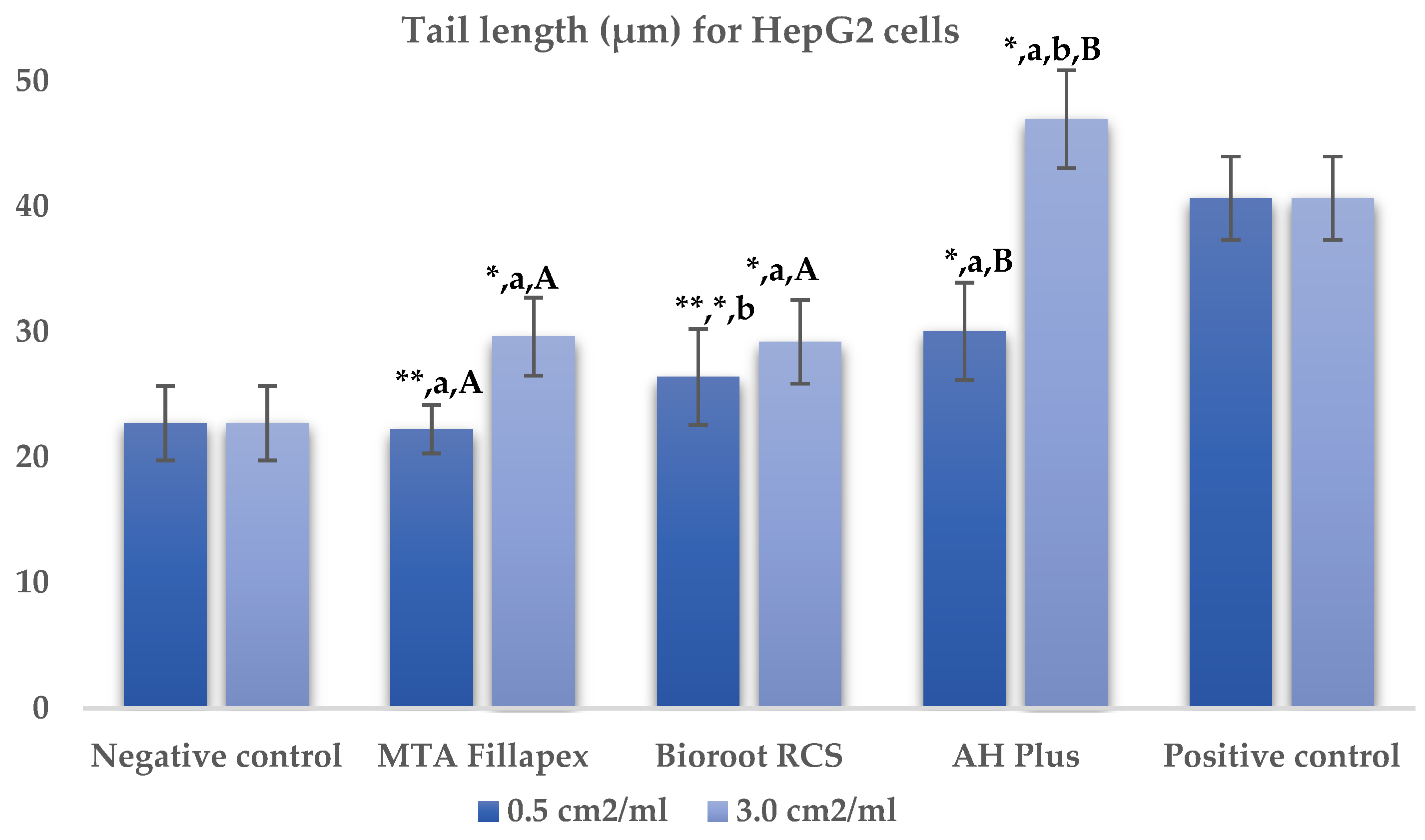
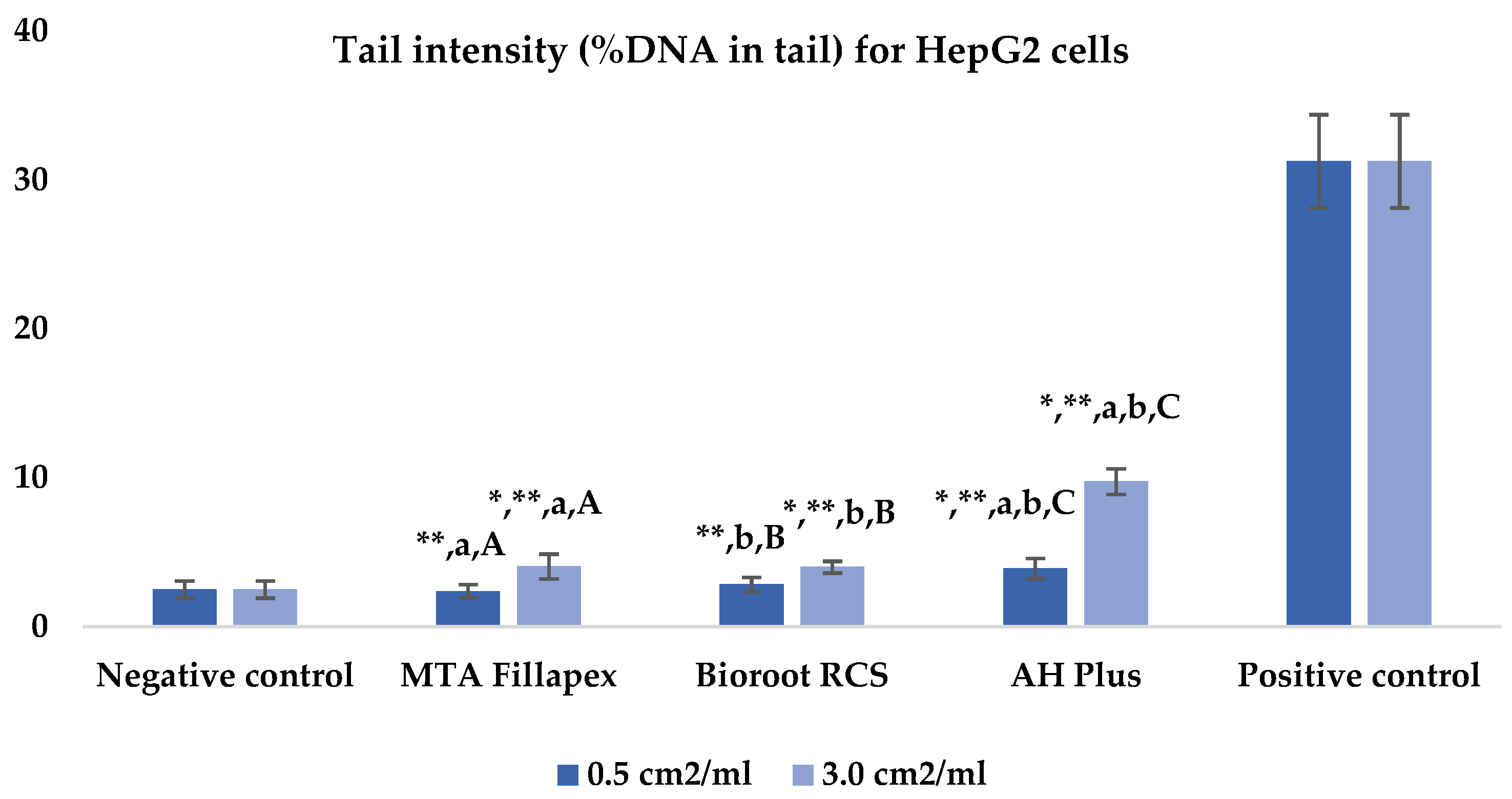
| Material | Tail Length (μm) | p-Value † | Tail Intensity (%DNA in Tail) | p-Value † | ||
|---|---|---|---|---|---|---|
| 0.5 cm2/mL | 3.0 cm2/mL | 0.5 cm2/mL | 3.0 cm2/mL | |||
| Negative control | 22.74 ± 2.96 | n. a. | 2.49 ± 0.58 | n. a. | ||
| MTA Fillapex | 22.26 ± 1.73 **,a | 29.64 ± 3.12 *,a | ≤0.001 | 2.38 ± 0.45 **,a | 4.04 ± 0.83 *,**,a | ≤0.001 |
| BioRoot RCS | 26.43 ± 3.82 ** | 29.22 ± 3.33 *,b | 0.190 | 2.83 ± 0.48 **,b | 4.00 ± 0.40 *,**,b | ≤0.001 |
| AH Plus | 30.06 ± 3.88 *,a | 46.98 ± 3.91 *,a,b | ≤0.001 | 3.90 ± 0.62 *,**,a,b | 9.75 ± 0.86 *,**, a,b | ≤0.001 |
| Positive control | 40.67 ± 3.31 | n. a. | 31.25 ± 3.13 | n. a. | ||
| p-value †† | ≤0.001 | ≤0.001 | ≤0.001 | ≤0.001 | ||
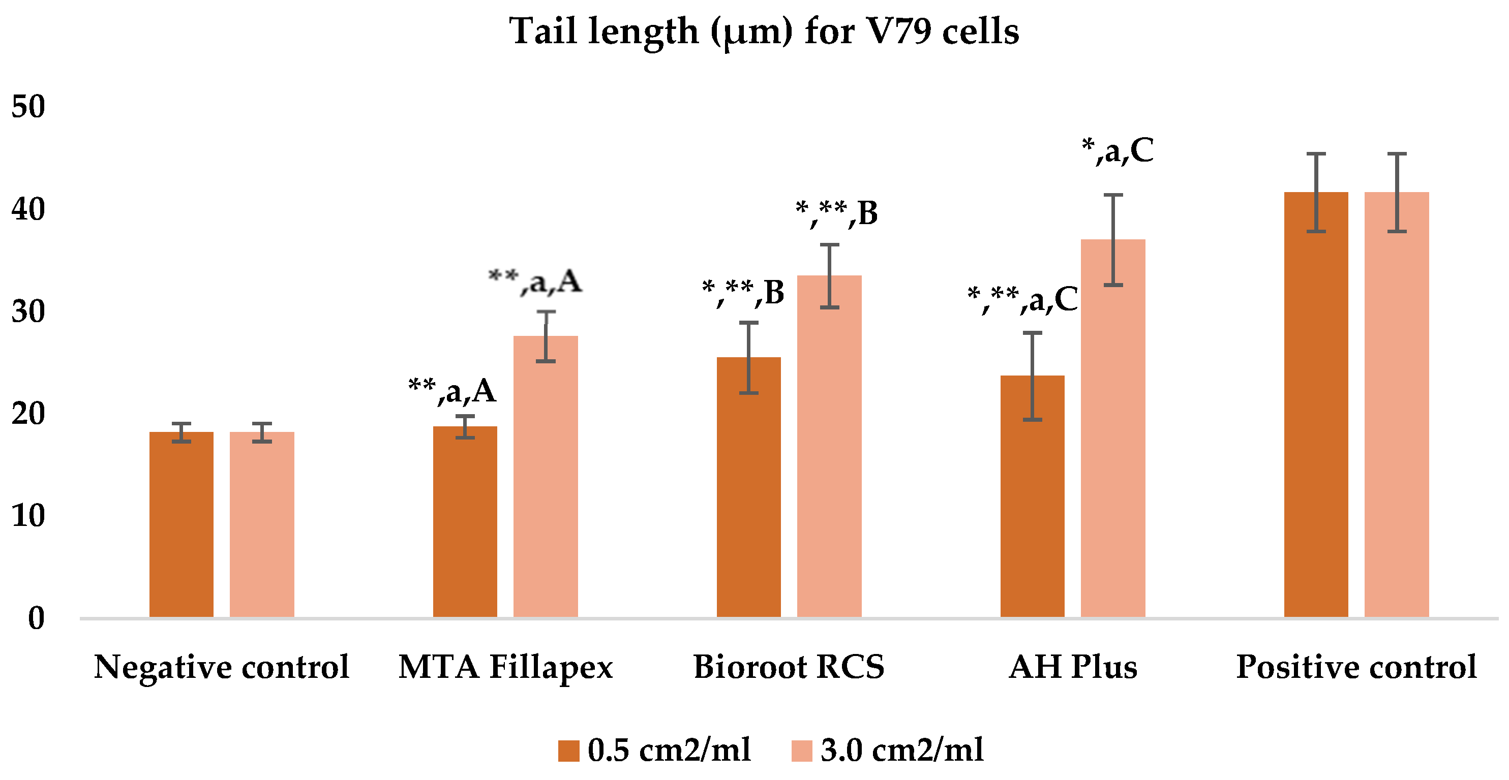
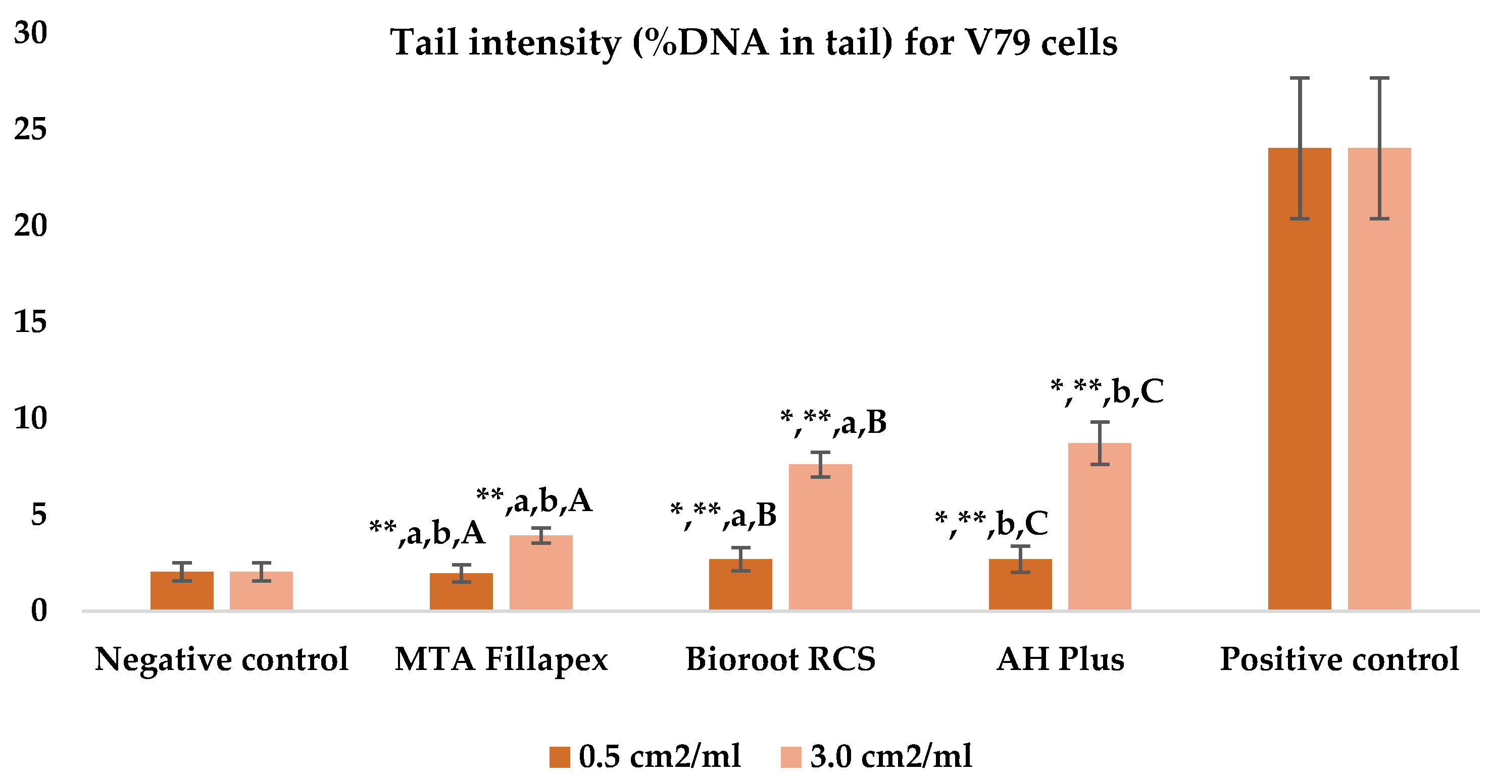
| Material | Tail Length (μm) | p-Value † | Tail Intensity (%DNA in Tail) | p-Value † | ||
|---|---|---|---|---|---|---|
| 0.5 cm2/mL | 3.0 cm2/mL | 0.5 cm2/mL | 3.0 cm2/mL | |||
| Negative control | 18.20 ± 0.88 | n. a. | 2.04 ± 0.48 | n. a. | ||
| MTA Fillapex | 18.74 ± 1.06 **,a | 27.63 ± 2.46 **,a | ≤0.001 | 1.96 ± 0.45 **,a,b | 3.93 ± 0.39 **,a,b | ≤0.001 |
| BioRoot RCS | 25.31 ± 3.44 *,** | 33.51 ± 3.07 *,** | ≤0.001 | 2.69 ± 0.60 *,**,a | 7.61 ± 0.64 *,**,a | ≤0.001 |
| AH Plus | 23.71 ± 4.26 *,**,a | 37.05 ± 4.41 *,a | ≤0.001 | 2.70 ± 0.68 *,**,b | 8.73 ± 1.10 *,**,b | ≤0.001 |
| Positive control | 41.67 ± 3.78 | n. a. | 24.40 ± 3.66 | n. a. | ||
| p-value †† | ≤0.001 | ≤0.001 | ≤0.001 | ≤0.001 | ||
| Cytogenetic Damage | Concentration | Negative Control | MTA Fillapex | Bioroot RCS | AH Plus | Positive Control | p-Value †† |
|---|---|---|---|---|---|---|---|
| Micronucleus | 0.5 cm2/mL | 7.50 ± 1.90 | 7.40 ± 2.84 **,a | 7.30 ± 2.87 **,b | 16.90 ± 4.53 *,a,b | 46.40 ± 3.41 | ≤0.001 |
| 3.0 cm2/mL | 7.10 ± 2.42 **,a | 11.60 ± 2.91 ** | 21.40 ± 3.81 *,a | ≤0.001 | |||
| p-value † | n. a. | 0.853 | 0.005 | 0.023 | n. a. | ||
| Nuclear buds | 0.5 cm2/mL | 20.18 ± 5.15 | 21.50 ± 6.59 **,a,b | 14.40 ± 2.84 **,a,c | 31.90 ± 4.95 *,b,c | 45.40 ± 4.13 | ≤0.001 |
| 3.0 cm2/mL | 39.80 ± 5.12 *,a | 24.90 ± 4.58 **,a,b | 42.30 ± 6.78 *,b | ≤0.001 | |||
| p-value † | n.a. | ≤0.001 | ≤0.001 | 0.002 | n. a. | ||
| Nucleoplasmic bridges | 0.5 cm2/mL | 1.60 ± 1.51 | 3.80 ± 1.48 *,a | 1.10 ± 0.74 **,a | 2.80 ± 2.78 *,** | 8.30 ± 2.34 | ≤0.001 |
| 3.0 cm2/mL | 2.30 ± 1.25 ** | 2.70 ± 1.42 ** | 4.30 ± 2.45 *,** | ≤0.001 | |||
| p-value † | n. a. | 0.043 | 0.007 | 0.190 | |||
| CBPI | 0.5 cm2/mL | 1.84 ± 0.07 | 1.86 ± 0.07 **,a | 1.84 ± 0.05 | 1.76 ± 0.07 **,a | 1.64 ± 0.04 | ≤0.001 |
| 3.0 cm2/mL | 1.77 ± 0.09 **,a | 1.80 ± 0.05 **,b | 1.64 ± 0.07 *,a,b | ≤0.001 | |||
| p-value † | n. a. | 0.019 | 0.123 | ≤0.001 | n. a. | ||
| Apoptosis | 0.5 cm2/mL | 3.90 ± 3.18 | 9.90 ± 3.63 *,** | 12.70 ± 3.50 *,** | 15.20 ± 3.94 *,** | 28.10 ± 3.05 | ≤0.001 |
| 3.0 cm2/mL | 21.90 ± 3.00 *,** | 17.40 ± 4.48 *,**,a | 23.70 ± 4.27 *,a | ≤0.001 | |||
| p-value † | n.a. | ≤0.001 | 0.019 | ≤0.001 | n. a. | ||
| Necrosis | 0.5 cm2/mL | 1.20 ± 0.79 | 1.00 ± 0.82 ** | 1.10 ± 1.10 ** | 2.00 ± 1.33 ** | 9.60 ± 3.25 | ≤0.001 |
| 3.0 cm2/mL | 3.20 ± 2.66 ** | 3.00 ± 1.49 ** | 3.50 ± 2.37 *,** | ≤0.001 | |||
| p-value † | n. a. | 0.019 | 0.009 | 0.143 | n. a. | ||
| Cells with undamaged chromatin | 0.5 cm2/mL | 1968.3 ± 5.42 | 1956.40 ± 10.66 **,a | 1963.40 ± 5.60 **,b | 1931.20 ± 7.44 **,a,b | 1841.30 ± 11.12 | ≤0.001 |
| 3.0 cm2/mL | 1925.70 ± 7.94 *,** | 1940.40 ± 10.12 **,a | 1904.80 ± 9.30 *,**,a | ≤0.001 | |||
| p-value † | n. a. | ≤0.001 | ≤0.001 | ≤0.001 | n. a. | ||
| Cytogenetic Damage | Concentration | Negative Control | MTA Fillapex | Bioroot RCS | AH Plus | Positive Control | p-Value †† |
|---|---|---|---|---|---|---|---|
| Micronucleus | 0.5 cm2/mL | 2.36 ± 1.29 | 3.40 ± 1.43 **,a | 5.10 ± 2.38 **,b | 6.20 ± 2.04 *,**, a,b | 34.40 ± 3.73 | ≤0.001 |
| 3.0 cm2/mL | 4.80 ± 2.66 **,a | 5.90 ± 1.66 **,b | 11.50 ± 1.96 *,**,a,b | ≤0.001 | |||
| p-value † | n. a. | 0.353 | 0.436 | ≤0.001 | n. a. | ||
| Nuclear buds | 0.5 cm2/mL | 2.82 ± 1.33 | 14.90 ± 4.09 *,**,a | 9.60 ± 2.88 *,**,a,b | 16.50 ± 3.10 *,**,b | 27.50 ± 3.85 | ≤0.001 |
| 3.0 cm2/mL | 24.30 ± 4.47 *,a | 15.40 ± 3.24 *,**,a,b | 20.86 ± 1.05 *,b | ≤0.001 | |||
| p-value † | n. a. | ≤0.001 | 0.002 | ≤0.001 | n. a. | ||
| Nucleoplasmic bridges | 0.5 cm2/mL | 0.91 ± 0.72 | 0.90 ± 0.88 ** | 0.80 ± 0.67 ** | 0.90 ± 0.57 ** | 6.00 ± 2.00 | ≤0.001 |
| 3.0 cm2/mL | 2.10 ± 1.29 ** | 2.20 ± 1.42 ** | 2.70 ± 1.48 ** | ≤0.001 | |||
| p-value † | n. a. | 0.043 | 0.002 | 0.043 | n. a. | ||
| CBPI | 0.5 cm2/mL | 1.81 ± 0.05 | 1.84 ± 0.06 ** | 1.83 ± 0.0 ** | 1.84 ± 0.04 ** | 1.63 ± 0.03 | ≤0.001 |
| 3.0 cm2/mL | 1.69 ± 0.04 **,a | 1.80 ± 0.05 **,a,b | 1.77 ± 0.08 b | ≤0.001 | |||
| p-value † | n. a. | ≤0.001 | 0.143 | 0.023 | n. a. | ||
| Apoptosis | 0.5 cm2/mL | 3.90 ± 3.18 | 9.90 ± 3.63 *,** | 12.70 ± 3.50 *,** | 15.20 ± 3.94 *,** | 28.10 ± 3.05 | ≤0.001 |
| 3.0 cm2/mL | 21.90 ± 3.00 *,** | 17.40 ± 4.48 *,**,a | 23.70 ± 4.27 *,**,a | ≤0.001 | |||
| p-value † | n. a. | ≤0.001 | 0.019 | ≤0.001 | n.a. | ||
| Necrosis | 0.5 cm2/mL | 1.20 ± 0.79 | 1.00 ± 0.82 ** | 1.10 ± 1.10 ** | 2.00 ± 1.33 *,** | 9.60 ± 3.25 | ≤0.001 |
| 3.0 cm2/mL | 3.20 ± 2.66 ** | 3.00 ± 1.49 ** | 3.50 ± 2.37 *,** | ≤0.001 | |||
| p-value † | n. a. | 0.019 | 0.009 | 0.143 | n. a. | ||
| Cells with undamaged chromatin | 0.5 cm2/mL | 1968.3 ± 5.42 | 1956.40 ± 10.66 *,**,a | 1963.40 ± 5.60 *,**,b | 1931.20 ± 7.44 *,**,a,b | 1841.30 ± 11.12 | ≤0.001 |
| 3.0 cm2/mL | 1925.70 ± 7.94 *,** | 1940.40 ± 10.12 **,b | 1904.80 ± 9.30 *,**,b | ≤0.001 | |||
| p-value † | n. a. | ≤0.001 | ≤0.001 | ≤0.001 | n. a. | ||
Disclaimer/Publisher’s Note: The statements, opinions and data contained in all publications are solely those of the individual author(s) and contributor(s) and not of MDPI and/or the editor(s). MDPI and/or the editor(s) disclaim responsibility for any injury to people or property resulting from any ideas, methods, instructions or products referred to in the content. |
© 2025 by the authors. Licensee MDPI, Basel, Switzerland. This article is an open access article distributed under the terms and conditions of the Creative Commons Attribution (CC BY) license (https://creativecommons.org/licenses/by/4.0/).
Share and Cite
Tadin, A.; Badrov, M.; Juric Kacunic, D.; Galic, N.; Macan, M.; Kovacic, I.; Zeljezic, D. Evaluation of the Genotoxicity and Cytotoxicity of Bioceramic Endodontic Sealers in HepG2 and V79 Cell Lines: An In Vitro Study Using the Comet and Micronucleus Assays. J. Funct. Biomater. 2025, 16, 169. https://doi.org/10.3390/jfb16050169
Tadin A, Badrov M, Juric Kacunic D, Galic N, Macan M, Kovacic I, Zeljezic D. Evaluation of the Genotoxicity and Cytotoxicity of Bioceramic Endodontic Sealers in HepG2 and V79 Cell Lines: An In Vitro Study Using the Comet and Micronucleus Assays. Journal of Functional Biomaterials. 2025; 16(5):169. https://doi.org/10.3390/jfb16050169
Chicago/Turabian StyleTadin, Antonija, Marija Badrov, Danijela Juric Kacunic, Nada Galic, Matea Macan, Ivan Kovacic, and Davor Zeljezic. 2025. "Evaluation of the Genotoxicity and Cytotoxicity of Bioceramic Endodontic Sealers in HepG2 and V79 Cell Lines: An In Vitro Study Using the Comet and Micronucleus Assays" Journal of Functional Biomaterials 16, no. 5: 169. https://doi.org/10.3390/jfb16050169
APA StyleTadin, A., Badrov, M., Juric Kacunic, D., Galic, N., Macan, M., Kovacic, I., & Zeljezic, D. (2025). Evaluation of the Genotoxicity and Cytotoxicity of Bioceramic Endodontic Sealers in HepG2 and V79 Cell Lines: An In Vitro Study Using the Comet and Micronucleus Assays. Journal of Functional Biomaterials, 16(5), 169. https://doi.org/10.3390/jfb16050169