
Clinical Survival Rate and Laboratory Failure of Dental Veneers: A Narrative Literature Review

Abstract
1. Introduction
2. Study Selection
3. Results
3.1. Laboratory Failures
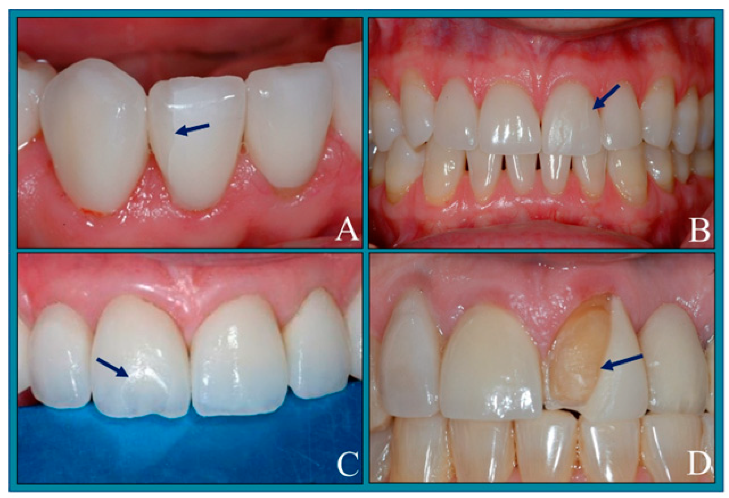
3.1.1. Fracture Failures
Die Spacer Thickness
Stiffness of Supporting Structures
Type of Veneer Ceramic Material
Tooth Preparation
Veneer Thickness
3.1.2. Debonding Failures
Veneer Surface Treatment
Tooth Preparation
Tooth Contamination
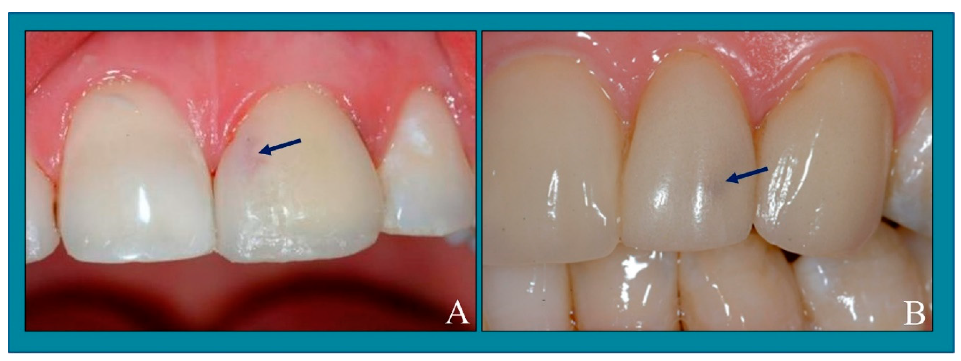
3.1.3. Color Failures
Literature Concerning Color Changes
3.2. Clinical Failures
4. Discussion
5. Conclusions
Funding
Conflicts of Interest
References
- Karaokutan, I.; Aykent, F.; Özdoğan, M.S. Comparison of the color change of porcelain laminate veneers produced by different materials after luting with three resin cements. Oper. Dent. 2023, 48, 166–169. [Google Scholar] [CrossRef] [PubMed]
- Fayad, B.R.; Zohdy, M.M.; Hussein, G.A.; Farag, E.A. Color stability and marginal adaptation of ceramic veneers cemented using different composite resins. Minerva Dent. Oral. Sci. 2023, 73, 88–95. [Google Scholar] [CrossRef] [PubMed]
- Villalobos-Tinoco, J.; Jurado, C.A.; Afrashtehfar, K.A.; Fischer, N. Combination of minimal- and non-preparation techniques with ceramic veneers for managing esthetic deficiencies. Int. J. Esthet. Dent. 2023, 18, 232–243. [Google Scholar]
- Yılmaz, D.; Sabatini, G.P.; Kahveci, Ç.; Yoon, H.I.; Yilmaz, B.; Çakmak, G.; Dönmez, M.B. Effect of material thickness and coffee thermocycling on the color stability and translucency of additively and subtractively manufactured resin-based materials for definitive restorations. Int. J. Prosthodont. 2024, 37, 143–150. [Google Scholar] [PubMed]
- Smielak, B.; Armata, O.; Bojar, W. A prospective comparative analysis of the survival rates of conventional vs no-prep/minimally invasive veneers over a mean period of 9 years. Clin. Oral. Investig. 2022, 26, 3049–3059. [Google Scholar] [CrossRef] [PubMed]
- Al-Ali, A.M.A.H.; Khalifa, N.; Hadj-Hamou, A.; Sheela, S.; El-Damanhoury, H.M. Effect of thickness and bonding technique on fatigue and fracture resistance of feldspathic ultra-thin laminate veneers. Eur. J. Dent. 2023, 17, 431–437. [Google Scholar] [CrossRef] [PubMed]
- Fotiadou, C.; Manhart, J.; Diegritz, C.; Folwaczny, M.; Hickel, R.; Frasheri, I. Longevity of lithium disilicate indirect restorations in posterior teeth prepared by undergraduate students: A retrospective study up to 8.5 years. J. Dent. 2021, 105, 103569. [Google Scholar] [CrossRef] [PubMed]
- Gao, J.; Jia, L.; Tan, X.; Yu, H. Three-dimensional quantification of enamel preservation in tooth preparation for porcelain laminate veneers: A fully digital workflow in vitro study. Oper. Dent. 2022, 47, 183–186. [Google Scholar] [CrossRef]
- Jurado, C.A.; Sadid-Zadeh, R.; Watanabe, H.; Robbins, C.E.; Afrashtehfar, K.I.; Fischer, N.G.; Lee, D.J. Effect of incisal preparation design on the fracture strength of monolithic zirconia-reinforced lithium silicate laminate veneers. J. Prosthodont. 2023, 33, 281–287. [Google Scholar] [CrossRef]
- Kintopp, C.C.A.; Diógenes, A.N.; Lopes, R.T.; Weber, K.R.; Rezende, C.E.E.; Kaizer, M.D.R.; Gonzaga, C.C. Stress distribution on teeth restored with veneers using various incisal preparation designs: A 3D finite element analysis study. J. Prosthet. Dent. 2024, in press. [Google Scholar] [CrossRef]
- Alghazzawi, T.F.; Lemons, J.; Liu, P.R.; Essig, M.E.; Janowski, G.M. The failure load of CAD/CAM generated zirconia and glass-ceramic laminate veneers with different preparation designs. J. Prosthet. Dent. 2012, 108, 386–387. [Google Scholar] [CrossRef]
- Tribst, J.P.M.; Dos Santos, A.F.C.; da Cruz Santos, G.; da Silva Leite, L.S.; Lozada, J.C.; Silva-Concílio, L.R.; Baroudi, K.; Amaral, M. Effect of cement layer thickness on the immediate and long-term bond strength and residual stress between lithium disilicate glass-ceramic and human dentin. Materials 2021, 14, 5153. [Google Scholar] [CrossRef] [PubMed]
- Magne, P.; Versluis, A.; Douglas, W.H. Effect of luting composite shrinkage and thermal loads on the stress distribution in porcelain laminate veneers. J. Prosthet. Dent. 1999, 81, 335–339. [Google Scholar] [CrossRef] [PubMed]
- Costa, V.L.S.; Tribst, J.P.M.; Uemura, E.S.; de Morais, D.C.; Borges, A.L.S. Influence of thickness and incisal extension of indirect veneers on the biomechanical behavior of maxillary canine teeth. Restor. Dent. Endod. 2018, 43, e48. [Google Scholar] [CrossRef]
- Gierthmuehlen, P.C.; Annika, J.A.; Fischer, J.B.; Bonfante, E.A.; Spitznagel, F.A. Posterior minimally invasive full-veneers: Effect of ceramic thicknesses, bonding substrate, and preparation designs on failure-load and -mode after fatigue. J. Esthet. Restor. Dent. 2022, 34, 145–148. [Google Scholar] [CrossRef]
- Mekled, S.; Elwazeer, S.; Jurado, C.A.; White, J.; Faddoul, F.; Afrashtehfar, K.I.; Fischer, N.G. Ultra-translucent zirconia laminate veneers: The Influence of restoration thickness and stump tooth-shade. Materials 2023, 16, 3030. [Google Scholar] [CrossRef]
- Khosravani, S.R.; Kahnamoui, M.A.; Kimyai, S.; Navimipour, E.J.; Mahounak, F.S.; Azar, F.P. Final colour of ultratranslucent multilayered zirconia veneers, effect of thickness, and resin cement shade. Biomed. Res. Int. 2022, 2022, 2555797. [Google Scholar] [CrossRef]
- May, M.M.; Fraga, S.; May, L.G. Effect of milling, fitting adjustments, and hydrofluoric acid etching on the strength and roughness of CAD-CAM glass-ceramics: A systematic review and meta-analysis. J. Prosthet. Dent. 2021, 128, 1190–1200. [Google Scholar] [CrossRef] [PubMed]
- Farag, S.M.; Ghoneim, M.M.; Afifi, R.R. Effect of die spacer thickness on the fracture resistance of CAD/CAM lithium disilicate veneers on maxillary first premolars. Clin. Cosmet. Investig. Dent. 2021, 13, 223–227. [Google Scholar] [CrossRef]
- Farag, S.M.; Ghoneim, M.M.; Afifi, R.R. Effect of die spacer thickness on the microshear bond strength of CAD/CAM lithium disilicate veneers. Int. J. Dent. 2021, 2021, 4593131. [Google Scholar] [CrossRef]
- Bencun, M.; Ender, A.; Wiedemeier, D.B.; Mehl, A. Fracture load of CAD/CAM feldspathic crowns influenced by abutment material. Materials 2020, 13, 3407. [Google Scholar] [CrossRef]
- Marcondes, R.L.; Moraes, R.R.; Pereira, J.; de Carvalho, M.A. Preheated restorative composite resin for luting ceramic laminate veneers: An optimized technique report. J. Clin. Exp. Dent. 2023, 15, e165–e168. [Google Scholar] [PubMed]
- Ustun, O.; Ozturk, A.N. The evaluation of stress patterns in porcelain laminate veneers with different restoration designs and loading angles induced by functional loads: A three-dimensional finite element analysis study. Niger. J. Clin. Pract. 2018, 21, 337–342. [Google Scholar] [CrossRef]
- Mihali, S.G.; Lolos, D.; Popa, G.; Tudor, A.; Bratu, D.C. Retrospective long-term clinical outcome of feldspathic ceramic veneers. Materials 2022, 15, 2150. [Google Scholar] [CrossRef] [PubMed]
- De Angelis, F.; D’Arcangelo, C.; Vadini, M. The effect of dentin bonding and material thickness on the flexural properties of a lithium-disilicate glass-ceramic. J. Adhes. Dent. 2021, 23, 309–318. [Google Scholar] [PubMed]
- Maunula, H.; Hjerppe, J.; Lassila, L.L.V.; Närhi, T.O. Optical properties and failure load of thin CAD/CAM ceramic veneers. Eur. J. Prosthodont. Restor. Dent. 2017, 25, 86–92. [Google Scholar] [PubMed]
- Blunck, U.; Fischer, S.; Hajtó, J.; Frei, S.; Frankenberger, R. Ceramic laminate veneers: Effect of preparation design and ceramic thickness on fracture resistance and marginal quality in vitro. Clin. Oral. Investig. 2020, 24, 2745–2749. [Google Scholar] [CrossRef] [PubMed]
- Sadighpour, L.; Geramipanah, F.; Rasaei, V.; Kharazi Fard, M.J. Fracture resistance of ceramic laminate veneers bonded to teeth with class V composite fillings after cyclic loading. Int. J. Dent. 2018, 2018, 1456745. [Google Scholar] [CrossRef] [PubMed]
- Zhu, J.; Gao, J.; Jia, L.; Tan, X.; Xie, C.; Yu, H. Shear bond strength of ceramic laminate veneers to finishing surfaces with different percentages of preserved enamel under a digital guided method. BMC Oral. Health 2022, 22, 3. [Google Scholar] [CrossRef]
- Gresnigt, M.M.M.; Cune, M.S.; Schuitemaker, J.; van der Made, S.A.M.; Meisberger, E.W.; Magne, P.; Özcan, M. Performance of ceramic laminate veneers with immediate dentine sealing: An 11 year prospective clinical trial. Dent. Mater. 2019, 35, 1042–1052. [Google Scholar] [CrossRef]
- Jurado, C.A.; Fischer, N.G.; Sayed, M.E.; Villalobos-Tinoco, J.; Tsujimoto, A. Rubber dam isolation for bonding ceramic veneers: A five-year post-insertion clinical report. Cureus 2021, 13, e20748. [Google Scholar] [CrossRef] [PubMed]
- Gao, J.; He, J.; Fan, L.; Lu, J.; Xie, C.; Yu, H. Accuracy of reduction depths of tooth preparation for porcelain laminate veneers assisted by different tooth preparation guides: An in vitro study. J. Prosthodont. 2022, 31, 593–597. [Google Scholar] [CrossRef] [PubMed]
- de Oliveira, D.; Souza, F.I.; Caixeta, M.T.; Duque, C.; Oliveira, S.H.P.; Rocha, E.P. Clinical and microbiologic outcomes of ceramic laminate veneers bonded to teeth without a finish line: 1-year results of a prospective study. Int. J. Prosthodont. 2023, 36, 244–248. [Google Scholar] [CrossRef]
- Walinski, C.J.; Gibson, J.E.; Colvert, D.S.; Redmond, D.C.; Jafarian, J.H.; Gregory, P.N.; Ou, K. Debonding of leucite-reinforced glass-ceramic veneers using Er, Cr:YSGG laser device: Optimizing speed with thermal safety. Oper. Dent. 2021, 46, 100–106. [Google Scholar] [CrossRef] [PubMed]
- Alves, L.M.M.; Campos, T.M.B.; Bergamo, E.T.P.; Benalcazar Jalkh, E.B.; Gierthmuehlen, P.C.; Sailer, I.; Thim, G.P.; Strazzi-Sahyon, H.B.; Celestrino, M.; Guimarães, C.C.L.; et al. Hydrofluoric acid concentration and etching time affect differently the microstructure and surface properties of pressed lithium disilicate glass ceramics. J. Esthet. Restor. Dent. 2023, 36, 47–55. [Google Scholar] [CrossRef] [PubMed]
- Klosa, K.; Boesch, I.; Kern, M. Long-term bond of glass ceramic and resin cement: Evaluation of titanium tetrafluoride as an alternative etching agent for lithium disilicate ceramics. J. Adhes. Dent. 2013, 15, 377–383. [Google Scholar]
- Sugai, R.; Kobayashi, M.; Niizuma, Y.; Iketani, Y.; Mizukami, H.; Hasegawa, M.; Toyama, T.; Manabe, A. Effect of chemical surface treatment of contaminated CAD-CAM resin composite blocks on surface free energy and bond strength. Am. J. Dent. 2022, 35, 79–83. [Google Scholar] [PubMed]
- Martins, J.D.; Moura, D.; Lima, C.M.; de Carvalho, R.; Leite, F.; Souza, R. Surface treatment and cementation of lithium silicate ceramics containing ZrO2. Oper. Dent. 2022, 47, 202–211. [Google Scholar] [CrossRef] [PubMed]
- Hardan, L.; Devoto, W.; Bourgi, R.; Cuevas-Suárez, C.E.; Lukomska-Szymanska, M.; Fernández-Barrera, M.Á.; Cornejo-Ríos, E.; Monteiro, P.; Zarow, M.; Jakubowicz, N.; et al. Immediate dentin sealing for adhesive cementation of indirect restorations: A systematic review and meta-analysis. Gels 2022, 8, 175. [Google Scholar] [CrossRef]
- Komagata, Y.; Ikeda, H.; Fujio, Y.; Nagamatsu, Y.; Shimizu, H. Effect of phosphoric acid and sodium hydroxide on cleaning and bonding of saliva-contaminated feldspar porcelain. J. Prosthodont. Res 2023, 67, 132–135. [Google Scholar] [CrossRef]
- Ayata, M.; Kilic, K.; Al-Haj Husain, N.; Özcan, M. Effect of thickness and translucency on color change and masking ability of ceramic materials used for laminate veneers. Eur. J. Prosthodont. Restor. Dent. 2023, 31, 383–387. [Google Scholar] [PubMed]
- Rinke, S.; Bettenhäuser-Hartung, L.; Leha, A.; Rödiger, M.; Schmalz, G.; Ziebolz, D. Retrospective evaluation of extended glass-ceramic ceramic laminate veneers after a mean observational period of 10 years. J. Esthet. Restor. Dent. 2020, 32, 487–488. [Google Scholar] [CrossRef] [PubMed]
- Alammar, A.; Blatz, M.B. The resin bond to high-translucent zirconia-A systematic review. J. Esthet. Restor. Dent. 2022, 34, 117–135. [Google Scholar] [CrossRef] [PubMed]
- Turkyilmaz, I.; Reiss, N. Maxillary rehabilitation of tetracycline-stained teeth with diastemas by using ceramic veneers and a digital workflow. J. Dent. Sci. 2023, 18, 1425–1426. [Google Scholar] [CrossRef]
- Campanelli de Morais, D.; de Oliveira Abu-Izze, F.; Rivoli Rossi, N.; Gallo Oliani, M.; de Assunção, E.; Souza, R.O.; de Siqueira Anzolini Saavedra, G. Effect of consecutive firings on the optical and mechanical properties of silicate and lithium disilicate based glass-ceramics. J. Prosthodont. 2021, 30, 776–782. [Google Scholar] [CrossRef] [PubMed]
- Machado, P.S.; Pereira, G.K.R.; Rodrigues, C.D.S.; Guilardi, L.F.; Valandro, L.F.; Rippe, M.P. Fatigue behavior and colorimetric differences of a porcelain-veneered zirconia: Effect of quantity and position of specimens during firing. J. Prosthodont. Res. 2021, 65, 202–205. [Google Scholar] [CrossRef] [PubMed]
- Kim, S.J.; Woo, J.M.; Jo, C.W.; Park, J.H.; Kim, S.K.; Kahm, S.H. Color changes of ceramic veneers following glazing with respect to their composition. J. Adv. Prosthodont 2019, 11, 16–22. [Google Scholar] [CrossRef] [PubMed]
- Lee, S.M.; Choi, Y.S. Effect of ceramic material and resin cement systems on the color stability of laminate veneers after accelerated aging. J. Prosthet. Dent. 2018, 120, 99–106. [Google Scholar] [CrossRef] [PubMed]
- Paken, G.; Yıldırım, B.; Ünal, M.; Tekeli, A.; Kırarslan, Ö. Colour agreement between try-in paste and resin cement: Effect of background on zirconia-reinforced lithium silicate. Aust. Dent. J. 2021, 66, 406–412. [Google Scholar] [CrossRef]
- Kavut, İ.; Uğur, M. The effect of amine-free initiator system and polymerization type on long-term color stability of resin cements: An in-vitro study. BMC Oral. Health 2022, 22, 426. [Google Scholar] [CrossRef]
- Haralur, S.B. Microleakage of porcelain laminate veneers cemented with different bonding techniques. J. Clin. Exp. Dent. 2018, 10, e166–e171. [Google Scholar]
- Sen, N.; Sermet, B. Masking ability of monolithic CAD-CAM laminate veneers over different resin cement shades and substrate colors. Int. J. Prosthodont. 2023. ahead of print. [Google Scholar] [CrossRef] [PubMed]
- Alghazzawi, T.F. The effect of extended aging on the optical properties of different zirconia materials. J. Prosthodont. Res. 2017, 61, 305–309. [Google Scholar] [CrossRef]
- De Angelis, F.; D’Arcangelo, C.; Angelozzi, R.; Vadini, M. Retrospective clinical evaluation of a no-prep porcelain veneer protocol. J. Prosthet. Dent. 2023, 129, 40–48. [Google Scholar] [CrossRef]
- Lim, T.W.; Tan, S.K.; Li, K.Y.; Burrow, M.F. Survival and complication rates of resin composite laminate veneers: A systematic review and meta-analysis. J. Evid. Based. Dent. Pract. 2023, 23, 101911. [Google Scholar] [CrossRef]
- Yıldırım, B.; Recen, D.; Paken, G. Two-year evaluation of porcelain laminate veneers using FDI criteria. J. Prosthodont. 2023, 32, 854–856. [Google Scholar] [CrossRef] [PubMed]
- Sen, N.; Olley, R.C. Retrospective evaluation of factors affecting long-term clinical performance of CAD-CAM laminate veneers. Int. J. Prosthodont. 2023. [Google Scholar] [CrossRef]
- Kam Hepdeniz, O.; Temel, U.B. Clinical survival of No-prep indirect composite laminate veneers: A 7-year prospective case series study. BMC Oral. Health 2023, 23, 257. [Google Scholar] [CrossRef] [PubMed]
- Silva, N.R.; Araújo, G.; Moura, D.; Araújo, L.; Gurgel, B.V.; Melo, R.M.; Bottino, M.; Özcan, M.; Zhang, Y.; Souza, R. Clinical performance of minimally invasive monolithic ultratranslucent zirconia veneers: A case series up to five years of follow-up. Oper. Dent. 2023, 48, 606–611. [Google Scholar] [CrossRef]
- Tekçe, N.; Demirci, M.; Tuncer, S.; Güder, G.; Sancak, E.I. Clinical performance of direct composite restorations in patients with amelogenesis imperfecta—Anterior restorations. J. Adhes. Dent. 2022, 24, 77–79. [Google Scholar]
- Demirekin, Z.B.; Turkaslan, S. Laminate veneer ceramics in aesthetic rehabilitation of teeth with fluorosis: A 10-year follow-up study. BMC Oral. Health 2022, 22, 42. [Google Scholar] [CrossRef] [PubMed]
- Mazzetti, T.; Collares, K.; Rodolfo, B.; da Rosa Rodolpho, P.A.; van de Sande, F.H.; Cenci, M.S. 10-year practice-based evaluation of ceramic and direct composite veneers. Dent. Mater. 2022, 38, 898–906. [Google Scholar] [CrossRef] [PubMed]
- Aslan, Y.U.; Uludamar, A.; Özkan, Y. Retrospective analysis of lithium disilicate laminate veneers applied by experienced dentists: 10-year results. Int. J. Prosthodont. 2019, 32, 471–473. [Google Scholar] [CrossRef] [PubMed]
- Morimoto, S.; Albanesi, R.B.; Sesma, N.; Agra, C.M.; Braga, M.M. Main clinical outcomes of feldspathic porcelain and glass-ceramic laminate veneers: A systematic review and meta-analysis of survival and complication rates. Int. J. Prosthodont 2016, 29, 38–39. [Google Scholar] [CrossRef] [PubMed]
- Lee, Y.K. Influence of filler on the difference between the transmitted and reflected colors of experimental resin composites. Dent. Mater 2008, 24, 1243–1247. [Google Scholar] [CrossRef] [PubMed]
- Castellanos, M.; Delgado, A.J.; Sinhoreti, M.A.C.; de Oliveira, D.C.R.S.; Abdulhameed, N.; Geraldeli, S.; Roulet, J.-F. Effect of thickness of ceramic veneers on color stability and bond strength of resin luting cements containing alternative photoinitiators. J. Adhes. Dent. 2019, 21, 67–69. [Google Scholar] [PubMed]
- Favarão, J.; Oliveira, D.; Zanini, M.M.; Rocha, M.G.; Correr-Sobrinho, L.; Sinhoreti, M. Effect of curing-light attenuation on color stability and physical and chemical properties of resin cements containing different photoinitiators. J. Mech. Behav. Biomed. Mater. 2021, 113, 104110. [Google Scholar] [CrossRef]
- Delgado, A.J.; Castellanos, E.M.; Sinhoreti, M.; Oliveira, D.C.; Abdulhameed, N.; Geraldeli, S.; Sulaiman, T.; Roulet, J.-F. The use of different photoinitiator systems in photopolymerizing resin cements through ceramic veneers. Oper. Dent. 2019, 44, 396–398. [Google Scholar] [CrossRef]

| Factor | Examples |
|---|---|
| Inappropriate case selection | Unfavorable occlusion [9]. |
| Endodontically treated teeth [7]. | |
| Patient with inherent parafunctionality, e.g., grinding (ice cubes), biting (nail and pencil), bruxism [7]. | |
| Improper material selection | Selection of resin cements with a low modulus of elasticity [10]. |
| Selection of a material with a low flexural strength (feldspathic porcelain) for cases that need a high strength, e.g., lingually tilted teeth, diastema closure, and/or correction of malformed anterior teeth [11]. | |
| Improper communication with the dental laboratory | Thicker veneer [12] and incorrect ratio of veneer thicknesses to die spacer (the die spacer thickness must not be more than 1/3 of the veneer thickness to prevent debonding or fracture) [13]. |
| Improper preparation design | Sharp angles or inadequate tooth reduction; extension of the preparation to the palatal surface [9]; incisal coverage for maxillary canine [14]; not restoring a cavity to obtain a thick cement layer [12]; labial thicknesses of ultrathin veneers should be 0.5/0.4 mm for premolar teeth [15]. |
| Improper cementation procedure | Improper veneer handling, especially for fragile feldspathic veneers. |
| Incomplete polymerization using light-cure based resin cements for thick (>1 mm) opaque cement and opaque veneer (e.max MO *, HO *, zirconia) [16,17]. | |
| Inappropriate finishing and polishing, leading to cracks [18]. | |
| Improper occlusion for post laminate veneer delivery | Inappropriate occlusion in centric relation, protrusive, and canine guided movements [14]. |
| Factor | Examples |
|---|---|
| Inappropriate case selection | The patient has poor oral hygiene and gingivitis, resulting in bleeding during cementation [30,31]. |
| Presence of large preexisting composite resin; insufficient enamel layer for bonding [28,29]. | |
| Severe erosion; completely dissolved enamel layer [32]. | |
| Improper diagnosis and treatment planning | Preparation of the enamel is not needed, e.g., lingually tilted tooth [8,29,32]. |
| Improper provisional veneers | Thick margins and rough surfaces promote food collection, leading to bleeding during cementation [33]. Additionally, residual cord fragments from impressions and residual resin cement promotes bleeding during cementation |
| Previous debonded veneer | Old veneers were removed mechanically, exposing the dentin, without using laser technology [34]. |
| Improper communication with the dental laboratory | Incorrect ratio of veneer thicknesses to die spacer (die spacer thickness must not be more than 1/3 the veneer thickness to prevent debonding or fracture) [13]. |
| Etching the veneer with hydrofluoric acid without the knowledge of the dentist [35]. | |
| Improper material selection | Use of polishing paste containing fluoride [36] or oil [37]. |
| Silane coupling agent used is not fresh [38]. | |
| Over-tooth preparation | Exposed dentin of ≥50%; IDS * is not used [30,39]. |
| Improper isolation and tissue management | The sulcular fluids can be controlled with retraction cords. Saliva can be controlled with lip retractors. Bleeding can be controlled by astringents (aluminum chloride) [40]. |
| Poor cementation techniques | Hydrofluoric acid is not used properly (the veneer is etched twice, is over-etched, or is not etched at all) [35]. |
| Contamination of the veneer after hydrofluoric acid etching and/or silane application; moisture/oil contamination from air syringe [37]. | |
| Incomplete polymerization using light-cure based resin cements for thick (>1 mm) opaque cement and opaque veneer (e.max MO *, HO *, zirconia) [16,17,41]. |
| Factor | Examples |
|---|---|
| Improper patient selection | The patient is a heavy smoker (marginal discoloration) [30] and has poor oral hygiene. |
| The tooth underwent a previous endodontic treatment [30]. | |
| Improper material selection | Selection of a material (feldspathic porcelain) for cases that need staining or are adjacent to crowns [16]. |
| Improper communication with dental laboratory | Failure to select the opacity (MO *, HO *, zirconia) of dark teeth occurring after tooth preparation due to poor communication with the dental laboratory [16,17,41,44]. |
| Making a thinner veneer for a dark substrate [16,17,41,44]. | |
| Producing a thick veneer without an appropriate reason (thicker veneers decrease the translucency) [16,17,41,44]. | |
| Large number of firing cycles are used, which will burn the coloring metallic oxides and the veneer will be darker [45]; the quantity and position of the veneers during firing [46]. | |
| Poor glazing and polishing [47]. | |
| Normal aging process of the tooth | The tooth has ability to change color over time [48]. |
| Improper cementation technique | No verification of the veneer color occurred before cementation by using try-paste [49]. |
| Use of dual-cure resin cement for thin veneers (≤1 mm), with HT *, LT *, and MT * glass-ceramics [41,50] | |
| Microleakage presented as a dark line at the gingival margin | Lack of bonding agent; use of a scaler to remove resin cement; subgingival margin at the dentin or root surface is more likely to be prone to leakage, poor isolation, and tissue management (proper subgingival margin isolation before and during bonding is vital to prevent interference from the sulcular fluids with the bonding surfaces, which causes yellowish discoloration.); use of thick adhesive layer; and lack of margin fit [51]. |
| Study | Failure Cause | Preparation Type | Survival/Success Rate and Material |
|---|---|---|---|
| De Angelis et al. [54] 2023 | Five relative failures (3 minimal fractures or chips and 2 limited marginal discolorations) and 2 absolute failures (unrepairable fractures) | No-prep porcelain laminate veneers | The mean observation period was 43.1 months, with an observation interval of 36 to 60 months, a survival rate of 97.4%, and a success rate of 91.0%. |
| Limet et al. [55] 2023 | Surface roughness, color mismatch, and marginal discoloration | NONE | The overall pooled survival rate of the randomized controlled trials was 88% (95% CI *: 81–94%), with the mean follow-up time ranging from 24 to 97 months. |
| Yıldırım et al. [56] 2023 | Small marginal fractures | NONE | It was found that 73% (n * = 22) of the PLVs * had perfect marginal adaptation, and 57% (n * = 17) of the PLVs were evaluated as a good color match (no difference in shade and/or translucency). |
| Sen et al. [57] 2023 | NONE | NONE | According to the ceramic system used, the estimated Kaplan–Meier survival rate was 92.7% for Emax-CAD * and 89.1% for feldspathic ceramic. Survival rates were significantly affected by the location of the veneer. |
| Kam Hepdeniz et al. [58] 2023 | Four debonding (marginal adaptation, score 4) and 3 fractures (fracture of restoration, score 3) | No tooth preparation | The overall survival rate was 91.3% after 7 years. |
| Silva et al. [59] 2023 | No absolute failures such as debonding, veneer fracture, or secondary caries. Superficial marginal discoloration was observed in one element (maxillary left lateral incisor) of one patient | NONE | After a mean follow-up of 4.33 years (4–5 years), a survival rate of 100% was detected for the 28 minimally invasive ultratranslucent zirconia veneers cemented in the 3 patients. |
| Mihali et al. [24] 2022 | In this retrospective survival analysis, the failures, including the fracture of veneers and dental hard tissue, occurred both in prep and no-prep teeth. No failures were observed in veneers with a maximum thickness of 0.5 mm compared to those with a maximum thickness of 1 mm, 1.5 mm, 2 mm, and 2.5 mm | Prep and no-prep | The overall survival rate was 91.77% for up to 7 years of function, with a failure rate of 8.23%. |
| Tekçe et al. [60] 2022 | Fracture (12.4%) | Prep for amelogenesis imperfecta | Survival rate: 80.5% after 4 years for nanohybrid and 92.5% for nanofill composite. |
| Smielak et al. [5] 2022 | Eight restoration chipping/fractures, one debonding, and one fracturing of the tooth | Conventional prep and no-prep/minimally prep | Survival rate: 9.67% for conventional veneers and 100% for no-prep/minimal prep veneers Mean success rate time for conventional veneers without absolute or relative failures was 9.32 years, and 10.28 years for no-prep/minimally invasive veneers. |
| Demirekin et al. [61] 2022 | Fracture and marginal discoloration | Incisal edge and part of the palatal/lingual side of the tooth | Survival rate: 99.7% after 10 years for IPS e.max Press. |
| Mazzetti et al. [62] 2022 | Composite veneers presented a higher risk of failure than ceramic veneers with higher HR * for survival [HR 4.00 (2.74–5.83)] and success [HR 5.16 (2.65–10.04)] | NONE | Considering success analysis, AFR * for veneers in 5 and 10 years were 9.1% and 10% for direct composite and 2.9% and 2.8% for ceramic, respectively. Survival analysis showed an AFR * of 3.9% and 4.1% for composite and 1.4% and 1.2% for ceramic over the same periods. |
| Fotiadou et al. [7] 2021 | Fracture, debonding, endodontic complications, and recurrence of caries | NONE | Survival and success rates of lithium disilicate indirect restorations were calculated at 6.6 years to be 96.3% and 93.8%, respectively. After 8.5 years, the survival rate was calculated at 94% and the success rate at 83.8%. |
| Gonzalez-Martin et al. [32] 2021 | Total fracture occurrence was 9.8% in 13 participants. No fractures were observed in prep veneers, while 16 out of 125 min-prep and 3 out of 57 no-prep veneers had fractures | Twelve veneers were prep, 125 were min-prep, and 57 were no-prep. | A generalized estimating equation model revealed that the OR * of veneer fracture was significantly higher in men (OR = 11.29), in patients who exhibited tooth wear at baseline (OR * = 5.54), and in central (OR * = 13.56) and lateral (OR * = 10.43) incisors compared to canines and premolars. |
| Rinke et al. [42] 2020 | Nine re-cementations, two endodontic treatments, two composite fillings, and one fracture polishing. The jaw position (maxilla/mandible, survival p = 0.578/success p = 0.056) had no influence on the clinical performance | NONE | The 10-year survival rate was 91.8% [95% CI *: 0.87;0.97]. Seventy-seven of the 101 restorations remained intervention-free in service (success rate: 78.6% [95% CI *: 0.70;0.88]). |
| Aslan et al. [63] 2019 | Failures were seen in 1.64% of the restorations (fractures and debonding in 0.55% and 1.09%, respectively) | NONE | Survival rate: 97.4% after 10 years. |
| Gresnig et al. [30] 2019 | Nineteen failures were observed in the form of debonding (n * = 3), fracture (n * = 15), and extraction due to endodontic complications (n * = 1) | NONE | Teeth with more than 50% dentin exposure significantly benefited from IDS *. Preexisting restorations or endodontic treatments did not have an effect on the survival rate of ceramic laminate veneers. However, smoking habits and previous endodontic treatments negatively affected the success rate due to color changes. |
| Morimoto et al. [64] 2016 | Debonding: 2% (95% CI *: 1% to 4%); fracture/chipping: 4% (95% CI *: 3% to 6%); secondary caries: 1% (95% CI *: 0% to 3%); severe marginal discoloration: 2% (95% CI *: 1% to 10%); endodontic problems: 2% (95% CI *: 1% to 3%); and incisal coverage (OR *: 1.25) (95% CI *: 0.33 to 4.73) | NONE | The estimated overall cumulative survival rate was 89% (95% CI *: 84% to 94%) for a median follow-up period of 9 years. The estimated survival for glass-ceramic was 94% (95% CI *: 87% to 100%), and for feldspathic porcelain veneers, 87% (95% CI *: 82% to 93%). |
Disclaimer/Publisher’s Note: The statements, opinions and data contained in all publications are solely those of the individual author(s) and contributor(s) and not of MDPI and/or the editor(s). MDPI and/or the editor(s) disclaim responsibility for any injury to people or property resulting from any ideas, methods, instructions or products referred to in the content. |
© 2024 by the author. Licensee MDPI, Basel, Switzerland. This article is an open access article distributed under the terms and conditions of the Creative Commons Attribution (CC BY) license (https://creativecommons.org/licenses/by/4.0/).
Share and Cite
Alghazzawi, T.F. Clinical Survival Rate and Laboratory Failure of Dental Veneers: A Narrative Literature Review. J. Funct. Biomater. 2024, 15, 131. https://doi.org/10.3390/jfb15050131
Alghazzawi TF. Clinical Survival Rate and Laboratory Failure of Dental Veneers: A Narrative Literature Review. Journal of Functional Biomaterials. 2024; 15(5):131. https://doi.org/10.3390/jfb15050131
Chicago/Turabian StyleAlghazzawi, Tariq F. 2024. "Clinical Survival Rate and Laboratory Failure of Dental Veneers: A Narrative Literature Review" Journal of Functional Biomaterials 15, no. 5: 131. https://doi.org/10.3390/jfb15050131
APA StyleAlghazzawi, T. F. (2024). Clinical Survival Rate and Laboratory Failure of Dental Veneers: A Narrative Literature Review. Journal of Functional Biomaterials, 15(5), 131. https://doi.org/10.3390/jfb15050131