Article

Macrophages Loaded with Fe Nanoparticles for Enhanced Photothermal Ablation of Tumors

1
Department of Dermatology, Zhujiang Hospital of Southern Medical University, No. 253 Gongye Avenue, Guangzhou 510282, China
2
The Key Laboratory of Resource Chemistry of the Ministry of Education, Shanghai Normal University, Shanghai 200234, China
3
The Shanghai Key Laboratory of Rare Earth Functional Materials, Shanghai Normal University, Shanghai 200234, China
4
The Shanghai Municipal Education Committee Key Laboratory of Molecular Imaging Probes and Sensors, Shanghai Normal University, Shanghai 200234, China
*
Author to whom correspondence should be addressed.
J. Funct. Biomater. 2022, 13(3), 94; https://doi.org/10.3390/jfb13030094
Submission received: 28 May 2022 / Revised: 5 July 2022 / Accepted: 12 July 2022 / Published: 14 July 2022

Abstract

:
Magnetic iron nanoparticle-based theranostics agents have attracted much attention due to their good magnetism and biocompatibility. However, efficiently enriching tumors with iron nanoparticles to enhance the treatment effect remains a pressing challenge. Herein, based on the targeting and high phagocytosis of macrophages, an Fe nanoparticle-loaded macrophage delivery system was designed and constructed to efficiently deliver iron nanoparticles to tumors. Hydrophilic Fe@Fe3O4 nanoparticles with a core-shell structure were synthesized by pyrolysis and ligand exchange strategy. Subsequently, they were loaded into macrophages (RAW264.7 cells) using a co-incubation method. After loading into RAW264.7, the photothermal performance of Fe@Fe3O4 nanoparticles were significantly enhanced. In addition, Fe@Fe3O4 nanoparticles loaded into the macrophage RAW264.7 (Fe@Fe3O4@RAW) exhibited a good T2-weighted MRI contrast effect and clear tumor imaging in vivo due to the tumor targeting tendency of macrophages. More importantly, after being intravenously injected with Fe@Fe3O4@RAW and subjected to laser irradiation, the tumor growth was effectively inhibited, indicating that macrophage loading could enhance the tumor photothermal ablation ability of Fe@Fe3O4. The macrophage mediated delivery strategy for Fe@Fe3O4 nanoparticles was able to enhance the treatment effect, and has great potential in tumor theranostics.

1. Introduction

Iron-based magnetic nanoparticles are considered excellent theranostics agents, which are widely used for tumor imaging and hyperthermia therapy due to their good biocompatibility and easy surface modification [1,2,3,4,5]. For example, iron-based nanoparticles are used in T2-weighted magnetic resonance imaging (MRI) and magnetic hyperthermia therapy due to their good magnetic properties, while their strong absorption in the near-infrared region also enables their use as good photoacoustic imaging and photothermal therapy agents [6,7,8,9]. A prerequisite for tumor theranostics is the efficient delivery of iron-based nanoparticles to tumors [10,11]. To date, a commonly used delivery strategy for iron-based nanoparticles is passive targeting delivery based on the enhanced permeability and retention (EPR) effect of tumors [12,13]. However, the delivery of iron-based nanoparticles based on the EPR effect is very easily obstructed by the reticuloendothelial system in vivo, resulting in the delivery efficiency of iron-based nanoparticles being very low [14,15]. Therefore, developing a new delivery strategy for iron-based nanoparticles to enhance the treatment effect of tumors is urgently required.
Cell-mediated nanoparticle delivery strategies have great potential in cancer theranostics, since they not only exhibit low biological toxicity but can also penetrate various biological barriers [16,17,18,19,20]. Among all the cells used for nanoparticle delivery, macrophages have attracted increasing attention due to two major advantages [21,22]. One is that macrophages can target cancer-related cytokines and chemokines, allowing them to specifically target and enrich tumors, and the other is that macrophages have a strong phagocytic ability, thus allowing them to load more nanoparticles compared with other cell carriers [23,24,25]. Therefore, macrophages are good candidate vehicles for the delivery of nanoparticles to cancer [26,27,28]. Based on this, Wang et al. reported that macrophages loaded with supramolecular aggregates of CuS nanoparticles can enhance tumor deposition and photothermal therapy [29]. Therefore, the macrophage-mediated delivery strategy will be ideal for the delivery of Fe nanoparticles; however, to the best of our knowledge, few references have been reported.
As a proof of concept, macrophages loaded with Fe-based nanoparticles were designed to demonstrate their enhanced photothermal therapy efficacy (Scheme 1). Firstly, hydrophobic Fe-based nanoparticles were prepared by a pyrolysis method. For bioapplications, the hydrophobic Fe nanoparticles required modification through ligand exchange to obtain hydrophilic Fe nanoparticles, which were subsequently loaded into macrophages (RAW264.7 cells) using a co-incubation method. Lastly, the photothermal and MRI performance of the RAW264.7 cells loaded with Fe nanoparticles were explored both in vitro and in vivo. This delivery strategy takes advantage of the targeting and high phagocytosis of macrophages, which can achieve the integration of tumor diagnosis and treatment, and has the potential to be applied to a variety of therapies.

2. Materials and Methods

2.1. Materials and Characterization

Fe(CO)5 was purchased from Kegonghua Chemical Technology Co., Ltd.(Beijing, China); 1-hexadecylamine, oleylamine and oleic acid were purchased from Sigma Aldrich (Taufkirchen, Germany); 1-octadecene and 3-(3,4-dihydroxyphenyl) propionic acid (DHCA) were obtained from Alfa Aesar (Lancashire, UK). All reagents (AR) were purchased and used directly without purification.
The crystalline phase of Fe@Fe3O4 was analyzed by X-ray powder diffraction (XRD, D/MAX2000, Rigaku, Tokyo, Japan) and the morphology was characterized by transmission electron microscopy (TEM, JEM-2100, JEOL, Tokyo, Japan) and high-resolution transmission electron microscopy (HR-TEM, JEM-2100, JEOL, Tokyo, Japan). The hydrodynamic size distribution and zeta potential of Fe@Fe3O4 were determined by Zetasizer (Nano-ZS90, Marlven Panalytical, Malvern, UK). The functional groups existing on the surface of Fe@Fe3O4 were characterized by Fourier transform infrared spectrum (FTIR, Nicolet-iS10, Thermo Fisher, Waltham, MA, USA). The absorbance of various concentrations of Fe@Fe3O4 at 808 nm was measured by a UV spectrophotometer (DU 730, Beckman Coulter, Indianapolis, IN, USA). The concentration of Fe was determined by inductively coupled plasma-atomic emission spectrometry (ICP-AES, VISTAMPXICP Varian, Palo Alto, CA, USA). Temperature changes were recorded by a thermal camera (A310, FLIR Systems, Wilsonville, OR, USA), magnetic resonance imaging (MRI) in vitro and the relaxivity were obtained by a nuclear magnetic resonance imaging analyzer (0.5 T, NMI20-Ananlyst, NIUMAG, Shanghai, China), and MRI performed in vivo was characterized by a 1.0 T small animal MRI system (MesoMR, NIUMAG, Shanghai, China).

2.2. Synthesis of Fe@Fe3O4 Nanoparticles

Hexadecylamine hydrochloride was prepared using an ice bath as described by [30]. Generally, hydrochloride was mixed with 1-hexadecylamine for 5 h until a white jelly was formed. After extraction with n-hexane and H2O, the product was filtrated and dried in a vacuum. The obtained hexadecylamine hydrochloride was mixed with 1-octadecene and oleylamine for 1 h under dehydrated and deoxygenated conditions at room temperature. This was then heated to 120 °C and continued to react for 40 min. Fe(CO)5 was added to the high-temperature pyrolysis for 30 min at 185 °C. Subsequently, oleic acid was added for another 10 min, then the reaction was cooled down to room temperature. Finally, the hydrophobic Fe@Fe3O4 was obtained after 1 h of air oxidation. The hydrophilic Fe@Fe3O4 was synthesized by a ligand exchange method. Briefly, DHCA was mixed with hydrophobic Fe@Fe3O4 in tetrahydrofuran for 2–3 h, then NaOH (0.5 M) was added, followed by washing with acetone and H2O and two rounds of centrifugation. The obtained hydrophilic Fe@Fe3O4 nanoparticles were dispersed in H2O for subsequent use.

2.3. The Cytotoxicity Analysis

The RAW264.7 cell line was provided by the Cell Bank of Chinese Academy of Sciences (Shanghai, China). The cells in the logarithmic phase (106 cells) were incubated with different concentrations of Fe@Fe3O4 nanoparticles (0, 10, 20, 50, 100, 200, 400, 600, 800, and 1000 μg/mL) for 24 h, then the supernatant was removed and the cells were washed with PBS. After that, the cell viability was determined according to a typical MTT assay (3-(4,5-dimethylthiazol-2-yl)-2,5-diphenyltetrazolium bromide) by the multi-mode microplate reader (Varioskan Flash, Thermo Fisher Scientific, Waltham, MA, USA).
For confocal imaging, RAW264.7 cells were seeded in a confocal petri dish. After the cells adhered to the dish, Fe@Fe3O4 nanoparticles were added for co-incubation for 4 h, then the supernatant was removed and the cells were stained with Calcein-AM/PI live/dead double staining kit.

2.4. Construction of Fe@Fe3O4@RAW

The hydrophilic Fe@Fe3O4 nanoparticles (1 mg/mL) were co-incubated with logarithmic macrophages (RAW264.7 cells, 106 cells) for 3 h, followed by the removal of the supernatant, and washing twice with PBS. After digestion by trypsin for 2 min, the suspension was centrifuged to collect Fe@Fe3O4@RAW. The Fe concentration was determined by ICP-AES.

2.5. MRI Analysis

The in vitro MRI, the longitudinal relaxation time (T1) and the transverse relaxation time (T2) of Fe@Fe3O4@RAW nanoparticles with different concentrations (0, 12.5, 25, 50, 100 and 200 μg/mL) were detected in a 0.5 T MRI analyzer. The parameters of T2-weighted MRI were: TR = 2500 ms, TE = 36 ms, 82 × 120 mm, 220 Hz/Px. The longitudinal relaxivity (r1) and the transverse relaxivity (r2) were defined as the slope of the fitting line, with Fe concentration as the abscissa and 1/T as the ordinate.
For in vivo MRI, the BALB/c mice were purchased from Shanghai SLAC Laboratory Animal Co., Ltd. (Shanghai, China). After intravenously injecting mice tumors with Fe@Fe3O4@RAW nanoparticles, MRI was performed in a 1.0 T MRI scanner at different times (0, 3, 5, and 7 h). The parameters of T2-weighted MRI were: TR = 1500 ms, TE = 50 ms.

2.6. Characterization of Photothermal Properties

The Fe@Fe3O4 suspension with different concentrations (0, 50, 100, 150 and 200 μg/mL) was irradiated with an 808 nm laser (1.0 W/cm2) for 900 s, and the temperature change was monitored by a photothermal imager. The photothermal conversion efficiency (η) was calculated using:
η = h S   T m a x T s u r r Q d i s I   1 10 A 808
where h is the heat conductivity, S is the surface area, Tmax is the equilibrium temperature, Tsurr is the initial temperature, Qdis is the energy converted by water absorbing the light, I is the laser power of 808 nm, and A808 is the absorbance of Fe@Fe3O4 at 808 nm.
The tumor growth was monitored for 16 days in 4T1 tumor-bearing mice. The mice were randomly divided into three groups (control, Fe@Fe3O4 and Fe@Fe3O4@RAW), and were intravenously injected with PBS, Fe@Fe3O4, and Fe@Fe3O4@RAW, respectively. At 5 h after injection, the tumors in the right hind flank of the mice were subjected to laser irradiation (the laser irradiation distance was 1 cm, and the light spot area was 1.13 cm2), and then one mouse in each group was sacrificed and the tumor was collected for histopathological staining slices. The tumor growths of the other mice were monitored to investigate the photothermal therapy (PTT) effect.

2.7. Ethical Statement for the Tumor Model

The construction and use of the 4T1 tumor bearing model was approved by the Animal Ethics Committee of Shanghai Normal University and the guidelines of the Animal Protection and Animal Use Committee were strictly adhered to (22007066, January 2020).

2.8. Statistical Analyses

Data were analyzed with one-way ANOVA statistical analyses (multiple comparisons). Statistical significance was set at p < 0.05 (* p < 0.05, ** p < 0.01, *** p < 0.001).

3. Results and Discussion

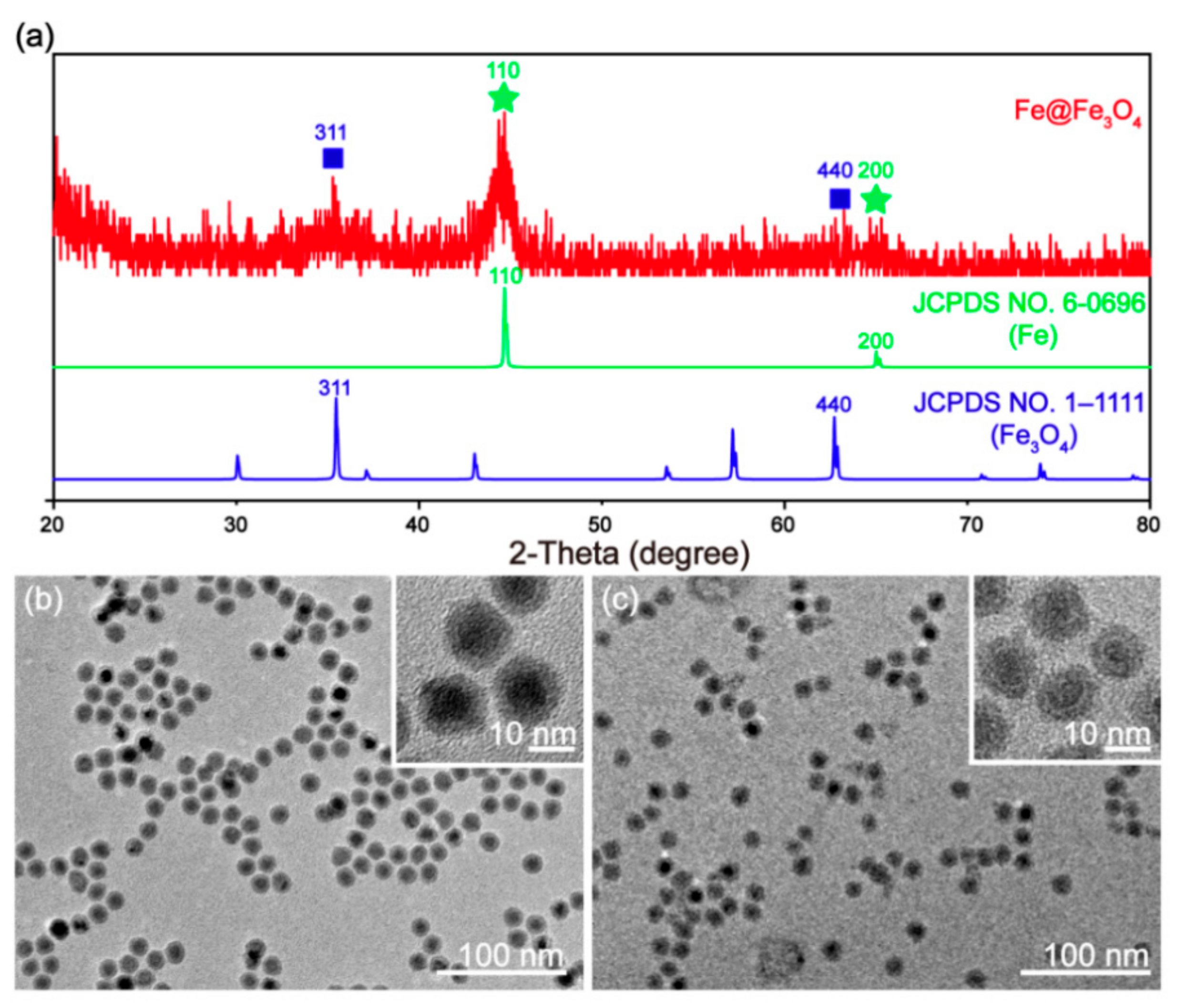
3.1. Synthesis and Characterization of Fe@Fe3O4 Nanoparticles

The hydrophobic Fe-based nanoparticles (Fe@Fe3O4) were prepared following our previous report [31]. In order to improve their biocompatibility, hydrophilic Fe@Fe3O4 nanoparticles were further synthesized by a ligand exchange strategy. Firstly, the crystal structure of the obtained Fe@Fe3O4 nanoparticles was characterized by XRD (Figure 1a). The XRD pattern of the obtained Fe@Fe3O4 exhibited two obvious peaks at 44.6° and 65.1° which matched well with the 110 and 200 crystal face of cubic Fe, respectively (JCPDS no. 6-0696). In addition, there were two weak peaks at 35.3° and 62.7°, which could be assigned to the 311 and 440 crystal face of cubic Fe3O4, respectively (JCPDS no. 1-1111). These results demonstrate that the obtained nanoparticles contained both Fe and Fe3O4 crystals, and that the crystallinity of Fe was good, whereas that of Fe3O4 was relatively poor. Further, the morphology of the obtained hydrophobic and hydrophilic Fe@Fe3O4 nanoparticles was observed by TEM. As shown in Figure 1b, the obtained nanoparticles exhibited a core-shell structure with a core diameter of ~10 nm and a shell thickness of ~2 nm (Figure S1a), which can be more clearly observed in the HR-TEM image (Figure 1b inset). The inner core comprised the Fe nanoparticles, and the shell was the Fe3O4 layer that prevented further oxidation of the Fe core, which was similar to previous results [32]. After ligand exchange (Figure 1c), the morphology and size (Figure S1b) did not change significantly, and the core-shell structure could still be maintained (Figure 1c inset). Due to the presence of DHCA on the surface of Fe@Fe3O4, the hydrodynamic size was slightly higher than TEM, and the zeta potential was negative, which was consistent with previous results (Figure S2) [31]. Moreover, the two strong vibration peaks at 1575 cm−1 and 1404 cm−1 originated from the benzene ring of DHCA ligand, and the typical Fe-O stretching vibration peak appeared at 593 cm−1 (Figure S3), indicating that the DHCA ligands were modified onto the Fe@Fe3O4. The above results demonstrate that the hydrophilic Fe@Fe3O4 nanoparticles were prepared successfully.

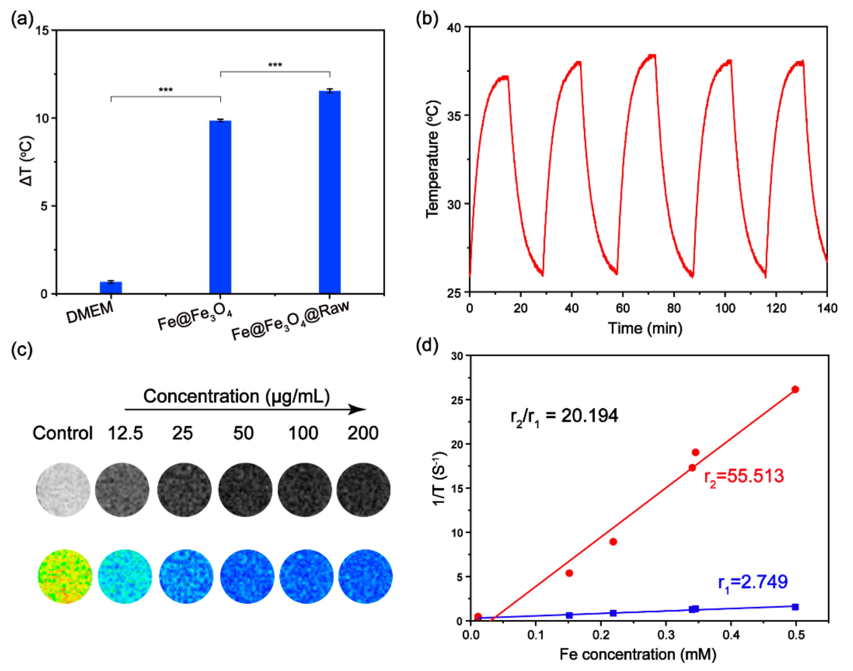
3.2. Photothermal Performance of Fe@Fe3O4 Nanoparticles

Considering that the photothermal performance of Fe@Fe3O4 nanoparticles is related with their absorbance, the absorbance of various concentrations of Fe@Fe3O4 was first explored (Figure S4a). As shown in Figure 2a, with the increasing concentration (50, 100, 150 and 200 μg/mL), the absorbance of Fe@Fe3O4 nanoparticles at 808 nm gradually increased. In addition, compared with the control group (water), the temperature of the Fe@Fe3O4 water dispersion increased dramatically after 808 nm irradiation (1.0 W/cm2), and when the concentration reached 100 μg/mL, the temperatures rose by more than 10 °C (Figure 2b and Figure S4b), which was high enough for photothermal therapy (PTT). Moreover, in order to verify the photothermal stability of Fe@Fe3O4 nanoparticles, the dispersion was irradiated for 15 min, then the irradiation was stopped to allow the sample to cool down to the initial temperature, followed by five cycles of re-irradiation. The heating effect and the maximum reachable temperature (Figure 2c) were essentially the same, indicating the good photothermal stability of the Fe@Fe3O4 nanoparticles. The absorbance of the Fe@Fe3O4 nanoparticles changed only slightly before and after laser irradiation (Figure S5), further demonstrating the good photothermal stability. The calculated photothermal conversion efficiency was 34.3% (Figure 2d), which is higher than typical Au nanorods (~22%) [33,34]. All of the above results demonstrate the good photothermal effect of Fe@Fe3O4 nanoparticles.

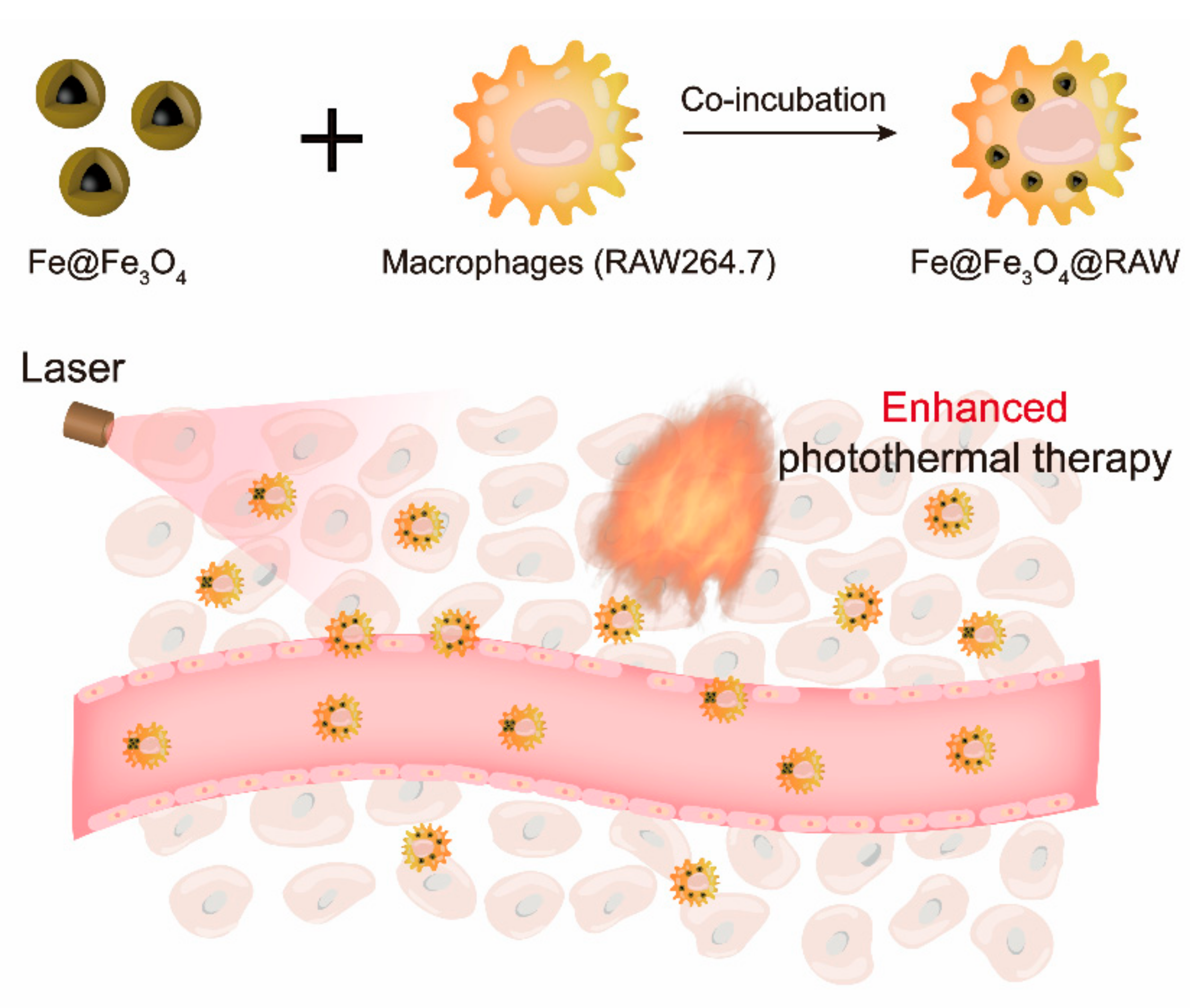
3.3. Macrophages Loaded with Fe Nanoparticles

To explore the imaging and treatment effect of the Fe@Fe3O4 nanoparticles delivered by macrophages, the MRI and photothermal performance in vitro should be explored. Before further investigation, the cytotoxicity of Fe@Fe3O4 nanoparticles to RAW264.7 cells must first be confirmed. As an iron-based material, Fe@Fe3O4 showed little toxicity, even at very high doses (Figure S6). Meanwhile, obvious green fluorescence (living cells) and weak red fluorescence (dead cells) were observed in the confocal images (Figure S7), indicating that Fe@Fe3O4 nanoparticles had good biocompatibility. Based on this, RAW 264.7 phagocytic cells loaded with Fe@Fe3O4 were constructed after 3 h of co-incubation (~717.1 pg/cell), and further irradiated with an 808 nm laser to investigate their photothermal performance (Figure S8). As expected, the macrophages loaded with Fe@Fe3O4 (Fe@Fe3O4@RAW) exhibited a greater increase in temperature than the Fe@Fe3O4 nanoparticles (Figure 3a). In contrast, the temperature of the control group (DMEM) containing cell medium only, barely increased, which ruled out the possibility of DMEM as a solvent interfering with the results. The greater heating effect may have been due to the aggregation of Fe@Fe3O4 in the macrophages, resulting in locally concentrated heating [35]. Compared with previous reports, the macrophage loading strategy exhibited superior photothermal effect at lower laser power, while macrophages are shown to be excellent carriers for transporting Fe@Fe3O4 to tumors [36,37]. Moreover, as for Fe@Fe3O4, after five cycles of laser on and off, the temperature rises of the Fe@Fe3O4@RAW dispersion remained stable (Figure 3b). All these results prove that Fe@Fe3O4@RAW is a potential PTT agent with good photothermal stability. Moreover, iron-based nanomaterials have been reported as magnetic resonance imaging (MRI) contrast agents, thus T2-weighted MRI of Fe@Fe3O4@RAW was investigated at 0.5 T field strength. As shown in Figure 3c, after being phagocytosed by RAW264.7 cells, Fe@Fe3O4@RAW with a lower concentration (0~50 μg/mL) exhibited T2-enhanced contrast enhancement when the concentration was increased. When the concentration was greater than 50 μg/mL, the MR image was no longer darkened, which may have been due to phagocytic saturation of Fe@Fe3O4 by RAW 264.7 cells. The transverse relaxivity (r2) and longitudinal relaxivity (r1) of Fe@Fe3O4@RAW were 55.513 mM−1 s−1 and 2.749 mM−1 s−1, respectively (Figure 3d), which also indicated that it was a good T2 contrast agent. In addition, r2 of different patches of Fe@Fe3O4@RAW with the same Fe concentration was similar (Figure S9), indicating the good reproducibility of RAW loading Fe@Fe3O4. The above results demonstrate that macrophage phagocytosis could enhance the local aggregation of Fe@Fe3O4, thus enhancing the photothermal effect. Moreover, because of its good T2 MRI effect, Fe@Fe3O4@RAW can be used for MRI-mediated photothermal therapy.

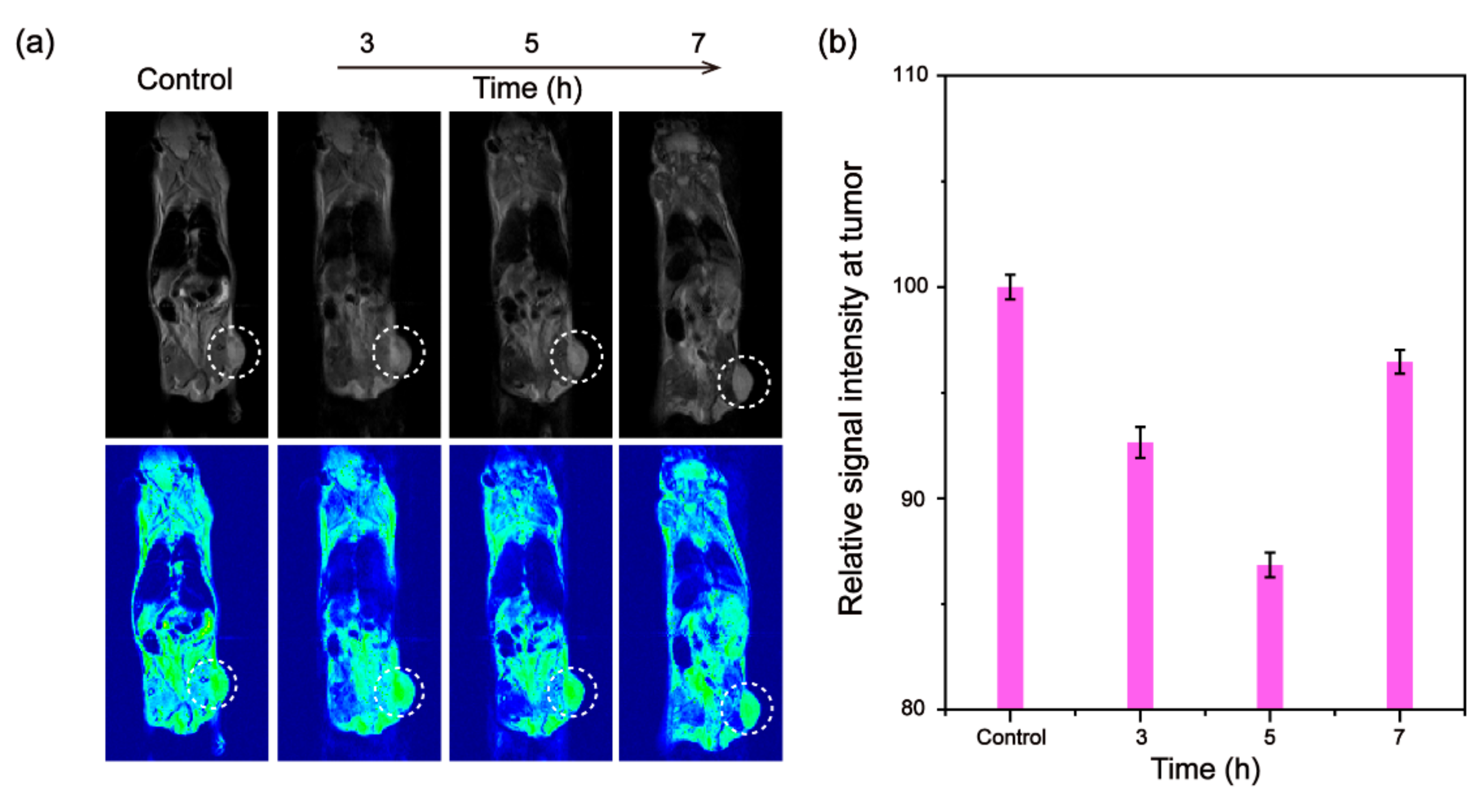
3.4. In Vivo Tumor MRI

Encouraged by the good T2 contrast effect of Fe@Fe3O4@RAW in vitro, the in vivo MRI performance was further explored in 4T1 tumor-bearing mice. As shown in Figure 4a, the tumor site (white circle) was darkened after intravenous injection of Fe@Fe3O4@RAW and continued to darken 5 h later, indicating the good T2-weighted MRI contrast effect in vivo. The brightness gradually restored 7 h following injection, which may have been related to the metabolic character of Fe@Fe3O4@RAW. This suggested that Fe@Fe3O4@RAW can target the tumor and can be metabolized out of the body. The relative signal intensity at the tumor site can better reflect the changing trend of tumor imaging quantitatively (Figure 4b). The SNR% after intravenous injection of Fe@Fe3O4@RAW gradually decreased until 5 h post-injection, then increased at 7 h post-injection, which further demonstrates that Fe@Fe3O4@RAW is a good targeted T2-weighted MRI contrast agent, and a benefit for tumor theranostics.

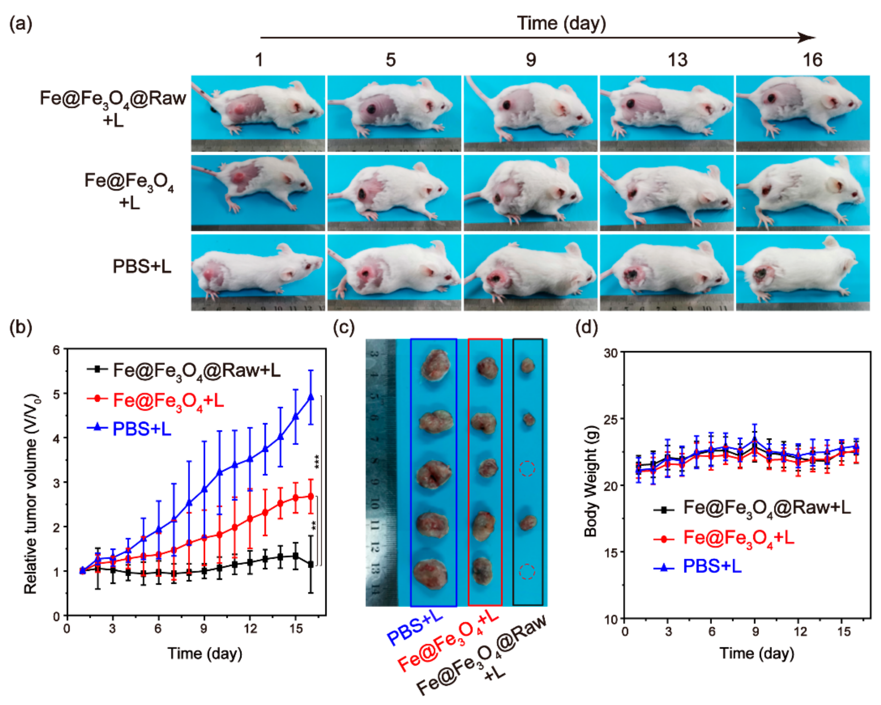
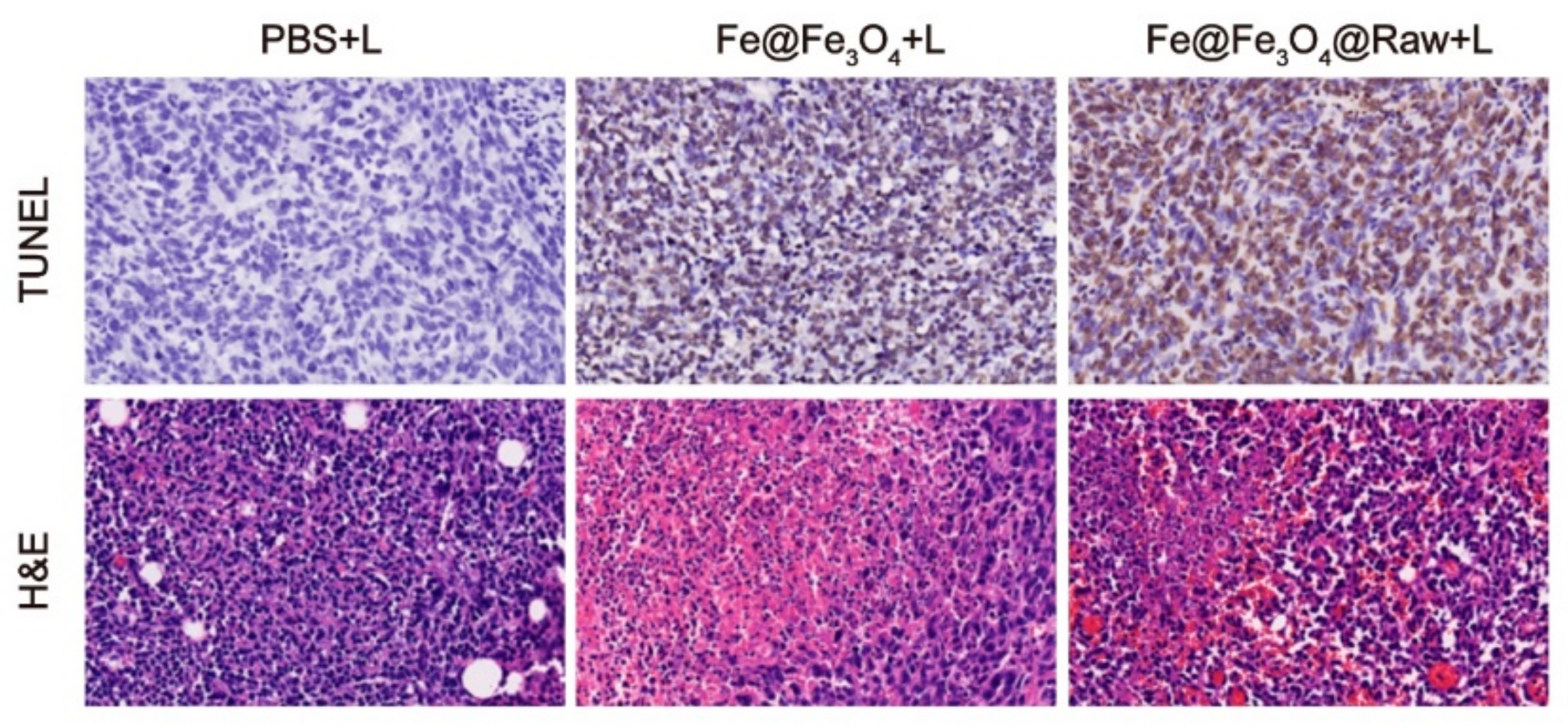
3.5. In Vivo Photothermal Tumor Ablation

As mentioned above, macrophage loading can enhance the photothermal effect of Fe@Fe3O4 in vitro and exhibit tumor-targeting imaging effects, therefore, Fe@Fe3O4@RAW should be used as a tumor-targeting PTT agent. To prove this speculation, 4T1 tumor-bearing mice were randomly divided into three groups, including control, Fe@Fe3O4 and Fe@Fe3O4@RAW groups, each of which was intravenously injected with PBS, Fe@Fe3O4 and Fe@Fe3O4@RAW, respectively. Five hours after injection (the best accumulation time at the tumor site), all groups were irradiated with an 808 nm laser for 10 min. Subsequently, one mouse from each group was sacrificed and the tumor tissues were collected and stained with TdT-mediated Dutp Nick-End Labeling (TUNEL) and hematoxylin-eosin (H&E) for histopathological analysis. As shown in Figure 5, the apoptotic cells (brown) appeared in both the Fe@Fe3O4 and Fe@Fe3O4@RAW groups, among which the apoptotic were more obvious (78.3%) in the Fe@Fe3O4@RAW group, demonstrating the more effective tumor PTT effect of Fe@Fe3O4@RAW. In contrast, after injection with PBS and laser irradiation, most of the cells were normal, indicating the safety of the laser. Similarly, in H&E tumor slices, the morphology of the cells in the Fe@Fe3O4 and Fe@Fe3O4@RAW groups was destroyed, and the nucleus was absent, which further proves that the photothermal effect of Fe@Fe3O4 is better after loading into macrophages.
In addition, the remaining mice from each group were further monitored for 16 days after laser irradiation to evaluate the tumor therapeutic effect. As shown in Figure 6a, after laser irradiation, the tumor sites of the mice in the Fe@Fe3O4 and Fe@Fe3O4@RAW groups were blackened, and the tumors were also ablated. However, the tumors in the Fe@Fe3O4 group began to recur over time, while in the Fe@Fe3O4@RAW group, tumor growth was completely inhibited and the tumor site was scabbed. In comparison, the tumor growth in the PBS group was still fast, suggesting that the laser alone could not kill tumors. Meanwhile, the change of the relative tumor volume in the Fe@Fe3O4@RAW group was almost negligible, while that of the Fe@Fe3O4 group showed a trend of slow growth, and that of the PBS group continued to grow (Figure 6b). Compared with the control group, the dissected tumors in the Fe@Fe3O4 group were smaller after treatment. It is clear that part of the collected tumors from the Fe@Fe3O4@RAW group disappeared, meaning that the tumor was ablated and, although they existed, the tumors of the remaining mice were far smaller than those of the other two groups (Figure 6c). All these results further prove the good tumor inhibition effect of Fe@Fe3O4@RAW. Moreover, the body weight of mice in each group (Figure 6d) remained within a stable range during the treatment period, and no abnormal behavior was observed, which proved that Fe@Fe3O4@RAW and the laser were safe. This evidence indicates that Fe@Fe3O4@RAW is a potential PTT agent with good photothermal properties and low biological toxicity.

4. Conclusions

In summary, macrophages loaded with Fe@Fe3O4 nanoparticles were constructed to enhance the photothermal ablation of tumors. Hydrophilic Fe@Fe3O4 nanoparticles with a core-shell structure were synthesized by a pyrolysis and ligand exchange strategy. After loading into RAW264.7 cells, the local aggregation of Fe@Fe3O4 nanoparticles resulted in an enhancement of the photothermal effect. In addition, Fe@Fe3O4@RAW exhibited a good T2-weighted MRI contrast effect and obvious tumor imaging in vivo due to the tumor targeting tendency of macrophages. More importantly, upon intravenous injection with Fe@Fe3O4@RAW and subjection to laser irradiation, tumor growth was inhibited effectively, indicating that macrophage loading could enhance the tumor photothermal ablation ability of Fe@Fe3O4. This delivery strategy takes advantage of the targeting and high phagocytosis of macrophages to achieve the integration of tumor diagnosis and treatment, and has the potential to be applied to a variety of therapies.

Supplementary Materials

The following supporting information can be downloaded at: https://www.mdpi.com/article/10.3390/jfb13030094/s1, Figure S1: Particle size statistics of Fe@Fe3O4 nanoparticles; Figure S2: DLS and zeta potential of Fe@Fe3O4-DHCA; Figure S3: FTIR spectra of Fe@Fe3O4 nanoparticles, DHCA and Fe@Fe3O4-DHCA; Figure S4: Absorbance and photothermal performance of hydrophilic Fe@Fe3O4 nanoparticles; Figure S5: Absorbance of the Fe@Fe3O4 nanoparticle before and after laser irradiation; Figure S6: The cytotoxicity; Figure S7: The confocal images stained with Calcein-AM/PI; Figure S8: Photothermal performance of DMEM, Fe@Fe3O4 and Fe@Fe3O4 @RAW under the irradiation of an 808 nm laser; Figure S9: The repeatability r2 of Fe@Fe3O4@RAW with same Fe concentration.

Author Contributions

Conceptualization, L.Y. and L.A.; data collecting and investigation, S.Z.; validation, K.Q. and X.F.; writing—original draft preparation, L.Y.; writing—review and editing, L.Y. and L.A. All authors have read and agreed to the published version of the manuscript.

Funding

This work was partially supported by the National Natural Science Foundation of China (22007066).

Institutional Review Board Statement

The animal study protocol was approved by the Ethics Committee of Shanghai Normal University (22007066, January 2020).

Informed Consent Statement

Not applicable.

Data Availability Statement

The data presented are available on request from the corresponding author.

Conflicts of Interest

The authors declare no conflict of interest.

References

  1. Ho, D.; Sun, X.; Sun, S. Monodisperse magnetic nanoparticles for theranostic applications. Acc. Chem. Res. 2011, 44, 875–882. [Google Scholar] [CrossRef] [PubMed] [Green Version]
  2. Wang, J.; Mei, T.; Liu, Y.; Zhang, Y.; Zhang, Z.; Hu, Y.; Wang, Y.; Wu, M.; Yang, C.; Zhong, X.; et al. Dual-targeted and MRI-guided photothermal therapy via iron-based nanoparticles-incorporated neutrophils. Biomater. Sci. 2021, 9, 3968–3978. [Google Scholar] [CrossRef]
  3. Alphandéry, E. Light-interacting iron-based nanomaterials for localized cancer detection and treatment. Acta Biomater. 2021, 124, 50–71. [Google Scholar] [CrossRef] [PubMed]
  4. Rajan, A.; Kaczmarek-Szczepańskac, B.; Sahu, N.K. Magneto-thermal response of Fe3O4@CTAB nanoparticles for cancer hyperthermia applications. Mater. Today Commun. 2021, 28, 102583. [Google Scholar] [CrossRef]
  5. Nicolás-Boluda, A.; Vaquero, J.; Laurent, G.; Renault, G.; Bazzi, R.; Donnadieu, E.; Roux, S.; Fouassier, L.; Gazeau, F. Photothermal depletion of cancer-associated fibroblasts normalizes tumor stiffness in desmoplastic cholangiocarcinoma. ACS Nano 2020, 14, 5738–5753. [Google Scholar] [CrossRef]
  6. Qin, J.; Liu, Q.; Zhang, J.; Chen, J.; Chen, S.; Zhao, Y.; Du, J. Rationally separating the corona and membrane functions of polymer vesicles for enhanced T2 MRI and drug delivery. ACS Appl. Mater. Interfaces 2015, 7, 14043–14052. [Google Scholar] [CrossRef]
  7. Zhou, P.; Zhao, H.; Wang, Q.; Zhou, Z.; Wang, J.; Deng, G.; Wang, X.; Liu, Q.; Yang, H.; Yang, S. Photoacoustic-enabled self-guidance in magnetic-hyperthermia Fe@Fe3O4 nanoparticles for theranostics in vivo. Adv. Healthc. Mater. 2018, 7, 1701201. [Google Scholar] [CrossRef]
  8. Atabaev, T.S. PEG-coated superparamagnetic dysprosium-doped Fe3O4 nanoparticles for potential MRI imaging. BioNanoScience 2018, 8, 299–303. [Google Scholar] [CrossRef]
  9. Nicolas-Boluda, A.; Laurent, G.; Bazzi, R.; Roux, S.; Donnadieu, E.; Gazeau, F. Two step promotion of a hot tumor immune environment by gold decorated iron oxide nanoflowers and light-triggered mild hyperthermia. Nanoscale 2021, 13, 18483–18497. [Google Scholar] [CrossRef]
  10. Zhu, Y.; Fang, Y.; Kaskel, S. Folate-conjugated Fe3O4@SiO2 hollow mesoporous spheres for targeted anticancer drug delivery. J. Phys. Chem. C 2010, 114, 16382–16388. [Google Scholar] [CrossRef]
  11. Cheng, K.; Peng, S.; Xu, C.; Sun, S. Porous hollow Fe3O4 nanoparticles for targeted delivery and controlled release of cisplatin. J. Am. Chem. Soc. 2009, 131, 10637–10644. [Google Scholar] [CrossRef] [PubMed] [Green Version]
  12. Cai, H.; Li, K.; Shen, M.; Wen, S.; Luo, Y.; Peng, C.; Zhang, G.; Shi, X. Facile assembly of Fe3O4@Au nanocomposite particles for dual mode magnetic resonance and computed tomography imaging applications. J. Mater. Chem. 2012, 22, 15110–15120. [Google Scholar] [CrossRef]
  13. Zhang, Y.; Li, X.; Zhang, Y.; Wei, J.; Wang, W.; Dong, C.; Xue, Y.; Liu, M.; Pei, R. Engineered Fe3O4-based nanomaterials for diagnosis and therapy of cancer. New J. Chem. 2021, 45, 7918–7941. [Google Scholar] [CrossRef]
  14. Yu, M.; Zheng, J. Clearance pathways and tumor targeting of imaging nanoparticles. ACS Nano 2015, 9, 6655–6674. [Google Scholar] [CrossRef] [Green Version]
  15. Wang, L.; Huang, J.; Chen, H.; Wu, H.; Xu, Y.; Li, Y.; Yi, H.; Wang, Y.A.; Yang, L.; Mao, H. Exerting enhanced permeability and retention effect driven delivery by ultrafine iron oxide nanoparticles with T1–T2 switchable magnetic resonance imaging contrast. ACS Nano 2017, 11, 4582–4592. [Google Scholar] [CrossRef] [Green Version]
  16. Xia, Y.; Rao, L.; Yao, H.; Wang, Z.; Ning, P.; Chen, X. Engineering macrophages for cancer immunotherapy and drug delivery. Adv. Mater. 2020, 32, 2002054. [Google Scholar]
  17. Lang, T.; Dong, X.; Huang, Y.; Ran, W.; Yin, Q.; Zhang, P.; Zhang, Z.; Yu, H.; Li, Y. Ly6Chi monocytes delivering pH-sensitive micelle loading paclitaxel improve targeting therapy of metastatic breast cancer. Adv. Funct. Mater. 2017, 27, 1701093. [Google Scholar] [CrossRef]
  18. Li, S.; Feng, S.; Ding, L.; Liu, Y.; Zhu, Q.; Qian, Z.; Gu, Y. Nanomedicine engulfed by macrophages for targeted tumor therapy. Int. J. Nanomed. 2016, 11, 4107–4124. [Google Scholar]
  19. Feng, Y.; Liu, Q.; Li, Y.; Han, Y.; Liang, M.; Wang, H.; Yao, Q.; Wang, Y.; Yang, M.; Li, Z.; et al. Cell relay-delivery improves targeting and therapeutic efficacy in tumors. Bioact. Mater. 2021, 6, 1528–1540. [Google Scholar] [CrossRef]
  20. Vercellino, S.; Kokalari, I.; Liz Cantoral, M.; Petseva, V.; Cursi, L.; Casoli, F.; Castagnola, V.; Boselli, L.; Fenoglio, I. Biological interactions of ferromagnetic iron oxide–carbon nanohybrids with alveolar epithelial cells. Biomater. Sci. 2022, 10, 3514–3526. [Google Scholar] [CrossRef]
  21. Choi, J.; Kim, H.-Y.; Ju, E.J.; Jung, J.; Park, J.; Chung, H.-K.; Lee, J.S.; Lee, J.S.; Park, H.J.; Song, S.Y.; et al. Use of macrophages to deliver therapeutic and imaging contrast agents to tumors. Biomaterials 2012, 33, 4195–4203. [Google Scholar] [CrossRef] [PubMed]
  22. Fu, J.; Wang, D.; Mei, D.; Zhang, H.; Wang, Z.; He, B.; Dai, W.; Zhang, H.; Wang, X.; Zhang, Q. Macrophage mediated biomimetic delivery system for the treatment of lung metastasis of breast cancer. J. Control Release 2015, 204, 11–19. [Google Scholar] [CrossRef] [PubMed]
  23. An, L.; Wang, Y.; Lin, J.; Tian, Q.; Xie, Y.; Hu, J.; Yang, S. Macrophages-mediated delivery of small gold nanorods for tumor hypoxia photoacoustic imaging and enhanced photothermal therapy. ACS Appl. Mater. Interfaces 2019, 11, 15251–15261. [Google Scholar] [CrossRef] [PubMed]
  24. Nguyen, V.D.; Min, H.-K.; Kim, H.Y.; Han, J.; Choi, Y.H.; Kim, C.-S.; Park, J.-O.; Choi, E. Primary macrophage-based microrobots: An effective tumor therapy in vivo by dual-targeting function and near-infrared-triggered drug release. ACS Nano 2021, 15, 8492–8506. [Google Scholar] [CrossRef] [PubMed]
  25. Zhang, W.; Wang, M.; Tang, W.; Wen, R.; Zhou, S.; Lee, C.; Wang, H.; Jiang, W.; Delahunty, I.M.; Zhen, Z.; et al. Nanoparticle-laden macrophages for tumor-tropic drug delivery. Adv. Mater. 2018, 30, 1805557. [Google Scholar] [CrossRef] [PubMed]
  26. Madsen, S.J.; Hirschberg, H. Macrophages as delivery vehicles for anticancer agents. Ther. Deliv. 2019, 10, 189–201. [Google Scholar]
  27. Alvarenga, B.M.; Melo, M.N.; Frézard, F.; Demicheli, C.; Gomes, J.M.M.; Borba da Silva, J.B.; Speziali, N.L.; Corrêa Junior, J.D. Nanoparticle phosphate-based composites as vehicles for antimony delivery to macrophages: Possible use in leishmaniasis. J. Mater. Chem. B 2015, 3, 9250–9259. [Google Scholar] [CrossRef]
  28. Burke, B.; Sumner, S.; Maitland, N.; Lewis, C.E. Macrophages in gene therapy: Cellular delivery vehicles and in vivo targets. J. Leukoc. Biol. 2002, 72, 417–428. [Google Scholar]
  29. Li, J.; Cheng, Q.; Yue, L.; Gao, C.; Wei, J.; Ding, Y.; Wang, Y.; Zheng, Y.; Wang, R. Macrophage-hitchhiking supramolecular aggregates of CuS nanoparticles for enhanced tumor deposition and photothermal therapy. Nanoscale Horiz. 2021, 6, 907–912. [Google Scholar] [CrossRef]
  30. Lacroix, L.-M.; Frey Huls, N.; Ho, D.; Sun, X.; Cheng, K.; Sun, S. Stable single-crystalline body centered cubic Fe nanoparticles. Nano Lett. 2011, 11, 1641–1645. [Google Scholar] [CrossRef]
  31. Liu, D.; Li, J.; Wang, C.; An, L.; Lin, J.; Tian, Q.; Yang, S. Ultrasmall Fe@Fe3O4 nanoparticles as T1–T2 dual-mode MRI contrast agents for targeted tumor imaging. Nanomed. NBM 2021, 32, 102335. [Google Scholar] [CrossRef]
  32. Zhou, Z.; Sun, Y.; Shen, J.; Wei, J.; Yu, C.; Kong, B.; Liu, W.; Yang, H.; Yang, S.; Wang, W. Iron/iron oxide core/shell nanoparticles for magnetic targeting MRI and near-infrared photothermal therapy. Biomaterials 2014, 35, 7470–7478. [Google Scholar] [CrossRef]
  33. Chen, H.; Shao, L.; Ming, T.; Sun, Z.; Zhao, C.; Yang, B.; Wang, J. Understanding the photothermal conversion efficiency of gold nanocrystals. Small 2010, 6, 2272–2280. [Google Scholar] [CrossRef] [PubMed]
  34. An, L.; Cao, M.; Zhang, X.; Lin, J.; Tian, Q.; Yang, S. pH and glutathione synergistically triggered release and self-assembly of Au nanospheres for tumor theranostics. ACS Appl. Mater. Interfaces 2020, 12, 8050–8061. [Google Scholar] [CrossRef] [PubMed]
  35. Estelrich, J.; Busquets, M.A. Iron oxide nanoparticles in photothermal therapy. Molecules 2018, 23, 1567. [Google Scholar] [CrossRef] [PubMed] [Green Version]
  36. Guo, X.; Wu, Z.; Li, W.; Wang, Z.; Li, Q.; Kong, F.; Zhang, H.; Zhu, X.; Du, Y.P.; Jin, Y.; et al. Appropriate size of magnetic nanoparticles for various bioapplications in cancer diagnostics and therapy. ACS Appl. Mater. Interfaces 2016, 8, 3092–3106. [Google Scholar] [CrossRef] [PubMed]
  37. Shen, S.; Wang, S.; Zheng, R.; Zhu, X.; Jiang, X.; Fu, D.; Yang, W. Magnetic nanoparticle clusters for photothermal therapy with near-infrared irradiation. Biomaterials 2015, 39, 67–74. [Google Scholar]
Scheme 1. Macrophages loaded with Fe nanoparticles for an enhanced photothermal ablation of tumors.
Jfb 13 00094 sch001
Figure 1. Characterization of Fe@Fe3O4 nanoparticles. (a) XRD pattern of the obtained Fe@Fe3O4 nanoparticles (red line) and the standard pattern of cubic Fe (green line, JCPDS no. 6-0696) and Fe3O4 (blue line, JCPDS no. 1-1111). The stars and the squares indicate the characteristic peaks of Fe and Fe3O4, respectively. TEM and HR-TEM (inset) of the (b) hydrophobic and (c) hydrophilic Fe@Fe3O4 nanoparticles.
Jfb 13 00094 g001
Figure 2. Photothermal performances of the Fe@Fe3O4 nanoparticles. (a) Absorbance at 808 nm of water (control) and Fe@Fe3O4 nanoparticles water dispersions of different concentrations. *** p < 0.001. (b) Increasing temperature of the water (control) and Fe@Fe3O4 nanoparticle water dispersions of different concentrations under 808 nm laser irradiation. (c) Photothermal stability of the Fe@Fe3O4 nanoparticle water dispersion under five cycles of laser on and off. (d) Time constant by applying the linear time data versus the negative natural logarithm of the driving force temperature (black square), which was obtained from (c).
Jfb 13 00094 g002
Figure 3. (a) Temperature changes (ΔT) of DMEM, Fe@Fe3O4 nanoparticles (Fe@Fe3O4) and macrophages loaded with Fe@Fe3O4 nanoparticles (Fe@Fe3O4@Raw) under 808 nm laser irradiation. *** p < 0.001. (b) Photothermal stability of the macrophages loaded with the Fe@Fe3O4 nanoparticle water dispersion under the five cycles of laser on and off. (c) T2-weighted MR images of water (control) and macrophages loaded with Fe@Fe3O4 nanoparticles of different concentrations. (d) Transverse (r2, red line) and longitudinal (r1, blue line) relaxivity of the macrophages loaded with Fe@Fe3O4 nanoparticles.
Jfb 13 00094 g003
Figure 4. In vivo MRI performance. (a) T2-weighted MR images, and (b) corresponding relative signal intensity of tumor, before (control) and after intravenous injection of macrophages loaded with Fe@Fe3O4 nanoparticles at different time points.
Jfb 13 00094 g004
Figure 5. Tumor slice stained by TUNEL and H&E after photothermal therapy.
Jfb 13 00094 g005
Figure 6. Photothermal therapy effects of the PBS, Fe@Fe3O4, and Fe@Fe3O4@RAW groups (n = 5). (a) Photographs of the mice in different groups on different days after photothermal therapy. (b) Relative tumor volume of the mice in the different groups after photothermal therapy (** p < 0.01, *** p < 0.001). (c) Photographs of the dissected tumors 16 days after photothermal therapy. (d) Body weight of the mice in different groups after photothermal therapy.
Jfb 13 00094 g006
Publisher’s Note: MDPI stays neutral with regard to jurisdictional claims in published maps and institutional affiliations.

