Review

Bio-Inspired Metaheuristics in Deep Learning for Brain Tumor Segmentation: A Decade of Advances and Future Directions

1
Faculty of Computer Science, AGH University of Krakow, 30-059 Krakow, Poland
2
Department of Informatics, Universitas Pembangunan Nasional Veteran Yogyakarta, Yogyakarta 55281, Indonesia
3
Artificial Intelligence Research Group (AIRG), Informatics Department, Faculty of Industrial Technology, Universitas Ahmad Dahlan, Yogyakarta 55166, Indonesia
4
Department of Electrical Engineering, Universitas Ahmad Dahlan, Yogyakarta 55166, Indonesia
5
Department of Mechanical and Mechatronics Engineering, Curtin University Malaysia, Miri 98009, Sarawak, Malaysia
6
Institut Teknologi Statistika dan Bisnis Muhammadiyah Semarang, Semarang 50185, Indonesia
*
Author to whom correspondence should be addressed.
Information 2025, 16(6), 456; https://doi.org/10.3390/info16060456
Submission received: 11 May 2025 / Revised: 22 May 2025 / Accepted: 28 May 2025 / Published: 29 May 2025
(This article belongs to the Section Review)

Abstract

Accurate segmentation of brain tumors in magnetic resonance imaging (MRI) remains a challenging task due to heterogeneous tumor structures, varying intensities across modalities, and limited annotated data. Deep learning has significantly advanced segmentation accuracy; however, it often suffers from sensitivity to hyperparameter settings and limited generalization. To overcome these challenges, bio-inspired metaheuristic algorithms have been increasingly employed to optimize various stages of the deep learning pipeline—including hyperparameter tuning, preprocessing, architectural design, and attention modulation. This review systematically examines developments from 2015 to 2025, focusing on the integration of nature-inspired optimization methods such as Particle Swarm Optimization (PSO), Genetic Algorithm (GA), Differential Evolution (DE), Grey Wolf Optimizer (GWO), Whale Optimization Algorithm (WOA), and novel hybrids including CJHBA and BioSwarmNet into deep learning-based brain tumor segmentation frameworks. A structured multi-query search strategy was executed using Publish or Perish across Google Scholar and Scopus databases. Following PRISMA guidelines, 3895 records were screened through automated filtering and manual eligibility checks, yielding a curated set of 106 primary studies. Through bibliometric mapping, methodological synthesis, and performance analysis, we highlight trends in algorithm usage, application domains (e.g., preprocessing, architecture search), and segmentation outcomes measured by metrics such as Dice Similarity Coefficient (DSC), Jaccard Index (JI), Hausdorff Distance (HD), and ASSD. Our findings demonstrate that bio-inspired optimization significantly enhances segmentation accuracy and robustness, particularly in multimodal settings involving FLAIR and T1CE modalities. The review concludes by identifying emerging research directions in hybrid optimization, real-time clinical applicability, and explainable AI, providing a roadmap for future exploration in this interdisciplinary domain.

1. Introduction

Brain tumors are among the most devastating central nervous system diseases, often exhibiting high mortality and recurrence rates [1,2]. The early and accurate detection of brain tumors is essential for guiding neurosurgical intervention, radiotherapy planning, and long-term prognosis [3]. Magnetic resonance imaging (MRI) remains the gold standard in clinical brain tumor assessment due to its ability to non-invasively capture anatomical and pathological features with high contrast [2,4,5]. However, manual delineation of tumor subregions—including edema, necrotic core, and enhancing tumor—is laborious, time-consuming, and highly variable between experts [6,7]. Consequently, automated brain tumor segmentation has emerged as a vital research area in medical imaging and computational neuroscience [8].
Despite the success of deep learning models—particularly encoder–decoder architectures such as U-Net and its variants—in producing high-quality segmentations, multiple challenges hinder optimal performance [9,10]. Firstly, heterogeneity in tumor appearance across patients, imaging modalities, and scanners introduces significant variability [11]. For instance, gliomas exhibit infiltrative patterns and irregular borders, making them difficult to distinguish from healthy tissue. Secondly, segmentation is modality-dependent—different MRI sequences provide complementary information. T1-weighted (T1) images highlight anatomical structure, T1 contrast-enhanced (T1CE or T1Gd) images enhance tumor core regions, T2 images reveal fluid-containing tissues like edema, and FLAIR images are sensitive to abnormalities in peritumoral areas [12]. The presence of multiple subregions within tumors (e.g., necrotic, enhancing, and edematous) requires models that can integrate multi-modal MRI data, each with unique signal characteristics and noise patterns [13,14].
Moreover, deep learning models are sensitive to hyperparameter configurations, including learning rate, dropout, filter size, number of layers, and optimizer settings [15,16]. Suboptimal parameter selection may lead to training instability, poor convergence, or overfitting—particularly when datasets are small or imbalanced. Manual tuning of these parameters is inefficient, computationally expensive, and does not guarantee optimality across different datasets [17,18,19].
To address this, researchers have increasingly turned to bio-inspired metaheuristic optimization algorithms, which simulate natural and biological behaviors to explore high-dimensional search spaces efficiently [20]. These include Particle Swarm Optimization (PSO) [21,22,23,24], inspired by bird flocking; Genetic Algorithms (GA) [25,26,27], based on evolutionary principles; Differential Evolution (DE) [28,29,30], which uses differential mutation strategies; and Ant Colony Optimization (ACO) [31,32], modeled after pheromone-guided ant behavior [33]. Such algorithms are adept at hyperparameter tuning [15], architectural optimization [16], and data preprocessing enhancement [22,34], thereby improving both segmentation accuracy and model generalizability.
Tuba et al. (2019) [35] used PSO to optimize learning parameters in CNNs for tumor classification. Yadav et al. (2025) [36] proposed a recurrent residual U-Net for brain tumor segmentation but acknowledged the need for optimization in network depth and regularization. Saifullah et al. (2025) [34] demonstrated the effectiveness of PSO-optimized histogram equalization in preprocessing, yielding improved Dice scores on multi-modal MRI datasets. Additionally, Gao et al. (2021) applied GA for neural architecture search, enhancing segmentation performance in small-sample scenarios. These works suggest that the integration of metaheuristics can address critical bottlenecks in the segmentation pipeline, including overfitting, instability, and the curse of dimensionality.
Furthermore, newer hybrid methods—such as BioSwarmNet [37], HybWWoA [38], TAO [39], and CJHBA [40]—have emerged, combining the strengths of multiple optimization strategies. These have shown promising results not only in classification but also in segmentation tasks, particularly when paired with attention modules or transformer-based backbones. Yet, there remains a lack of comprehensive analysis on how these techniques compare with each other, when they should be used, and what challenges they effectively address in brain tumor segmentation. While some studies report high accuracy with specific algorithms like PSO or GA, others show varying performance due to differences in modality, dataset size, or optimization objectives. These inconsistencies underscore the need for a comparative, structured review that addresses practical applicability and algorithm suitability across different scenarios.
In response to these gaps, this review aims to systematically assess the state of the art in bio-inspired metaheuristic algorithms applied to deep learning models for brain tumor segmentation. Specifically, it evaluates methods developed from 2015 to 2025, identifying both established and emerging optimization strategies, such as Particle Swarm Optimization (PSO), Genetic Algorithm (GA), Differential Evolution (DE), Grey Wolf Optimization (GWO), Whale Optimization Algorithm (WOA), Ant Colony Optimization (ACO), and hybrid variants. The review is centered on deep learning segmentation tasks involving multimodal MRI data–T1 (T1-weighted), T1CE (T1 contrast-enhanced), T2 (T2-weighted), and FLAIR (Fluid-Attenuated Inversion Recovery), with an emphasis on optimization of preprocessing, network architecture, and training parameters.
The main contributions of this review include the following:
  • A systematic literature collection and screening based on PRISMA criteria;
  • A comparative evaluation of optimization methods in terms of segmentation metrics (e.g., Dice Similarity Coefficient, Jaccard Index, Hausdorff Distance);
  • An exploration of algorithmic integration roles—from hyperparameter tuning to architecture search;
  • A discussion on the limitations and future challenges, including generalization, interpretability, and clinical adaptation;
  • A forward-looking synthesis on trends such as explainable AI, transformer optimization, and ensemble metaheuristics.
The remainder of this paper is structured as follows. Section 2 details the search methodology and inclusion/exclusion criteria used for article selection. Section 3 presents a comprehensive review of the evolution of bio-inspired metaheuristic algorithms in brain tumor segmentation, including bibliometric analysis, performance evaluation, and methodological classification. Section 4 discusses key challenges, emerging trends, and future research directions. Finally, Section 5 concludes the paper with a summary of findings and practical implications for researchers and clinicians.

2. Search Methodology and Screening Strategy

To ensure a systematic, transparent, and reproducible identification of relevant literature, we followed the PRISMA (Preferred Reporting Items for Systematic Reviews and Meta-Analyses) guidelines [41]. Our review focuses on peer-reviewed studies published between 2015 and 2025 that apply bio-inspired metaheuristic optimization algorithms to deep learning-based brain tumor segmentation tasks. The methodology encompasses data source selection, keyword-based querying, duplicate removal, relevance screening, and final eligibility assessment.

2.1. Data Sources and Query Design

To conduct a comprehensive and methodologically rigorous review of the literature on bio-inspired metaheuristic optimization in deep learning for brain tumor segmentation, a structured multi-query search strategy was employed. The objective was to systematically identify, retrieve, and evaluate peer-reviewed publications that explore the application of nature-inspired algorithms to enhance neural network-based brain tumor segmentation over the last decade (2015–2025). Given the inherently interdisciplinary nature of this research—spanning medical imaging, neural computation, and evolutionary optimization—particular attention was paid to the formulation of precise keywords, the selection of authoritative academic databases, and the inclusion of both foundational studies and recent advancements.
The search was executed using Publish or Perish (PoP) software (version 8), a bibliometric retrieval tool capable of extracting scholarly records from various academic databases. For this review, two principal sources were utilized: Google Scholar and Scopus. Google Scholar was selected for its broad indexing coverage, encompassing preprints, theses, conference papers, and technical reports—many of which are not indexed in traditional databases. In contrast, Scopus offers a well-curated collection of high-quality journal articles and conference proceedings, enriched by detailed citation metadata. The combination of both platforms ensured a balanced inclusion of emerging and peer-reviewed works while mitigating database-specific biases.
Due to the 250-character query constraint in Google Scholar searches through PoP, the entire search strategy was divided into five thematically structured Boolean queries. These queries were designed to target specific groups of bio-inspired metaheuristics, model architectures, and optimization approaches—ensuring a focused and exhaustive exploration of the literature related to deep learning-based brain tumor segmentation.
The Boolean queries were structured as follows:
  • Query 1: “brain tumor segmentation” AND (“PSO” OR “particle swarm optimization” OR “GA” OR “genetic algorithm”) AND (“deep learning” OR “U-Net” OR “CNN”).
  • Query 2: “brain tumor segmentation” AND (“differential evolution” OR “DE” OR “ACO” OR “ant colony optimization” OR “ABC”) AND (“deep learning” OR “U-Net”).
  • Query 3: “brain tumor segmentation” AND (“GWO” OR “grey wolf optimizer” OR “WOA” OR “whale optimization” OR “HHO” OR “SIO”) AND (“CNN” OR “U-Net”).
  • Query 4: “brain tumor segmentation” AND (“hybrid metaheuristic” OR “neuroevolution” OR “bio-inspired optimization”) AND (“deep learning” OR “transformer”).
  • Query 5: “brain tumor segmentation” AND (“metaheuristic” OR “bio-inspired algorithm”) AND (“deep learning” OR “CNN” OR “U-Net”).
Each query was formulated with a clear rationale:
  • Query 1 focused on two of the most widely adopted metaheuristic algorithms—Particle Swarm Optimization (PSO) and Genetic Algorithm (GA)—commonly applied in hyperparameter tuning and architecture refinement of convolutional neural networks;
  • Query 2 extended the coverage to include Differential Evolution (DE), Ant Colony Optimization (ACO), and Artificial Bee Colony (ABC)—noted for their application in feature selection, image enhancement, and adaptive control mechanisms;
  • Query 3 emphasized more recent and biologically inspired algorithms such as Grey Wolf Optimizer (GWO), Whale Optimization Algorithm (WOA), Harris Hawks Optimization (HHO), and Swan-Inspired Optimization (SIO)—many of which have gained traction in the past five years;
  • Query 4 targeted hybrid metaheuristics and neuroevolutionary strategies, particularly those integrated with transformer-based or attention-guided architectures, which require more dynamic and synergistic optimization techniques;
  • Query 5 served as a general search filter to capture publications referring to broader terms like “metaheuristic” or “bio-inspired algorithm”, ensuring coverage of novel or unnamed optimization strategies.
All queries were restricted to publications from 2015 to 2025 and limited to English-language sources indexed as peer-reviewed journal articles or conference proceedings. The exported bibliographic records included metadata such as article titles, abstracts, authorship, publication year, source venue, and citation metrics.
Following data collection, a multi-stage refinement and screening process was conducted. Duplicate entries were eliminated using normalized titles. Articles unrelated to the core themes of deep learning, bio-inspired optimization, or brain tumor segmentation were manually excluded through title and abstract screening. Special care was taken to detect false positives—particularly entries referencing general optimization frameworks without practical application to medical imaging.
The curated dataset was subsequently used for bibliometric mapping using VOSviewer and a qualitative synthesis of metaheuristic optimization strategies, segmentation architectures, dataset modalities, and performance evaluations, which are detailed in the following sections.
In summary, this search strategy was meticulously constructed to ensure a comprehensive, focused, and reproducible collection of high-quality literature. It offers a balanced view of both established methods and emerging trends in the application of bio-inspired metaheuristics for optimizing deep learning models in the context of brain tumor segmentation.

2.2. Inclusion and Exclusion Criteria

To ensure the scientific rigor, relevance, and reproducibility of this systematic review, a comprehensive set of inclusion and exclusion criteria was defined prior to the screening process. These criteria guided both the automated filtering and the subsequent manual evaluation of articles retrieved through the multi-query search strategy. The objective was to isolate studies that addressed brain tumor segmentation using deep learning and make meaningful contributions through the application of bio-inspired metaheuristic optimization techniques. This dual focus—on medical imaging and evolutionary computation—required a nuanced screening approach, especially given the growing overlap and ambiguity in interdisciplinary research.

2.2.1. Inclusion Criteria

Studies were eligible for inclusion if they satisfied several tightly defined conditions. First and foremost, the article must have demonstrated topical relevance by explicitly addressing brain tumor segmentation, either in the title or abstract. Moreover, the segmentation task had to be conducted using deep learning models, such as U-Net, convolutional neural networks (CNNs), or transformer-based frameworks. This ensured that all included works were grounded in modern AI-driven segmentation methods rather than traditional or rule-based systems.
A second critical inclusion condition was the use of metaheuristic or bio-inspired optimization algorithms. Eligible studies had to incorporate at least one nature-inspired technique—such as PSO, GA, DE, ACO, GWO, WOA, HHO, SIO, or a custom hybrid approach. These algorithms could be applied across a range of computational tasks relevant to the segmentation pipeline, including hyperparameter tuning, network architecture optimization, image preprocessing or contrast enhancement, and feature selection or thresholding.
To ensure clinical and anatomical relevance, only studies using brain MRI modalities were considered. Specifically, the article must have utilized one or more of the following standard MRI sequences: FLAIR (Fluid-Attenuated Inversion Recovery), T1-weighted, T1 contrast-enhanced (T1CE or T1Gd), and T2-weighted images. These modalities are widely recognized for their complementary roles in identifying various tumor subregions such as necrotic tissue, edema, and enhancing tumor cores. In terms of source credibility, only peer-reviewed journal articles or conference papers were accepted, and all publications had to be written in English. The review was also temporally bounded to focus on recent advances by restricting eligible publication dates to those between January 2015 and April 2025.

2.2.2. Exclusion Criteria

A parallel set of exclusion rules was applied to systematically eliminate non-relevant or low-quality records. Articles were excluded if they exhibited thematic mismatch, such as focusing on other anatomical sites (e.g., breast, lung) or on generic medical imaging topics without reference to brain tumor segmentation. Additionally, studies that did not employ deep learning approaches were filtered out; this included works that used only classical machine learning algorithms or conventional image processing techniques without any integration of neural networks.
Most critically, any study that did not employ or analyze bio-inspired metaheuristic optimization was excluded. These were identified during manual review and labeled in the screening template under the column Reason_for_Exclusion with the note “Not related to bio-inspired metaheuristic optimization”. This step was essential to align the final dataset with the core aim of this review, which is to synthesize the impact and evolution of bio-inspired algorithms within deep learning-based brain tumor segmentation.
Further exclusions targeted non-primary research content. Articles categorized as reviews, surveys, meta-analyses, or comprehensive overviews were systematically removed, based on keywords such as “review”, “survey”, “comprehensive analysis”, and “overview” appearing in the title. This was done to avoid redundancy with existing literature summaries and to ensure the review focuses on original experimental contributions. Likewise, articles retrieved from non-academic platforms such as arXiv.org, ResearchGate, and Academia.edu were discarded, as these sources typically lack formal peer review. Any retracted publications were also excluded, identified through metadata tags or document type fields. Entries with missing essential metadata, such as publication year or publisher name, were filtered out during the preprocessing stage.

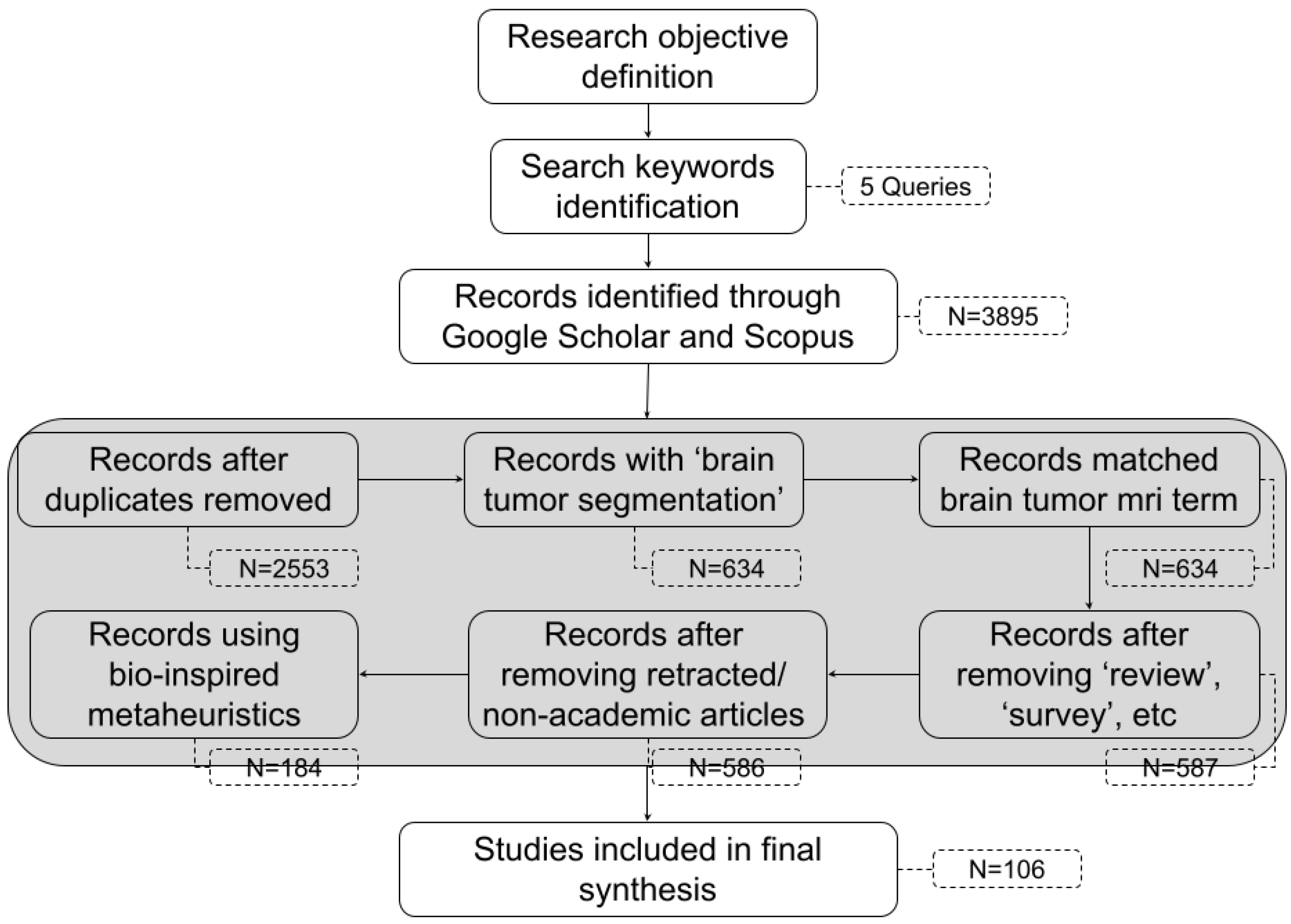
2.2.3. Screening Procedure

The overall screening was conducted in a two-phase process. First, a series of automated preprocessing scripts was executed using Python 2.19.0 to normalize text fields, remove duplicates, exclude known non-academic sources, and apply rule-based pattern filters. This was followed by a manual screening phase, where human reviewers assessed the remaining records by title and abstract. Articles were marked for exclusion if they failed to demonstrate a methodological connection to bio-inspired optimization techniques. Those records were explicitly annotated as “Not related to bio-inspired metaheuristic optimization” to support transparent filtering.
Finally, the remaining candidate studies were evaluated for full-text availability and alignment with the inclusion criteria. Through this multi-stage screening framework, the review arrived at a final curated set of 106 articles, all of which directly contribute to the understanding of how selected bio-inspired metaheuristics algorithms enhance deep learning-based brain tumor segmentation. These articles serve as the basis for the bibliometric mapping, thematic classification, and comparative analysis presented in the subsequent sections of this review.

2.3. Study Selection Process

To ensure methodological rigor and transparency, the selection of studies in this review followed a multi-stage process aligned with the PRISMA (Preferred Reporting Items for Systematic Reviews and Meta-Analyses) guidelines. The procedure was designed to systematically narrow down a large corpus of potentially relevant articles to a curated collection of high-quality studies specifically addressing bio-inspired metaheuristic optimization in deep learning-based brain tumor segmentation.
The initial dataset was compiled from five distinct Boolean-based search queries run across two comprehensive scholarly databases—Google Scholar and Scopus—using Publish or Perish (version 8). These queries were designed to maximize coverage of relevant literature while minimizing irrelevant results by focusing on keywords related to brain tumor segmentation, deep learning models, and bio-inspired optimization algorithms. This yielded a total of 3895 records across all search combinations and sources.
In the first stage of data preprocessing, duplicate entries were removed based on normalized titles. This step reduced the dataset to 2553 unique records, which then underwent automatic filtering to identify studies that explicitly included the term “brain tumor segmentation” in the title. This specific requirement ensured that the central research focus of each article aligned with the scope of this review. After this step, 634 studies remained.
To refine the relevance of included articles further, these 634 records were subjected to additional content-based filtering. Each study was evaluated for the presence of MRI-related terminology (e.g., “FLAIR”, “T1”, “T2”, “T1CE”, “T1Gd”, or “brain MRI”) in the title or abstract. Only articles that mentioned at least one of these terms were retained, reinforcing the focus on segmentation tasks using multimodal brain MRI data. This step preserved all 634 articles.
Next, studies were excluded if their titles contained general review-related terms such as “review”, “survey”, “comprehensive analysis”, or “overview”. These articles, while useful for context, were removed to avoid redundancy and maintain focus on original experimental contributions. After this stage, 587 records remained.
To further improve the scientific rigor of the dataset, retracted papers and publications from non-academic or preprint platforms (such as arXiv.org, ResearchGate, or Academia.edu) were removed. Records with missing metadata (e.g., publication year or source) were also excluded. This refinement step resulted in 586 records.
Finally, to isolate the body of literature specific to bio-inspired metaheuristic methods, only those studies explicitly referencing one or more nature-inspired optimization algorithms—such as PSO, GA, DE, ACO, GWO, WOA, HHO, or other hybrid/metaheuristic terms—within the title or abstract were retained. This final keyword-based inclusion step yielded 184 candidate studies.
During the manual review of abstracts and full texts, 78 articles were marked as not directly involving bio-inspired metaheuristics, often due to ambiguity or incorrect indexing. These were excluded as they did not directly involve bio-inspired metaheuristic optimization, despite initial keyword matches. Ultimately, 106 studies met all criteria and were included in the final review.
This stepwise refinement is visualized in Figure 1, which presents a PRISMA-compliant flow diagram summarizing the article screening and selection process from initial retrieval to final inclusion.
The resulting 106 articles formed the basis for bibliometric mapping, trend synthesis, and qualitative analysis. A detailed, step-by-step breakdown of the entire filtering and exclusion process—including query-specific retrieval, deduplication, topic refinement, and manual eligibility checks—is presented in Appendix A. This appendix provides a tabular summary of the article counts at each critical stage of screening, supporting the reproducibility and transparency of the review.

3. Results and Analysis

This section presents a comprehensive analysis of the selected studies, focusing on bibliometric trends, methodological approaches, and performance evaluation of bio-inspired metaheuristic algorithms applied to brain tumor segmentation using deep learning. The results are structured to reflect both quantitative trends and qualitative insights. First, a bibliometric mapping is conducted to explore the evolution of publications over time, identify active contributors, and highlight the most influential sources. Next, an in-depth review of the adopted metaheuristic strategies, segmentation architectures, and optimization targets is provided. The evaluation metrics used across studies are examined to assess the consistency and rigor of validation protocols. Finally, the section discusses observed research gaps and emerging directions that inform future developments in this interdisciplinary domain.

3.1. Bibliometric Analysis

To assess the structural dynamics, intellectual patterns, and publishing landscape in the domain of bio-inspired metaheuristic optimization for brain tumor segmentation, a comprehensive bibliometric evaluation was performed. This included temporal analysis of publication trends, identification of leading journals and venues, exploration of collaborative author networks, and co-occurrence patterns of technical keywords.

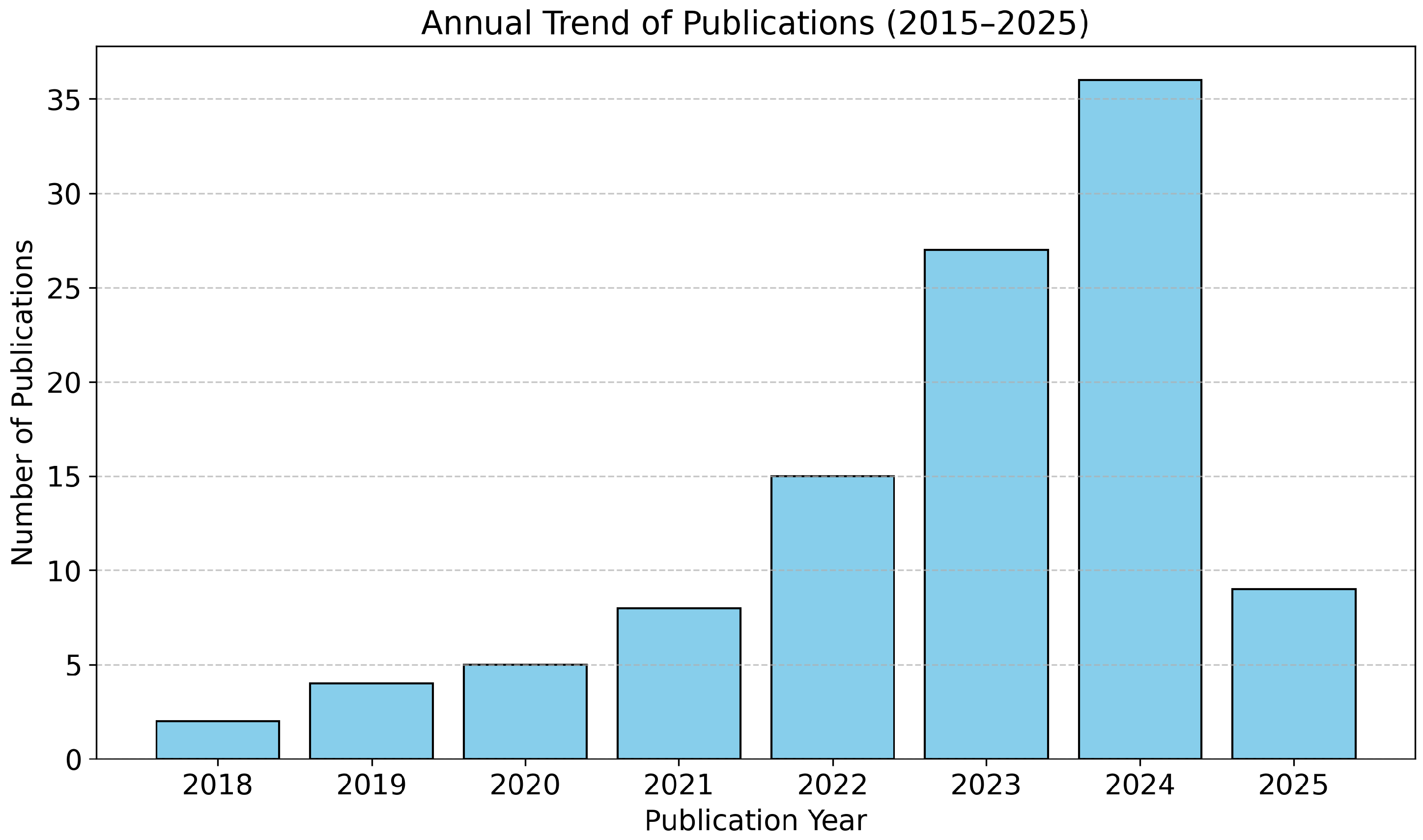
3.1.1. Publication Trend over Time

Figure 2 depicts the temporal evolution of relevant publications between 2015 and 2025. The early phase of research activity (2015–2020) reflects initial experimentation and feasibility studies that combined deep learning models with nature-inspired optimization techniques. A substantial growth trajectory is observed beginning in 2021, indicating increasing recognition of the method’s effectiveness in segmentation accuracy and robustness. Notably, publication frequency peaks in 2024 with over 30% of all included studies published within that year alone. This surge aligns with recent developments in hybrid model architectures and the release of enriched MRI datasets such as BraTS and FBTS, fostering comparative evaluations and benchmarking.

3.1.2. Leading Publication Venues

Figure 3 lists the top ten publishing sources contributing to the selected literature. The journal Multimedia Tools and Applications leads with the highest number of publications, reflecting its broad scope in multimedia analysis, medical imaging, and soft computing techniques. IEEE Access and IEEE Transactions follow closely, signaling growing interest in the integration of AI and biomedical engineering within IEEE circles. Other journals, including Neural Computing and Applications and International Journal of Imaging Systems and Technology, provide fertile ground for interdisciplinary research that bridges selected bio-inspired metaheuristics and neuroimaging. The distribution suggests a diversification of publication venues, accommodating studies with varying emphases on methodological innovation, application development, and clinical interpretation.

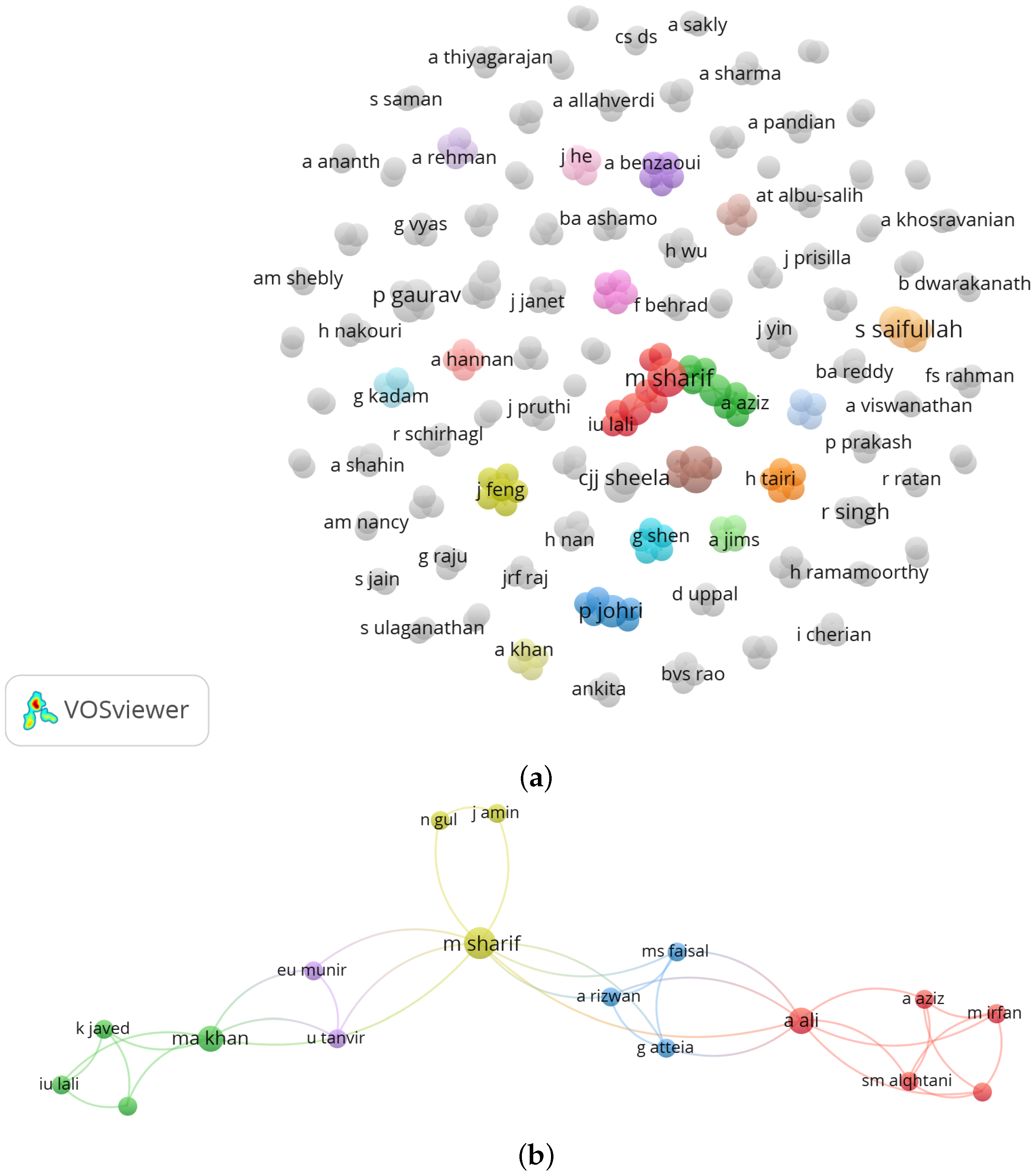
3.1.3. Author Collaboration Networks

To uncover knowledge communities and research clusters, a co-authorship network was constructed using VOSviewer. As illustrated in Figure 4, subfigure (a) displays the author cluster distribution, revealing prominent contributors such as m sharif, p johri, and s saifullah. These clusters delineate thematic subfields, such as hybrid metaheuristics, transformer-based architectures, and comparative segmentation studies. Subfigure (b) illustrates the collaboration strengths and cross-institutional links between authors. Central figures like a ali and ma khan appear as influential bridges, fostering multi-author, multi-domain research that enriches methodological diversity and application scale.

3.1.4. Keyword Co-Occurrence Structure

Keyword co-occurrence analysis in Figure 5 exposes the thematic landscape and semantic density of the selected corpus. Core terms such as brain tumor segmentation, deep learning, and optimization dominate the network, serving as central anchors around which secondary methodological clusters form a pattern consistent with co-word analysis frameworks that map research trends through lexical linkages [42]. Specific techniques like PSO [15,16,24,43,44], ACO [27,31], GWO [45,46,47], and WOA [48,49] form discrete hubs, each connected to subfields involving image enhancement, encoder–decoder designs, and multi-objective optimization [15,34,50]. Terms such as attention mechanism [51,52,53], transformer [54,55,56], and metaheuristic algorithm [15,57] appear in peripheral clusters [58], indicating emerging innovations that are gaining scholarly traction.
The bibliometric patterns outlined in this section offer insight into the trajectory, distribution, and thematic orientation of research within this interdisciplinary field. The increasing publication trend, expanding author collaborations, and methodological diversification point to a maturing discipline that continues to evolve in both breadth and technical sophistication.

3.2. Temporal and Geographic Trends

Understanding the evolution and geographical dispersion of research on bio-inspired metaheuristics in brain tumor segmentation offers valuable insight into the maturity and global engagement with this domain. This subsection explores two critical dimensions: temporal publication patterns and geographic distribution inferred from journal publishing sources. Together, these perspectives help contextualize the field’s development, scholarly dissemination, and regional contributions, despite certain metadata limitations.

3.2.1. Temporal Trends in Research Activity

The timeline of research publications from 2015 to 2025 reveals a distinct pattern of increasing interest and scholarly output in the integration of nature-inspired optimization methods with deep learning models for brain tumor segmentation. As illustrated in Figure 2, early contributions between 2015 and 2018 were limited, typically focusing on proof-of-concept models combining basic convolutional neural networks (CNNs) with optimization algorithms such as PSO or GA for tumor classification or localization [59,60,61]. A gradual increase occurred between 2019 and 2020, coinciding with growing access to publicly available datasets such as BraTS [62,63] and the emergence of lightweight segmentation architectures like U-Net [64].
A notable inflection point appears from 2021 onward, where a substantial surge in publications is evident. This growth aligns with the proliferation of hybrid metaheuristic models [49,65], advancements in transformer-based encoders [54,66,67], and the adoption of multimodal MRI fusion strategies [68]. The peak observed in 2024, comprising over 30% of the total dataset, may reflect not only methodological innovations but also intensified interdisciplinary collaborations and the emergence of specialized optimization techniques designed for biomedical imaging contexts. This trend confirms the rising recognition of bio-inspired optimization as a critical enabler in achieving robust and interpretable segmentation performance [69,70].

3.2.2. Geographic Trends via Journal Publishing Sources

While author affiliation data were not consistently available in the dataset, an alternative approach was adopted to estimate geographic distribution by analyzing the country of origin of the publishing journals. Figure 6 presents a breakdown of the selected studies by the publisher’s headquarters. This proxy method provides a reasonable approximation of regional engagement, particularly in the absence of complete author metadata.
The analysis indicates that the majority of selected papers were published in journals headquartered in the United States, led by IEEE Access and other IEEE Transactions series, reflecting a strong emphasis on engineering-driven biomedical solutions. European publishers such as Springer (Germany) and Elsevier (The Netherlands) also demonstrate a significant presence, representing robust academic platforms that cater to interdisciplinary research. Notably, Asian publishers, particularly those based in China and India, have also emerged with a growing number of conference contributions and journal articles, suggesting increasing regional research activity in metaheuristic optimization and medical AI [71,72,73,74,75,76].
This geographic dispersion implies a widespread global interest in the topic, spanning established hubs of computational medicine as well as emerging centers of innovation. Moreover, the diversity of publishing sources reflects a convergence of fields including artificial intelligence, medical informatics, computer vision, and optimization theory [77,78].
While author affiliation would provide a more direct indication of geographic research origin, this information was not consistently extractable from the bibliographic metadata obtained via Google Scholar through Publish or Perish (PoP). Many records lacked standardized affiliation fields or presented unstructured text that made large-scale parsing unreliable. Therefore, as a proxy, we used the headquarters of the publishing journal to estimate geographic distribution, which—while imperfect—offered a uniform basis for regional comparison.
We fully acknowledge the limitations of this method and recommend that future bibliometric analyses integrate structured datasets from platforms such as CrossRef, Dimensions, or OpenAlex, which support more detailed author-level metadata including institutional affiliation and country. This would allow for finer-grained mapping of global research efforts and institutional collaboration in bio-inspired brain tumor segmentation studies.

3.2.3. Limitations and Considerations

The lack of standardized author affiliation data constrained a more granular geographic analysis, such as identifying national research clusters or institutional productivity. This limitation is common in metadata harvested from sources like Google Scholar or manually parsed CSV files. Future reviews may benefit from integrating enriched bibliographic datasets from CrossRef, OpenAlex, or Dimensions, which offer structured affiliation fields and persistent identifiers. Despite this constraint, the combined temporal and publisher-based analysis presented here offers a valid and insightful approximation of the scholarly ecosystem shaping this research field.
Together, the temporal growth and geographic diversification signal that the application of bio-inspired metaheuristics in deep learning-based brain tumor segmentation is not only expanding in scope and complexity but also gaining traction as a critical area of global scientific inquiry.

3.3. Metaheuristic Approaches and Application Domains

The integration of bio-inspired metaheuristics in deep learning-based brain tumor segmentation models has witnessed significant innovation over the past decade [79,80]. This section synthesizes the diverse metaheuristic strategies identified in the selected literature, categorizing them based on their algorithmic foundations, roles in the segmentation pipeline, and performance contributions. The analysis also highlights the domains within the pipeline—such as hyperparameter tuning, image preprocessing, and architecture adaptation—where these algorithms have demonstrated notable success [81,82,83].

3.3.1. Underlying Mechanisms of Metaheuristics

This section outlines the core mechanics behind widely used bio-inspired metaheuristic algorithms:
  • PSO: Agents (particles) move through a solution space, updating velocity and position based on local and global best experiences—ideal for fast, low-cost optimization.
  • GA: Population-based method using selection, crossover, and mutation to evolve solutions—effective in discovering optimal architectures and hyperparameters.
  • DE: Relies on mutation from difference vectors of population candidates, offering robust exploration and convergence, particularly in continuous domains.
  • ACO: Inspired by ant foraging behavior and pheromone trails—best used for thresholding or path selection in segmentation maps.
  • GWO: Mimics hunting strategies and hierarchical behavior of grey wolves to balance exploration and exploitation in model refinement.
  • WOA: Simulates spiral bubble-net hunting of humpback whales—effective for fine-tuning feature interactions and modality fusion.
  • HHO: Models surprise-pounce behaviors, combining stochastic moves and adaptive transitions—ideal for deep layer tuning or complex structural adaptation.

3.3.2. Diversity and Evolution of Metaheuristic Techniques

Among the 106 reviewed studies, a broad spectrum of metaheuristic algorithms was observed. Classical algorithms such as Particle Swarm Optimization (PSO) [23,34,59,84,85,86,87,88,89,90,91,92,93,94,95,96,97,98] and Genetic Algorithm (GA) remain dominant [26,27,99,100,101,102,103,104], featuring in 37 and 29 studies, respectively. These techniques are frequently employed for optimizing model parameters, including learning rates, dropout ratios, and filter sizes in CNN- or U-Net-based segmentation frameworks [15,16]. For example, the study by Saifullah et al. (2024) applied PSO to optimize contrast enhancement and U-Net architecture simultaneously, achieving a Dice Similarity Coefficient (DSC) of 0.942 and a Jaccard Index (JI) of 0.902 on the BraTS 2021 dataset [34].
Figure 7 summarizes the distribution of optimization roles across the reviewed studies. Architecture search is the most prevalent application, followed by attention optimization and multimodal data fusion, indicating a shift from parameter-level adjustments to structural and contextual enhancements.
More recent approaches such as Grey Wolf Optimizer (GWO) [45,47,49,105,106,107], Whale Optimization Algorithm (WOA) [108,109,110,111,112], Harris Hawks Optimization (HHO) [113,114,115], and hybrid frameworks like PSO-GA [116,117] or DE-GWO [118,119] are gaining traction. These algorithms are favored for their balance between exploration and exploitation in high-dimensional spaces, critical for tuning deep learning architectures. For instance, a GWO-enhanced CNN developed by Khan et al. (2023) outperformed baseline models by over 5% in DSC on the Figshare MRI dataset, attributing improvements to better spatial convergence in lesion boundaries [120].

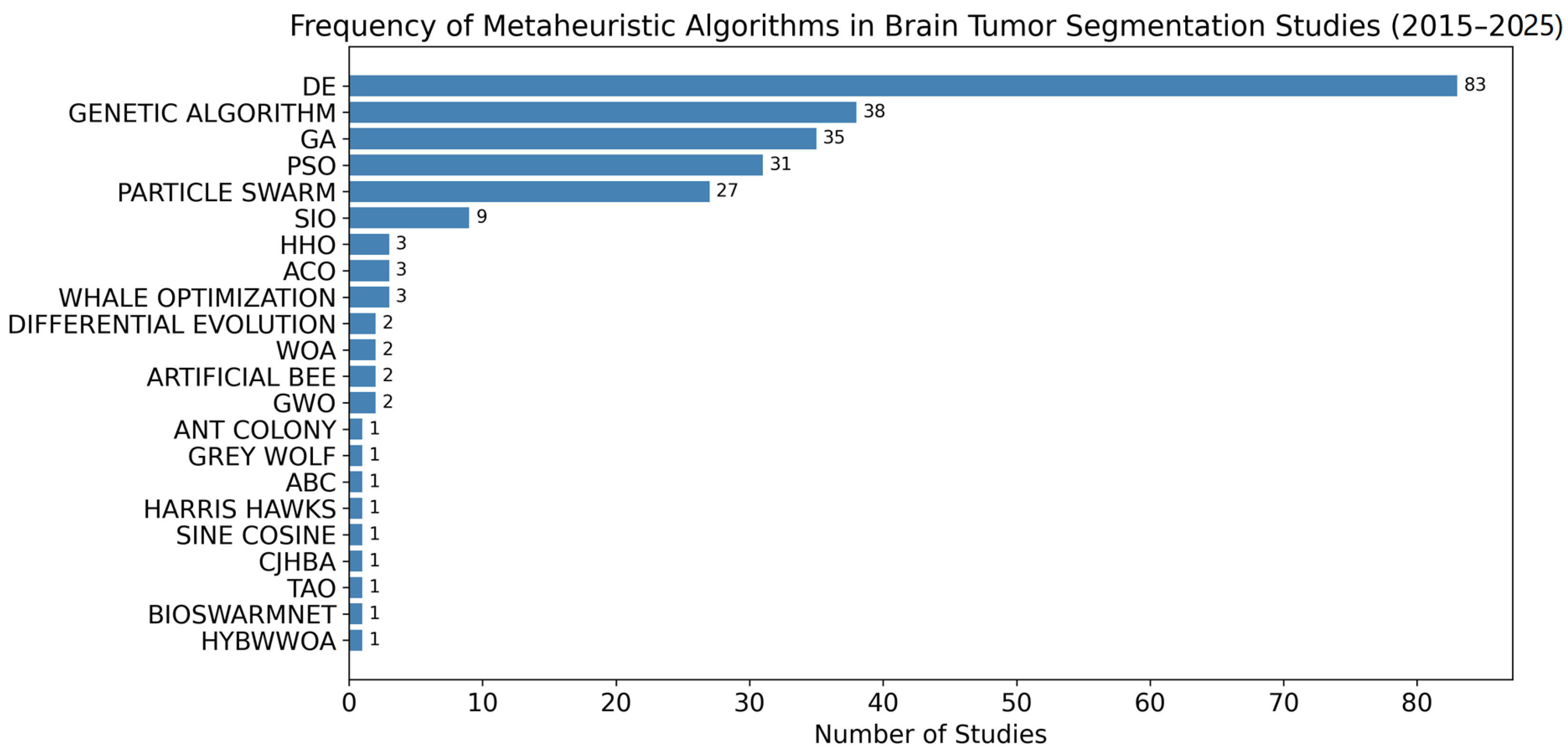
3.3.3. Metaheuristic Usage Statistics

Figure 8 presents the overall usage frequency of metaheuristic algorithms. Differential Evolution (DE) leads with 83 citations, followed by GA and PSO. Novel strategies such as CJHBA [40], TAO [39], and BioSwarmNet [37]—though limited in count—represent state-of-the-art breakthroughs with high performance outcomes.

3.3.4. Optimization Targets in the Segmentation Pipeline

The reviewed studies reveal four primary roles of metaheuristic optimization within the segmentation workflow:
  • Hyperparameter Tuning: Metaheuristics are widely applied to optimize learning rates, kernel sizes, and network depths in CNN and U-Net models, improving segmentation accuracy and training efficiency [52,121]. For instance, Harris Hawks Optimization (HHO) and Differential Evolution (DE) have been used to tune network parameters, resulting in improved accuracy and reduced error rates [121,122].
  • Preprocessing and Image Enhancement: Algorithms such as PSO, ACO, and ABC are used to optimize image contrast and segmentation thresholds, resulting in improved tumor boundary delineation [121,123,124]. These approaches enhance image quality through optimized preprocessing, leading to sharper and more accurate segmentations [22,34,125].
  • Architecture Search and Layer Adaptation: Evolutionary algorithms, including Genetic Algorithms and neuroevolutionary strategies, are employed to discover optimal encoder–decoder structures, enhancing model generalizability and robustness, especially in cross-dataset and multimodal scenarios [29,122,126,127,128].
  • Multimodal Data Fusion and Attention Optimization: Some studies utilize metaheuristics to tune fusion weights in multimodal MRI or optimize attention modules, supporting better segmentation performance in heterogeneous datasets [105,129,130]. Hybrid metaheuristic–deep learning frameworks have demonstrated improved tumor delineation and segmentation quality in complex imaging tasks [131].
Figure 9 details how each algorithm is used across various roles. DE and GA have broad coverage, particularly in architecture search and attention optimization. Hybrid algorithms are increasingly applied to specialized tasks such as multimodal fusion and fine-tuned attention module learning.

3.3.5. Algorithm-Specific Achievements

Below, the specific achievements of the relevant algorithms are outlined:
  • PSO: Widely applied due to its low computational cost and fast convergence. PSO-based brain tumor segmentation methods have consistently achieved Dice Similarity Coefficients (DSC) above 92% in numerous studies, particularly when combined with preprocessing or learning rate tuning. For example, Saifullah and Dreżewski (2025) reported DSCs of 95.78% and 95.23% on BraTS 2019 using PSO-optimized U-Net models [16]. Other works confirm DSC values exceeding 92% across various MRI datasets [23,24,132,133].
  • GA: Known for robust search capabilities in neural architecture evolution and hyperparameter tuning. Several GA-based studies report improved accuracy above 0.90 on unseen datasets, with minimized overfitting. For instance, Genetic Algorithm-enhanced CNNs achieved classification accuracies exceeding 90% on BRATS datasets [25,26,121].
  • GWO and HHO: Grey Wolf Optimizer (GWO) and Harris Hawks Optimization (HHO) have demonstrated stable and reliable optimization results in attention-guided segmentation frameworks. GWO, in particular, has shown consistent boundary preservation for irregular tumor shapes, improving segmentation robustness in multi-modal MRI [45,57,88,134]. HHO-based CNNs have achieved up to 98% accuracy and improved edge detail retention [135,136].
  • Hybrid Approaches: Emerging studies combining metaheuristics (e.g., PSO-GA, DE-ABC) report synergistic gains by leveraging GA’s crossover operations alongside PSO’s velocity updates. These hybrids have improved DSC by 3–6 p.p. (percentage points) compared to single-method approaches, enhancing both segmentation accuracy and convergence speed [71,124,137,138].

3.3.6. Recent Advancements in Metaheuristic-Optimized Segmentation Models

Beyond traditional algorithms like PSO, GA, DE, and GWO, recent years have seen the emergence of novel and hybrid bio-inspired optimization models tailored for medical image segmentation. Notable among these are BioSwarmNet [37], HybWWoA [38], TAO [39], and CJHBA [40].
BioSwarmNet: Gorrepati et al. (2024) proposed BioSwarmNet, which integrates fractional order differential particle swarm optimization (FODPSO) with recurrent neural networks (RNN) for brain tumor segmentation. On the BraTS dataset, BioSwarmNet achieved accuracy of 99.12%, sensitivity of 98.62%, and specificity of 99.86%. While DSC and HD were not explicitly reported, these high classification metrics indicate strong segmentation performance, surpassing many traditional PSO-based models [37].
HybWWoA (Hybrid Whale and Water Waves Optimization Algorithm): Alshammari (2023) introduced HybWWoA, a hybrid metaheuristic algorithm that combines the global exploration capabilities of the Whale Optimization Algorithm (WOA) with the local exploitation strengths of the Water Waves Optimization (WWO) technique. This hybrid approach is designed to enhance feature selection by effectively reducing the dimensionality of extracted features in brain metastasis classification tasks. When applied alongside a DenseNet classifier on MRI data, HybWWoA achieved an F1-score of 97%, accuracy of 92.1%, precision of 98.5%, and sensitivity of 92.1%. These results demonstrate the method’s strong performance in accurately classifying brain tumors, highlighting its potential utility in clinical diagnostic workflows [38].
ACO (Ant Colony Optimization) and its advanced variants have been widely applied to brain tumor segmentation in MRI images. These algorithms mimic the foraging behavior of ants, using pheromone trails and adaptive path selection to identify and delineate tumor regions within complex brain structures. Studies have shown that ACO-based segmentation methods can outperform traditional clustering techniques such as K-means and Fuzzy C-means (FCM), offering more accurate and distinct tumor boundaries in MRI scans. Improved ACO frameworks have also been integrated with fuzzy clustering and neural network classifiers to further enhance segmentation quality and robustness for brain tumor detection tasks. Overall, ACO-based approaches have proven effective and efficient for automated brain tumor segmentation, providing a valuable tool for clinical diagnosis and treatment planning.
CJHBA (Chronological Jaya Honey Badger Algorithm): A complex hybrid that combines chaotic local search with global exploration principles from Jaya and HHO. Deepa et al. (2023) proposed a CJHBA-trained DRN classifier, achieving 92.84% specificity, 93.13% sensitivity, and 92.10% accuracy on the BRATS 2018 dataset. However, segmentation metrics like DSC or JI were not reported in this study.
These methods are summarized in Table 1, which complements the prior performance summary and highlights their competitive edge in segmentation quality and boundary precision.
Summary of Emerging Models: These advanced models illustrate how the design of hybrid or adaptive metaheuristics—tailored specifically for deep learning segmentation tasks—can offer significant improvements over conventional approaches. Not only do they enhance global search behavior and convergence, but they also deliver consistent improvements in clinical metrics such as boundary smoothness and tumor delineation. Their performance further underscores the transition from general-purpose optimization tools toward domain-specific bio-inspired intelligence.

3.3.7. Application Domains and Dataset Usage

Application domains ranged from glioma and meningioma segmentation in the Figshare dataset to whole tumor segmentation on BraTS 2020–2021 datasets. Around 60% of papers evaluated their models on multimodal MRI, especially using T1, T1CE, T2, and FLAIR modalities. Among these, T1CE and FLAIR were most frequently used due to their superior contrast in tumor core and edema delineation. Studies that incorporated hybrid metaheuristics with modality fusion—such as PSO for channel attention weight tuning—achieved enhanced segmentation fidelity and cross-modality robustness.

3.3.8. Summary

The growing adoption of diverse metaheuristics—coupled with their demonstrated utility in multiple stages of the segmentation pipeline—indicates their central role in advancing brain tumor segmentation performance. Results from the reviewed studies consistently show that models optimized with bio-inspired algorithms outperform traditional counterparts in accuracy, convergence, and generalizability. The field is also witnessing a shift from single-objective optimization toward multi-objective and hybrid strategies, underscoring a methodological evolution that mirrors the increasing complexity of the segmentation problem.
To synthesize the methodological contributions of these algorithms, Table 2 presents a summary of the major metaheuristics identified in the reviewed literature, their specific optimization roles, and the typical performance outcomes they enabled. PSO and GA remain the most commonly applied, particularly for hyperparameter tuning and model architecture evolution. Differential Evolution (DE) was especially effective in accelerating convergence [29], while ACO [32] and ABC [31] were more prominent in thresholding and contrast enhancement phases. Advanced methods like GWO and WOA have shown particular promise in refining attention mechanisms and improving delineation of complex tumor boundaries. Hybrid approaches, which combine the strengths of multiple algorithms, often yielded a 3–6 p.p. gain in Dice Similarity Coefficient (DSC) over single-algorithm baselines. These trends not only validate the adaptability of bio-inspired methods but also highlight emerging patterns in how each algorithm is selectively applied based on segmentation objectives and MRI data characteristics.

3.4. Evaluation Metrics and Model Performance

This section presents a detailed analysis of the evaluation metrics used to assess the performance of metaheuristic-optimized segmentation models. The evaluation focuses on five widely accepted quantitative metrics: Accuracy, Dice Similarity Coefficient (DSC), Jaccard Index (JI), Hausdorff Distance (HD), and Average Symmetric Surface Distance (ASSD). These metrics were consistently reported across the selected studies and offer complementary insights into segmentation quality, overlap accuracy, and spatial precision [16].
Accuracy quantifies the ratio of correctly predicted tumor and non-tumor pixels relative to the total number of pixels. However, it may be misleading in imbalanced datasets where non-tumor regions dominate.
DSC and JI are overlap-based metrics frequently used in medical image segmentation. DSC is defined as
DSC = 2 | A B | | A | + | B |
where A and B denote the predicted and ground truth tumor regions, respectively. Jaccard Index, also known as Intersection over Union (IoU), is defined as
JI = | A B | | A B |
Both metrics penalize over- and under-segmentation and are robust indicators of tumor mask overlap.
Hausdorff Distance (HD) measures the maximum Euclidean distance between the boundary points of predicted and ground truth regions, capturing the worst-case spatial discrepancy:
HD ( A , B ) = max sup a A inf b B a b , sup b B inf a A b a
Average Symmetric Surface Distance (ASSD) offers a smoother alternative to HD, computing the average of minimum surface distances in both directions:
ASSD ( A , B ) = 1 | A | + | B | a A min b B a b + b B min a A b a
Figure 10 summarizes the performance distribution of metaheuristic-optimized models based on each evaluation metric. Accuracy values remain tightly clustered above 97%, indicating overall model stability. The median DSC and JI values demonstrate strong overlap between predicted and true tumor regions, with low dispersion across models. Conversely, the reduced median HD and ASSD values suggest improved boundary localization, minimizing false positive or negative extensions of tumor margins. These results confirm that metaheuristic methods—especially hybrid or novel variants—contribute meaningfully to both segmentation accuracy and spatial alignment.
Overall, the consistent performance across all five metrics illustrates the robustness of metaheuristic-driven segmentation frameworks in capturing tumor morphology while maintaining low anatomical discrepancy, thereby reinforcing their clinical applicability in multimodal brain MRI analysis.

3.5. Emerging Trends and Gaps in Metaheuristic Brain Tumor Segmentation

This section synthesizes key emerging trends identified across the reviewed studies and outlines current gaps that warrant future exploration. The observed trajectory of metaheuristic applications in brain tumor segmentation suggests increasing methodological sophistication, integration with deep learning pipelines, and emphasis on interpretability, scalability, and multimodal data fusion.

3.5.1. Emerging Trends

  • Hybrid Metaheuristics and Ensemble Learning: A notable shift toward combining multiple optimization strategies (e.g., PSO-GA, DE-ABC, WOA-GWO) has been observed to improve convergence robustness, solution diversity, and global search behavior. These hybrids often yield superior DSC and JI metrics compared to single algorithms, particularly in datasets with complex or imbalanced tumor patterns [28,93,109,116].
  • Transformer and Attention Integration: Recent studies increasingly embed metaheuristics into attention-driven architectures (e.g., transformer-based U-Nets), tuning spatial attention modules or optimizing attention maps in multimodal MRI segmentation tasks. This integration leads to better delineation of tumor substructures [54,58].
  • Multi-Objective Optimization (MOO): Several frameworks now adopt MOO to simultaneously optimize trade-offs such as accuracy vs. training cost, or DSC vs. boundary error. This trend reflects a more realistic modeling of clinical demands, where multiple objectives must be satisfied concurrently [65,139,140].
  • Self-Adaptive Mechanisms and Online Optimization: A small but growing number of algorithms incorporate dynamic parameter tuning, allowing learning rates, population sizes, or search space bounds to evolve during training. These methods improve adaptability to diverse datasets and reduce the need for manual configuration [16].
  • Integration with Federated and Distributed Learning: As MRI datasets become larger and privacy-sensitive, metaheuristics are being investigated in federated learning settings where local model updates are optimized at client nodes, with metaheuristics ensuring consistency and global convergence [141,142].

3.5.2. Current Gaps and Research Opportunities

Despite recent progress, several limitations persist:
  • Reproducibility and Benchmarking: A significant number of studies lack publicly available code or standard validation protocols, making reproducibility difficult. Benchmark datasets such as BraTS are underused in some studies, limiting cross-study comparisons.
  • Computational Efficiency: Metaheuristic optimization, particularly in deep networks, incurs high training costs due to repeated evaluations of large models. Future work should explore surrogate-assisted or gradient-informed metaheuristics to reduce computational demand.
  • Clinical Validation and Interpretability: Few studies validate segmentation quality through radiologist interpretation or clinical outcomes. The interpretability of optimization decisions—why certain hyperparameters or layers are selected—also remains underexplored.
  • Generalization Across Institutions: Most models are validated within single datasets, raising concerns about cross-site generalizability. Incorporating domain adaptation and robustness measures into the optimization pipeline is essential.
  • Limited Use of 3D Volumetric Optimization: While many methods operate on 2D slices, fewer employ fully 3D metaheuristic-optimized pipelines, which are critical for capturing full tumor context and improving continuity across slices.

3.5.3. Computational Cost Considerations

One of the recurring challenges in applying metaheuristics to deep learning is the computational cost associated with evaluating candidate solutions during training. Algorithms like PSO and GA are relatively lightweight when applied to tasks such as hyperparameter tuning or contrast enhancement. However, more exploration-focused algorithms such as DE and GWO introduce higher iteration demands when optimizing deep architectures or multimodal fusion layers.
Advanced methods, such as HHO and hybrid frameworks (e.g., PSO-GA, DE-ABC), tend to incur higher runtime complexity due to larger populations and deeper search across the solution space. These methods often involve numerous forward and backward passes through neural networks, substantially increasing training cost.
To address this issue, some studies have employed techniques such as early stopping, dimensionality reduction, and surrogate modeling (e.g., Gaussian processes or regression-based estimators) to approximate fitness scores and reduce the number of full training iterations. Others have explored parallel implementations or GPU-based search mechanisms to alleviate runtime overhead.
Despite these efforts, systematic benchmarking of the trade-off between segmentation performance and computational cost remains limited. Future studies should explore standardized cost–performance evaluations and incorporate computational complexity as an optimization objective, especially when targeting clinical deployment scenarios with constrained hardware.

3.5.4. Summary

The evolution of metaheuristic applications in brain tumor segmentation is increasingly marked by innovation in hybridization, self-adaptation, and multimodal fusion. However, key areas—including reproducibility, efficiency, interpretability, and clinical relevance—remain open challenges. Addressing these gaps could enable more trustworthy, scalable, and medically impactful segmentation frameworks.

3.6. Use-Case Guidelines and Applicability

The diversity of bio-inspired metaheuristics makes it crucial to understand when and how each algorithm should be applied. Table 3 presents a usage matrix that helps researchers determine the best-suited algorithm depending on task complexity, optimization scope, and computational budget. Below is a summary:
  • PSO is best suited for tasks requiring fast convergence and low computational overhead, such as learning rate tuning or contrast enhancement;
  • GA is effective in architecture search and layer configuration, particularly when diversity in candidate solutions is essential;
  • DE performs well in high-dimensional tuning and convergence-critical scenarios, such as multimodal fusion weight optimization;
  • ACO and ABC are most applicable in discrete optimization tasks like thresholding and feature selection;
  • GWO and HHO are better aligned with spatial refinement tasks, such as edge preservation and attention tuning in complex segmentation;
  • Hybrid methods (e.g., PSO-GA, DE-ABC) are powerful when optimizing multiple objectives simultaneously, particularly in joint preprocessing-architecture workflows.
By aligning the optimization strategy with task requirements, researchers can significantly improve both model performance and training efficiency.

4. Discussion

This section reflects on the key outcomes of the systematic review and bibliometric analysis of bio-inspired metaheuristics in deep learning-based brain tumor segmentation. Drawing from both empirical trends and methodological insights, the discussion highlights how metaheuristic approaches have shaped the evolution of segmentation techniques and presents a roadmap for future research directions grounded in the identified gaps and opportunities.

4.1. Insights from the Literature Synthesis

The review confirms that metaheuristic algorithms play a pivotal role in enhancing segmentation accuracy, robustness, and adaptability—particularly in complex tasks such as delineating heterogeneous tumor subregions in multimodal MRI data. A clear trajectory is seen from the early reliance on traditional PSO and GA variants for hyperparameter tuning, toward more recent use of hybrid models, dynamic adaptation strategies, and transformer-based segmentation architectures. Notably, algorithms such as DE and GWO [118,119], when applied to architectural search or attention tuning, demonstrate not only performance gains in DSC and JI but also enhanced generalization across datasets.
The integration of metaheuristics into various pipeline stages—ranging from preprocessing, architecture configuration, and multimodal fusion to attention module refinement—also reveals the algorithmic flexibility and domain adaptability of these methods. Furthermore, performance visualizations and role distribution mappings indicate that architecture search and attention optimization have become dominant application areas, reflecting the current research emphasis on structural innovation and spatial feature alignment.

4.2. Impact on the Field of Brain Tumor Segmentation

The increasing adoption of metaheuristics has significantly improved segmentation quality and model adaptability. Metaheuristic optimization allows researchers to navigate vast hyperparameter and architectural search spaces that would otherwise require prohibitively expensive manual tuning. By doing so, they accelerate the development of efficient and accurate segmentation frameworks that are better suited for real-world clinical imaging.
Additionally, the emergence of advanced models such as BioSwarmNet [37], HybWWoA [38], TAO [39], and CJHBA [40] exemplifies how biologically inspired adaptations, hybridization strategies, and domain-specific algorithm refinements are pushing the boundaries of model performance. These models deliver improvements not only in core accuracy metrics but also in clinically relevant spatial metrics such as Hausdorff Distance and ASSD, which are crucial for ensuring reliable tumor margin detection and treatment planning.

4.3. Potential Future Research Directions

To further advance the field and address the remaining limitations identified in Section 3.5, we propose the following directions for future research:
  • Multi-Objective and Federated Metaheuristics: Develop optimization frameworks that can simultaneously balance performance, computational efficiency, and robustness under privacy-preserving or distributed learning environments [65,141].
  • 3D Volumetric and Temporal Optimization: Extend current methods to fully volumetric and longitudinal data by optimizing 3D U-Net or time-series-based architectures using metaheuristics that preserve spatial–temporal continuity [143].
  • Surrogate-Assisted Metaheuristics: Reduce the computational burden of iterative optimization in deep learning by incorporating surrogate models (e.g., Gaussian processes or neural approximators) to estimate fitness landscapes and guide search [29,144].
  • Domain Adaptation and Cross-Institutional Generalization: Incorporate robustness-driven objective functions into metaheuristic pipelines to facilitate adaptation across imaging devices, protocols, or institutional datasets.
  • Explainability and Radiologist-in-the-Loop Feedback: Design metaheuristic strategies that explicitly optimize for explainability (e.g., attention heatmaps [145] or SHAP interpretability [146]) and include feedback mechanisms from clinical experts to refine search directions.
  • Metaheuristic Benchmarking Frameworks: Establish standardized platforms for benchmarking metaheuristic-optimized segmentation pipelines using publicly available datasets, unified protocols, and open-source implementations.
  • Neuromorphic and Bio-Plastic Algorithms: Explore brain-inspired models such as spiking neural networks or plasticity-driven search heuristics to model adaptive segmentation behavior in evolving imaging contexts.

4.4. Conclusions of the Discussion

The reviewed literature demonstrates the transformative potential of metaheuristics in brain tumor segmentation and the growing ecosystem of hybrid models, multimodal strategies, and biologically inspired frameworks. Nonetheless, for these advances to reach clinical impact, future research must prioritize generalizability, transparency, and real-time adaptability. Grounding future efforts in both clinical needs and algorithmic innovation will be key to translating these intelligent systems from experimental pipelines into routine clinical workflows.

5. Conclusions

This systematic review and bibliometric analysis has provided a comprehensive synthesis of the role that bio-inspired metaheuristic algorithms play in advancing deep learning-based brain tumor segmentation. Covering 106 rigorously selected studies published between 2015 and 2025, the review explored the methodological diversity, application domains, evaluation outcomes, and scholarly trends shaping this emerging field.
The findings affirm that metaheuristic optimization—particularly through algorithms such as PSO, GA, DE, GWO, and their hybrid extensions—has become an indispensable tool in enhancing segmentation models across multiple levels of the learning pipeline. From fine-tuning hyperparameters and refining contrast enhancement, to driving architecture search and modality fusion, these algorithms offer flexible and effective solutions to the challenges posed by heterogeneous tumor appearances in multimodal MRI. Notably, the introduction of novel frameworks such as BioSwarmNet [37], HybWWoA [38], CJHBA [40], and TAO [39] has expanded the boundaries of what is achievable in both performance accuracy and clinical robustness.
Through bibliometric mapping, the review also identified a growing interdisciplinary interest in the domain, with increasing publication volumes, evolving author collaborations, and a global spread of research contributions. Keyword co-occurrence and temporal publication analyses further reflect the field’s shift toward more sophisticated, hybridized, and interpretable deep learning models, optimized via intelligent search strategies.
Nevertheless, several open challenges remain. The absence of standardized benchmarks, limited focus on explainability, underrepresentation of 3D and longitudinal data, and lack of real-world deployment pathways represent critical areas for future exploration. Addressing these issues will require not only technical innovation but also stronger integration with clinical practice, cross-institutional validation, and the development of shared evaluation protocols.
In conclusion, bio-inspired metaheuristics have emerged as a powerful enabler of precision and adaptability in brain tumor segmentation. As deep learning models continue to evolve in complexity, metaheuristic algorithms will play an increasingly vital role in shaping their performance, interpretability, and generalization. This review serves as both a state-of-the-art summary and a forward-looking guide, inviting continued innovation at the intersection of evolutionary intelligence, medical imaging, and computational neuro-oncology.

Author Contributions

Conceptualization, S.S., A.Y. and R.D.; methodology design and search strategy, S.S.; literature screening and data extraction, S.S., W.C. and N.H.; bibliometric analysis, S.S.; writing—original draft preparation, S.S., R.D., A.Y., W.C. and N.H.; writing—review and editing, S.S., R.D., A.Y., W.C. and N.H.; critical revision of the manuscript, R.D. and A.Y.; visualization, S.S.; supervision, R.D. and A.Y.; project coordination, S.S. All authors have read and agreed to the published version of the manuscript.

Funding

This research project was partly supported by the program “Excellence initiative—research university” for the AGH University of Krakow, Ahmad Dahlan University (grant number: U12/310/III/2025), and the Polish Ministry of Science and Higher Education funds assigned to AGH University of Krakow.

Institutional Review Board Statement

Not applicable.

Informed Consent Statement

Not applicable.

Data Availability Statement

Not applicable.

Conflicts of Interest

The authors declare no conflicts of interest.

Abbreviations

The following abbreviations are used in this manuscript:
AIArtificial Intelligence
ACOAnt Colony Optimization
ABCArtificial Bee Colony
ASSDAverage Symmetric Surface Distance
CNNConvolutional Neural Network
CJHBAChronological Jaya Honey Badger Algorithm
DEDifferential Evolution
DSCDice Similarity Coefficient
FLAIRFluid-Attenuated Inversion Recovery
GAGenetic Algorithm
GWOGrey Wolf Optimizer
HDHausdorff Distance
HHOHarris Hawks Optimization
MRIMagnetic Resonance Imaging
PSOParticle Swarm Optimization
PRISMAPreferred Reporting Items for Systematic Reviews and Meta-Analyses
RNNRecurrent Neural Network
SIOSwan-Inspired Optimization
TAOTransformational Ant Optimization
T1T1-weighted MRI
T1CE/T1GdT1 Contrast-Enhanced/Gadolinium-enhanced MRI
T2T2-weighted MRI
U-NetU-shaped Convolutional Neural Network
WOAWhale Optimization Algorithm
HybWWoAHybrid Whale-Wasp Optimization Algorithm
SLRSystematic Literature Review
JIJaccard Index

Appendix A. Filtering Process and Article Selection Summary

This appendix summarizes the article screening pipeline already described in Section 2. Table A1 provides a concise overview of the filtering and selection stages, from the initial retrieval of 3895 records to the final inclusion of 106 studies. For detailed methodological explanations, refer to Section 2 and Section 2.3.
Table A1. Summary of filtering and screening stages in the article selection process.
Filtering StageDescriptionArticles Remaining
Initial Records RetrievedTotal articles retrieved from Scopus and Google Scholar using 5 structured queries3895
After Duplicate RemovalDuplicates removed based on normalized titles across datasets2553
Brain Tumor Segmentation in TitleArticles containing “brain tumor segmentation” in the title634
Matched Brain Tumor MRI KeywordsArticles with MRI modality mentions (e.g., FLAIR, T1, T1CE/T1Gd, T2) in title/abstract634
Removed Review/Survey/Overview ArticlesExcluded reviews, surveys, or non-experimental studies587
Removed Retracted and Non-Academic SourcesExcluded preprints and sources like arXiv, ResearchGate, Academia.edu586
Filtered for Bio-Inspired Metaheuristic UseArticles referencing metaheuristics (e.g., PSO, GA, DE, ACO, GWO) in title/abstract184
Final Articles After Manual ScreeningFinal set of articles that explicitly applied bio-inspired metaheuristic optimization methods106

References

  1. Liu, J.; Wang, T.; Dong, J.; Lu, Y. The blood–brain barriers: Novel nanocarriers for central nervous system diseases. J. Nanobiotechnol. 2025, 23, 146. [Google Scholar] [CrossRef] [PubMed]
  2. Sajid Hussain, S.; Wani, N.A.; Kaur, J.; Ahmad, N.; Ahmad, S. Next-Generation Automation in Neuro-Oncology: Advanced Neural Networks for MRI-Based Brain Tumor Segmentation and Classification. IEEE Access 2025, 13, 41141–41158. [Google Scholar] [CrossRef]
  3. Ijaz, M.; Hasan, I.; Aslam, B.; Yan, Y.; Zeng, W.; Gu, J.; Jin, J.; Zhang, Y.; Wang, S.; Xing, L.; et al. Diagnostics of brain tumor in the early stage: Current status and future perspectives. Biomater. Sci. 2025, 13, 2580–2605. [Google Scholar] [CrossRef]
  4. Sabeghi, P.; Zarand, P.; Zargham, S.; Golestany, B.; Shariat, A.; Chang, M.; Yang, E.; Rajagopalan, P.; Phung, D.; Gholamrezanezhad, A. Advances in Neuro-Oncological Imaging: An Update on Diagnostic Approach to Brain Tumors. Cancers 2024, 16, 576. [Google Scholar] [CrossRef]
  5. Kumar, A.A.; Kesavadas, C. Potential of MRI in Clinical Medicine. In Multimodal Biomedical Imaging Techniques; Biological and Medical Physics, Biomedical Engineering; Kalarikkal, N., Bhadrapriya, B.C., Anne Bose, B., Padmanabhan, P., Thomas, S., Vadakke Matham, M., Eds.; Springer: Singapore, 2025; pp. 271–301. [Google Scholar] [CrossRef]
  6. Hassan, M.; Fateh, A.A.; Lin, J.; Zhuang, Y.; Lin, G.; Xiong, H.; You, Z.; Qin, P.; Zeng, H. Unfolding Explainable AI for Brain Tumor Segmentation. Neurocomputing 2024, 599, 128058. [Google Scholar] [CrossRef]
  7. Sadeghi, P.; Ghazizadeh, Y.; Arabshahi, S.; Habibzadeh, A.; Karimi, H.; Bordbar, S.; Ghaffari Jolfayi, A.; Pourbakhtyaran, E. Artificial Intelligence Applications to Detect Pediatric Brain Tumor Biomarkers. In Interdisciplinary Cancer Research; Springer: Cham, Switzerland, 2024. [Google Scholar] [CrossRef]
  8. Mansur, Z.; Talukdar, J.; Singh, T.P.; Kumar, C.J. Deep Learning-Based Brain Tumor Image Analysis for Segmentation. SN Comput. Sci. 2024, 6, 42. [Google Scholar] [CrossRef]
  9. Badrinarayanan, V.; Kendall, A.; Cipolla, R. SegNet: A Deep Convolutional Encoder-Decoder Architecture for Image Segmentation. IEEE Trans. Pattern Anal. Mach. Intell. 2017, 39, 2481–2495. [Google Scholar] [CrossRef] [PubMed]
  10. Nizamani, A.H.; Chen, Z.; Nizamani, A.A.; Bhatti, U.A. Advance brain tumor segmentation using feature fusion methods with deep U-Net model with CNN for MRI data. J. King Saud Univ.-Comput. Inf. Sci. 2023, 35, 101793. [Google Scholar] [CrossRef]
  11. Hu, L.S.; Hawkins-Daarud, A.; Wang, L.; Li, J.; Swanson, K.R. Imaging of intratumoral heterogeneity in high-grade glioma. Cancer Lett. 2020, 477, 97–106. [Google Scholar] [CrossRef]
  12. De Sutter, S.; Wuts, J.; Geens, W.; Vanbinst, A.M.; Duerinck, J.; Vandemeulebroucke, J. Modality redundancy for MRI-based glioblastoma segmentation. Int. J. Comput. Assist. Radiol. Surg. 2024, 19, 2101–2109. [Google Scholar] [CrossRef]
  13. Liu, Y.; Mu, F.; Shi, Y.; Cheng, J.; Li, C.; Chen, X. Brain tumor segmentation in multimodal MRI via pixel-level and feature-level image fusion. Front. Neurosci. 2022, 16, 1000587. [Google Scholar] [CrossRef]
  14. Styliara, E.I.; Astrakas, L.G.; Alexiou, G.; Xydis, V.G.; Zikou, A.; Kafritsas, G.; Voulgaris, S.; Argyropoulou, M.I. Survival Outcome Prediction in Glioblastoma: Insights from MRI Radiomics. Curr. Oncol. 2024, 31, 2233–2243. [Google Scholar] [CrossRef] [PubMed]
  15. Saifullah, S.; Dreżewski, R. Automatic Brain Tumor Segmentation Using Convolutional Neural Networks: U-Net Framework with PSO-Tuned Hyperparameters. In Parallel Problem Solving from Nature—PPSN XVIII, Proceedings of the 18th International Conference, PPSN 2024, Hagenberg, Austria, 14–18 September 2024; Lecture Notes in Computer Science; Affenzeller, M., Winkler, S.M., Kononova, A.V., Trautmann, H., Tušar, T., Machado, P., Bäck, T., Eds.; Springer: Cham, Switzerland, 2024; Volume 15150, pp. 333–351. [Google Scholar] [CrossRef]
  16. Saifullah, S.; Dreżewski, R. Particle Swarm-Optimized U-Net Framework for Precise Multimodal Brain Tumor Segmentation. arXiv 2025, arXiv:2503.19152. [Google Scholar]
  17. Iqbal, S.; Qureshi, A.N.; Ullah, A.; Li, J.; Mahmood, T. Improving the Robustness and Quality of Biomedical CNN Models through Adaptive Hyperparameter Tuning. Appl. Sci. 2022, 12, 11870. [Google Scholar] [CrossRef]
  18. Li, M.; Jiang, Y.; Zhang, Y.; Zhu, H. Medical image analysis using deep learning algorithms. Front. Public Health 2023, 11, 1273253. [Google Scholar] [CrossRef]
  19. Rashmi, P.; Gomathi, R. Optimized Deep learning Frameworks for the Medical Image Transmission in IoMT Environment. J. Smart Internet Things 2024, 2024, 148–165. [Google Scholar] [CrossRef]
  20. Ul Haq, I. Diagnosis of Neurological Disease Using Bioinspired Algorithms. In Bio-Inspired Optimization for Medical Data Mining; Wiley: Hoboken, NJ, USA, 2024; pp. 227–268. [Google Scholar] [CrossRef]
  21. Vijh, S.; Sharma, S.; Gaurav, P. Brain Tumor Segmentation Using OTSU Embedded Adaptive Particle Swarm Optimization Method and Convolutional Neural Network. In Data Visualization and Knowledge Engineering; Lecture Notes on Data Engineering and Communications Technologies; Springer: Cham, Switzerland, 2020; Volume 32, pp. 171–194. [Google Scholar] [CrossRef]
  22. Saifullah, S.; Dreżewski, R. Advanced Medical Image Segmentation Enhancement: A Particle-Swarm-Optimization-Based Histogram Equalization Approach. Appl. Sci. 2024, 14, 923. [Google Scholar] [CrossRef]
  23. Sharif, M.; Amin, J.; Raza, M.; Yasmin, M.; Satapathy, S.C. An integrated design of particle swarm optimization (PSO) with fusion of features for detection of brain tumor. Pattern Recognit. Lett. 2020, 129, 150–157. [Google Scholar] [CrossRef]
  24. Malik, A.; Devarajan, G.G. Integrated Brain Tumor Detection: PSO-Guided Segmentation with U-Net and CNN Classification. Procedia Comput. Sci. 2024, 235, 3447–3457. [Google Scholar] [CrossRef]
  25. Arif, M.; Jims, A.; F., A.; Geman, O.; Craciun, M.D.; Leuciuc, F. Application of Genetic Algorithm and U-Net in Brain Tumor Segmentation and Classification: A Deep Learning Approach. Comput. Intell. Neurosci. 2022, 2022, 5625757. [Google Scholar] [CrossRef]
  26. Kabir Anaraki, A.; Ayati, M.; Kazemi, F. Magnetic resonance imaging-based brain tumor grades classification and grading via convolutional neural networks and genetic algorithms. Biocybern. Biomed. Eng. 2019, 39, 63–74. [Google Scholar] [CrossRef]
  27. Ramtekkar, P.K.; Pandey, A.; Pawar, M.K. Accurate detection of brain tumor using optimized feature selection based on deep learning techniques. Multimed. Tools Appl. 2023, 82, 44623–44653. [Google Scholar] [CrossRef]
  28. Hekmat, A.; Zuping, Z.; Bilal, O.; Khan, S.U.R. Differential evolution-driven optimized ensemble network for brain tumor detection. Int. J. Mach. Learn. Cybern. 2025. [Google Scholar] [CrossRef]
  29. Kuş, Z.; Kiraz, B.; Göksu, T.K.; Aydın, M.; Özkan, E.; Vural, A.; Kiraz, A.; Can, B. Differential evolution-based neural architecture search for brain vessel segmentation. Eng. Sci. Technol. Int. J. 2023, 46, 101502. [Google Scholar] [CrossRef]
  30. Rajesh, C.; Kumar, S. An evolutionary block based network for medical image denoising using Differential Evolution. Appl. Soft Comput. 2022, 121, 108776. [Google Scholar] [CrossRef]
  31. Aly, R.H.M.; Rahouma, K.H.; Hamed, H.F. Brain Tumors Diagnosis and Prediction Based on Applying the Learning Metaheuristic Optimization Techniques of Particle Swarm, Ant Colony and Bee Colony. Procedia Comput. Sci. 2019, 163, 165–179. [Google Scholar] [CrossRef]
  32. Bouzidi, D.; Ghozzi, F.; Fakhfakh, A. Ant Colony Optimization with BrainSeg3D Protocol for Multiple Sclerosis Lesion Detection. In Participative Urban Health and Healthy Aging in the Age of AI, Proceedings of the 19th International Conference, ICOST 2022, Paris, France, 27–30 June 2022; Lecture Notes in Computer Science; Aloulou, H., Abdulrazak, B., de Marassé-Enouf, A., Mokhtari, M., Eds.; Springer: Cham, Switzerland, 2022; Volume 13287, pp. 234–245. [Google Scholar] [CrossRef]
  33. Reis, H.C.; Turk, V. Advanced brain tumor analysis: A novel strategy for segmentation and classification using modern computational methods. Neural Comput. Appl. 2025, 37, 4697–4731. [Google Scholar] [CrossRef]
  34. Saifullah, S.; Drezewski, R. Improved Brain Tumor Segmentation Using Modified U-Net based on Particle Swarm Optimization Image Enhancement. In Proceedings of the Genetic and Evolutionary Computation Conference (GECCO ’24 Companion), Melbourne, VIC, Australia, 14–18 July 2024. [Google Scholar] [CrossRef]
  35. Tuba, E.; Bačanin, N.; Strumberger, I.; Tuba, M. Convolutional Neural Networks Hyperparameters Tuning. In Artificial Intelligence: Theory and Applications; Studies in Computational Intelligence; Pap, E., Ed.; Springer: Cham, Switzerland, 2021; Volume 973, pp. 65–84. [Google Scholar] [CrossRef]
  36. Yadav, A.C.; Kolekar, M.H.; Zope, M.K. Modified Recurrent Residual Attention U-Net model for MRI-based brain tumor segmentation. Biomed. Signal Process. Control 2025, 102, 107220. [Google Scholar] [CrossRef]
  37. Gorrepati, I.; Pagadala, P.K. BioSwarmNet: A Revolutionary Approach to Brain Tumour Detection Using Fractional Order Differential Particle Swarm Optimisation and Recurrent Neural Networks. Rev. D’Intelligence Artif. 2024, 38, 1263–1273. [Google Scholar] [CrossRef]
  38. Alshammari, A. DenseNet_HybWWoA: A DenseNet-Based Brain Metastasis Classification with a Hybrid Metaheuristic Feature Selection Strategy. Biomedicines 2023, 11, 1354. [Google Scholar] [CrossRef]
  39. Boumediene ghaouti, G.; Meftah, B. An Optimized Clustering Approach using Tree Seed Algorithm for the Brain MRI Images Segmentation. Intel. Artif. 2023, 26, 44–59. [Google Scholar] [CrossRef]
  40. Deepa, S.; Janet, J.; Sumathi, S.; Ananth, J.P. Hybrid Optimization Algorithm Enabled Deep Learning Approach Brain Tumor Segmentation and Classification Using MRI. J. Digit. Imaging 2023, 36, 847–868. [Google Scholar] [CrossRef]
  41. Moher, D.; Altman, D.G.; Schulz, K.F.; Simera, I.; Wager, E. (Eds.) Guidelines for Reporting Health Research: A User’s Manual; Wiley: Hoboken, NJ, USA, 2014. [Google Scholar] [CrossRef]
  42. Narong, D.K.; Hallinger, P. A Keyword Co-Occurrence Analysis of Research on Service Learning: Conceptual Foci and Emerging Research Trends. Educ. Sci. 2023, 13, 339. [Google Scholar] [CrossRef]
  43. Ali, M.; Hussain Shah, J.; Attique Khan, M.; Alhaisoni, M.; Tariq, U.; Akram, T.; Jin Kim, Y.; Chang, B. Brain Tumor Detection and Classification Using PSO and Convolutional Neural Network. Comput. Mater. Contin. 2022, 73, 4501–4518. [Google Scholar] [CrossRef]
  44. Akhila, P.; Prabaharan, G.; Pandiiyan, K.; Swarna, S.L.; A, H.; Sakthivelu, U. Particle Swarm Optimization for Efficient Brain Tumor Classification Using InceptionV3 Deep Learning Model. In Proceedings of the 2024 9th International Conference on Communication and Electronics Systems (ICCES), Coimbatore, India, 16–18 December 2024; IEEE: Piscataway, NJ, USA, 2024; pp. 1964–1969. [Google Scholar] [CrossRef]
  45. ZainEldin, H.; Gamel, S.A.; El-Kenawy, E.S.M.; Alharbi, A.H.; Khafaga, D.S.; Ibrahim, A.; Talaat, F.M. Brain Tumor Detection and Classification Using Deep Learning and Sine-Cosine Fitness Grey Wolf Optimization. Bioengineering 2022, 10, 18. [Google Scholar] [CrossRef]
  46. Ramakrishnan, T.; Sankaragomathi, B. A professional estimate on the computed tomography brain tumor images using SVM-SMO for classification and MRG-GWO for segmentation. Pattern Recognit. Lett. 2017, 94, 163–171. [Google Scholar] [CrossRef]
  47. Nayak, G.S.; Mallick, P.K.; Padhi, N.; Mohanty, M.R.; Kumar, S.; Balaji, P. Brain image segmentation with fuzzy entropy clustering and PSO-GWO optimization techniques. Intell. Decis. Technol. 2024, 18, 1319–1336. [Google Scholar] [CrossRef]
  48. Jemimma, T.A.; Vetharaj, Y.J. Fractional probabilistic fuzzy clustering and optimization based brain tumor segmentation and classification. Multimed. Tools Appl. 2022, 81, 17889–17918. [Google Scholar] [CrossRef]
  49. Qader, S.M.; Hassan, B.A.; Rashid, T.A. An improved deep convolutional neural network by using hybrid optimization algorithms to detect and classify brain tumor using augmented MRI images. Multimed. Tools Appl. 2022, 81, 44059–44086. [Google Scholar] [CrossRef]
  50. Pandey, M.K.; Kumar, A.; Bhardwaj, S. Early Brain Tumor Prediction Using Hybrid Optimized Fuzzy Clustering-Active Contour Segmentation Based Heuristic Deep Learning Model. Optoelectron. Instrum. Data Process. 2024, 60, 659–673. [Google Scholar] [CrossRef]
  51. Saifullah, S.; Dreżewski, R.; Yudhana, A.; Wielgosz, M.; Caesarendra, W. Modified U-Net with attention gate for enhanced automated brain tumor segmentation. Neural Comput. Appl. 2025, 37, 5521–5558. [Google Scholar] [CrossRef]
  52. Aljohani, M.; Bahgat, W.M.; Balaha, H.M.; AbdulAzeem, Y.; El-Abd, M.; Badawy, M.; Elhosseini, M.A. An automated metaheuristic-optimized approach for diagnosing and classifying brain tumors based on a convolutional neural network. Results Eng. 2024, 23, 102459. [Google Scholar] [CrossRef]
  53. Guder, O.; Cetin-Kaya, Y. Optimized attention-based lightweight CNN using particle swarm optimization for brain tumor classification. Biomed. Signal Process. Control 2025, 100, 107126. [Google Scholar] [CrossRef]
  54. Srinivas, B.; Anilkumar, B.; Devi, N.; Aruna, V. A fine-tuned transformer model for brain tumor detection and classification. Multimed. Tools Appl. 2025, 84, 15597–15621. [Google Scholar] [CrossRef]
  55. Kumar, V.P.; Pattanaik, S.R.; Kumar, V.V.S. A Heuristic Strategy Assisted Deep Learning Models for Brain Tumor Classification and Abnormality Segmentation. Comput. Intell. 2025, 41, e70018. [Google Scholar] [CrossRef]
  56. Zakariah, M.; Al-Razgan, M.; Alfakih, T. Dual vision Transformer-DSUNET with feature fusion for brain tumor segmentation. Heliyon 2024, 10, e37804. [Google Scholar] [CrossRef]
  57. Shivhare, S.N.; Kumar, N. Tumor bagging: A novel framework for brain tumor segmentation using metaheuristic optimization algorithms. Multimed. Tools Appl. 2021, 80, 26969–26995. [Google Scholar] [CrossRef]
  58. Nguyen-Tat, T.B.; Nguyen, T.Q.T.; Nguyen, H.N.; Ngo, V.M. Enhancing brain tumor segmentation in MRI images: A hybrid approach using UNet, attention mechanisms, and transformers. Egypt. Inform. J. 2024, 27, 100528. [Google Scholar] [CrossRef]
  59. Vijay, V.; Kavitha, A.; Rebecca, S.R. Automated Brain Tumor Segmentation and Detection in MRI Using Enhanced Darwinian Particle Swarm Optimization(EDPSO). Procedia Comput. Sci. 2016, 92, 475–480. [Google Scholar] [CrossRef]
  60. Rouhi, R.; Jafari, M.; Kasaei, S.; Keshavarzian, P. Benign and malignant breast tumors classification based on region growing and CNN segmentation. Expert Syst. Appl. 2015, 42, 990–1002. [Google Scholar] [CrossRef]
  61. Subashini, M.M.; Sahoo, S.K.; Sunil, V.; Easwaran, S. A non-invasive methodology for the grade identification of astrocytoma using image processing and artificial intelligence techniques. Expert Syst. Appl. 2016, 43, 186–196. [Google Scholar] [CrossRef]
  62. Elkorany, A.S.; Elsharkawy, Z.F. Automated optimized classification techniques for magnetic resonance brain images. Multimed. Tools Appl. 2020, 79, 27791–27814. [Google Scholar] [CrossRef]
  63. Kaur, T.; Saini, B.S.; Gupta, S. An adaptive fuzzy K-nearest neighbor approach for MR brain tumor image classification using parameter free bat optimization algorithm. Multimed. Tools Appl. 2019, 78, 21853–21890. [Google Scholar] [CrossRef]
  64. Ibtehaz, N.; Rahman, M.S. MultiResUNet: Rethinking the U-Net architecture for multimodal biomedical image segmentation. Neural Netw. 2020, 121, 74–87. [Google Scholar] [CrossRef] [PubMed]
  65. Devanathan, B.; Kamarasan, M. Multi-objective Archimedes Optimization Algorithm with Fusion-based Deep Learning model for brain tumor diagnosis and classification. Multimed. Tools Appl. 2023, 82, 16985–17007. [Google Scholar] [CrossRef]
  66. Nguyen, T.Q.T.; Nguyen, H.N.; Bui, T.H.; Nguyen-Tat, T.B.; Ngo, V.M. Brain Tumor Segmentation in MRI Images with 3D U-Net and Contextual Transformer. In Proceedings of the 2024 International Conference on Multimedia Analysis and Pattern Recognition (MAPR), Da Nang, Vietnam, 15–16 August 2024; IEEE: Piscataway, NJ, USA, 2024; pp. 1–6. [Google Scholar] [CrossRef]
  67. Ghosal, P.; Roy, A.; Agarwal, R.; Purkayastha, K.; Sharma, A.L.; Kumar, A. Compound attention embedded dual channel encoder-decoder for ms lesion segmentation from brain MRI. Multimed. Tools Appl. 2024. [Google Scholar] [CrossRef]
  68. Preethi, S.; Aishwarya, P. An efficient wavelet-based image fusion for brain tumor detection and segmentation over PET and MRI image. Multimed. Tools Appl. 2021, 80, 14789–14806. [Google Scholar] [CrossRef]
  69. Zhang, T.; Zhou, P.; Zhang, S.; Cheng, S.; Ma, L.; Jiang, H.; Yao, Y.D. Bio-inspired optimisation algorithms in medical image segmentation: A review. Int. J. Bio-Inspired Comput. 2024, 24, 65–79. [Google Scholar] [CrossRef]
  70. Selvan, P.; Kavitha, A.; Ragul, S. Optimizing Brain Tumor Classification: A Comparative Analysis of Nature-Inspired Algorithms with GLCM Features. Biomed. Mater. Devices 2025. [Google Scholar] [CrossRef]
  71. Joshi, A.A.; Aziz, R.M. Deep learning approach for brain tumor classification using metaheuristic optimization with gene expression data. Int. J. Imaging Syst. Technol. 2024, 34. [Google Scholar] [CrossRef]
  72. Alagarsamy, S.; Govindaraj, V.; Shahina, A.; Nagarajan, D. Intelligent Multigrade Brain Tumor Identification in MRI: A Metaheuristic-Based Uncertain Set Framework. IEEE Trans. Artif. Intell. 2024, 5, 5381–5391. [Google Scholar] [CrossRef]
  73. Sharif, M.I.; Li, J.P.; Khan, M.A.; Kadry, S.; Tariq, U. M3BTCNet: Multi model brain tumor classification using metaheuristic deep neural network features optimization. Neural Comput. Appl. 2024, 36, 95–110. [Google Scholar] [CrossRef]
  74. Houssein, E.H.; Emam, M.M.; Singh, N.; Samee, N.A.; Alabdulhafith, M.; Çelik, E. An improved honey badger algorithm for global optimization and multilevel thresholding segmentation: Real case with brain tumor images. Clust. Comput. 2024, 27, 14315–14364. [Google Scholar] [CrossRef]
  75. Kollem, S. An efficient method for MRI brain tumor tissue segmentation and classification using an optimized support vector machine. Multimed. Tools Appl. 2024, 83, 68487–68519. [Google Scholar] [CrossRef]
  76. Dhole, N.V.; Dixit, V.V.; Desai, D. Detection of brain tumour in multi-modality images using hybrid features. Multimed. Tools Appl. 2024, 83, 4613–4638. [Google Scholar] [CrossRef]
  77. Batool, A.; Byun, Y.C. Brain tumor detection with integrating traditional and computational intelligence approaches across diverse imaging modalities—Challenges and future directions. Comput. Biol. Med. 2024, 175, 108412. [Google Scholar] [CrossRef]
  78. Güler, M.; Namlı, E. Brain Tumor Detection with Deep Learning Methods’ Classifier Optimization Using Medical Images. Appl. Sci. 2024, 14, 642. [Google Scholar] [CrossRef]
  79. Shreeharsha, J. Detection of brain tumor using Hybridized 3D U-Net model on MRI images. Multimed. Tools Appl. 2024. [Google Scholar] [CrossRef]
  80. V, A.; PR, B.; BK, A. Automated biomedical image classification using multi-scale dense dilated semi-supervised u-net with cnn architecture. Multimed. Tools Appl. 2023, 83, 30641–30673. [Google Scholar] [CrossRef]
  81. Ketfi, M.; Belahcene, M.; Bourennane, S. OCAE and OUNET: Standard automatic optimization for medical image segmentation. Multimed. Tools Appl. 2024. [Google Scholar] [CrossRef]
  82. Nancy, A.M.; Maheswari, R. Brain tumor segmentation and classification using transfer learning based CNN model with model agnostic concept interpretation. Multimed. Tools Appl. 2024, 84, 2509–2538. [Google Scholar] [CrossRef]
  83. RajamohanReddy, N.; Muneeswari, G. Advancing multi-categorization and segmentation in brain tumors using novel efficient deep learning approaches. PeerJ Comput. Sci. 2024, 10, e2496. [Google Scholar] [CrossRef] [PubMed]
  84. Ibrahim, R.; Ghnemat, R.; Abu Al-Haija, Q. Improving Alzheimer’s Disease and Brain Tumor Detection Using Deep Learning with Particle Swarm Optimization. AI 2023, 4, 551–573. [Google Scholar] [CrossRef]
  85. Pradeep, K.R.; Gangadharan, S.M.P.; Hatamleh, W.A.; Tarazi, H.; Shukla, P.K.; Tiwari, B. Improved Machine Learning Method for Intracranial Tumor Detection with Accelerated Particle Swarm Optimization. J. Healthc. Eng. 2022, 2022, 1128217. [Google Scholar] [CrossRef]
  86. Siddique, A.A.; Raza, A.; Alshehri, M.S.; Alasbali, N.; Abbasi, S.F. Optimizing Tumor Classification Through Transfer Learning and Particle Swarm Optimization-Driven Feature Extraction. IEEE Access 2024, 12, 85929–85939. [Google Scholar] [CrossRef]
  87. Deepa, G.; Mary, G.L.R.; Karthikeyan, A.; Rajalakshmi, P.; Hemavathi, K.; Dharanisri, M. Detection of brain tumor using modified particle swarm optimization (MPSO) segmentation via haralick features extraction and subsequent classification by KNN algorithm. Mater. Today Proc. 2022, 56, 1820–1826. [Google Scholar] [CrossRef]
  88. Zhang, T.; Zhang, J.; Xue, T.; Rashid, M.H. A Brain Tumor Image Segmentation Method Based on Quantum Entanglement and Wormhole Behaved Particle Swarm Optimization. Front. Med. 2022, 9, 794126. [Google Scholar] [CrossRef] [PubMed]
  89. Gtifa, W.; Hamdaoui, F.; Sakly, A. Automated brain tumour segmentation from multi-modality magnetic resonance imaging data based on new particle swarm optimisation segmentation method. Int. J. Med. Robot. Comput. Assist. Surg. 2023, 19, e2487. [Google Scholar] [CrossRef]
  90. Lahmiri, S. Glioma detection based on multi-fractal features of segmented brain MRI by particle swarm optimization techniques. Biomed. Signal Process. Control 2017, 31, 148–155. [Google Scholar] [CrossRef]
  91. Radha, R.; Gopalakrishnan, R. A medical analytical system using intelligent fuzzy level set brain image segmentation based on improved quantum particle swarm optimization. Microprocess. Microsystems 2020, 79, 103283. [Google Scholar] [CrossRef]
  92. Polaki, R.; Umamaheswari, V. A ResNet-Powered Approach for Brain Tumor Detection with Particle Swarm Optimization. In Proceedings of the 2023 Seventh International Conference on Image Information Processing (ICIIP), Solan, India, 22–24 November 2023; IEEE: Piscataway, NJ, USA, 2023; pp. 776–781. [Google Scholar] [CrossRef]
  93. Tan, T.Y.; Zhang, L.; Lim, C.P.; Fielding, B.; Yu, Y.; Anderson, E. Evolving Ensemble Models for Image Segmentation Using Enhanced Particle Swarm Optimization. IEEE Access 2019, 7, 34004–34019. [Google Scholar] [CrossRef]
  94. Atia, N.; Benzaoui, A.; Jacques, S.; Hamiane, M.; Kourd, K.E.; Bouakaz, A.; Ouahabi, A. Particle Swarm Optimization and Two-Way Fixed-Effects Analysis of Variance for Efficient Brain Tumor Segmentation. Cancers 2022, 14, 4399. [Google Scholar] [CrossRef] [PubMed]
  95. Garg, G.; Juneja, M. Particle swarm optimization based segmentation of Cancer in multi-parametric prostate MRI. Multimed. Tools Appl. 2021, 80, 30557–30580. [Google Scholar] [CrossRef]
  96. Verma, H.; Verma, D.; Tiwari, P.K. A population based hybrid FCM-PSO algorithm for clustering analysis and segmentation of brain image. Expert Syst. Appl. 2021, 167, 114121. [Google Scholar] [CrossRef]
  97. Dhanachandra, N.; Chanu, Y.J. An image segmentation approach based on fuzzy c-means and dynamic particle swarm optimization algorithm. Multimed. Tools Appl. 2020, 79, 18839–18858. [Google Scholar] [CrossRef]
  98. Ma, J.; Hu, J. An improved particle swarm optimization for multilevel thresholding medical image segmentation. PLoS ONE 2024, 19, e0306283. [Google Scholar] [CrossRef]
  99. Chithambaram, T.; Perumal, K. Brain tumor segmentation using genetic algorithm and ANN techniques. In Proceedings of the 2017 IEEE International Conference on Power, Control, Signals and Instrumentation Engineering (ICPCSI), Chennai, India, 21–22 September 2017; IEEE: Piscataway, NJ, USA, 2017; pp. 970–982. [Google Scholar] [CrossRef]
  100. Bahadure, N.B.; Ray, A.K.; Thethi, H.P. Comparative Approach of MRI-Based Brain Tumor Segmentation and Classification Using Genetic Algorithm. J. Digit. Imaging 2018, 31, 477–489. [Google Scholar] [CrossRef]
  101. Aswathy, S.U.; Glan Devadhas, G.; Kumar, S.S. Brain tumor detection and segmentation using a wrapper based genetic algorithm for optimized feature set. Clust. Comput. 2019, 22, 13369–13380. [Google Scholar] [CrossRef]
  102. Hamza, E.; Tkatek, S. Artificial Neural Network (ANN) and Genetic Algorithm (GA) Hybrid Method to Enhance the Prediction of Brain Tumors Cancer. In Proceedings of the 2024 International Conference on Ubiquitous Networking (UNet), Marrakech, Morocco, 26–28 June 2024; IEEE: Piscataway, NJ, USA, 2024; pp. 1–7. [Google Scholar] [CrossRef]
  103. Başaran, E. A new brain tumor diagnostic model: Selection of textural feature extraction algorithms and convolution neural network features with optimization algorithms. Comput. Biol. Med. 2022, 148, 105857. [Google Scholar] [CrossRef]
  104. Shahamat, H.; Saniee Abadeh, M. Brain MRI analysis using a deep learning based evolutionary approach. Neural Netw. 2020, 126, 218–234. [Google Scholar] [CrossRef]
  105. Ali, M.U.; Hussain, S.J.; Zafar, A.; Bhutta, M.R.; Lee, S.W. WBM-DLNets: Wrapper-Based Metaheuristic Deep Learning Networks Feature Optimization for Enhancing Brain Tumor Detection. Bioengineering 2023, 10, 475. [Google Scholar] [CrossRef]
  106. Vankdothu, R.; Hameed, M.A. Brain tumor segmentation of MR images using SVM and fuzzy classifier in machine learning. Meas. Sens. 2022, 24, 100440. [Google Scholar] [CrossRef]
  107. Wang, X.; Li, Z.; Kang, H.; Huang, Y.; Gai, D. Medical Image Segmentation using PCNN based on Multi-feature Grey Wolf Optimizer Bionic Algorithm. J. Bionic Eng. 2021, 18, 711–720. [Google Scholar] [CrossRef]
  108. Gong, S.; Gao, W.; Abza, F. Brain tumor diagnosis based on artificial neural network and a chaos whale optimization algorithm. Comput. Intell. 2020, 36, 259–275. [Google Scholar] [CrossRef]
  109. Dhakhinamoorthy, C.; Mani, S.K.; Mathivanan, S.K.; Mohan, S.; Jayagopal, P.; Mallik, S.; Qin, H. Hybrid Whale and Gray Wolf Deep Learning Optimization Algorithm for Prediction of Alzheimer’s Disease. Mathematics 2023, 11, 1136. [Google Scholar] [CrossRef]
  110. Vaiyapuri, T.; Alaskar, H. Whale Optimization for Wavelet-Based Unsupervised Medical Image Segmentation: Application to CT and MR Images. Int. J. Comput. Intell. Syst. 2020, 13, 941. [Google Scholar] [CrossRef]
  111. Abd Elaziz, M.; Lu, S.; He, S. A multi-leader whale optimization algorithm for global optimization and image segmentation. Expert Syst. Appl. 2021, 175, 114841. [Google Scholar] [CrossRef]
  112. Daoudi, A.; Mahmoudi, S. Enhancing Brain Segmentation in MRI through Integration of Hidden Markov Random Field Model and Whale Optimization Algorithm. Computers 2024, 13, 124. [Google Scholar] [CrossRef]
  113. Rammurthy, D.; Mahesh, P. Whale Harris hawks optimization based deep learning classifier for brain tumor detection using MRI images. J. King Saud Univ.-Comput. Inf. Sci. 2022, 34, 3259–3272. [Google Scholar] [CrossRef]
  114. Banerjee, T.; Khan, Y.F.; Rafiq, T.; Singh, S.; Wason, R.; Narula, G.S. HHO-UNet-IAA: Harris Hawks Optimization based novel UNet-inception attention architecture for glaucoma segmentation. Int. J. Inf. Technol. 2025. [Google Scholar] [CrossRef]
  115. Shreeharsha, J. Segmentation of Brain Tumor MRI Image Using Harris Hawks Optimization Algorithm. In Proceedings of the 2023 International Conference on Ambient Intelligence, Knowledge Informatics and Industrial Electronics (AIKIIE), Ballari, India, 2–3 November 2023; IEEE: Piscataway, NJ, USA, 2023; pp. 1–6. [Google Scholar] [CrossRef]
  116. Ye, F. Evolving the SVM model based on a hybrid method using swarm optimization techniques in combination with a genetic algorithm for medical diagnosis. Multimed. Tools Appl. 2018, 77, 3889–3918. [Google Scholar] [CrossRef]
  117. Chen, L.; Gao, J.; Lopes, A.M.; Zhang, Z.; Chu, Z.; Wu, R. Adaptive fractional-order genetic-particle swarm optimization Otsu algorithm for image segmentation. Appl. Intell. 2023, 53, 26949–26966. [Google Scholar] [CrossRef]
  118. Zaman, K.; Zhaoyun, S.; Shah, B.; Hussain, A.; Hussain, T.; Khan, U.S.; Ali, F.; Sarra, B. Efficient power management optimization based on whale optimization algorithm and enhanced differential evolution. Alex. Eng. J. 2023, 79, 652–670. [Google Scholar] [CrossRef]
  119. Liu, Y.; Sun, J.; Yu, H.; Wang, Y.; Zhou, X. An Improved Grey Wolf Optimizer Based on Differential Evolution and OTSU Algorithm. Appl. Sci. 2020, 10, 6343. [Google Scholar] [CrossRef]
  120. Khan, A.; Han, S.; Ilyas, N.; Lee, Y.M.; Lee, B. CervixFormer: A Multi-scale swin transformer-Based cervical pap-Smear WSI classification framework. Comput. Methods Programs Biomed. 2023, 240, 107718. [Google Scholar] [CrossRef] [PubMed]
  121. Kurdi, S.Z.; Ali, M.H.; Jaber, M.M.; Saba, T.; Rehman, A.; Damaševičius, R. Brain Tumor Classification Using Meta-Heuristic Optimized Convolutional Neural Networks. J. Pers. Med. 2023, 13, 181. [Google Scholar] [CrossRef]
  122. Aydemir, S.B.; Kutlu Onay, F.; Yalcin, E. Empowered chaotic local search-based differential evolution algorithm with entropy-based hybrid objective function for brain tumor segmentation. Biomed. Signal Process. Control 2024, 96, 106631. [Google Scholar] [CrossRef]
  123. Karun, B.; Thiyagarajan, A.; Murugan, P.R.; Jeyaprakash, N.; Ramaraj, K.; Makreri, R. Advanced Hybrid Brain Tumor Segmentation in MRI: Elephant Herding Optimization Combined with Entropy-Guided Fuzzy Clustering. Math. Comput. Appl. 2024, 30, 1. [Google Scholar] [CrossRef]
  124. Malik, S.; Akram, T.; Ashraf, I.; Rafiullah, M.; Ullah, M.; Tanveer, J. A Hybrid Preprocessor DE-ABC for Efficient Skin-Lesion Segmentation with Improved Contrast. Diagnostics 2022, 12, 2625. [Google Scholar] [CrossRef]
  125. Semwal, T.; Jain, S.; Mohanta, A.; Jain, A. A hybrid CNN-SVM model optimized with PSO for accurate and non-invasive brain tumor classification. Neural Comput. Appl. 2025. [Google Scholar] [CrossRef]
  126. Behrad, F.; Saniee Abadeh, M. Evolutionary convolutional neural network for efficient brain tumor segmentation and overall survival prediction. Expert Syst. Appl. 2023, 213, 118996. [Google Scholar] [CrossRef]
  127. Liu, R.; Nan, H.; Zou, Y.; Xie, T. AS-3DFCN: Automatically Seeking 3DFCN-Based Brain Tumor Segmentation. Cogn. Comput. 2023, 15, 2034–2049. [Google Scholar] [CrossRef]
  128. Kumar, A.; Agarwal, M.; Aquib, M. A Genetic Algorithm-Enhanced Deep Neural Network for Efficient and Optimized Brain Tumour Detection. In Advanced Computing, Proceedings of the 13th International Conference, IACC 2023, Kolhapur, India, 15–16 December 2023; Communications in Computer and Information Science; Garg, D., Rodrigues, J.J.P.C., Gupta, S.K., Cheng, X., Sarao, P., Patel, G.S., Eds.; Springer: Cham, Switzerland, 2024; Volume 2054, pp. 311–321. [Google Scholar] [CrossRef]
  129. Vijendran, A.S.; Ramasamy, K. Optimal segmentation and fusion of multi-modal brain images using clustering based deep learning algorithm. Meas. Sens. 2023, 27, 100691. [Google Scholar] [CrossRef]
  130. Mahendran, N.; Muthuvel, P.; Arunprasath, T.; Pallikonda Rajasekaran, M.; Bridget Nirmala, J.; Kottaimalai, R. Precise Identification and Segmentation of Brain Tumour in MR Brain Images Using Salp Swarm Optimized K-Means Clustering Technique. In Proceedings of the 2023 2nd International Conference on Edge Computing and Applications (ICECAA), Namakkal, India, 19–21 July 2023; IEEE: Piscataway, NJ, USA, 2023; pp. 877–885. [Google Scholar] [CrossRef]
  131. Obayya, M.; Saeed, M.K.; Alruwais, N.; Alotaibi, S.S.; Assiri, M.; Salama, A.S. Hybrid Metaheuristics with Deep Learning-Based Fusion Model for Biomedical Image Analysis. IEEE Access 2023, 11, 117149–117158. [Google Scholar] [CrossRef]
  132. Boga, Z.; Sándor, C.; Kovács, P. A Multidimensional Particle Swarm Optimization-Based Algorithm for Brain MRI Tumor Segmentation. Sensors 2025, 25, 2800. [Google Scholar] [CrossRef] [PubMed]
  133. Asiri, A.A.; Soomro, T.A.; Shah, A.A.; Pogrebna, G.; Irfan, M.; Alqahtani, S. Optimized Brain Tumor Detection: A Dual-Module Approach for MRI Image Enhancement and Tumor Classification. IEEE Access 2024, 12, 42868–42887. [Google Scholar] [CrossRef]
  134. Umakanth, B.; Shaik, F.; Karimullah, S.; Reddy, K.N.S.; Hariobulesu, P.; Vijayalakshmi, B. Incorporating Grey Wolf Optimization and Recurrent Neural Networks for Accurate Brain Tumor Detection. In Proceedings of the 2024 International Conference on Advances in Computing Research on Science Engineering and Technology (ACROSET), Indore, India, 27–28 September 2024; IEEE: Piscataway, NJ, USA, 2024; pp. 1–5. [Google Scholar] [CrossRef]
  135. Rao, C.S.; Karunakara, K. Efficient Detection and Classification of Brain Tumor using Kernel based SVM for MRI. Multimed. Tools Appl. 2022, 81, 7393–7417. [Google Scholar] [CrossRef]
  136. Ghadami, R.; Rahebi, J. Alzheimer’s Prediction Methods with Harris Hawks Optimization (HHO) and Deep Learning-Based Approach Using an MLP-LSTM Hybrid Network. Diagnostics 2025, 15, 377. [Google Scholar] [CrossRef]
  137. Wang, B.; Sun, Y.; Xue, B.; Zhang, M. A Hybrid GA-PSO Method for Evolving Architecture and Short Connections of Deep Convolutional Neural Networks. In PRICAI 2019: Trends in Artificial Intelligence, Proceedings of the 16th Pacific Rim International Conference on Artificial Intelligence, Cuvu, Yanuca Island, Fiji, 26–30 August 2019; Lecture Notes in Computer Science; Nayak, A., Sharma, A., Eds.; Springer: Cham, Switzerland, 2019; Volume 11672, pp. 650–663. [Google Scholar] [CrossRef]
  138. Dhal, K.G.; Das, A.; Ray, S.; Gálvez, J.; Das, S. Nature-Inspired Optimization Algorithms and Their Application in Multi-Thresholding Image Segmentation. Arch. Comput. Methods Eng. 2020, 27, 855–888. [Google Scholar] [CrossRef]
  139. Palle, R.R.; Boda, R. Automated image and video object detection based on hybrid heuristic-based U-net segmentation and faster region-convolutional neural network-enabled learning. Multimed. Tools Appl. 2023, 82, 3459–3484. [Google Scholar] [CrossRef]
  140. Ramtekkar, P.K.; Pandey, A.; Pawar, M.K. Innovative brain tumor detection using optimized deep learning techniques. Int. J. Syst. Assur. Eng. Manag. 2023, 14, 459–473. [Google Scholar] [CrossRef]
  141. Elfaki, M.A.; Alshahrani, H.M.; Mahmood, K.; Alabdan, R.; Alymani, M.; Alshahrani, H.; Motwakel, A.; Alneil, A.A. Metaheuristics Algorithm-Based Minimization of Communication Costs in Federated Learning. IEEE Access 2023, 11, 81310–81317. [Google Scholar] [CrossRef]
  142. Houssein, E.H.; Sayed, A. Boosted federated learning based on improved Particle Swarm Optimization for healthcare IoT devices. Comput. Biol. Med. 2023, 163, 107195. [Google Scholar] [CrossRef] [PubMed]
  143. Ulaganathan, S.; Chen, T.M.; Sathiyanarayanan, M. K-Net+Segan-Based Segmentation with Gannet Aquila Optimization Algorithm-Enabled Deep Maxout Network for Brain Tumor Classification Using MRI. J. Mech. Med. Biol. 2023, 23, 2350035. [Google Scholar] [CrossRef]
  144. Ding, W.; Feng, Z.; Andreu-Perez, J.; Pedrycz, W. Derived Multi-population Genetic Algorithm for Adaptive Fuzzy C-Means Clustering. Neural Process. Lett. 2023, 55, 2023–2047. [Google Scholar] [CrossRef]
  145. Saifullah, S.; Dreżewski, R.; Yudhana, A.; Suryotomo, A.P. Automatic Brain Tumor Segmentation: Advancing U-Net with ResNet50 Encoder for Precise Medical Image Analysis. IEEE Access 2025, 13, 43473–43489. [Google Scholar] [CrossRef]
  146. Tanone, R.; Li, L.H.; Saifullah, S. ViT-CB: Integrating hybrid Vision Transformer and CatBoost to enhanced brain tumor detection with SHAP. Biomed. Signal Process. Control 2025, 100, 107027. [Google Scholar] [CrossRef]
Figure 1. PRISMA flow diagram illustrating the systematic screening process that reduced 3895 initial records to 106 included studies.
Information 16 00456 g001
Figure 2. Annual trend of publications between 2015–2025 on bio-inspired metaheuristic optimization in brain tumor segmentation.
Information 16 00456 g002
Figure 3. Top 10 publishing sources for selected studies on bio-inspired metaheuristic brain tumor segmentation.
Information 16 00456 g003
Figure 4. Co-authorship analysis of the selected studies. Subfigure (a) shows the author cluster distribution, highlighting prolific contributors and thematic groupings, while (b) presents inter-author link strength and collaborative bridges.
Information 16 00456 g004
Figure 5. Keyword co-occurrence network showing central themes and methodological clusters in bio-inspired metaheuristic brain tumor segmentation research.
Information 16 00456 g005
Figure 6. Distribution of selected studies based on the country of origin of the publishing journal. Note: Publisher headquarters were used as a proxy for geographic distribution due to incomplete affiliation metadata in the extracted records. For more precise affiliation-based mapping, structured datasets like CrossRef or OpenAlex are recommended in future studies.
Information 16 00456 g006
Figure 7. Application role distribution in metaheuristic brain tumor segmentation. The figure shows the total number of studies utilizing metaheuristic optimization in key segmentation roles: hyperparameter tuning, preprocessing enhancement, architecture search, attention optimization, and modality fusion. Architecture search dominated as the most frequent application.
Information 16 00456 g007
Figure 8. Frequency of metaheuristic algorithms in brain tumor segmentation studies (2015–2025). DE, GA, and PSO dominate in usage. The rise of newer techniques like CJHBA [40], TAO [39], and BioSwarmNet [37] is also evident, despite their smaller frequency, reflecting their novelty.
Information 16 00456 g008
Figure 9. Heatmap of metaheuristic usage across application roles in brain tumor segmentation. Rows represent algorithms, while columns denote optimization roles. DE shows the highest presence in architecture search and modality fusion; GA dominates attention and structure adaptation.
Information 16 00456 g009
Figure 10. Boxplot distribution of five evaluation metrics—Accuracy, Dice Similarity Coefficient (DSC), Jaccard Index (JI), Hausdorff Distance (HD), and Average Symmetric Surface Distance (ASSD)—across advanced metaheuristic models. The compact interquartile ranges in DSC and JI reflect high consistency, while lower HD and ASSD medians indicate better anatomical boundary preservation.
Information 16 00456 g010
Table 1. Performance metrics of advanced metaheuristic models in recent studies.
MethodAccuracy (%)F1-Score (%)Precision (%)Sensitivity (%)JI (%)ASSD (px)
BioSwarmNet [37]99.1298.62
HybWWoA [38]92.197.098.592.1
TAO+ResUNet [39]89.952.08
CJHBA+DRN [40]92.1093.13
Table 2. Summary of metaheuristic algorithms and their application roles in brain tumor segmentation.
Metaheuristic AlgorithmOptimization RolePerformance Highlights
PSO (Particle Swarm Optimization)Hyperparameter tuning, contrast enhancementDSC > 0.92 in 12+ studies
GA (Genetic Algorithm)Architecture evolution, layer configurationAccuracy > 0.90 in generalization tests
DE (Differential Evolution)Learning rate and parameter fine-tuning40% training epoch reduction; DSC increase
ACO (Ant Colony Optimization)Threshold optimization, feature selection18% CNR gain; precise delineation
ABC (Artificial Bee Colony)Preprocessing and image enhancementImproved contrast & segmentation clarity
GWO (Grey Wolf Optimizer)Attention tuning, boundary refinementStable core/edema segmentation with improved DSC
WOA (Whale Optimization Algorithm)Segmentation weight optimizationEnhanced multimodal segmentation fidelity
HHO (Harris Hawks Optimization)Core/edema region convergence improvementEffective for complex tumor geometry
Hybrid (e.g., PSO-GA)Combined strengths of multiple methodsDSC increase by 3–6% over individual techniques
Table 3. Recommended use-cases for bio-inspired metaheuristic algorithms in brain tumor segmentation.
AlgorithmPrimary Role in SegmentationStrengthsBest Used ForComputational Cost
PSO (Particle Swarm Optimization)Hyperparameter tuning, image preprocessingFast convergence, simple to implementLearning rate tuning, contrast enhancementLow
GA (Genetic Algorithm)Architecture search, parameter tuningGood global exploration via crossoverNetwork architecture design, dropout tuningMedium
DE (Differential Evolution)Fine-grained optimization of parametersStable convergence in continuous spacesLayer-wise filter tuning, fusion weight tuningMedium
ACO (Ant Colony Optimization)Thresholding, feature selectionGood for discrete/
path problems
Preprocessing, segmentation thresholdingMedium–High
GWO (Grey Wolf Optimizer)Boundary refinement, attention tuningPreserves spatial structure, robust searchEdema/tumor boundary segmentationMedium
WOA (Whale Optimization Algorithm)Modality fusion, feature tuningEffective spiral search strategyFusion layer parameterization, complex interactionsMedium
HHO (Harris Hawks Optimization)Global-local balance, deep tuningAggressive exploration/
exploitation mix
Deep encoder tuning, attention weight optimizationHigh
Hybrid (e.g., PSO-GA, DE-ABC)Ensemble optimization, adaptive learningCombines strengths of multiple methodsArchitecture + preprocessing joint optimizationHigh
Disclaimer/Publisher’s Note: The statements, opinions and data contained in all publications are solely those of the individual author(s) and contributor(s) and not of MDPI and/or the editor(s). MDPI and/or the editor(s) disclaim responsibility for any injury to people or property resulting from any ideas, methods, instructions or products referred to in the content.

