Article

An Acoustic Treatment to Mitigate the Effects of the Apple Snail on Agriculture and Natural Ecosystems

1
Laboratory of Applied Bioacoustics, Technical University of Catalonia-BarcelonaTech, 08800 Vilanova i la Geltrú, Spain
2
Institute of Marine Sciences, Spanish National Research Council, 08003 Barcelona, Spain
*
Authors to whom correspondence should be addressed.
J. Mar. Sci. Eng. 2021, 9(9), 969; https://doi.org/10.3390/jmse9090969
Submission received: 12 June 2021 / Revised: 24 August 2021 / Accepted: 25 August 2021 / Published: 6 September 2021
(This article belongs to the Special Issue Ocean Noise: From Science to Management)

Abstract

:
Global change is the origin of increased occurrence of disturbance events in natural communities, with biological invasions constituting a major threat to ecosystem integrity and functioning. The apple snail (Pomacea maculata) is a freshwater gastropod mollusk from South America. Considered one of the 100 most harmful invasive species in the world, due to its voracity, resistance, and high reproductive rate, it has become a global problem for wetland crops. In Catalonia, it has affected the rice fields of the Ebre Delta since 2010 with significant negative impact on the local economy. As a gastropod mollusc it possesses statocysts consisting of a pair of sacs, one located on each side of the foot, that contain multiple calcium carbonate statoconia. This study shows the first ultrastructural images of pathological changes in the sensory epithelium of the statocyst of apple snail adults with an increase in the severity of the lesions over time after exposure to low frequency sounds. Sound-induced damage to the statocyst could likely result in an inhibition of its vital functions resulting in a potential reduction in the survival ability of the apple snail and lead to an effective mitigation method for reducing damage to rice fields.

1. Introduction

The introduction of invasive biological species as a result of globalization represents a worldwide threat to the integrity of ecosystems. Invasive species are one of the main causes of biodiversity loss, as they are the second main cause of species extinction, especially in ecosystems that are geographically and evolutionarily isolated, such as small islands [1,2]. To understand the factors that determine the success of the invasion and its effects on native species most studies have focused on individual species or taxonomic groups. As a result of trophic and non-trophic interactions (e.g., mutualism) invasions can profoundly alter the structure of the entire food web [3]. A holistic view of the problem that considers species interactions within trophic networks can contribute to a better understanding of the effects of invasions on complex communities. Aspects to be considered in this global view are the local biodiversity, diet amplitude, number of predators, or bioenergetic thresholds below which invasive and native species become extinct [3,4]. Simpler food webs are more vulnerable to invasions and relatively isolated mammals are amongst the most successful invaders. Invasive species modify the food web structure by decreasing biodiversity [4]. Resident species, on the other hand, evolve to try to tolerate or exploit invaders, a process that can lead to a more well-adjusted food web and may help to avoid extinctions [3]. A comprehensive approach identifies combinations of trophic factors that facilitate or prevent the introduction of new species and provides contrasting predictions about their effects on the structure and dynamics of ecosystems [4], enabling global problem management.
Another aspect to consider is climate change. Global warming allows alien species to settle in new ecosystems by locally increasing temperatures or eliminating the winter hypoxia that prevented their survival in the past [5]. In addition, global warming is increasing the competitive and predatory effects of invasive species on native ones and will enhance the virulence of some diseases [5]. Invasive species may constitute a health risk, such as the introduction of disease and parasite vectors that can lead to global pandemics, and/or an economic risk, as in the case of agricultural pests. Predation is the most dramatic damage to invaded ecosystems and often affects primary producers [3].
The apple snail (Pomacea maculata) is a freshwater gastropod mollusc (Ampullariidae) from the Amazon basin (Brazil, Bolivia and Argentina). Considered one of the 100 most harmful invasive species in the world, due to its voracity, resistance, and high reproductive rate it has become a global problem (USA, Southeast Asia (China, Japan, Taiwan, Cambodia, Malaysia, Pakistan, and the Philippines), Israel and Europe (Spain and Belgium)) for wetland crops [6,7,8] (Figure 1).
There are two vectors that increase the risk of introduction of P. maculata in new habitats, the aquaculture industry and the aquarium trade. The necessity of replacing expensive sources of protein with cheaper alternatives at the global level has resulted in the deliberate introduction of apple snail into new areas (from South America to Asia) as a potential source of food [9]. But in Asia the market for P. maculata did not develop because it was not well liked as a food [10]. The deliberate release into agricultural or natural ecosystems and the abandonment of snail farms allowed P. maculata specimens to escape and become agricultural pests. The pet trade has been another cause of the introduction of apple snails. Pet stores receive freshwater snails from multiple sources. P. maculata was probably introduced in the southeastern USA, Belgium, Israel, and Spain via the aquarium trade [11,12,13,14]. Once introduced, P. maculata spreads naturally by floating in canals and rivers, during flooding or by attaching to birds, as has been reported in Hawaii [15]. People may also act as an introduction vector by accidentally transporting eggs on boats [12].
As most ampullariids, adult and juvenile P. maculata can be considered a particularly voracious generalist herbivore that feeds on diverse aquatic plants [16]. They can affect two aquatic crops, taro (Colocasia esculenta) and rice (Oryza sativa) and can feed on macroalgae, submerged plants or freely floating macrophytes. Typically, the P. maculata female size exceeds the male in size. They reproduce by internal fertilization and oviparous development [6]. Their female lays clutches of pink eggs out of the water on an emergent substrate. Their reproductive capacity is bigger than other Pomacea species [17]. Egg clutches take 10–14 days to hatch [6] and hatchlings fall into the water trying to adhere to some type of substrate. P. maculata can survive long periods without access to water [18] under muddy substrates by closing the shell with the operculum. They can breathe with their lung rather than their gill and survive brief periods out of the water (e.g., during egg laying).
Herbivory by apple snails impacts habitats and the biodiversity of ecosystems. As generalist herbivores they can opportunistically consume available resources very quickly (e.g., can quickly eliminate aquatic machrophytes). In populations with high densities this behaviour increases its ecological impact, for example changing the stable state of a lake from a clear to turbid condition [19]. The impact of P. maculata on biodiversity could occur through several mechanisms like competition [20,21,22] or hybridization with local species, or influencing the foraging behaviour of native species and consequently negatively influencing higher trophic levels [23]. In addition, this snail acts as a vector of various parasites including Angiostrongyulus cantonensis, a nematode which causes human eosinophillic meningitis [10].
Early detection and eradication together with transport regulatory legislation are the best measures to fight against the introduction of invasive species and to prevent their evolution to a pest. The first sign of P. maculata infestation is the presence of pink eggs highly visible above the water line. Eradication by removing the eggs manually or by trapping the snails is only possible when the population is small and restricted to one area. But when a bigger population is stablished this control treatment is ineffective [11].
Biological control of P. maculata has been tried in Alabama by introducing native redear sunfish (Lepomis microlophus) which eat the snail hatchlings [11]. The species eradication was unsuccessful but this trial suggested that some species of fishes could help in the control of the snail hatchlings. In Asia, fire ants (Solenopsis geminate) feed on eggs and juveniles of apple snails and have been suggested as possible biocontrol agents [24], but the introduction of a new invasive pest species would be inappropriate.
Limited success has been achieved by chemical control of P. maculata. Traditional pesticides failed because the apple snails close the operculum for long periods of time. The use of chelated copper or copper sulphate in the USA provoked high snail mortality but some large, adult snails still survived by filling their shells with air and floating away from the pesticide, and the eggs survived until the next year [11]. The copper treatments are very expensive and there is very little research on the indirect impacts of the pesticide on ecosystems, by poisoning not only the snails but also other invertebrates in the ecosystem, desirable or not, native or introduced.
In Catalonia, the species has affected the rice fields of the left hemi-delta of the Ebre Delta since 2010 [25]. P. maculata moves, actively, against the current on the bottom or, passively, closing the operculum and floating in the direction of the current. It is currently occupying part of the hydraulic network and could also affect the river itself in the near future. As a gastropod mollusc, the apple snail presents statocysts, the organs responsible for determining position with respect to gravity, as well as acoustic perception [26]. The ability of aquatic invertebrates, both larvae and adults, to perceive gravity and live under gravitational load is critical to their survival. In snails, statocysts consist of a pair of sacs, each located on a side of the foot, which contain multiple calcium carbonate statoconia in adulthood. Noteworthy in terms of their physiological relevance, they are the first neural structures to appear in larval development. A change in the snail position causes movement of the statoconia which in turn mechanically press the hair cells leading to a modification of the perception of gravity. These cilia act as transducers of the information that reaches the snail’s nervous system, triggering a regulatory response based on its needs [27,28].
Research on the sensitivity of aquatic invertebrates to noise have determined ultrastructural effects of anthropogenic sound on cephalopods, cnidarians, and crustaceans even though they lack proper auditory receptors [29,30,31,32,33]. Only a few works reported the effects on behavior and development of gastropods and bivalves after sound exposure [34,35,36,37], but there are no studies reporting lesions on sensory epithelia of snail statocyst. Given our previous experience on the determination of the effects produced at ultrastructural and physiological levels in the statocyst of invertebrates exposed to artificial sounds [30,31,32], we hypothesized the possibility, by applying the same approach, of affecting snail sensory epithelia through exposure to sound. We present here the first images of P. maculata sensory epithelia after sound exposure. Presumably, ultrastructural effects, which cause behavioural effects, as observed in work on cephalopods—loss of appetite and decreased reproductive rate (see [38])—would be an effective and applicable measure in the fight against the apple snail plague in the Ebre Delta.

2. Methods

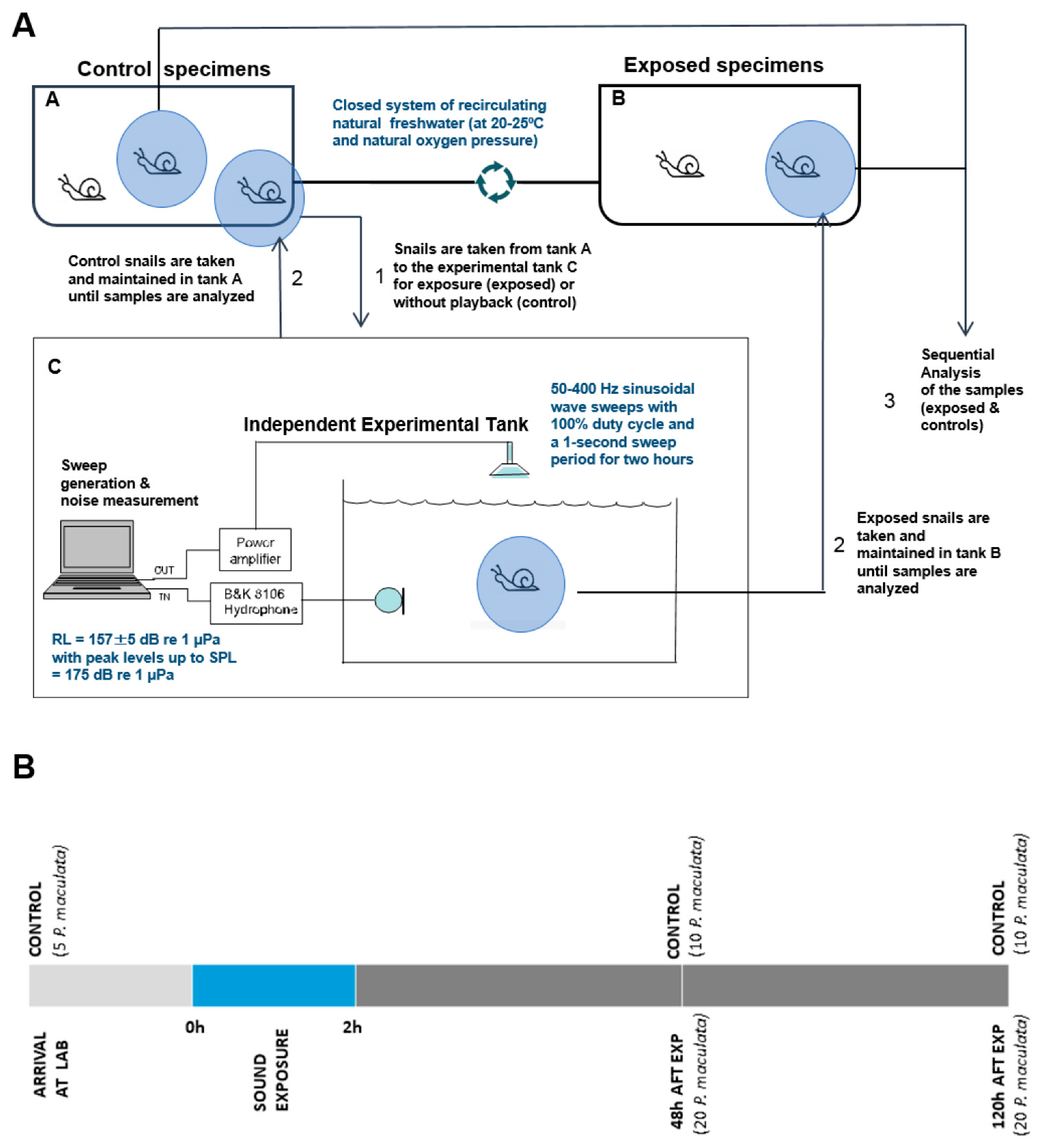
2.1. Sound Exposure Protocol

Sequential Controlled Exposure Experiments (CEE) were conducted on individuals of P. maculata (n = 40). An additional set of individuals (n = 25) was used as control. Individuals were maintained in the LAB maintenance tank system (see detailed system description and exposure protocol in [38]. The maintenance tanks consisted of a closed system where the two tanks (control and exposed animals) were connected. Controls and exposed animals were kept in the same conditions (natural freshwater at 20–25 °C and natural oxygen pressure). The exposure consisted of 50–400 Hz sinusoidal wave sweeps with 100% duty cycle and a 1 s sweep period for two hours. The sweep was produced and amplified through an in-air loudspeaker (QSC KW153) while the level received was measured by a calibrated B&K 8106 hydrophone. Figure 2 displays the power spectral density of the signal received by the hydrophone (RL). The average level calculated over the sweep was 157 ± 5 dB re 1 μ Pa2/Hz with peak levels up to 175 dB re 1 μ Pa. The controls were placed in the exposure tank for as long as those exposed (2 h), without any sound playback (Figure 3A). The sequential sacrificing process after exposure was identical for controls and exposed animals. Five animals were sacrificed to obtain statocyst samples upon arrival at the LAB as initial controls. Following exposure, samples were obtained from individuals (exposed, n = 20; and controls, n = 10) at 48 h and 120 h after sound exposure (Figure 3B).

2.2. Removal of Statocysts

The animals were euthanized with anaesthesia (magnesium chloride 25 g/L), extracted from the shell and the anterior portion of the body was sectioned through a dorso-ventral cut that revealed the statocysts. This anterior body section was detached and chemically fixed for observation and analysis. The two statocysts were analysed for each snail, taking special care to prevent mechanical damage to the tissues and making sure to expose the sensory epithelia overlaid by multiple statoconias.

2.3. Scanning Electron Microscopy

One hundred and thirty statocysts from sixty five P. maculata were used for this study. Fixation was performed in 2.5% glutaraldehyde for 24–48 h at 4 °C. Statocysts embedded on the snail muscular mass were dehydrated in graded ethanol solutions and critical-point dried with CO2 in a Bal-Tec CPD 030 unit (Leica Mycrosystems, Austria). The dried samples were mounted on specimen stubs with double-sided tape. The mounted tissues were gold coated with a Quorum Q150R S sputter coated unit (Quorum Technologies, Ltd. Laughton, East Sussex, United Kingdom) and viewed with a variable pressure Hitachi S-3500N scanning electron microscope (Hitachi High-Technologies Co., Ltd., Tokyo, Japan) at an accelerating voltage of 5 kV at the Institute of Marine Sciences of the Spanish Research Council (CSIC) facilities.

2.4. Quantification and Data Analysis

We selected the sensory areas of the statocyst. The surface of these statocyst areas comprising hair cells was determined for each sample. Sampling squares of 400 µm2 (20 µm × 20 µm) were placed in this area at 20%, 40%, 60%, and 80% along its centre axis (Figure 4). Hair cell damage was analysed by classifying them as intact (hair cell undamaged), damaged (kinocilium or surrounding stereocilia partially or entirely missing or fused), extruded (hair cell partially extruded of the epithelium) and missing (hole in the epithelium caused by the total extrusion of the hair cell). The severity of the lesions was quantified as the percentage of extruded and missing hair cells with respect to the total hair cell count of the sampling square. The damaged category encompassed a wide range of different types of lesions with different severities; this made direct comparison between animals more difficult. The extruded and missing categories were well-defined and easily compared, and the presence of extruded cells showed the limit of severe damage after sound exposure. Two statocysts of each exposed animal were analysed; the hair cell counts of the statocysts were combined to obtain a single measurement per region per animal.
The influence of recovery time after sound exposure in groups of animals sacrificed 48 h vs. 120 h after sound exposure was tested comparing the mean damage count between groups using permutation tests repeated multiple times with N = 1000. To evaluate the difference in damage between regions, the median hair cell counts between all regions (for control and exposed animals at 48 h and 120 h separately) were compared using a Kruskal–Wallis test.

3. Results

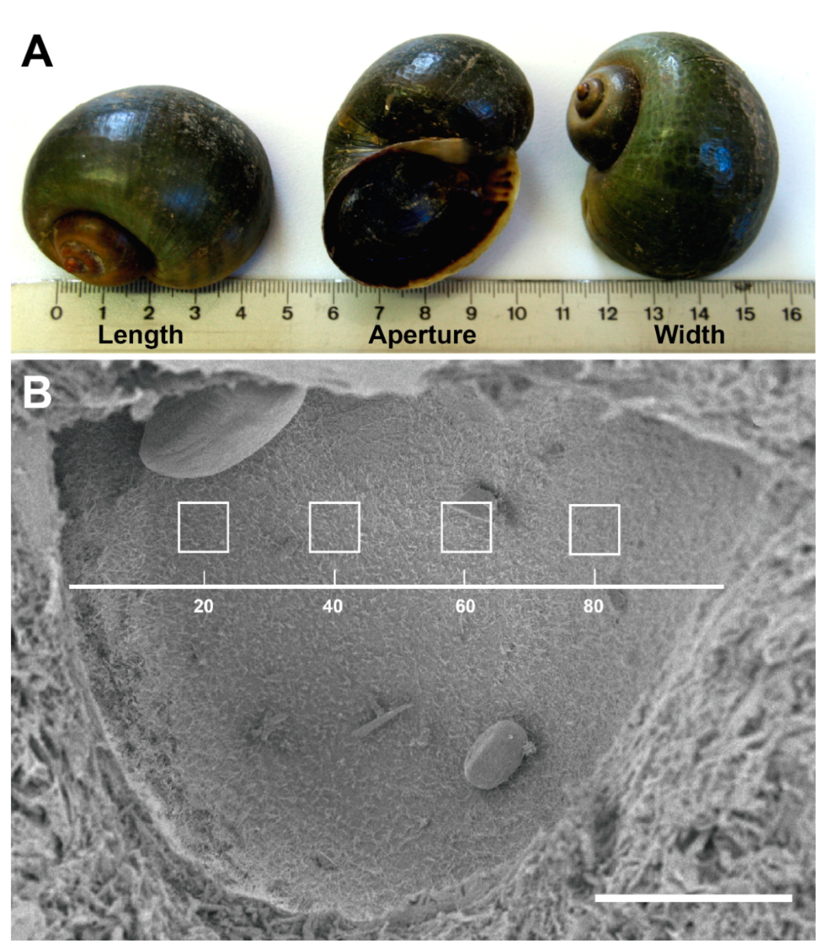
3.1. P. maculata Statocyst Morphology

All the animals used in this study were adult snails with 3–5 cm shell length, 2–4 cm shell width and 4–5 cm shell aperture length (Figure 4A). P. maculata individuals presented two statocysts, 340–350 µm in diameter, located in the foot and linked ventrolaterally to each pleural ganglion (Figure 5A). Statocysts consisted essentially of a cavity, covered by an epithelium of mechanosensitive hair cells, which contained a fluid and a large number of highly morphologically dissimilar aragonite (calcium carbonate) crystals, statoconia (Figure 5B–D). Two types of epithelial cells were found on the statocyst: small supporting cells carrying microvilli and sensory hair cells which in addition of a crown of stereocilia bear one large kinocilia (Figure 5E). The hair cells projected the kinocilia into the lumen of the statocyst, which also contained endolymph and statoconia. The majority of statoconia were oval shaped with smoothened edges (Figure 5B,C). The individual statoconia filled all the statocyst cavity and were constantly moving independently of each other.

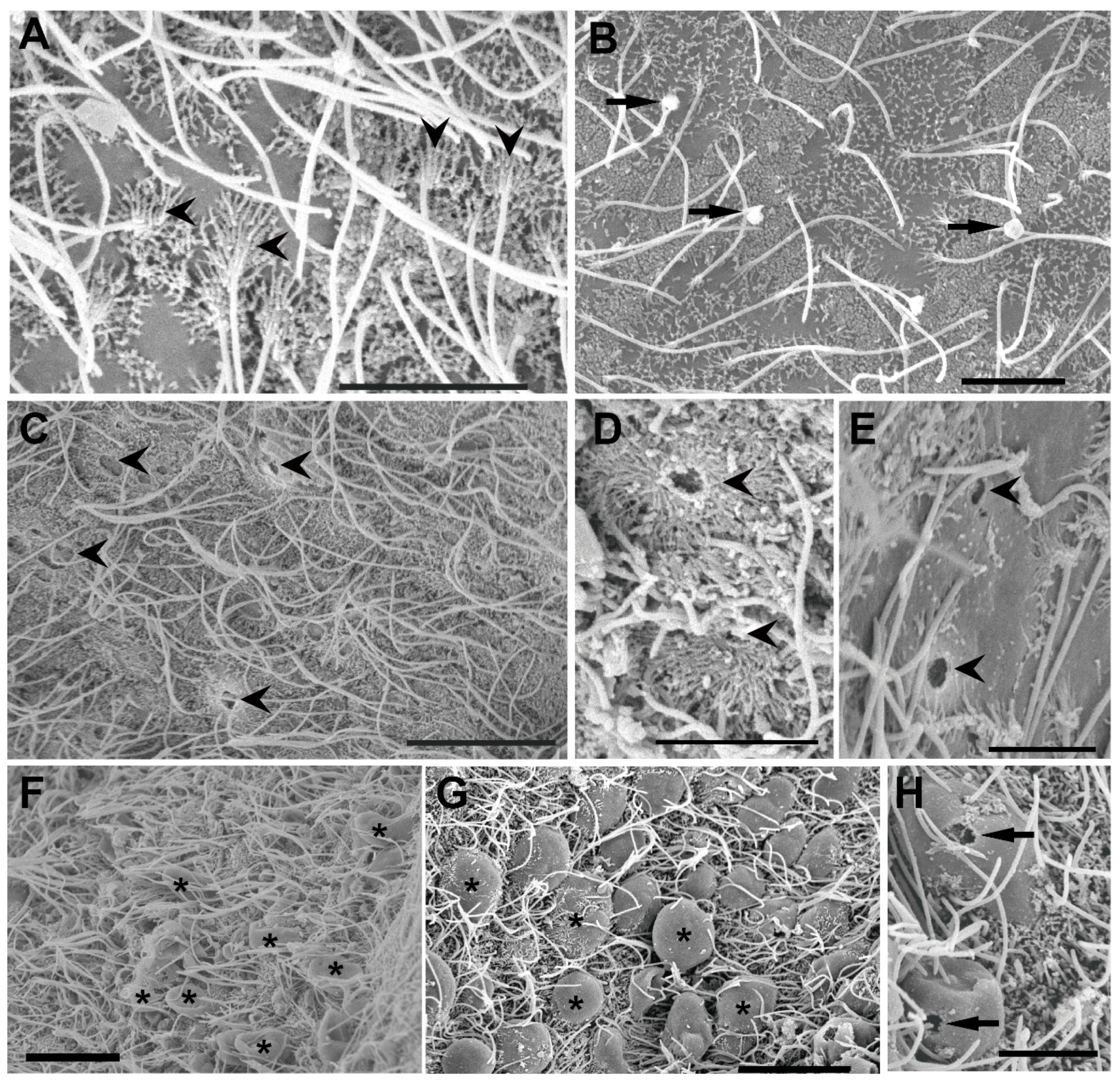
3.2. Ultrastructural Analysis of the Statocyst Sensory Epithelium

The exposed animals presented lesions in the statocyst sensory epithelium that incremented against time. In comparison with the control animals (Figure 5E and Figure 6A), damaged hair cells on exposed animals presented swelling (Figure 6B) or missing (Figure 6C–E) kinocilia or the whole hair cell was missing or extruded (ejected) from the sensory epithelium into the statocysts cavity (Figure 6F–H). A considerable number of hair cells had totally lost the unique kinocilium (Figure 6E) and the crown of stereocilia surrounding the large kinocilium was totally fused (Figure 6D) or lost (Figure 6E). Some ejected hair cells presented spherical holes (Figure 6H), probably due to the cell swelling and the extrusion of the inner cellular material.

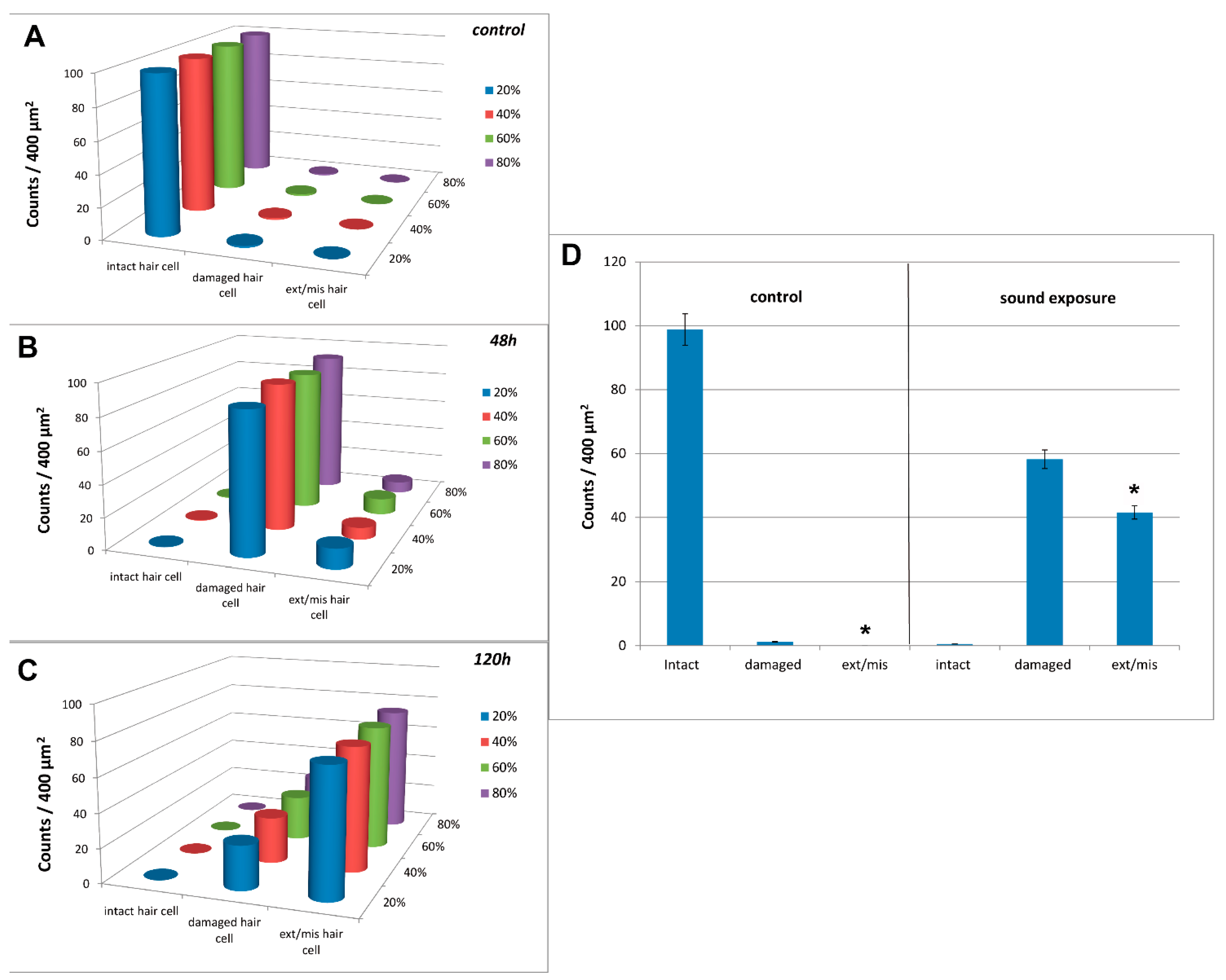
3.3. Image and Data Analysis

The abnormal features we identified on the surface of sound exposed epithelia included damaged (kinocilium or surrounding stereocilia partially or entirely missing or fused), extruded (hair cell extruded from the epithelium) and missing hair cells (hole in the epithelium caused by the total extrusion of the hair cell). The number of damaged, extruded and missing hair cells was counted for each image. The severity of the lesions was chosen to be quantified as the percentage of extruded and missing hair cells because these were well-defined and easy-to-compare categories. The damaged category was not useful for this purpose as with increasing physical damage it was increasing and then decreasing again as a damaged hair cell was at an intermediate state. Therefore, in order to be able to quantify the magnitude of overall damage to the sensory organs “lesion severity” was based on the combination of extruded and missing hair cells. The presence of extruded cells showed the start of severe damage after sound exposure. None of the control animals showed extruded or missing hair cells on the sensory epithelia and all exposed animals had various degrees of damage.
First, we analysed if there was a difference in lesion severity between the different regions. No difference in hair cell counts would allow us to sum the counts over all the regions for each animal, without the need to consider weighting factors. Comparing the median counts between the regions of all control animals (in this case summing the counts across all categories for each region together as the categories considered for lesion severity have a 0 count), each region appeared to have a similar median count with p = 0.074. Performing the same test for the exposed animals, but now considering the extruded and missing categories, we found for the animals sacrificed at 48 h that median counts were dissimilar with p = 0.027 and at 120 h they were similar again with p = 0.73. Taking a closer look at 48 h, we performed the KW test with the regions 40% to 80%, leaving out the 20% region resulting in p = 0.91. Considering these outcomes, we concluded that there was no significant difference in hair cell count or hair cell damage between regions (Figure 7A–C; Figure 8B,C).
Secondly, we tested for a difference in lesions between the control and exposed groups. For this we used the measure of lesion severity as described above. The measurements of all control animals sacrificed at 48 h and 120 h were combined into one group, and similarly all exposed animals were combined into another group. For each animal, all sampling squares in both statocysts were added together to obtain a single measurement per animal. Note that the measure of the lesion severity in the control group (number of extruded missing cells) was 0 for each animal. A permutation test (N = 1000) was performed to test the difference of the mean between the two groups. We concluded that statocyst epithelium was affected by exposure to sound (p = 9.99 × 10⁻⁴) (Figure 7).
Third, we analysed if the measured damage was increasing over time. For this we compared the lesion severity between the groups sacrificed at 48 h and 120 h. Following the approach above to obtain a single measurement per animal, we found a significant difference between these two groups (p = 9.99 × 10⁻⁴; t = −108). The t-value indicated higher damage at 120 h than at 48 h (Figure 8A).
The almost complete expulsion of the sensory cells is a clear sign that the acoustic impact was acute and that the lesion in the sensory epithelium was immediate.

4. Discussion

Our study not only shows the first ultrastructural images of P. maculata statocyst sensory epithelium but also describes pathological changes after sound exposure in these tissues.
The apple snail sensory epithelia and statoconia presented a similar structure as in other pulmonate gastropods [39,40,41]. As in terrestrial pulmonate snails [42] the presence of small and very small statoconia together with large statoconia in the same statocyst suggest their state of generation and growth during the whole life of the animal.
The sensory epithelium hair cell death and damage in vertebrates after loud or prolonged noise exposure or ototoxic drugs can consist of a dysfunction of mechanotransduction complex, loss of ribbon synapses or hair cell death by apoptosis or necrosis [43]. Noise trauma produces an increase in calcium levels in hair cells, which leads to the stimulation of reactive oxygen species (ROS) production. In addition, higher metabolic demand in hair cells during exposure to sound could also lead to increased ROS levels, leading to various forms of damage in the cell (including apoptosis or necrosis) [44]. These dying cells are eliminated from the sensory epithelium by extrusion, a mechanism used to eliminate unfit, excess, or dying cells [45]. The apex of the hair cell or its entire body is expelled from the epithelium [44] and the lost hair cells are replaced by expansion of adjacent supporting cells, which form a scar. This process has been observed in vertebrates [44] and some invertebrates, specifically in cephalopods and cnidarians [32,38]. The lesions observed on P. maculata statocysts sensory epithelium showed similarities in their nature and intensity to those described in cephalopod statocysts (hair cells partially or totally ejected from the sensory epithelium into the statocyst cavity; swollen, bent, fused or lost kinocilia) after being exposed to the same low-frequency sounds [30,46], as well as in cnidarians [32] and crabs [47]. The consequences for the cephalopod’s species exposed to low-frequency sounds were an immediate loss of the ability to orient, mate, and feed. Probably due the similarity of their sensory systems, because of their taxonomic proximity (both are molluscs) sound exposure in P. maculata would affect the physiology and functioning of the P. maculata statocyst. Further investigation is needed to determine threshold levels and to explain these effects in apple snail species.
In regards to pathology findings, there was an increase in the severity of the lesions over time after exposure to low frequency sounds. The increase of the damage severity with time after sound exposure in the sensory epithelia affected by acoustic trauma is a common process described in vertebrate and invertebrate species. In mammals this can provoke the entire destruction of the Corti organ [48]. In birds, hair cells which are affected by apoptosis within the basilar papilla after sound exposure continue degenerating after the beginning of the cell regeneration [44,49]. In invertebrates [30,32,38,50] and plants [33] similar degeneration processes of hair cells increases with time after the exposure.
The Ebre Delta is a particularly sensitive area that has experienced successive introductions of invasive species that have become real pests to their ecosystems. A non-exhaustive list of the most recently introduced species includes red swamp crayfish (Procambarus clarkii), which was initially introduced to the river in 1979 for commercial purposes. Artificial introduction of wels catfish (Silurus glanis), the largest-bodied European freshwater fish, ocurred in 1983. Since 1993, black fly (Simuliidae) have had periodic episodes in the area. Since 1997, piranha (Serrasalmus sp.) individuals have been detected in the Ebre River, probably originating from aquarofilia installations. To date, zebra mussel (Dreissena polymorpha) has been the most aggressive freshwater invader worldwide and is present in Ebre Delta since 2001. Since the beginning of the twentieth century red palm weevil (Rhynchophorus ferrugineus) has been reported as one of the most important pests of ornamental and oil palms and it represents a plague for the palms on the Delta since 2003. In 2008, the first apple snail (P. maculata) was detected. Since 2012 the American bullfrog (Lithobates catesbeianus) and American blue crab (Callinectes sapidus) have become new plagues in the Ebre Delta [8]. After some years of detection of the P. maculata pest in the Ebre Delta a new invasive species was established in the same area, the American blue crab Callinectes sapidus. Although this species has been present in Europe (Atlantic cost of France, North Sea, Mediterranean Sea, Baltic Sea, Black Sea) since the early years of the 20th century [51], where multiple independent introductions may have taken place through ballast water, it was not detected at the Ebre Delta until 2012 [52]. The future impact of this new pest on the Delta ecosystem is unclear, but at this moment the crab is acting as a biocontrol agent of P. maculata, reducing its population by feeding on them. In any case, the introduction into the ecosystem of a foreign species that is particularly voracious and has a high reproduction rate, such as C. sapidus, cannot be a long-term solution because of the problems that accompany such an invasion.
Different treatments have been used to fight the P. maculata rice crops invasion of Ebre Delta: sea water flooding of the rice fields, chemical treatments with saponin, winter drying, channel cleaning, manual removal of the eggs clutches, traps, and advance obstructions [53]. None of these treatments had an acceptable success, and due to the severe infestation of the species in the area a total eradication is non-viable. In this context, the use of an alternative treatment such as exposure to sound would be an innovative and ecological option. The results obtained in the present study are consistent with the hypothesis that exposure to sound may impair the sensory perception of P. maculata. Sound-induced damage to the statocyst could likely result, as in cephalopods [38], in an inhibition of its vital functions, which would result in a dramatic reduction of the survival ability of the apple snail. Cellular effects, which can trigger behavioral effects (e.g., in cephalopods, loss of appetite and a decrease in the reproductive rate) would be an effective measure to fight apple snail plagues over the world. However, further efforts should analyse the possible interactions of the method with other trophic levels in the ecosystem, in order to manage the problem holistically and avoid damage to other native species. This is essential to provide global benefits for complex communities. The choice of the sound parameters (frequencies, amplitudes, and exposure times) used during this study was made for the sole purpose of verifying the possible harmful effects of exposure to acoustic sources on apple snails and not to determine a precise threshold for inducing effective lesions. Further investigation is also needed to determine the levels that produce lesions in association with behavioural changes, which would result in a pest reduction, while at the same time being innocuous for the other species and the ecosystem as a whole.
This method is patented (WO 2018/167003 A1) and could become a valuable approach to mitigate the invasions of any aquatic alien species that are expected to interfere with ecological communities due to the increasing negative effects of climate change.

5. Patent

André M., Solé M., Van der Schaar, De Vreese S (International Patent WO 2018/167003 A1). 20-09-2018. A method for inducing lethal lesions in sensory organs of undesirable aquatic organisms by use of sound. Licensed to SEASEL SOLUTIONS AS [NO/NO]; P.O.BOX 93 N-6282 BRATTVÅG (NO).

Author Contributions

M.S. and M.A. planned the research and designed the study. M.S., M.A. and M.v.d.S. conducted experimental/lab work. M.S. and J.-M.F. performed SEM analysis. M.S. and M.v.d.S. analysed the data. M.S. prepared the figures. M.S. wrote the article and all authors reviewed the manuscript. All authors have read and agreed to the published version of the manuscript.

Funding

This research received no external funding.

Institutional Review Board Statement

Not applicable.

Informed Consent Statement

Not applicable.

Data Availability Statement

The authors declare that the data supporting the findings of this study are available within the paper.

Conflicts of Interest

The authors declare no conflict of interest.

References

  1. Clarke, B.; Murray, J.; Johnson, M.S. The extinction of endemic species by a program of biological control. Pac. Sci. 1984, 38, 97–104. [Google Scholar]
  2. Savidge, J.A. Extinction of an Island Forest Avifauna by an Introduced Snake. Ecology 1987, 68, 660–668. [Google Scholar] [CrossRef]
  3. David, P.; Thébault, E.; Anneville, O.; Duyck, P.-F.; Chapuis, E.; Loeuille, N. Impacts of Invasive Species on Food Webs; Elsevier: Amsterdam, The Netherlands, 2017; pp. 1–60. [Google Scholar] [CrossRef]
  4. Galiana, N.; Lurgi, M.; Montoya, J.M.; López, B.C. Invasions cause biodiversity loss and community simplification in vertebrate food webs. Oikos 2014, 123, 721–728. [Google Scholar] [CrossRef]
  5. Rahel, F.J.; Olden, J. Assessing the Effects of Climate Change on Aquatic Invasive Species. Conserv. Biol. 2008, 22, 521–533. [Google Scholar] [CrossRef]
  6. Barnes, M.A.; Fordham, R.K.; Burks, R.L.; Hand, J.J. Fecundity of the exotic applesnail, Pomacea insularum. J. North Am. Benthol. Soc. 2008, 27, 738–745. [Google Scholar] [CrossRef]
  7. Rawlings, T.A.; Hayes, K.A.; Cowie, R.H.; Collins, T.M. The identity, distribution, and impacts of non-native apple snails in the continental United States. BMC Evol. Biol. 2007, 7, 97. [Google Scholar] [CrossRef] [PubMed] [Green Version]
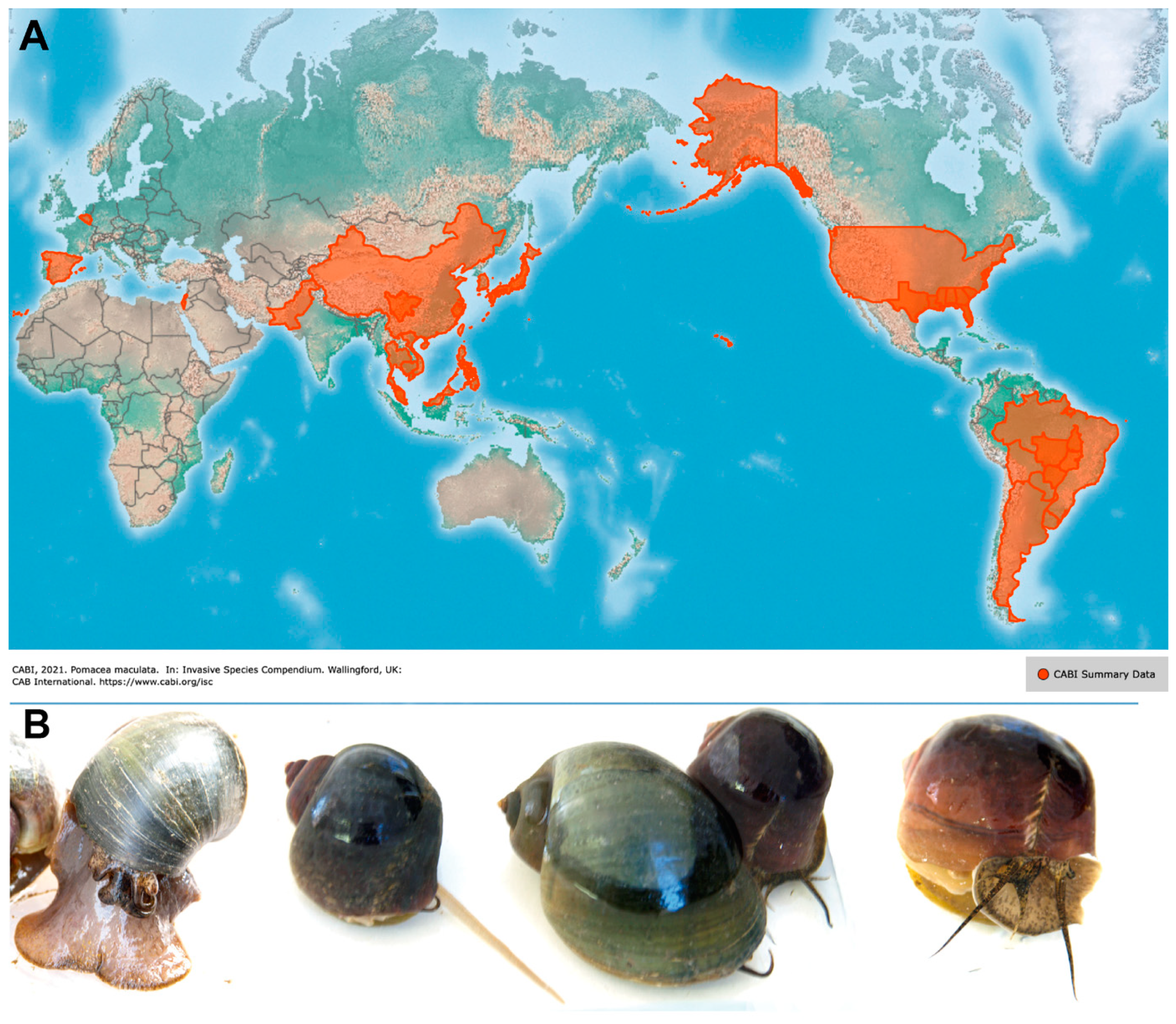
  8. CABI. Pomacea maculata. In Invasive Species Compendium; CAB International: Wallingford, UK, 2019; Available online: https://www.cabi.org/isc/datasheet/116486#toDistributionMaps (accessed on 24 August 2021).
  9. Naylor, R. Invasions in agriculture: Assessing the cost of the golden apple snail in Asia. Ambio 1996, 25, 443–448. [Google Scholar]
  10. Yang, T.-B.; Wu, Z.-D.; Lun, Z.-R. The Apple Snail Pomacea canaliculata, A Novel Vector of the Rat Lungworm, Angiostrongylus cantonensis: Its Introduction, Spread, and Control in China. Hawai'i. J. Med. Public Health J. Asia Pac. Med. Public Health 2013, 72, 23–25. [Google Scholar]
  11. Martin, C.W.; Bayha, K.M.; Valentine, J.F. Establishment of the Invasive Island Apple Snail Pomacea insularum (Gastropoda: Ampullaridae) and Eradication Efforts in Mobile, Alabama, USA. Gulf Mexico Sci. 2012, 1–2, 30–38. [Google Scholar] [CrossRef]
  12. Panel, E.; Plh, H.; Baker, R.; Candresse, T.; Simon, E.D.; Gilioli, G.; Grégoire, J.; Puglia, P.; Rafoss, T.; Rossi, V.; et al. Scientific Opinion on the evaluation of the pest risk analysis on Pomacea insularum, the island apple snail, prepared by the Spanish Ministry of Environment and Rural and Marine Affairs. EFSA J. 2012, 10. [Google Scholar] [CrossRef]
  13. Hayes, K.A.; Joshi, R.C.; Thiengo, S.; Cowie, R.H. Out of South America: Multiple origins of non-native apple snails in Asia. Divers. Distrib. 2008, 14, 701–712. [Google Scholar] [CrossRef]
  14. Roll, U.; Dayan, T.; Simberloff, D.; Mienis, H.K. Non-indigenous land and freshwater gastropods in Israel. Biol. Invasions 2008, 11, 1963–1972. [Google Scholar] [CrossRef]
  15. Levin, P.; Cowie, R.; Taylor, J.; Hayes, K.; Burnett, K.; CA, F. Apple snail invasions and the slow road to control: Ecological, economic, agricultural, and cultural perspectives in Hawaii. In Global Advances in Ecology and Management of Golden Apple Snails; Joshi, R.C., Sebastian, L.S., Eds.; Philippine Rice Research Institute (PhilRice): Los Baños, Philippines, 2006; pp. 325–335. [Google Scholar]
  16. Morrison, W.E.; Hay, M.E. Herbivore Preference for Native vs. Exotic Plants: Generalist Herbivores from Multiple Continents Prefer Exotic Plants That Are Evolutionarily Naïve. PLoS ONE 2011, 6, e17227. [Google Scholar] [CrossRef] [PubMed] [Green Version]
  17. Cowie, R. Apple snails (Ampullariidae) as agricultural pests: Their biology, impacts and management. In Molluscs as Crop Pests; Barker, G.M., Ed.; CABI: Los Baños, Philippines, 2002; pp. 145–192. [Google Scholar]
  18. Ramakrishnan, V. Salinity, pH, Temperature, Desiccation and Hypoxia Tolerance in the Invasive Freshwater Apple Snail Pomacea insularum. Ph.D. Thesis, University of Texas at Arlington, Arlington, TX, USA, 2007. [Google Scholar]
  19. Carlsson, N.; Kestrup, Å.; Mårtensson, M.; Nyström, P. Lethal and non-lethal effects of multiple indigenous predators on the invasive golden apple snail (Pomacea canaliculata). Freshw. Biol. 2004, 49, 1269–1279. [Google Scholar] [CrossRef]
  20. Warren GL Nonindigenous freshwater invertebrates. In Strangers in Paradise; Simberloff, D.; Schmitz, D.C.; Brown, T.C. (Eds.) Island Press: Washington, DC, USA, 1997; pp. 101–108. [Google Scholar]
  21. Wood, T.S.; Anurakpongsatorn, P.; Chaichana, R.; Mahujchariyawong, J.; Satapanajaru, T. Heavy Predation on Freshwater Bryozoans by the Golden Apple Snail, Pomacea canaliculata Lamarck, 1822 (Ampullariidae). Nat. Hist. J. Chulalongkorn Univ. 2006, 6, 31–36. [Google Scholar]
  22. Carlsson, N.O.L.; Brönmark, C.; Hansson, L.-A. Invading herbivory: The golden apple snail alters ecosystem functioning in asian wetlands. Ecology 2004, 85, 1575–1580. [Google Scholar] [CrossRef] [Green Version]
  23. Cattau, C.E.; Martin, J.; Kitchens, W.M. Effects of an exotic prey species on a native specialist: Example of the snail kite. Biol. Conserv. 2010, 143, 513–520. [Google Scholar] [CrossRef]
  24. Way, M.; Islam, Z.; Heong, K.; Joshi, R. Ants in tropical irrigated rice: Distribution and abundance, especially of Solenopsis geminata (Hymenoptera: Formicidae). Bull. Èntomol. Res. 1998, 88, 467–476. [Google Scholar] [CrossRef]
  25. Departament D’agricultura Alimentació i Acció Rural. Generalitat de Catalunya Ordre AAR/404/2010, de 27 de Juliol, per la Qual es Declara Oficialment l’existència d’un Focus del Cargol Poma (Pomacea sp.) A L’hemidelta Esquerra del Delta de l’Ebre; DOGC: Catalonia, Spain, 2010; Available online: https://portaljuridic.gencat.cat/eli/es-ct/o/2010/07/27/aar404 (accessed on 24 August 2021).
  26. Budelmann, B.U. Hearing in N onarthropod Invertebrates. In The Evolutionary Biology of Hearing; Webster, D.B., Fay, R.R., Eds.; Springer: New York, NY, USA, 1992; pp. 141–155. [Google Scholar]
  27. Wiederhold, M.L.; Sharma, J.S.; Driscoll, B.P.; Harrison, J.L. Development of the statocyst in Aplysia californica I. Observations on statoconial development. Hear. Res. 1990, 49, 63–78. [Google Scholar] [CrossRef]
  28. Balaban, P.M.; Malyshev, A.Y.; Ierusalimsky, V.; Aseyev, N.; Korshunova, T.A.; Bravarenko, N.I.; Lemak, M.S.; Roshchin, M.; Zakharov, I.S.; Popova, Y.; et al. Functional Changes in the Snail Statocyst System Elicited by Microgravity. PLoS ONE 2011, 6, e17710. [Google Scholar] [CrossRef] [Green Version]
  29. Day, R.D.; McCauley, R.D.; Fitzgibbon, Q.P.; Hartmann, K.; Semmens, J.M. Assessing the Impact of Marine Seismic Surveys on Southeast Australian Scallop and Lobster Fisheries; University of Tasmania: Hobart, Tasmania, 2016; ISBN 9780646959108. [Google Scholar]
  30. Solé, M.; Sigray, P.; Lenoir, M.; Van Der Schaar, M.; Lalander, E.; André, M. Offshore exposure experiments on cuttlefish indicate received sound pressure and particle motion levels associated with acoustic trauma. Sci. Rep. 2017, 7, 45899. [Google Scholar] [CrossRef] [Green Version]
  31. Solé, M.; Monge, M.; André, M.; Quero, C. A proteomic analysis of the statocyst endolymph in common cuttlefish (Sepia officinalis): An assessment of acoustic trauma after exposure to sound. Sci. Rep. 2019, 9, 1–12. [Google Scholar] [CrossRef]
  32. Solé, M.; Lenoir, M.; Fontuño, J.M.; Durfort, M.; Van Der Schaar, M.; André, M. Evidence of Cnidarians sensitivity to sound after exposure to low frequency noise underwater sources. Sci. Rep. 2016, 6, 37979. [Google Scholar] [CrossRef] [Green Version]
  33. Solé, M.; Lenoir, M.; Durfort, M.; Fortuño, J.-M.; van der Schaar, M.; De Vreese, S.; André, M. Seagrass Posidonia is impaired by human-generated noise. Commun. Biol. 2021, 4, 1–11. [Google Scholar] [CrossRef]
  34. La Bella, G.; Cannata, S.; Froglia, C.; Ratti, S.; Rivas, G. First assessment of effects of air-gun seismic shooting on marine resources in the Central Adriatic Sea. Int. Conf. Heal. Saf. Environ. Oil Gas Explor. Prod. 1996, 1, 227–238. [Google Scholar] [CrossRef]
  35. Stocks, J.R. Response of Marine Invertebrate Larvae to Natural and Anthropogenic Sound: A Pilot Study. Open Mar. Biol. J. 2012, 6, 57–61. [Google Scholar] [CrossRef]
  36. Nedelec, S.L.; Radford, A.N.; Simpson, S.D.; Nedelec, B.; Lecchini, D.; Mills, S.C. Anthropogenic noise playback impairs embryonic development and increases mortality in a marine invertebrate. Sci. Rep. 2014, 4, srep05891. [Google Scholar] [CrossRef] [PubMed] [Green Version]
  37. De Soto, N.A.; Delorme, N.; Atkins, J.; Howard, S.; Williams, J.M.; Johnson, M.J.A. Anthropogenic noise causes body malformations and delays development in marine larvae. Sci. Rep. 2013, 3, 2831. [Google Scholar] [CrossRef] [PubMed] [Green Version]
  38. Solé, M.; Lenoir, M.; Durfort, M.; López-Bejar, M.; Lombarte, A.; van der Schaar, M.; André, M. Does exposure to noise from human activities compromise sensory information from cephalopod statocysts? Deep. Sea Res. Part II Top. Stud. Oceanogr. 2013, 95, 160–181. [Google Scholar] [CrossRef]
  39. Stahlschmidt, V.; Wolff, H.G. The fine structure of the statocyst of the prosobranch mollusc Pomacea paludosa. Z. Mikroskop. Anat. 1972, 133, 529–537. [Google Scholar] [CrossRef] [PubMed]
  40. Zaitseva, O. Structural organization of the sensory systems of the snail. Neurosci. Behav. Physiol. 1994, 24, 47–57. [Google Scholar] [CrossRef]
  41. Kononenko, N.L.; Kiss, T.; Elekes, K. The neuroanatomical and ultrastructural organization of statocyst hair cells in the pond snail, Lymnaea stagnalis. Acta Biol. Hung. 2012, 63, 99–113. [Google Scholar] [CrossRef] [PubMed]
  42. Gorgiladze, G.I.; Bukia, R.D.; Davitashvili, M.T.; Taktakishvili, A.D.; Gelashvili, N.S.; Kalandarishvili, E.L.; Satdykova, G.P. Morphological peculiarities statoconia in statocysts of terrestrial pulmonary snail Helix lucorum. Bull. Exp. Biol. Med. 2010, 149, 269–272. [Google Scholar] [CrossRef]
  43. Wagner, E.L.; Shin, J.-B. Mechanisms of Hair Cell Damage and Repair. Trends Neurosci. 2019, 42, 414–424. [Google Scholar] [CrossRef]
  44. Leonova, E.V.; Raphael, Y. Organization of cell junctions and cytoskeleton in the reticular lamina in normal and ototoxically damaged organ of Corti. Hear. Res. 1997, 113, 14–28. [Google Scholar] [CrossRef]
  45. Atieh, Y.; Wyatt, T.; Zaske, A.M.; Eisenhoffer, G.T. Pulsatile contractions promote apoptotic cell extrusion in epithelial tissues. Curr. Biol. 2021, 31, 1129–1140. [Google Scholar] [CrossRef] [PubMed]
  46. Solé, M.; Lenoir, M.; Fortuño, J.-M.; van der Schaar, M.; André, M. A critical period of susceptibility to sound in the sensory cells of cephalopod hatchlings? Biol. Open 2018, 7, bio033860. [Google Scholar] [CrossRef] [PubMed] [Green Version]
  47. Day, R.D.; McCauley, R.D.; Fitzgibbon, Q.; Hartmann, K.; Semmens, J.M. Exposure to seismic air gun signals causes physiological harm and alters behavior in the scallop Pecten fumatus. Proc. Natl. Acad. Sci. USA 2017, 114, E8537–E8546. [Google Scholar] [CrossRef] [PubMed] [Green Version]
  48. Spoendlin, H. Primary Structural Changes in the Organ of Corti After Acoustic Overstimulation. Acta Oto Laryngolog. 1971, 71, 166–176. [Google Scholar] [CrossRef]
  49. Corwin, J.; Cotanche, D. Regeneration of sensory hair cells after acoustic trauma. Science 1988, 240, 1772–1774. [Google Scholar] [CrossRef] [PubMed]
  50. Solé, M.; Lenoir, M.; Fortuño, J.-M.; De Vreese, S.; van der Schaar, M.; André, M. Sea Lice Are Sensitive to Low Frequency Sounds. J. Mar. Sci. Eng. 2021, 9, 765. [Google Scholar] [CrossRef]
  51. Nehring, S. Invasion History and Success of the American Blue Crab Callinectes sapidus in European and Adjacent Waters. In the Wrong Place Alien Marine Crustaceans: Distribution, Biology and Impacts; Springer: Amsterdam, The Netherlands, 2011; pp. 607–624. [Google Scholar]
  52. Castejón, D.; Guerao, G. A new record of the American blue crab, Callinectes sapidus Rathbun, 1896 (Decapoda: Brachyura: Portunidae), from the Mediterranean coast of the Iberian Peninsula. BioInvasions Rec. 2013, 2, 141–143. [Google Scholar] [CrossRef]
  53. Generalitat de Catalunya. Memòria D’actuacions de Control del Cargol Poma. Periode 2015–2018. 2018. Available online: http://mediambient.gencat.cat/web/.content/home/ambits_dactuacio/patrimoni_natural/especies_exotiques_medinatural/llista_sp_catalogades/invertebrat/cargol/Memoria-cargol-poma.pdf (accessed on 24 August 2021).
Figure 1. (A): P. maculata world distribution. Reproduced with permission from CABI 2021 [8] (B): Different images of P. maculata specimens used in the experiments.
Jmse 09 00969 g001
Figure 2. Power spectral density of the 50 to 400 Hz sweep as received by the hydrophone on top with below the background noise level recorded just before exposure.
Jmse 09 00969 g002
Figure 3. Sound exposure protocol, sampling collection, and analysis. (A): Sound exposure protocol. P. maculata were maintained in tank A until some were transferred to an independent experimental tank C where they were exposed to sound (1). At the end of the exposure experiments, the snails were transferred to tank B (2) that presented the same environmental conditions as tank A. Control specimens were transferred to tank C for 2 h without any playback and after that, they were taken back to tank A (2). Samples of control and exposed plants were sequentially taken for analysis (3). (Figure modified from 38). (B): Sampling collection and analysis. Before the sound exposure started, control specimens were taken and analyzed at the arrival of the laboratory facilities. Samples of control and exposed snails were sequentially analyzed at 48 h and 120 h after sound exposure.
Jmse 09 00969 g003
Figure 4. (A): Each individual of P. maculata was characterized through its shell dimensions (length, width and aperture length). (B): Hair cell bundle count locations (sampling squares) on the P. maculata statocyst. Hair cell counts were sampled at four predetermined locations: 20%, 40%, 60% and 80% of the total statocysts length. A 400 µm2 box was placed at each sampling area and hair cells were counted within each box. Scale bar: (B) = 100 µm.
Jmse 09 00969 g004
Figure 5. SEM. Internal morphology of P. maculata statocyst. Control animal. (A): Dorso-ventral cutting of the body anterior portion showing the statocyst location in the snail foot and linked ventrolaterally to each pleural ganglion (PG: pleural ganglion, St: statocyst). (B): Opened statocyst completely filled with statoconia (asterisk). (C): Differently sized statoconia. (D): Internal cavity of the statocyst covered by the sensory epithelium. Some of the aragonite crystals are visible (asterisk). (E): Inner statocyst sensory epithelia. Arrowheads point to the hair cells exhibiting their lonely kinocilia surrounded by a crown of stereocilia. Between them microvilli of the supporting cells are visible (arrows). Scale bar: (A) = 1 mm. (D) = 200 µm. (B) = 100 µm. (C) = 50 µm. (E) = 5 µm.
Jmse 09 00969 g005
Figure 6. SEM. P. maculata statocyst sensory epithelium. (A): Control animal. (BD): 48 h after sound exposure. (FH): 120 h after sound exposure. (A): Healthy hair cells show the kinocilia surrounded by a crown of stereocilia (arrowheads). (B): Some hair cells present swollen kinocilia (arrows). (C): Arrowheads point to the holes left by the single kinocilia after their ejection. (D): Detail from (C). Surrounding the hole left by the ejected kinocilia a crown of stereocilia are fused in a compact structure. (E): Detail from (C). The hair cells have lost the entire kinocilia and the crown of stereocilia (arrowheads). (F): The hair cells show severe damage. Most hair cells are (asterisks) ejected from the sensory epithelium. (G): Detail from (F), shows the cellular body of the sensory cells extruded from the epithelium (asterisks). (H): Two extruded hair cells show spherical holes in the body due to the extrusion of the inner cellular material (arrows). Their kinocilia has lost the stereocilia crown. Scale bar: (A,B,D,E,H) = 5 µm. (C,G) = 10 µm. (F) = 20 µm.
Jmse 09 00969 g006
Figure 7. (AC): Mean intact, damaged, and extruded/missing hair cells at 20%, 40%, 60%, and 80% of the total length of sensory epithelium on P. maculata statocyst (48 h and 120 h after sound exposure versus control animals). Note the increase of damaged, extruded, and missing cells versus controls with increase in time. (D): Mean (±SE) intact, damaged and extruded/missing hair cells after sound exposure (n = 80) versus control (n = 50) on statocyst sensory epithelium of P. maculata. Each bar is the average over the 4 regions with the line indicating the standard error. The percentage was computed by dividing by the total count for each individual sample. The lesion severity (see text for definition) of the sound exposure group was found to be significantly higher than that of the control group with p = 9.99 × 10⁻⁴ (* significant difference).
Jmse 09 00969 g007
Figure 8. (A): Mean (±SE) intact, damaged, extruded/missing hair cells over the total length of sensory epithelium on P. maculata statocyst (48 h versus 120 h after sound exposure). The lesion severity was found to be significantly higher at 120 h than at 48 h (p = 9.99 × 10⁻⁴; n = 40). (* significant difference). (B): Hair cell counts between the different regions in animals 48 h after sound exposure. The lesion severity was found to be similar between 40%, 60%, and 80% regions (p = 0.91; n = 40; see text for 20% region consideration). (C): Hair cell count between the different regions in animals 120 h after sound exposure. Each bar is the average over the 4 regions with the line indicating the standard error. The percentage was computed by dividing with the total count for each individual sample. The lesion severity was found to be similar between all regions (p = 0.73; n = 40).
Jmse 09 00969 g008
Publisher’s Note: MDPI stays neutral with regard to jurisdictional claims in published maps and institutional affiliations.

