Communication

Proteome Response of Meretrix Bivalves Hepatopancreas Exposed to Paralytic Shellfish Toxins Producing Dinoflagellate Gymnodinium catenatum

Department of Science, School of Science and Technology, Hong Kong Metropolitan University, Hong Kong, China
*
Author to whom correspondence should be addressed.
J. Mar. Sci. Eng. 2021, 9(9), 1039; https://doi.org/10.3390/jmse9091039
Submission received: 15 August 2021 / Revised: 14 September 2021 / Accepted: 15 September 2021 / Published: 21 September 2021
(This article belongs to the Special Issue Marine Omics & Biotechnology)

Abstract

:
Paralytic shellfish toxins (PSTs) contamination of seafood has become a growing global problem. However, the molecular response of bivalves, some of the most popular seafoods, to PSP toxins has seldom been reported and the underlying molecular mechanisms of the interactions between Meretrix meretrix bivalves and PSTs-producing dinoflagellates are scarcely known. This study compared the protein expression profiles between PSP toxin-contaminated and non-PSP toxin contaminated M. meretrix, determined proteome responses and identified potential biomarkers based on feeding experiments. Results showed that the content of total PSP toxins in contaminated bivalves was 40.63 ± 4.08 μg saxitoxin (STX) equivalents per gram, with 95.3% in hepatopancreas, followed by gill (1.82%) and foot (1.79%). According to two-dimensional gel electrophoresis (2-DE), 15 differentially expressed proteins (at least 2-fold difference) between the hepatopancreas of bivalves with and without PSP toxins were detected. Eight of them were successfully identified by MALDI-TOF MS. These were catalase, protein ultraspiracle homolog, G2 and S phase-expression protein, paramyosin, Mn-superoxide dismutase, response regulator receiver domain-containing protein, sarcoplasmic calcium-binding protein and major facilitator superfamily transporters. The differences in the expression levels of the last three proteins involving in cell signaling, structure and membrane transport were 4.2, 5.3 and 4.9-fold, respectively. These proteins could be further developed as potential biomarkers. The other two up-regulated proteins, Mn-superoxide dismutase and catalase, were involved in cell defence mechanisms against oxidative stress, suggesting PSP toxin acts as xenobiotics and poses oxidative stress in bivalves. This study gives insights into the response of bivalves to PSP toxin-producing dinoflagellate at the proteomic level and the potential of using 2-DE to develop specific protein markers in bivalves.

1. Introduction

Many bivalves like Meretix, Ruditapes, Saccostrea and Mytilus are major foods in our diets. Marine bivalve mollusks feed on phytoplankton, but some phytoplankton are toxigenic species producing bioactive toxins that can lead to shellfish poisoning [1,2]. Toxigenic microalgae are also one of the causative agents for harmful algal blooms (HABs). Bivalves have the ability and capacity to accumulate high levels of shellfish poisoning in their tissues during a toxigenic HAB event [3,4]. Such contaminated shellfishes are then toxic to human [5,6]. The outbreaks of HABs occurring in coastal water globally cause serious health problems through the consumption of highly toxic bivalve species and also lead to severe annual losses of several billions of US dollars in aquaculture areas.
Majority of shellfish poisoning is produced by marine microalgae, includinig paralytic shellfish poisoning (PSP), ciguatera fish poisoning (CFP), diarrheic shellfish poisoning (DSP), amnesic shellfish poisoning (ASP), neurotoxic shellfish poisoning (NSP) and azaspiracid shellfish poisoning (AZP) [7]. Among these, PSP is the most common shellfish poisoning in bivalves because of their wide distribution, where saxitoxin (STX) is the main PSP toxin. STX acts as blockages of voltage-gated sodium channels of neuronal cell membranes, causing partial paralysis or cardiorespiratory arrest [8]. It has been reported that some of the phytoplankton species in dinoflagellate genera such as Alexandrium, Gymnodinium and Pyrodinium are the main PSP toxins producers with more than 20 analogues of paralytic shellfish toxins (PSTs), that are commonly divided into three groups, namely carbamate, N-sulfocarbamoyl and decarbamoyl groups [7,8,9,10,11].
It is difficult to know whether bivalves are contaminated with PSTs or not in Nature, as the contaminated bivalve cannot be easily identified by their appearance, smell or taste [12]. Bivalves have certain tolerance to PSTs as they have corresponding defence systems. They can accumulate, detoxify, metabolize and eliminate PSTs from their body (depuration) during a certain exposure period [12,13,14]. It has been found that the digestive gland of bivalves contains a high amount of total PSTs content, but PSTs decrease rapidly if the bivalve stops its ingestion of PST-producing dinoflagellates [15,16]. The composition of PSTs toxins in bivalves can be different from that of the PST-producing dinoflagellate [17,18]. In recent decades, mouse bioassays have been used as a standard detection method for PSTs, however, living animals have relatively low specificity and sensitivity [19]. Different analytical methods such as liquid chromatography mass spectrometry and enzyme-linked immunosorbent assay (ELISA) have been developed, but these methods require high amounts of standard solutions [20,21]. So far, studies on PSTs focus mainly on the occurrence, exposure, biotransformation, physiological and behavioural responses of PSTs in bivalves, but little is known about the molecular responses of the bivalves such as the change of protein profiles or gene expressions after exposure to PSTs contamination [12,22].
Two-dimensional gel electrophoresis (2-DE) has been described as an important tool for proteome, food quality and safety assessment for marine mussels [23]. It remains widely useful to characterize complex biologically functional proteins. In recent years, proteomics based on two-dimensional gel electrophoresis and mass spectrometry have been used to assess the effects in protein expression of mussels and other aquatic crustaceans exposed to toxins produced by cyanobacteria and other environmental stresses, as well as to search protein biomarkers in the field of pollution monitoring [24,25,26]. Previous studies mainly focused on proteomic responses of aquatic organisms exposed to metal stresses [27], bacterial or viral pathogens [28,29], and parasites [30]. To the best of our knowledge, proteomic responses of Meretrix bivalves exposed to the toxin-producing dinoflagellates Gymnodinium catenatum have never been studied, despite the fact they are common cultured species and are often reported as HAB species in Mainland China [31,32] and other parts of the world [33]. The present study therefore aims to to explore the molecular response of bivalves upon exposure to PST-producing HAB species through elucidating the PSTs composition and protein profile of Meretrixmeretrix fed with G. catenatum based on proteomics. The study also attempts to identify possible protein biomarkers (the differentially expressed proteins) by comparing protein profiles between treated and non-PSTs treated bivalves based on 2-DE and matrix-assisted laser desorption/ionization time-of-flight mass spectrometry (MALDI-TOF MS). M. meretrix (Bivalvia, Veneridae) is commonly distributed in East and Southeast Asia, and lives in sediments of the neritic intertidal zone along the coast of China [34]. This hard clam is both farmed and captured wild in China, with significant ecological and economic values (the price for one kilogram of the clams has increased from ¥10 to more than ¥100 in recent years) [35,36,37]. This research not only improves our understanding of possible defense mechanism and proteome changes of bivalves against the stress introduced by HAB species, the identified differentially expressed proteins also serve as potential biomarkers for the rapid detection of shellfish toxin contamination in aquatic ecosystems.

2. Materials and Methods

2.1. Experimental Animals

Marine bivalves, M. meretrix (64–82 mm shell length), were collected from Hong Kong during the period of no PSP toxicity in shellfish. The species was identified and confirmed by the gene sequence based on the partial region of cytochrome C oxidase subunit I (COI) DNA (around 710 bp). The bivalves were transferred to the laboratory and rinsed with natural seawater to remove attached macro-algae and epibionts. The cleaned bivalves were then placed in a 400 L glass tank containing 250 L of natural seawater at 21–22 °C for two-weeks acclimation. The seawater was collected from coastal environment in Hong Kong with a salinity 30–31 ppt and pH 7.8, filtered with 0.2 µm pore size cellulose nitrate filter membrane and autoclaved at 1–2 bar, 125 °C in saturated steam before use. The tank was equipped with a recirculation and aeration system. During the acclimation period, all bivalves were fed daily with a non-toxic green microalgae species, Dunaliella tertiolecta, at a rate of 1% of their tissues dry biomass in order to maintain the algal cell density in 104 cells per mL [38], and the natural seawater was renewed once a week.

2.2. Cultivation of Microalgae

The monocultures of toxic G. catenatum (GYM) and non-toxic D. tertiolecta microalgae were both isolated from the coastal water in Hong Kong. G. catenatum was used in this study because our preliminary work revealed that this dinoflagellate produced a higher amount of PSTs than other PST-producing species such as Alexandrium spp. (data not shown). D. tertiolecta, the non-toxic green microalga, was selected and used as the control because of its high growth rate and easiness to culture in the laboratory. Both species were cultivated in natural seawater-based L1 medium following the standard cultivation method [3]. The cultures were kept in an environmental growth chamber under 22 ± 1 °C, 12:12 h of light:dark cycle at a light intensity of 120 μE m−1s−1 provided by cool white fluorescent tubes.

2.3. Experimental Design

A group of 10 bivalves after acclimation was placed in a glass tank filled with 20 L of natural filtered seawater. During the exposure period, bivalves in the treatment group were fed with the GYM cells (at exponential growth phase) at 2.6 × 10⁷ cells per tank each day. This set-up was repeated except the bivalves were fed with the same amount of biomass of D. tertiolecta that served as the control group. All bivalves were harvested after exposed to algal cells for 7 days based on previous studies [18,39]. To monitor the feeding performance, algal cells used to feed the bivalves were harvested by plankton nets and counted under light microscopy with a Sedgwick-Rafter cell counter. The cells in the culture were harvested into a 50 mLcentrifuge tube and centrifuged at 3500× g for 5 min (Rotina 420R, Hettich, Tuttlingen, Germany). Cell pellets were placed in a 1.7 mL micro-centrifuge tube and store at 80 °C for toxin analysis. Bivalves were then washed, and different organs including gills, hepatopancreas, foot, adductor muscle, mantle margin and siphon were isolated. Each type of organ from 10 bivalve samples in the same group was pooled together for PSTs determination. The organ having the highest concentration of PSTs was selected for subsequent 2-DE analysis and protein analyses. The feeding experiment was done in triplicates, and no mortality was observed in all tanks during the feeding experiment.

2.4. Protein Extraction from Hepatopancreas

One hundred mg wet biomass of hepatopancreas were used to extract proteins using the 10% TCA/acetone precipitation method described in previous studies [40]. In brief, sample was added with 0.5 mL lysis buffer (7 M urea, 2 M thiourea, 4% CHAPS, 40 mM Tris, pH 8.7), lysed by sonication (10 s per pulse for 5 min) on ice (Model cv18, SONICS, Newtown, CT, USA), and centrifuged (14,000× g, 15 min, 4 °C) to collect the supernatant (protein extract). Subsequently, 0.5 mL protein extracts in lysis buffer was added with 2.5 mL of ice-cold 10% TCA/acetone and kept at −20 °C to allow protein precipitation for overnight. Precipitated proteins were then washed twice with ice-cold acetone. Finally, 0.5 mL lysis buffer was added to solubilize the protein pellet and store at −20 °C until further use in the proteomic analysis.

2.5. Protein Determination

Protein concentration was determined by modified Bradford assay Bio-Rad, Hercules, CA, USA) [41]. In brief, the extracted protein in 10 μL of lysis buffer was added into 240 μL distilled water and mixed with 50 μL Bradford reagent. After incubation for 15 min, protein quantitation was conducted by absorbance read at 595 nm. Standard curve was generated using bovine serum albumin (BSA) in concentrations of ranging from 0–12 μg/μL.

2.6. Toxin Determination

PSTs from fresh shellfish tissues/organs and GYM cells were extracted according to the standard method published by the Association of Official Analytical Chemists (AOAC) 2005.06 method. Briefly, bivalve or GYM samples were mixed with 3 mL 1% acetic acid and heated at 100 °C for 5 min. Suspension was centrifuged (3600× g, 10 min) after cooling, the supernatant was adjusted to a final volume of 10 mL with ddH2O, then passed through a 3 mL (500 mg sorbent) Solid-Phase Extraction (SPE) C18 cartridge column (Waters, Milford, MA, USA) for clean-up. The column was conditioned with 6 mL methanol, followed by 6 mL ddH2O. One mL of PSTs extract was added to C18 cartridge with the elution flow rate of 2–3 mL/min, the eluent was then injected to ultra-performance liquid chromatography (UPLC) with fluorescence detector (FLD) for toxin determination.
Toxin determination was performed according to Oshima’s post-column derivatization method by a Waters H-class UPLC system linked with a Waters Post Column Reaction Module [21]. Eleven carbamate toxins were determined by utilizing four different mobile phases. The separation of PSTs derivatives was performed on a Waters Nova-Pak C18 HPLC column (150 mm × 3.9 mm, 4 um particle) with a flow rate of 0.5 mL/min at 40 °C column temperature. After separation, the PSTs derivatives were oxidized by a solution containing 7 mM periodic acid and 50 mM sodium phosphate (pH 9.0 with 1 N KOH) in a biocompatible mixing chamber and passed through a reaction coil at 75 °C (Waters Temperature Control Module II) at a flow rate of 0.15 mL/min (adjusted by Waters Reagent Manager). The eluent was acidified in another mixing chamber with 0.5 M acetic acid (0.15 mL/min flow rate at ambient temperature) and monitored by a fluorescence detector with 330 and 390 nm excitation-emission wavelengths. Each PSTs derivative in the sample was identified by their corresponding retention time of the certified reference standards purchased from The National Research Council of Canada’s Metrology Research Centre (Ottawa, Canada), and the amount of each toxin derivative was quantified based on the respectively standard curve. The UPLC chromatograms showing different toxin derivative of the standard and algal sample were summarized in Supplementary Materials Figure S1.

2.7. Two-Dimensional Gel Electrophoresis and Imaging Analysis

Two-dimensional gel (2-DE gel) with a pH range of 4–7 strip was used since most of the bivalve proteins were found to be located in pH 4–7 [27]. A previous method based on 2D-gel electrophoresis and analysis was adopted [40]. Briefly, 18 cm immobilized pH gradient (IPG) strip of pH range 4–7 (Bio-Rad) was rehydrated with 340 μL rehydration buffer (containing 7 M urea, 2 M thiourea, 4% CHAPS, 0.2% dithiothreitol (DDT) and 3.4 μL of IPG buffer pH 4–7) for 16 h. 100 μg of protein samples were added into the cup after rehydration of IPG strip. Isoelectric focusing was achieved in the following condition: 1 h at 100 V, 2 h at 300 V, 2 h at 1000 V, 2 h at 4000 V and 5 h at 8000 V. Subsequently, IPG strip was first equilibrated with equilibration buffer (50 mM Tris, pH 8.8, 6 M urea, 30% glycerol, 2% SDS, 1% DDT and trace amount of bromophenol blue) for 15 min, then the IPG strip was placed in fresh equilibration buffer containing 1% iodoacetamine (IAA) (instead of DDT) for 15 min. 10% sodium dodecyl sulfate-polyacrylamide gel electrophoresis (SDS-PAGE) second dimension was performed using a constant current of 30 mA/gel, followed by silver stain. Visualization of 2-DE gels were scanned and the images were analyzed by Melanie VII (GeneBio, Geneva, Switzerland). All protein samples were done in triplicates.

2.8. In-Gel Digestion and MALDI-TOP Mass Spectrometry Analysis

2D gel spots containing the proteins of interests were excised from the gel, washed with 25 mM NH4HCO3 in 50% ACN three times, and dehydrated with 100% ACN for 15 min. The gel pieces were reduced and alkylated by incubating with 10 mM DDT (55 °C, 45 min) and 55 mM IAA (in dark, 30 min) at room temperature, respectively. Trypsin (20 ng/mL) in 25 mM NH4HCO3 was added to the sample for overnight at 37 °C to allow trypsin digestion. Samples were then analyzed with MALDI-TOF/TOF MS (Autoflex III; Bruker, Billerica, MA, USA) in reflection mode over a mass range of 700–3000 Da after calibrating with a peptide calibration standard (Bruker) as described in previous study [40].

2.9. N-Terminal Sulfonation

De novo peptide sequencing was applied with the aid of N-terminal sulfonation [42]. Sulfonation was performed by adding 4-sulfophenyl isothiocyanate (SPITC) in 20 mM sodium bicarbonate solution at pH 9.5 into tryptic peptides and incubated at 55 °C for 30 min. The reaction was stopped by adding 5% TFA. Sulfonated peptides were absorbed onto C18 zip-tip (Millipore, Burlington, MA, USA), followed by eluted with 2 μL 0.1% TFA with ACN (1:1) for MALDI-TOF/TOF MS analysis.

2.10. Statistical Analysis

All data were presented as means ± standard derivations of three replicates. One way analysis of variance (ANOVA) was used to determine any significant differences in toxin content between organs of M. meretrix.

3. Results and Discussion

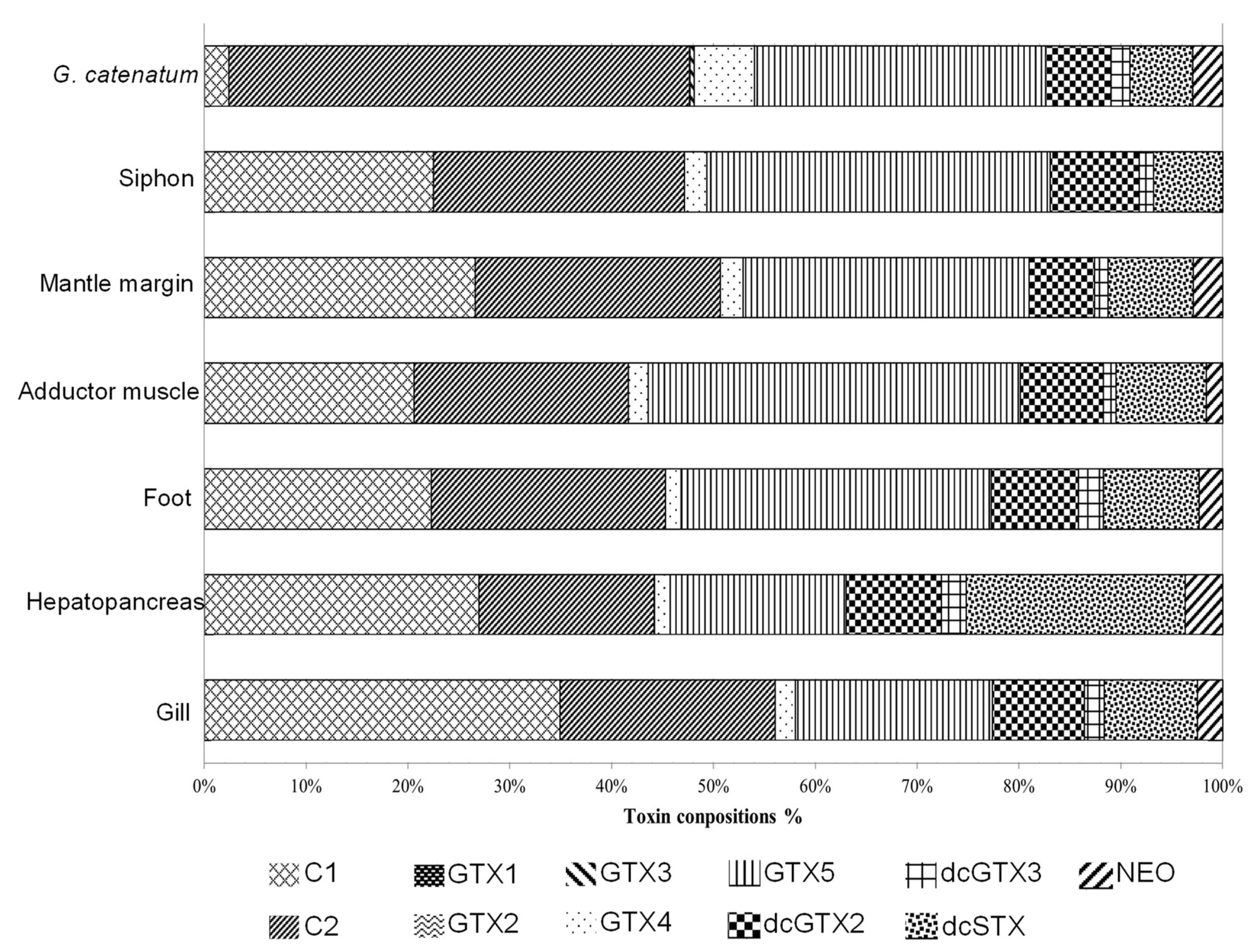
3.1. Toxin Composition in Dinoflagellate and Bivalve

The toxin composition of PSTs-producing dinoflagellate GYM isolate at its exponential phase included C1 (2.42%), C2 (45.24%), GTX2 (0.03%), GTX3 (0.38%), GTX4 (5.92%), GTX5 (28.60%), dcGTX2 (6.42%), dcGTX3 (1.86%), dcSTX (6.15%) and NEO (2.93%) (Figure 1). The total toxicity level of the GYM was 12.33 ± 19 pg STX eq cell−1, with C2 toxin being the most dominant toxin, followed by GTX5. STX and GTX1 were not detected in the GYM, same as in previous study [17]. A wide variation in cell toxicity among GYM isolates, even from the same geographical location, has been reported [17,43]. In previous reports, cell toxicity could vary from high values (9 to 17 µg STX eq cell−1) in Korean isolates [44] to very low levels (0.25 to 2.31 STX eq cell−1) in Mexican isolates [43]. It has been speculated that a high content of C1 or C2 seems to be a characteristic of GYM isolates in general [43]. However, more and more exceptions have also been found. For examples, isolates from Singapore [45] and Japan [46] produce the highest proportions of GTX1-4 toxins. Different studies have shown that the types and proportions of different PST analogues vary among isolates and environmental conditions. Changes of toxin profile in GYM have been detected when the cells are exposed to changes in temperature [43], culture media [17,47], chain length of the cells [47], growth phases [47,48] and growth rate [48].
The average toxin compositions in six organs of the bivalve include C1 (27.07%), C2 (17.43%), GTX2 (0.05%), GTX4 (1.53%), GTX5 (17.76%), dcGTX2 (9.30%), dcGTX3 (2.41%), dcSTX (20.83%) and NEO (3.58%). The toxin profiles of the six organs were highly similar, except NEO toxin was not detected in Siphon (Figure 1). The dominant PSTs in the bivalve after 7 days exposure were mainly the N-sulforcarbamoyl compounds including C1, C2 and GTX5, although previous research reported that all N-sulforcarbamoyl toxins could be transformed to carbamate toxins after a certain detoxification time in bivalves [18]. The second predominant toxin analogues were dcGTX2 and dcSTX. In terms of PSTs analogues, the toxin profile in the GYM-contaminated bivalve was comparable to that of GYM cell, except GTX3 was not detected in the bivalve sample and the dominant toxin was C1 instead of C2 (Figure 1). According to previous field studies and laboratory experimental work on PSTs content in bivalves, the toxin profile and the ratio of C1 and C2 in the digestive gland of the bivalve sample after exposure to PSTs-producing dinoflagellate were different from that in PSTs-producing dinoflagellate [16,18]. Such difference in toxin compositions between GYM and the bivalve is affected by several factors, including rates of feeding, accumulation, transformation such as reductive cleavage, hydrolysis, epimerization and enzyme conversion, detoxification and elimination of PSTs [16,18,21,49]. Mohamad et al. 2006 reported that the ratio of GTX5 and 6, and C3 and C4 toxins were eliminated after 96 h when the toxic dinoflagellate Alexandrium catenella was fed only once to the bivalve Tapes japonica [18]. In addition, the concentration of PSTs decreased rapidly if the bivalve without further ingestion of the PSTs producing dinoflagellates. The toxin profile of PSTs in bivalve tissues is changing incessantly, as the toxin decreases rapidly and excretes from the body like xenobiotics or exogenous compounds [50]. In the present study, however, the detoxification rate of the bivalves might not be able to immediately eliminate PSTs from their body due to the continuous feeding with GYM cells. The average of total toxin content obtained from the six organs of the contaminated bivalve was 40.63 ± 4.08 μg STX equivalents per gram (Table 1), which was much higher than the regulatory limitation in shellfish which set at 80 μg STX equivalents per 100 g shellfish meat [51].
More than 95% of PSTs was found in hepatopancreas, with toxin content at 38.71 ± 3.63 μg STX equivalents per gram, while the toxin content in the other five organs was less than 5% ranging from 0.107 ± 0.06 to 0.74 ± 0.14 μg STX equivalents per gram (Table 1). Previous studies demonstrated that PSTs were not evenly distributed in bivalve tissues, with higher toxin content in hepatopancreas than the other tissues [1,52]. The hepatopancreas in bivalves is an organ analogous to the liver in vertebrates and plays important roles in several metabolic processes and in reducing or eliminating the toxicity of cadmium and other heavy metals [34,53]. It is an important organ for the production of enzymes in the response to toxins, particularly digestion and detoxification processes [39,54]. The hepatopancreas is also involved in the immune responses in invertebrates [55]. The hepatopancreas of Mytilus galloprovincialis had an effective capacity to deal with PSTs, as it had the ability to cope with pro-oxidant challenges associated with PSTs [56]. The hepatopancreas of the bivalves with and without PSTs introduced were selected for the subsequent 2-DE analysis in the present study.

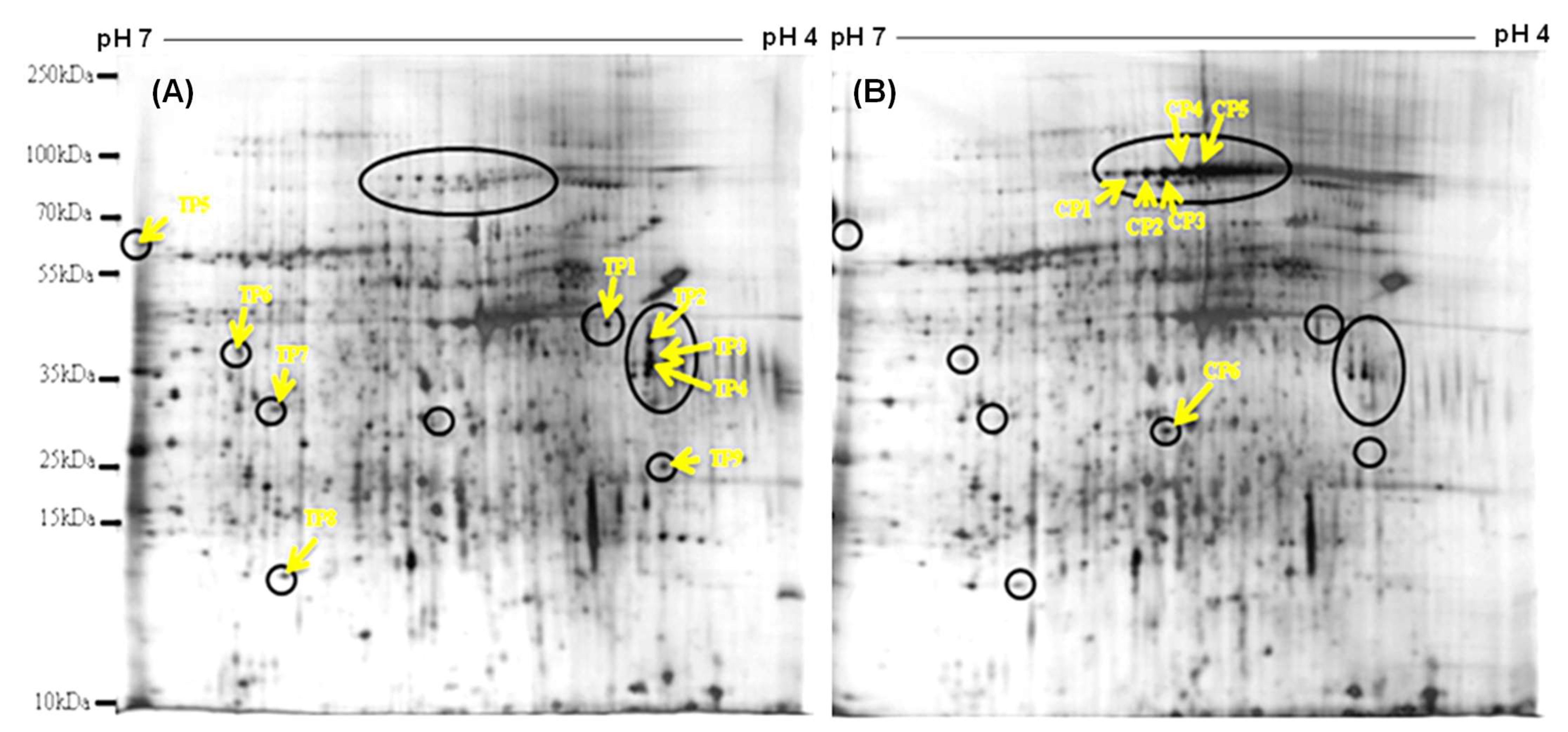
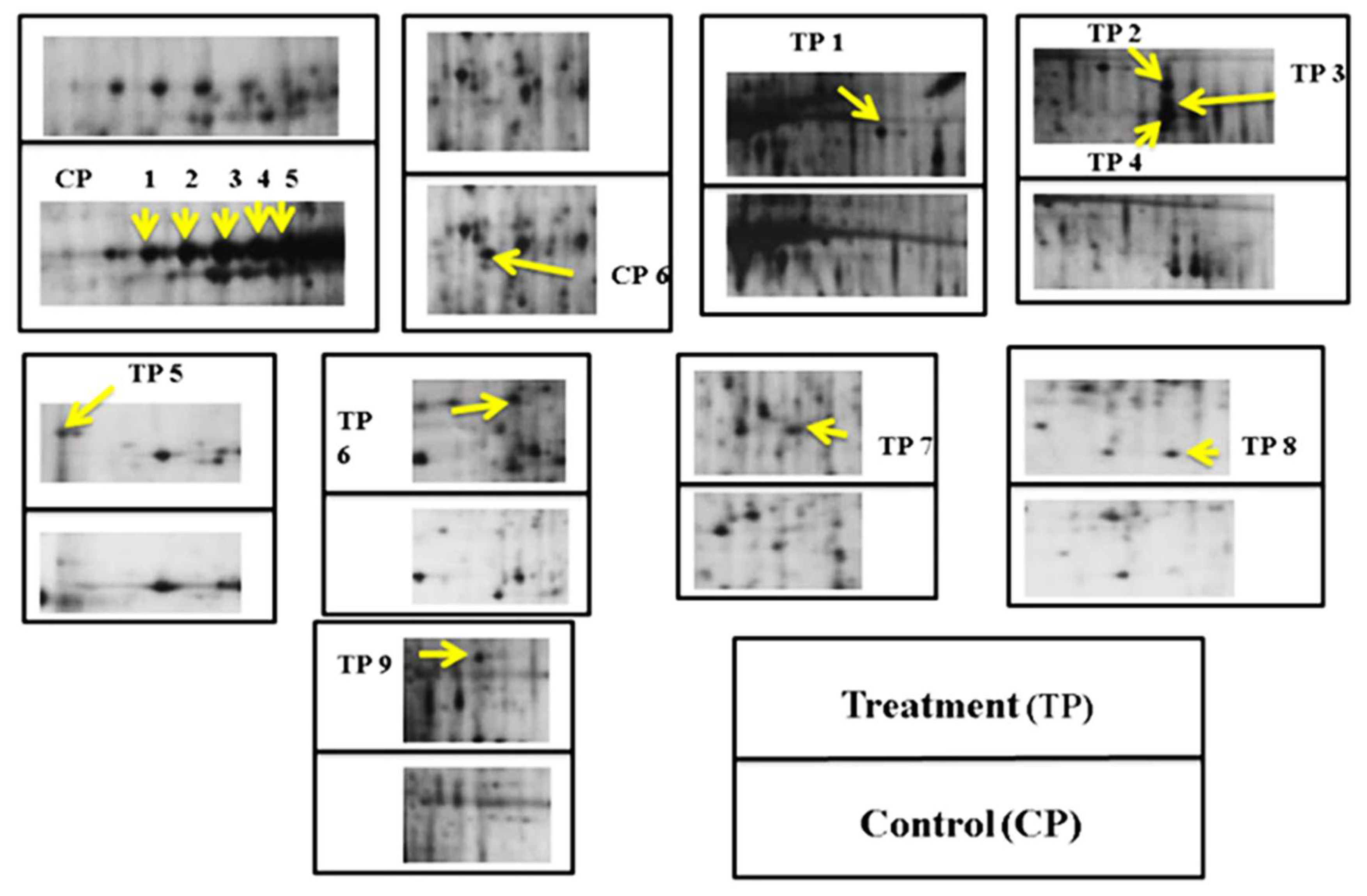
3.2. Protein Profile in the Hepatopancreas of Bivalves

In our study, a total of 745 and 681 protein spots were detected on 2-DE gels from PSTs-treated (exposed to G. catenatum) and non-PSTs treated (control) hepatopancreas, respectively. Protein profiles were reproducible among replicated 2-DE gels from the same group. Fifteen differentially expressed protein spots, each with at least 2-fold expression differences between PSTs-treated and non-treated hepatopancreas, were annotated (Figure 2). The selected areas are magnified in Figure 3. Nine of these differentially expressed proteins, named as the “toxic hepatopancreas” (TP) were up-regulated and the other six, the “control hepatopancreas” (CP), were down-regulated. Although proteomic analysis on the hepatopancreas of bivalves after GYM/PSTs exposure has yet to be explored, several relevant studies have been conducted. Martins and his group detected quantitative variations in 13 and 16 protein spots in digestive gland tract and gill samples, respectively, when the freshwater clam Corbicula fluminea was exposed to live Microcystis aeruginosa cells for 24 h [57]. Huang et al. also reported 12 proteins were up-regulated and 16 were down-regulated in Perna viridis after exposed to Prorocentrum lima, a dinoflagellate producing DSP toxins [58]. A total of 25 spots resolved in 2-DE gels were differentially expressed (>1.5 folds) in the hepatopancreas of Mytilus galloprovincialis challenged by Vibro anguillarum and Micrococcus luteus [59]. This study also found that out of the 25 differentially expressed protein spots, only four of them had 2- or more than 2-fold of change and the highest fold change was 3.27-fold (Putative C1q domain containing protein). In our case, protein expression levels of identified proteins ranged from 2.3 to 5.3 folds, and most of them had 3- or more fold of changes (Table 2).
Each of the differentially expressed protein spots, consistently present in the triplicate gels, was digested into peptides by in-gel digestion. To identify the proteins, each peptide mass was generated to form the “peptide mass fingerprints” (PMFs) by MALDI-TOF MS. Three up-regulated protein spots (TP5, TP6 and TP9) were successfully identified through bioinformatics searches from databases which were catalase, protein ultraspiracle homolog, and G2 and S phase-expressed protein 1, respectively (Table 2). Some of the PMFs were not successfully identified through the database searches due to the limitation of M. meretrix DNA and protein sequences information in the database. De novo amino acid sequencing was therefore applied. Five protein spots (TP1, TP2, TP3, TP7 and TP8) were identified by N-terminal sulfonation coupled with MALDI-TOF/TOF MS. Peptide sequence (m/z 1634.9) of TP1 protein spot matched with AIAKAVNHHNWAR of Ancylobacter rudongensis response regulator receiver domain-containing protein (accession no. SCW67799.1). The sequencing tags of peptide ion (m/z 1154.3) in TP2 matched with ALFSFWIDR Caballeronia sordidicola major facilitator superfamily transporters (accession no. WP 060818641.1). Peptide sequence (m/z 842.2) of TP3 matched with VATVSLPR Chionoecetes opilio sarcoplasmic calcium-binding protein (accession no. P86909.1). Peptide ion (m/z 1839.8) of TP7 and peptide ion (m/z 1646.6) of TP8 matched with the sequence DIEIAVISHESAEASIR of Haliotis discus discus paramyosin (accession no. BAJ61596.1) and the sequence AIFKIANWEEVGNR of the Ruditapes philippinarum manganese (Mn) superoxide dismutase (accession no. AFO64928.1), respectively.

3.3. Biological Functions of the Identified Proteins

All nine up-regulated proteins in PSTs-treated hepatopancreas group were identified except one (TP4), while all the six down-regulated proteins could not be identified in this study. The eight proteins successfully identified by MALDI-TOF with either PMFs or de novo amino acid sequencing were found to involve in structural (TP3 and TP7), cell signalling (TP1, TP6), cell cycle (TP9), membrane transport (TP2) and anti-oxidative system (TP5 and TP8).
Among the eight identified up-regulated proteins, the expression of TP1, TP2 and TP3 proteins (for cell signalling, membrane transport and structural) in the bivalve samples with PSP toxins was 4-fold more than that without PSP toxin. TP1 (response regulator receiver domain-containing protein) is one of the essential components in signal transduction systems. These systems allow an organism to sense, respond and adapt to a wide range of environmental and growth conditions, including nutrients, cellular redox state, changes in osmolality, quorum signals, antibiotics, temperature, chemoattractants, pH and more [60,61]. For instance, it was reported to involve in cellular response to oxidative stress in the fungal pathogen Candida glabrata [62]. The TP2, major facilitator superfamily transporters, are the proteins responsible to import or export of various drugs and metabolites, and they are important for controlling chemical exchange between cells and their environments [63]. Several studies have speculated that the overexpression of such membrane transporters may be attributed to the need of uptake, absorption and/or excretion of the HAB toxins. Pazos and co-author reported that several membrane transporters were over-expressed in the digestive gland of bivalve Mytilus galloprovincialis when exposed to domoic acid producing microalgae Pseudo-nitzschia [64]. Several membrane transporters related to the loading of saxitoxin (a derivative of PST) were detected in Pacific oyster when exposed to a PST-producing dinoflagellate Alexandrium minutum [65]. A number of membrane transporters were also highly expressed in the hepatopancreas of scallops after feeding with Alexandrium catenella and their expression changes aligned with the changes of PST accumulation in the hepatopancreas [66]. Such close association between the two expressions further indicated the potential role of the membrane transporters in PST absorption and distribution, which probably affected the accumulation of the toxin in hepatopancreas. On the other hand, membrane transporters were speculated to play a role in neural signalling and homeostasis in scallops to protect the nervous system from the PST attack. TP3, Sarcoplasmic calcium-binding protein (SCPs), can be commonly found in skeletal muscles and they are members of EF-hand Ca2+ binding protein family [67]. The high expression of SCPs in bivalve Laternula elliptica strongly associated with the increased of cytoskeletal activity, Ca2+ transport and homeostasis, as well as stresses such as heat [68]. Ca2+ binding proteins are also involved in hypoxia sensing to which the activation of Ca2+ signalling and alterations of intracellular Ca2+ concentrations constitute an early response of low levels of hypoxia in different types of cells [69]. Because of the similarity in metal binding properties, it has been suggested that SCPs can play a similar role in invertebrate muscle analogous to that of vertebrate parvalbumin, that can be differentially expressed in response to environmental stresses. For instance, parvalbumin was induced by cold exposure in skeletal muscle [70]. In the present study, significant up-regulations of TP1 (4.2-fold), TP2 (4.9-fold) and TP3 (5.3-fold) indicated the putative defensive response of hepatopancreas of M. meretrix bivalves to the challenge by GYM. The high expression levels suggested that these three proteins could be further developed as the potential biomarkers for bivalves in response to PST-producing algae. More studies are required to evaluate their applicability as biomarkers for rapid detection of shellfish toxin contamination in future.
PSTs are neurotoxins that can induce oxidative stress in bivalves, which occurs when the generation of reactive oxygen species (ROS) exceeds the capacity of neutralizing these molecules in the organism. An increase of ROS in bivalves when exposed to PST-producing Alexandrium cells have been reported in scallops (Patinopecten yessoensis), clams (Ruditapes philippinarum) and mussels (Mytilus galloprovincialis) [39,54]. Many organisms are known to produce antioxidative enzymes to scavenge excess ROS, prevent the accumulation of ROS and minimise oxidative damage to the cells. In the presnt study, TP5 (catalase, CAT) is an important enzyme that catalyzes the dismutation reaction of hydrogen peroxide to water and oxygen, while TP8 (Mn-superoxide dismutase, SOD) is involved in the dismutation of superoxide radical into either hydrogen peroxide or oxygen [71,72]. The overexpression of these two enzymes (TP5 and TP8) indicated the potential detoxication of PSTs in M. meretrix. A significant increase in gene expression levels of several antioxidant genes was observed in Pacific oysters (Crassostrea gigas) when they were fed with GYM cells [73]. Recently, a study demonstrated that antioxidant responses in the hepatopancreas of mussels Mytilus galloprovincialis could be modulated by various environmental conditions when the mussels were exposed to GYM cells [56].
Protein ultraspiracle homolog, USP (TP6) is the nuclear hormone receptor heterodimerizes with its partner ecdysone receptor (EcR) to transduce its signal and mediate the ecdysone response which is essential for metamorphosis in insects [74]. However, the functional characterization of this protein has not been described in mussels or bivalves yet, the observed increase in the USP expression level in the present study suggests it may be involved in signaling or other functions in the hepatopancreas of bivalve that deserves further research.
Paramyosin (TP7) is a fibrous thick filament protein which is one of the major structural components in smooth muscles of bivalves and it is involved in generating muscle contraction [75]. One of the possible reasons for the alternations of such cytoskeletal protein is the disorganization of cytoskeleton induced by oxidative stress [76]. Puerto et al. reported that the majority of the differentially expressed proteins in two bivalve species, the Mediterranean mussel M. galloprovincialis and the Asian clam C. fluminea, exposed to Cylindrospermopsis raciborskii cells producing cylindrospermopsin toxin, belonged to the group of structural proteins, especially actin and tubulin isoforms, and these alterations in actin cytoskeleton were linked to cellular stress [26]. Two proteins related to cytoskeleton were also up-regulated in Perna viridis exposed to Prorocentrum lima, a dinoflagellate that produces DSP toxins [58]. The structural proteins and cytoskeleton are essential to maintain cell shape and intracellular structures, the upregulation of paramyosin might imply that this protein could play a role in the resistance to the cellular damage of hepatopancreas caused by the GYM cells.
G2 and S phase-expressed protein 1 (TP9) is involved in cell proliferation and cycling. This is a well-studied protein in human cells, which is an enzyme expressed in the S and G2 phases of the cell cycle. This protein responds to DNA damage and binds to the tumor suppressor protein p53 [77]. Although its specific function and role in bivalves have not been reported, it is possible that the cell cycle of bivalves may be affected when exposed to stress. Important cell cycle regulators such as mitogen-activated protein kinase (MAPK) were upregulated in the gill cells of Mytilus galloprovincialis undergo cell cycle arrest in response to salinity stress [78]. The alternation of this cell cycle protein in the hepatopancreas of M. meretrix upon GYM exposure suggested the disturbance in cell cycle process could be critical for the response to PSP toxins.
Protein database in bivlaves is limited and protein identification is mainly relied on the search of sequence homologies against other species [24]. This makes the identification very difficult, which may explain why the remaining six down-regulated proteins could not be identified in the present study. More, the largest category of affected proteins in bivalves after exposure to toxins and stressed conditions belonging to the metabolism of carbohydrate, fat and protein, particularly glycolysis and energy production processes [58,59] were not identified in this study. Similarly, our study also did not detect the differentially expressed proteins involved in immunological processes, such as C1q domain containing proteins and fibrinogen C-terminal globular domain containing proteins. These two proteins were highly expressed when Mytilus galloprovincialis were exposed to pathogen in a proteomic study [59] and to HAB species in a transcriptomic study [64]. The lipopolysaccharide and beta-1,3-glucan binding protein (LGBP), suggested to play essential role in protective immune response, was overexpressed when oysters Crassostrea gigas exposed to GYM cells [73], but it was not found in the present bivalve exposure. It is essential to promote proteonmic research in bivalves.

4. Conclusions

The present study reports for the first time changes of toxin composition and protein expression in the hepatopancreas of M. meretrix bivalves after exposure to the PSTs-producing dinoflagellate G. catenatum using a gel-based proteomics approach. A total of 15 proteins exhibit significant alterations in their expression upon exposure to PSTs, and most have important biological functions, including detoxification and antioxidant enzymes such as catalase and dismutase. The three identified proteins, namely response regulator receiver domain-containing protein, major facilitator superfamily transporters, and sarcoplasmic calcium binding proteins, are potential biomarkers for the rapid detection of shellfish toxin contamination because of their significant up-regulation (4-fold increases in protein expression levels) when exposed to PSTs producing G. catenatum. Although the study is limited by a low representativeness of bivalve proteins in the database, we provide a good starting point and an initial insightful view into the effects of harmful algal bloom causative species to marine bivalves. More research efforts are needed to increase the protein database in bivalves, to validate the applicability of the putative biomarkers and to characterize the molecular responses of bivalves in the exposure to PSTs-producing dinoflagellates using gel-free high-throughput proteomic approach in future.

Supplementary Materials

The following are available online at https://www.mdpi.com/article/10.3390/jmse9091039/s1, Figure S1: Paralytic Shellfish Toxin (PST) derivatives (a) C1 and C2, (b) dcGTX2 and dcGTX3, (c) GTX 1-5 and (d) NEO and dcSTX of the standard and algal sample using post-column oxidation reaction. The amount of each toxin derivative was quantified based on the respectively standard curve.

Author Contributions

Conceptualization, E.T.-P.S. and F.W.-F.L.; methodology, K.-K.C., C.S.-N.K. and S.J.-L.X.; formal analysis, K.-K.C. and C.S.-N.K.; investigation, K.-K.C.; resources, F.W.-F.L.; data analysis, N.F.-Y.T., S.J.-L.X., F.W.-F.L. and K.-K.C.; writing—original draft preparation, K.-K.C.; writing—review and editing, C.N., N.F.-Y.T., S.J.-L.X. and F.W.-F.L.; supervision, F.W.-F.L. and E.T.-P.S.; project administration, F.W.-F.L.; funding acquisition, F.W.-F.L. All authors have read and agreed to the published version of the manuscript.

Funding

This work described in this paper was partially supported by grants from the Research Grants Council of the Hong Kong Special Administrative Region, China (UGC/FDS/M01/18) and (UGC/IDS(R)16/19).

Conflicts of Interest

The authors declare no conflict of interest.

References

  1. Taylor, S.L. Marine toxins of microbial origin. Food Technol. 1988, 42, 94–98. [Google Scholar]
  2. Yoosukh, W.; Matsukuma, A. Taxonomic study on Meretrix (Mollusca: Bivalvia) from Thailand. Phuket Mar. Biol. Cent. Spec. Publ. 2001, 25, 451–460. [Google Scholar]
  3. Hallegraeff, G. Harmful algal blooms: A global overview, in manual on harmful marine microalgae. Monogr. Oceanogr. Methodol 2003, 11, 25–49. [Google Scholar]
  4. Ho, K.C.; Hodgkiss, I.J. Red tides in sub-tropical waters: An overview of their occurrence. Asian Mar. Biol. 1991, 8, 5–23. [Google Scholar]
  5. Fleming, L.; Kirkpatrick, B.; Backer, L.; Walsh, C.; Nierenberg, K.; Clark, J.; Reich, A.; Hollenbeck, J.; Benson, J.; Cheng, Y.; et al. Review of Florida red tide and human health effects. Harmful Algae 2011, 10, 224–233. [Google Scholar] [CrossRef] [PubMed] [Green Version]
  6. McLeroy, S. Paralytic shellfish poisoning: Seafood safety and human health perspectives. Toxicon 2010, 56, 108–122. [Google Scholar]
  7. Daranas, A.; Norte, M.; Fernández, J. Toxic marine microalgae. Toxicon 2001, 39, 1101–1132. [Google Scholar] [CrossRef]
  8. Thottumkara, A.; Parsons, W.; Bois, J. Saxitoxin. Angew. Chem. Int. Ed. 2014, 53, 5760–5784. [Google Scholar] [CrossRef] [PubMed]
  9. Asakawa, M.; Miyazawa, K.; Takayama, H.; Noguchi, T. Dinoflagellate Alexandrium tamarense as the source of Paralytic shellfish poison (PSP) contained in bivalves from Hiroshima Bay, Hiroshima Prefecture, Japan. Toxicon 1995, 33, 691–697. [Google Scholar] [CrossRef]
  10. Estrada, N.; Lagos, N.; Garcia, C.; Maeda-Martinez, A.; Ascencio, F. Effects of the toxic dinoflagellate Gymnodinium catenatum on uptake and fate of paralytic shellfish poisons in the Pacific giant lions-paw scallop Nodipecten subnodosus. Mar. Biol. 2007, 151, 1205–1214. [Google Scholar] [CrossRef]
  11. Zhang, F.; Xu, X.; Li, T.; Liu, Z. Shellfish toxins targeting voltage-gated sodium channels. Mar. Drugs 2013, 11, 4698–4723. [Google Scholar] [CrossRef] [PubMed] [Green Version]
  12. Bricelj, V.; Shumway, S. Paralytic shellfish toxins in bivalve molluscs: Occurrence, transfer kinetics, and biotransformation. Rev. Fish Sci. 1998, 6, 315–383. [Google Scholar] [CrossRef]
  13. Bricelj, V.M.; Lee, J.H.; Cembella, A.D.; Anderson, D.M. Uptake kinetics of paralytic shellfish toxins from the dinoflagellate Alexandrium fundyense in the mussel Mytilus edulis. Mar. Ecol. Progs. Ser. 1990, 63, 177–188. [Google Scholar] [CrossRef]
  14. Xie, W.; Liu, X.; Yang, X.; Zhang, C.; Bian, Z. Accumulation and depuration of paralytic shellfish poisoning toxins in the oyster Ostrea rivularis Gould—Chitosan facilitates the toxin depuration. Food Control 2013, 30, 446–452. [Google Scholar] [CrossRef]
  15. Li, S.-C.; Hsieh, D. Feeding and absorption of the toxic dinoflagellate Alexandrium tamarense by two marine bivalves from the South China Sea. Mar. Biol. 2001, 139, 617–624. [Google Scholar] [CrossRef]
  16. Mohamad, S.; Takatani, T.; Yamaguchi, Y.; Sagara, T.; Noguchi, T.; Arakawa, O. Accumulation and elimination profiles of paralytic shellfish poison in the short-necked clam tapes japonica fed with the toxic dinoflagellate Gymnodinium catenatum. Shokuhin Eiseigaku Zasshi J. Food Hyg. Soc. Jpn. 2007, 48, 13–18. [Google Scholar]
  17. Band-Schmidt, C.; Bustillos, J.; Gárate-Lizárraga, I.; Lechuga-Devéze, C.; Erler, K.; Luckas, B. Paralytic shellfish toxin profile in strains of the dinoflagellate Gymnodinium catenatum Graham and the scallop Argopecten ventricosus G.B. Sowerby II from Bahía Concepción, Gulf of California, Mexico. Harmful Algae 2005, 4, 21–31. [Google Scholar] [CrossRef]
  18. Mohamad, S.; Yamaguchi, Y.; Sagara, T.; Takatani, T.; Arakawa, O.; Noguchi, T. Accumulation and depuration profiles of PSP toxins in the short-necked clam Tapes japonica fed with the toxic dinoflagellate Alexandrium catenella. Toxicon 2006, 48, 323–330. [Google Scholar]
  19. Sassolas, A.; Hayat, A.; Catanante, G.; Marty, J. Detection of the marine toxin okadaic acid: Assessing seafood safety. Talanta 2013, 105, 306–316. [Google Scholar] [CrossRef]
  20. Cao, J.; Jiang, Z.; Bing, Y.; Wang, Q.; Xu, J.; Aifeng, L. Evaluation of mouse bioassay results in an inter-laboratory comparison for paralytic shellfish poisoning toxins. Chin. J. Oceanol. Limnol. 2011, 29, 912–916. [Google Scholar] [CrossRef]
  21. Oshima, Y. Postcolumn derivation liquid chromatographic method for paralytic shellfish toxins. J. AOAC Int. 1995, 88, 1714–1732. [Google Scholar]
  22. Ronzitti, G.; Milandri, A.; Scortichini, G.; Poletti, R.; Rossini, G. Protein markers of algal toxin contamination in shellfish. Toxicon 2008, 52, 705–713. [Google Scholar] [CrossRef] [PubMed]
  23. Campos, A.; Puerto, M.; Prieto, A.; Cameán, A.; Almeida, A.; Coelho, A.; Vasconcelos, V. Protein Extraction and Two-dimensional gel electrophoresis of proteins in the marine mussel Mytilus galloprovincialis: An important tool for protein expression studies, food quality and safety assessment. J. Sci. Food Agric. 2013, 93, 1779–1787. [Google Scholar] [CrossRef] [PubMed]
  24. Campos, A.; Tedesco, S.; Vasconcelos, V.; Cristobal, S. Proteomic research in bivalves: Towards the identification of molecular markers of aquatic pollution. J. Proteom. 2012, 75, 4346–4359. [Google Scholar] [CrossRef] [PubMed]
  25. López-Pedrouso, M.; Varela, Z.; Franco, D.; Fernández, J.A.; Aboal, J.R. Can proteomics contribute to biomonitoring of aquatic pollution? A critical review. Environ. Pollut. 2020, 267, 115473. [Google Scholar] [CrossRef]
  26. Puerto, M.; Campos, A.; Prieto, A.; Cameán, A.; de Almeida, A.M.; Coelho, A.V.; Vasconcelos, V. Differential protein expression in two bivalve species; Mytilus galloprovincialis and Corbicula fluminea; exposed to Cylindrospermopsis raciborskii cells. Aquat. Toxicol. 2011, 101, 109–116. [Google Scholar] [CrossRef]
  27. Gomes, T.; Chora, S.; Guerreiro Pereira, C.; Cardoso, C.; Bebianno, M. Proteomic response of mussels Mytilus galloprovincialis exposed to CuO NPs and Cu2+: An exploratory biomarker discovery. Aquat. Toxicol. 2014, 155, 327–336. [Google Scholar] [CrossRef]
  28. Corporeau, C.; Tamayo, D.; Pernet, F.; Quéré, C.; Madec, S. Proteomic signatures of the oyster metabolic response to herpesvirus OsHV-1 μVar infection. J. Proteom. 2014, 109, 176–187. [Google Scholar] [CrossRef] [PubMed] [Green Version]
  29. Ji, C.; Wu, H.; Wei, L.; Zhao, J.; Wang, Q.; Lu, H. Responses of Mytilus galloprovincialis to bacterial challenges by metabolomics and proteomics. Fish Shellfish Immunol. 2013, 35, 489–498. [Google Scholar] [CrossRef]
  30. Fernández-Boo, S.; Villalba, A.; Cao, A. Protein expression profiling in haemocytes and plasma of the Manila clam Ruditapes philippinarum in response to infection with Perkinsus olseni. J. Fish Dis. 2016, 39, 1369–1385. [Google Scholar] [CrossRef]
  31. Gu, H.; Wu, Y.; Lu, S.; Lu, D.; Tang, Y.Z.; Qi, Y. Emerging harmful algal bloom species over the last four decades in China. Harmful Algae 2021, 102059. [Google Scholar] [CrossRef]
  32. Zhang, C.; Lim, P.T.; Li, X.; Gu, H.; Li, X.; Anderson, D. Wind-driven development and transport of Gymnodinium catenatum blooms along the coast of Fujian, China. Reg. Stud. Mar. Sci. 2020, 39, 101397. [Google Scholar] [CrossRef] [PubMed]
  33. Sierra-Beltrán, A.P.; Lluch-Cota, D.B.; Lluch-Cota, S.E.; Cortés-Altamirano, R.; Cortés-Lara, M.C.; Castillo-Chávez, M.; Carrillo, L.; Pacas, L.; Víquez, R.; García-Hansen, I. Spatial-temporal dynamics of red tide precursor organisms at the Pacific coast of North and Central America. Rev. Biol. Trop. 2004, 52 (Suppl. 1), 99–107. [Google Scholar]
  34. Xia, L.; Chen, S.; Dahms, H.U.; Ying, X.; Peng, X. Cadmium induced oxidative damage and apoptosis in the hepatopancreas of Meretrix meretrix. Ecotoxicology 2016, 25, 959–969. [Google Scholar] [CrossRef]
  35. Chen, L.S.; Wang, X.B.; Chen, D.Q. Research on the development of hard clam market in China mainland. J. Shanghai Ocean Univ. 2004, 283–287. [Google Scholar]
  36. Ho, J.S.; Zheng, G.X. Ostrincola koe (Copepoda, Myicolidae) and mass mortality of cultured hard clam (Meretrix meretrix) in China. Hydrobiologia 1994, 284, 169–173. [Google Scholar] [CrossRef]
  37. Li, H.; Liu, W.; Gao, X.G.; Zhu, D.; Wang, J.; Li, Y.F.; He, C.B. Identification of host-defense genes and development of microsatellite markers from ESTs of hard clam Meretrix meretrix. Mol. Biol. Rep. 2010, 38, 769–775. [Google Scholar] [CrossRef]
  38. Haoujar, I.; Abrini, J.; Chadli, H.; Essafi, A.; Nhhala, H.; Chebbaki, K.; Cacciola, F.; Skali-Senhaji, N. Effect of four diets based on three microalgae on the growth performance and quality of Mediterranean mussel flesh, Mytilus galloprovincialis. Int. Aquat. Res. 2020, 12, 137–145. [Google Scholar]
  39. Choi, N.; Yeung, L.; Siu, W.; So, I.; Jack, R.; Hsieh, D.; Wu, R.; Lam, P. Relationships between tissue concentrations of paralytic shellfish toxins and antioxidative responses of clams, Ruditapes philippinarum. Mar. Pollut. Bull. 2006, 52, 572–578. [Google Scholar] [CrossRef] [PubMed]
  40. Lee, F.W.F.; Lo, S.C.L. The use of Trizol reagent (phenol/guanidine isothiocyanate) for producing high quality two-dimensional gel electrophoretograms (2-DE) of dinoflagellates. J. Microbiol. Methods 2008, 73, 26–32. [Google Scholar] [CrossRef]
  41. Ramagli, L.; Rodriguez, L. Quantitation of Microgram Amounts of Protein in two-dimensional polyacrylamide-gel electrophoresis sample buffer. Electrophoresis 1985, 6, 559–563. [Google Scholar] [CrossRef]
  42. Chen, P.; Nie, S.; Mi, W.; Wang, X.C.; Liang, S.P. De Novo Sequencing of tryptic peptides sulfonated by 4-sulfophenyl isothiocyanate for unambiguous protein identification using post-source decay matrix-assisted laser desorption/ionization mass spectrometry. Rapid Commun. Mass Spectrom. 2004, 18, 191–198. [Google Scholar] [CrossRef]
  43. Band-Schmidt, C.J.; Bustillos-Guzmán, J.J.; Hernández-Sandoval, F.E.; Núñez-Vázquez, E.J.; López-Cortés, D.J. Effect of temperature on growth and paralytic toxin profiles in isolates of Gymnodinium catenatum (Dinophyceae) from the Pacific coast of Mexico. Toxicon 2014, 90, 199–212. [Google Scholar] [CrossRef] [PubMed]
  44. Park, T.G.; Kim, C.H.; Oshima, Y. Paralytic shellfish toxin profiles of different geographic populations of Gymnodinium catenatum (Dinophyceae) in Korean coastal waters. Phycol. Res. 2004, 52, 300–305. [Google Scholar] [CrossRef]
  45. Holmes, M.J.; Bolch, C.J.; Green, D.H.; Cembella, A.D.; Teo, S.L.M. Singapore isolates of the dinoflagellate Gymnodinium catenatum (Dinophyceae) produce a unique profile of paralytic shellfish poisoning toxins 1. J. Phycol. 2002, 38, 96–106. [Google Scholar] [CrossRef]
  46. Seok Jin, O.; Matsuyama, Y.; Yoon, Y.H.; Miyamura, K.; Choi, C.G.; Yang, H.S.; Kang, I.J. Comparative analysis of paralytic shellfish toxin content and profile produced by dinoflagellate Gymnodinium catenatum isolated from Inokushi Bay, Japan. J. Fac. Agric. Kyushu 2010, 55, 47–54. [Google Scholar]
  47. Band-Schmidt, C.; Bustillos-Guzmán, J.; Morquecho, L.; Gárate-Lizárraga, I.; Alonso-Rodríguez, R.; Reyes-Salinas, A.; Erler, K.; Luckas, B. Variations of psp toxin profiles during different growth phases in Gymnodinium catenatum (dinophyceae) strains isolated from three locations in the Gulf of California, Mexico. J. Phycol. 2006, 42, 757–768. [Google Scholar] [CrossRef]
  48. Bustillos-Guzman, J. Variations in growth and toxicity in Gymnodinium catenatum Graham from the Gulf of California under different ratios of nitrogen and phosphorus. Ciencias Marinas 2012, 38, 101–117. [Google Scholar] [CrossRef]
  49. Oshima, Y.; Blackburn, S.I.; Hallegraeff, G. Comparative study on paralytic shellfish toxin profiles of the dinoflagellate Gymnodinium catenatum from three different countries. Mar. Biol. 1993, 116, 471–476. [Google Scholar] [CrossRef]
  50. Kwong, R.; Lam, P.; Yu, P. The uptake, distribution and elimination of paralytic shellfish toxins in mussels and fish exposed to toxic dinoflagellates. Aquat. Toxicol. 2006, 80, 82–91. [Google Scholar] [CrossRef] [PubMed]
  51. Wekell, J.C.; Hurst, J.; Lefebvre, K. The origin of the regulatory limits for PSP and ASP toxins in shellfish. J. Shellfish Res. 2004, 23, 927–930. [Google Scholar]
  52. Reqia, S.; Amanhir, R.; Taleb, H.; Vale, P.; Blaghen, M.; Loutfi, M. Comparative study on differential accumulation of PSP toxins between cockle (Acanthocardia tuberculatum) and sweet clam (Callista chione). Toxicon 2005, 46, 612–618. [Google Scholar]
  53. Yasmeen, S. Changes in hepatopancreas of the bivalve molluscs Lamellidens marginalis exposed to acute toxicity of cadmium in summer. World J. Fish Mar. Sci. 2013, 5, 437–440. [Google Scholar]
  54. Qiu, J.; Ma, F.; Fan, H.; Aifeng, L. Effects of feeding Alexandrium tamarense, a paralytic shellfish toxin producer, on antioxidant enzymes in scallops (Patinopecten yessoensis) and mussels (Mytilus galloprovincialis). Aquaculture 2013, 396, 76–81. [Google Scholar] [CrossRef]
  55. Rőszer, T. The invertebrate midintestinal gland (“hepatopancreas”) is an evolutionary forerunner in the integration of immunity and metabolism. Cell Tissue Res. 2014, 358, 685–695. [Google Scholar] [CrossRef] [PubMed]
  56. Braga, A.C.; Pereira, V.; Marçal, R.; Marques, A.; Guilherme, S.; Costa, P.R.; Pacheco, M. DNA damage and oxidative stress responses of mussels Mytilus galloprovincialis to paralytic shellfish toxins under warming and acidification conditions—Elucidation on the organ-specificity. Aquat. Toxicol. 2020, 228, 105619. [Google Scholar] [CrossRef] [PubMed]
  57. Martins, J.C.; Leão, P.N.; Vasconcelos, V. Differential protein expression in Corbicula fluminea upon exposure to a Microcystis aeruginosa toxic strain. Toxicon 2009, 53, 409–416. [Google Scholar] [CrossRef]
  58. Huang, L.; Zou, Y.; Weng, H.W.; Li, H.Y.; Liu, J.S.; Yang, W.D. Proteomic profile in Perna viridis after exposed to Prorocentrum lima, a dinoflagellate producing DSP toxins. Environ. Pollut. 2015, 196, 350–357. [Google Scholar] [CrossRef]
  59. Wu, H.; Ji, C.; Wei, L.; Zhao, J.; Lu, H. Proteomic and metabolomic responses in hepatopancreas of Mytilus galloprovincialis challenged by Micrococcus luteus and Vibrio anguillarum. J. Proteom. 2013, 94, 54–67. [Google Scholar] [CrossRef]
  60. Bourret, R. Receiver domain structure and function in response regulator proteins. Curr. Opin. Microbiol. 2010, 13, 142–149. [Google Scholar] [CrossRef] [Green Version]
  61. Wolanin, P.; Thomason, P.; Stock, J. Histidine protein kinases: Key signal transducers outside the animal kingdom. Genome Biol. 2002, 3, 1–8. [Google Scholar] [CrossRef] [PubMed] [Green Version]
  62. Roetzer, A.; Klopf, E.; Gratz, N.; Marcet-Houben, M.; Hiller, E.; Rupp, S.; Gabaldón, T.; Kovarik, P.; Schüller, C. Regulation of Candida glabrata oxidative stress resistance is adapted to host environment. FEBS Lett. 2011, 585, 319–327. [Google Scholar] [CrossRef] [PubMed] [Green Version]
  63. Jack, D.L.; Yang, N.M.; Saier, M.H., Jr. The drug/metabolite transporter superfamily. Eur. J. Biochem. 2001, 268, 3620–3639. [Google Scholar] [CrossRef]
  64. Pazos, A.J.; Ventoso, P.; Martínez-Escauriaza, R.; Pérez-Parallé, M.L.; Blanco, J.; Triviño, J.C.; Sánchez, J.L. Transcriptional response after exposure to domoic acid-producing Pseudo-nitzschia in the digestive gland of the mussel Mytilus galloprovincialis. Toxicon 2017, 140, 60–71. [Google Scholar] [CrossRef]
  65. Mat, A.; Klopp, C.; Payton, L.; Jeziorski, C.; Chalopin, M.; Amzil, Z.; Tran, D.; Wikfors, G.; Hégaret, H.; Soudant, P.; et al. Oyster transcriptome response to Alexandrium exposure is related to saxitoxin load and characterized by disrupted digestion, energy balance, and calcium and sodium signaling. Aquat. Toxicol. 2018, 199, 127–137. [Google Scholar] [CrossRef] [PubMed]
  66. Xun, X.; Cheng, J.; Wang, J.; Li, Y.; Li, X.; Moli, L.; Lou, J.; Kong, Y.; Bao, Z.; Hu, X. Solute carriers in scallop genome: Gene expansion and expression regulation after exposure to toxic dinoflagellate. Chemosphere 2019, 241, 124968. [Google Scholar] [CrossRef]
  67. Hermann, A.; Cox, J. Sarcoplasmic calcium-binding protein. Comp. Biochem. Physiol. B Biochem. Mol. Biol. 1995, 111, 337–345. [Google Scholar] [CrossRef]
  68. Truebano, M.; Burns, G.; Thorne, M.A.S.; Hillyard, G.; Peck, L.S.; Skibinski, D.O.F.; Clark, M.S. Transcriptional response to heat stress in the Antarctic bivalve Laternula elliptica. J. Exp. Mar. Biol. Ecol. 2010, 391, 65–72. [Google Scholar] [CrossRef]
  69. Toescu, E.C. Hypoxia sensing and pathways of cytosolic Ca2+ increases. Cell Calcium 2004, 36, 187–199. [Google Scholar] [CrossRef]
  70. Nelson, T.; McEachron, D.; Freedman, W.; Yang, W.-P. Cold acclimation increases gene transcription of two calcium transport molecules, calcium transporting ATPase and parvalbumin beta, in carassius auratus lateral musculature. J. Therm. Biol. 2003, 28, 227–234. [Google Scholar] [CrossRef]
  71. Lian, S.; Zhao, L.; Xun, X.; Lou, J.; Moli, L.; Li, X.; Wang, S.; Zhang, L.; Hu, X.; Bao, Z. Genome-wide identification and characterization of SODs in zhikong scallop reveals gene expansion and regulation divergence after toxic dinoflagellate exposure. Mar. Drugs 2019, 17, 700. [Google Scholar] [CrossRef] [Green Version]
  72. Vitolo, M. Decomposition of hydrogen peroxide by catalase. World J. Pharm. Pharm. Sci. 2021, 10, 47. [Google Scholar]
  73. Garcia Lagunas, N.; Romero-Geraldo, R.; Hernandez Saavedra, N. Genomics study of the exposure effect of Gymnodinium catenatum, a paralyzing toxin producer, on Crassostrea gigas’ defense system and detoxification genes. PLoS ONE 2013, 8, e72323. [Google Scholar] [CrossRef] [PubMed] [Green Version]
  74. Cao, J.; Liu, Y.; Yang, Y.; Zhang, H.; Li, Z.; Yang, Q.; Zhang, S.; Zhang, Q.; Liu, X. Molecular characterization and functional analysis of the ultraspiracle (USP) in the oriental fruit moth Grapholita molesta (Lepidoptera: Olethreutidae). Comp. Biochem. Physiol. B Biochem. Mol. Biol. 2015, 190, 54–62. [Google Scholar] [CrossRef] [PubMed]
  75. Grøsvik, B.E.; Jonsson, H.; Rodríguez-Ortega, M.J.; Roepstorff, P.; Goksøyr, A. CYP1A-immunopositive proteins in bivalves identified as cytoskeletal and major vault proteins. Aquat. Toxicol. 2006, 79, 334–340. [Google Scholar] [CrossRef] [PubMed]
  76. Rodríguez-Ortega, M.J.; Grøsvik, B.E.; Rodríguez-Ariza, A.; Goksøyr, A.; López-Barea, J. Changes in protein expression profiles in bivalve molluscs (Chamaelea gallina) exposed to four model environmental pollutants. Proteomics 2003, 3, 1535–1543. [Google Scholar] [CrossRef] [PubMed]
  77. Liu, X.S.; Li, H.; Song, B.; Liu, X. Polo-like kinase 1 phosphorylation of G2 and S-phase-expressed 1 protein is essential for p53 inactivation during G2 checkpoint recovery. EMBO Rep. 2010, 11, 626–632. [Google Scholar] [CrossRef] [PubMed] [Green Version]
  78. Lockwood, B.L.; Somero, G.N. Transcriptomic responses to salinity stress in invasive and native blue mussels (genus Mytilus). Mol. Ecol. 2011, 20, 517–529. [Google Scholar] [CrossRef]
Figure 1. Toxin compositions of G. catenatum and six organs of M. meretrix.
Jmse 09 01039 g001
Figure 2. Representative 2-DE images of proteins from the hepatopancreas of M. meretrix after exposed to G. catenatum (A) and Control sample (B). Circled protein spots indicate the differentially expressed proteins with at least 2-fold changes.
Jmse 09 01039 g002
Figure 3. Magnifications of differentially expressed proteins of the hepatopancreas of M. meretrix. The upper panel is the Treatment group: M. meretrix after exposed to G. catenatum and the bottom panel is the control group.
Jmse 09 01039 g003
Table 1. Toxin content detected in six organs of M. meretrix by UPLC coupled with post column oxidation method. Different letters (a and b) in supercript position denote significant difference among organs according to one-way analysis of variation at p ≤ 0.05.
OrganToxin Content (μg STX Equivalents Per Gram)Relative Abundant
(% Molar)
Gill0.74 ± 0.14 a1.82
Hepatopancreas38.71 ± 3.63 b95.27
Foot0.73 ± 0.10 a1.79
Adductor muscle0.212 ± 0.05 a0.52
Mantle margin0.107 ± 0.06 a0.26
Sighon0.131 ± 0.03 a0.32
Table 2. Eight differentially expressed protein spots identified by PMF and de novo amino acid sequencing.
SpotpIMW (kDa)Fold ChangesProtein NameSourceAmino Acid Sequence
TP14.8464.2Response regulator receiver domain-containing proteinDe novo peptide sequencing
1634.9 m/z
FAI/LAKAVNHHNWAR
TP24.6424.9Major facilitator superfamily transportersDe novo peptide sequencing
1154.3 m/z
AI/LFSFWI/LDR
TP34.6405.3Sarcoplasmic calcium-binding proteinDe novo peptide sequencing
842.2 m/z
VATVSI/LPR
TP57.0602.3CatalasePMF--
TP66.5183.2Protein ultraspiracle homologPMF--
TP76.3343.0ParamyosinDe novo peptide sequencing
1839.8 m/z
DI/LEI/LAVI/LSHESAEASI/LR
TP86.1203.8Mg superoxide dismutaseDe novo peptide sequencing
1646.6 m/z
AI/LFKI/LANWEEVGNR
TP94.5293.3G2 and S phase-expressed protein 1PMF--
PMF: Peptide Mass Fingerprint.
Publisher’s Note: MDPI stays neutral with regard to jurisdictional claims in published maps and institutional affiliations.

