Article

Homeostatic Functions of Tecrem, a CD46-Like Regulatory Protein of Complement Activation, on Epithelial Cells in Carp Fish

1
Department of Bioscience and Biotechnology, Graduate School of Bioresource and Bioenvironmental Sciences, Kyushu University, Fukuoka 819-0395, Japan
2
Laboratory of Marine Biochemistry, Faculty of Agriculture, Kyushu University, Fukuoka 819-0395, Japan
*
Author to whom correspondence should be addressed.
J. Mar. Sci. Eng. 2021, 9(7), 687; https://doi.org/10.3390/jmse9070687
Submission received: 31 May 2021 / Revised: 18 June 2021 / Accepted: 20 June 2021 / Published: 22 June 2021
(This article belongs to the Section Marine Biology)

Abstract

:
Fish mucosal surface is a significant interface for pathogens to infect from an aqueous environment. In addition to mucosal innate and adaptive immune factors, epithelial cells are considered as a significant physical barrier against microbial invasion. Previously, we identified a mammalian CD46-like complement regulatory protein (Tecrem) in teleost and detected its expression on epithelial cells derived from fin, suggesting its physiological role on the fish surface. This study examines the homeostatic roles of Tecrem in maintaining the fish epithelium, by analyzing the expression behavior of Tecrem on the fin-derived epithelial cell lines (KF-1 from the common carp and CFS from ginbuna crucian carp) using monoclonal and polyclonal anti-Tecrem antibodies. Expression of KF-1 protein was associated with the adhesion of KF-1, and the adhesion was enhanced by anti-Tecrem treatments of the cells. Stimulation of the epithelial cells with anti-Tecrem enhanced wound healing, protein expression of tight-junction proteins, and cell density of the KF-1 and CFS monolayer culture. These results suggest that Tecrem on epithelial cells play a homeostatic role in maintaining intactness of the surface epithelial barrier, implying that modification of Tecrem expression may develop a novel tool to improve the first-line defense against pathogens in aquaculture.

1. Introduction

Aquaculture, a rapidly developing industry for animal protein supply, has been hampered by the persistent occurrence of infectious diseases caused by pathogenic microbes. High densities of the livestock under stressed conditions in tanks, ponds, and fish cages are considered as a major risk factor to increase the susceptibility of fish to pathogen infection. Fish body surfaces, including skin, gills, and intestine, face to microbe-rich environmental water through mucosal tissues. Therefore, mucosal immunity, including humoral and cellular factors, and physical barriers, has been regarded to be particularly important for defense [1].
As a humoral factor of innate immunity, the complement system has been recognized to play crucial roles in the removal and destruction of pathogens by opsonic, inflammatory, and direct-killing mechanisms [2]. These defensive responses are well conserved in vertebrates from fish to mammals [3]. The majority of the physiological activities of the complement system are attributable to C3, the central component of the system. C3 protein generates various active fragments, such as C3a, C3b, iC3b, and C3d, upon complement activation through one of three activation cascades, the classical, the lectin, and the alternative pathways [2]. On the other hand, excessive or misdirected activation of C3 may lead to inflammatory and autoimmune damages to bystander host tissue [4]. Therefore, the complement system is equipped with strict regulatory factors to limit C3-activation at the proper level and sites. CD46, or membrane cofactor protein (MCP), in mammals, has originally been identified as a membrane-bound regulator of complement activation (RCA) to avoid the activation of C3 on host cells [5]. A CD46-like protein designated as teleost complement-regulatory membrane protein or Tecrem has already been identified and characterized in teleost fishes, such as common carp, zebrafish, and ginbuna crucian carp [6,7,8,9]. We have clarified that Tecrem is a functional orthologue of human CD46 using common carp Tecrem (cTecrem) as a model [6]. A recent study has shown the cofactor activity of fish CD46 in association with factor I in the tongue sole as well [10]. These findings suggest that host cell protection is an evolutionarily conserved role of CD46.
Mammalian CD46 has also been reported to play a role in regulating adaptive immune response by supporting proliferation and cytokine production of T cells, inducing a Th1-type response of cellular immunity [5,11]. Evolutionary conservation of the CD46 function associated with T cell activation has been explored using a fish model by preceding works by our group [7,8]. Tecrem expressed on the T cells modulates mitogen-induced T cell proliferation in ginbuna crucian carp, indicating that modulation of adaptive immunity is one of the evolutionarily conserved functions of the CD46-like complement regulator [8].
In recent years, a novel function of human CD46 in maintaining the epithelial barrier integrity, as well as the barrier repair, has been demonstrated using mammalian cell lines established from mucosal tissues, suggesting that CD46 may play a role in homeostasis [12]. It seems reasonable to consider that such a homeostatic role should have an ancient evolutionary origin. However, the roles of Tecrem in fish epithelial cell homeostasis have remained to be studied.
The entire body surface of fish, including skin, gills, and digestive tract, is totally covered by nonkeratanized epithelial sheets or mucosal tissues. So, the maintenance of a healthy epithelium should be extremely important for fish. Interestingly, the expression of genes encoding complement components and regulatory factors, such as C3 and CD46, have been detected in fish skin and other mucosal tissue [13,14,15]. These findings raise a possibility that the complement activation cascades, the regulatory mechanisms, and the homeostatic epithelial maintenance play important roles as immune and physical barriers in the first-line defense against invading pathogens. This prompted us to investigate the homeostatic roles of Tecrem in maintaining epithelial cells in fishes. The present study focuses on the possible role of Tecrem on epithelial cell adhesion and proliferation using two cyprinid fish species as models.

2. Materials and Methods

2.1. Materials

Epithelial cells, KF-1 cells (a cell line derived from the fin of Koi carp, Cyprinus carpio) and CFS cells (Ginbuna crucian carp, Carassius auratus langsdorfii, fin cell line) were cultured in MEM supplemented with 10% FBS, 14 mM HEPES and 2 mM L-glutamine, 7.5% NaHCO3 at 20 °C. Red blood cells were obtained from heparinized blood taken from the common carp (bodyweight of 0.2–1 kg), which were maintained in our laboratory. Monoclonal mouse IgG antibody (whole molecule) conjugated with FITC against 6×His tag was purchased from SIGMA, and anti-carp Tecrem (cTecrem) mAb (1F12) was established earlier in our laboratory [6].

2.2. Detection of Tecrem Protein by ELISA and Immunoprecipitation

For ELISA, KF-1 cells were seeded on a 96-well plate at 2 × 10⁴ cells/well. After the adhesion of the cells by culture, the cells were treated with 2 µg/mL of anti-cTecrem mAb and then with 1/1000-diluted anti-mouse IgG conjugated with peroxidase, followed by color development with a chromogenic substrate (ABTS). Absorbance at 405 nm (A405) was measured using an ELISA reader (Multiscan FC, Thermo Scientific).
For immunoprecipitation, KF-1 cells forming monolayer were biotin-labeled using Biotin Sulfo-OSu kit (Dojinodo, Kumamoto, Japan) according to the manufacturer’s instruction. The biotinylated cells were scraped off and homogenized by sonication in 500 µL of NP-40 lysis buffer (150 mM NaCl, 50 mM Tris-HCl, 1% NP-40, pH 8.0), and the lysate was centrifuged. The supernatant (480 µL), after absorption with 20 μL of protein G-Sepharose, was incubated with 1 µg of anti-cTecrem mAb at 4 °C for 1 h. The immune complexes formed were trapped with 20 μL of protein G-Sepharose. The beads were washed with 1 mL of TBS (pH 7.5) and boiled in 50 mM Tris-HCl, pH 6.8, containing 2% SDS, 5% 2-mercaptoethanol (2ME), and 10% glycerol (pH 6.8) for SDS-PAGE analysis. The antigen bands were then visualized on nitrocellulose membrane (Hybond-C Extra) with biotin-avidin complex and 3,3-diaminobenzidine using Vectastain ABC kit (Vector Laboratories, USA).

2.3. Expression of Recombinant SCR1-2 and SCR3-4 Domains of cTecrem

Gene-specific primers with restriction sites, shown in Table 1, were used to amplify cDNA fragments encoding SCR1-2 and SCR3-4 domains, respectively, of cTecrem for PCR from its full-length cDNA clone with Phusion DNA polymerase (New England Biolabs, Japan). The amplified products were subcloned into the pGEM-T Easy vector (Promega, USA), and after sequence confirmation, each insert was excised out with XhoI/PstI and transferred into pCold-I, followed by transformation of Origami B strain. Recombinant SCR1-2 and SCR3-4 were expressed for 22 h at 15 °C by adding 0.1 mM isopropyl β-D-thiogalactoside, and the culture supernatant was subjected to affinity chromatography on 1 mL columns of Ni-NTA-agarose (Qiagen, USA), following the manufacturer’s instructions. The rSCR1-2 and rSCR3-4 were further purified by gel-filtration on HiLoad 16/600 Superdex 200pg equilibrated with PBS to remove trace contaminants.

2.4. Preparation of Rabbit Polyclonal Anti-cTecrem Domains

The purified rSCR1-2 and rSCR3-4 proteins were digested with factor Xa protease (Promega, Madison, MI, USA) and passed through the Ni-NTA column to remove 6xHis tag. The untagged proteins (about 1 mg each) were emulsified in Freund’s complete adjuvant for rabbit immunization, which was performed by SCRUM Co. (Tokyo, Japan). After four subcutaneous injections and a booster at weekly intervals, the rabbits were bled to separate antisera. IgG was purified from the antiserum using the HiTrap protein A column (GE Health Science, Tokyo, Japan), according to the manufacturer’s protocol.

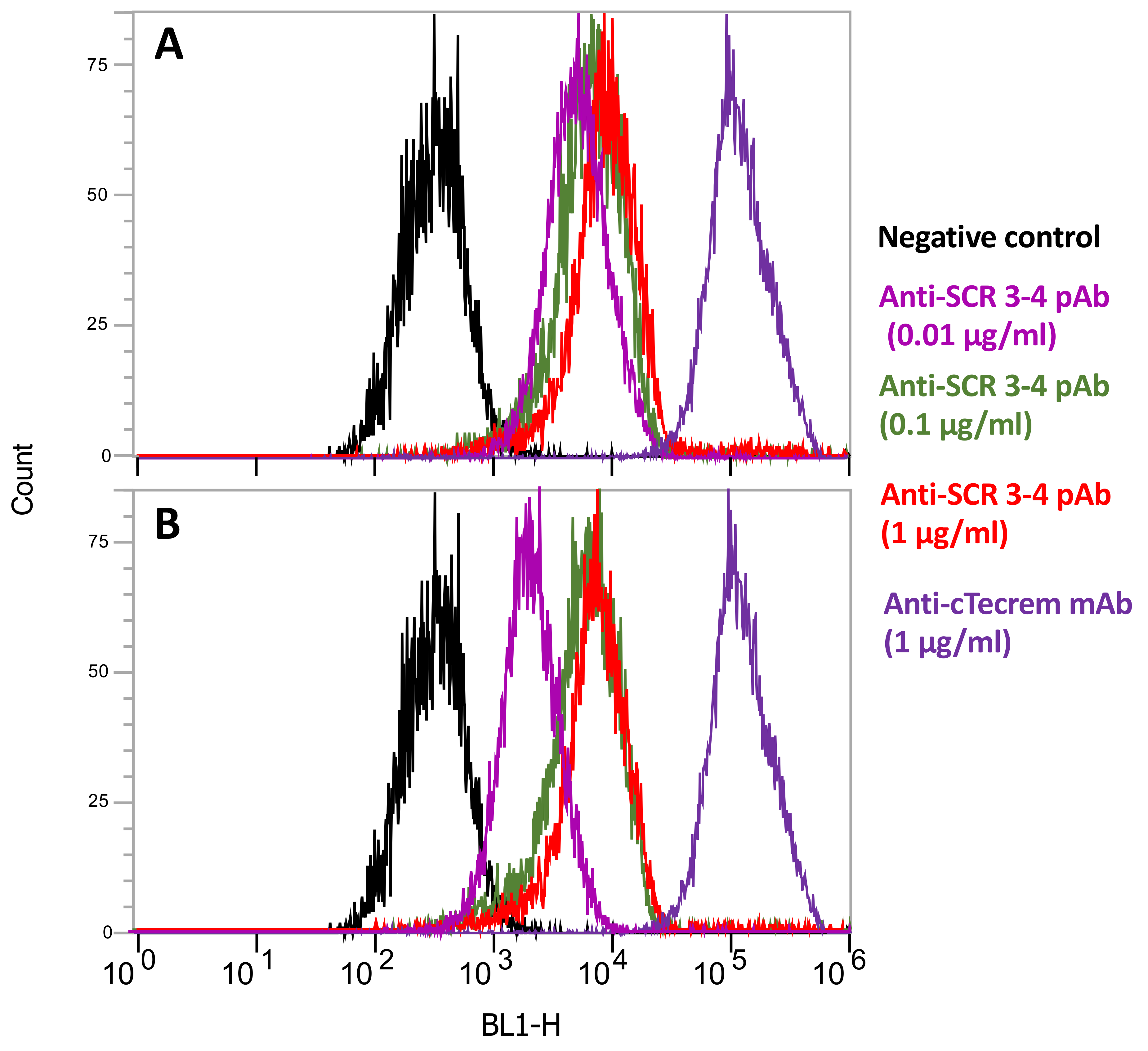
2.5. Flow Cytometric Analysis of Tecrem Expression on KF-1 Cells

KF-1 cells were passaged, harvested by scraping, and adjusted to 1 × 10⁶ cells/mL in MEM. The cells were treated with varying concentrations (10, 100 and 1000 ng/mL) of anti-SCR1-2 pAb, anti-SCR3-4 pAb or anti-cTecrem mAb (1µg/mL), and then with secondary FITC-labeled antibodies at 1/300 dilution), followed by analysis using Epics XL Flow Cytometer equipped with System II software (Beckman Coulter). And the obtained histograms were analyzed using EXPO 32 MultiCOMP software (Beckman Coulter).

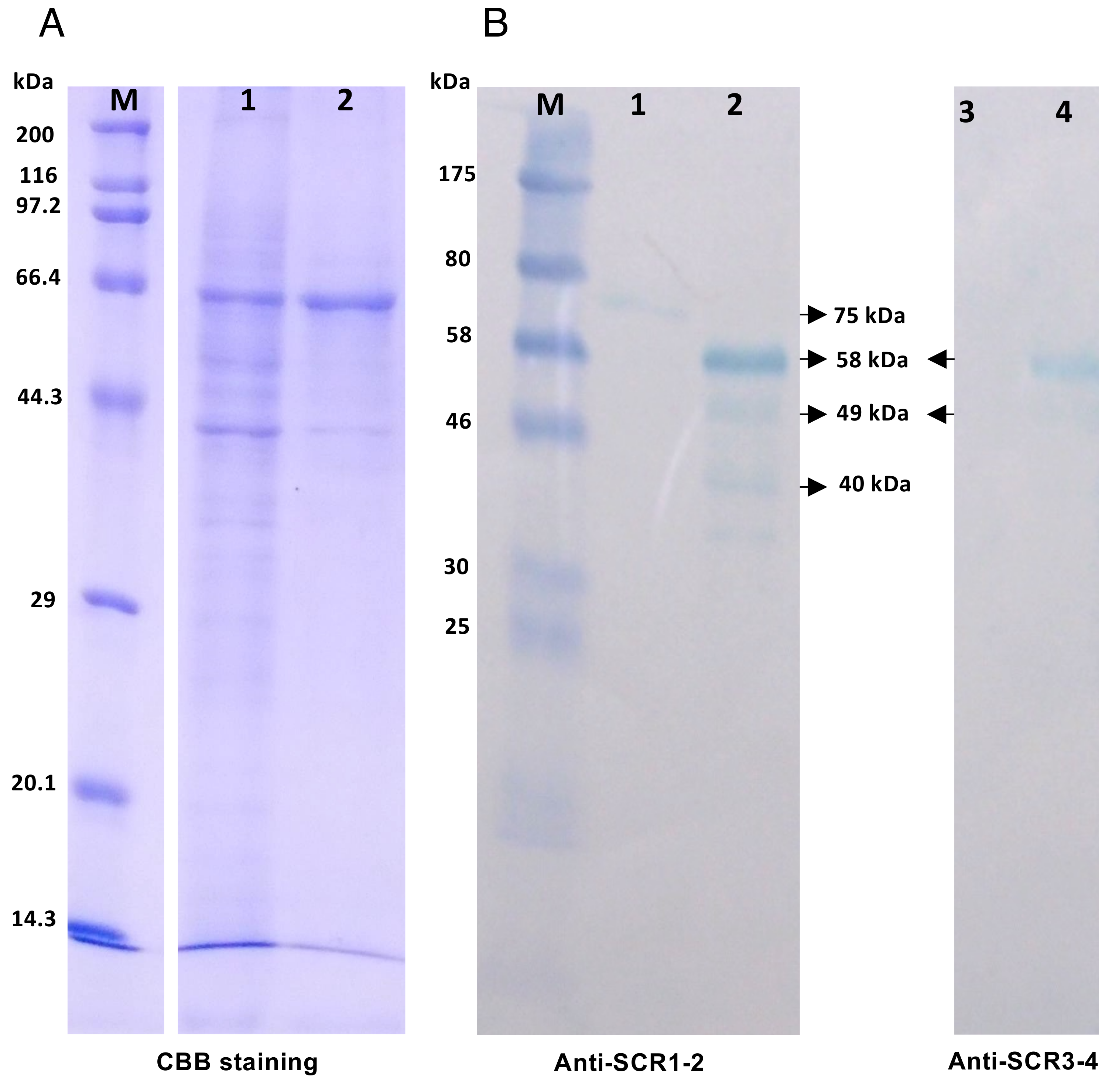
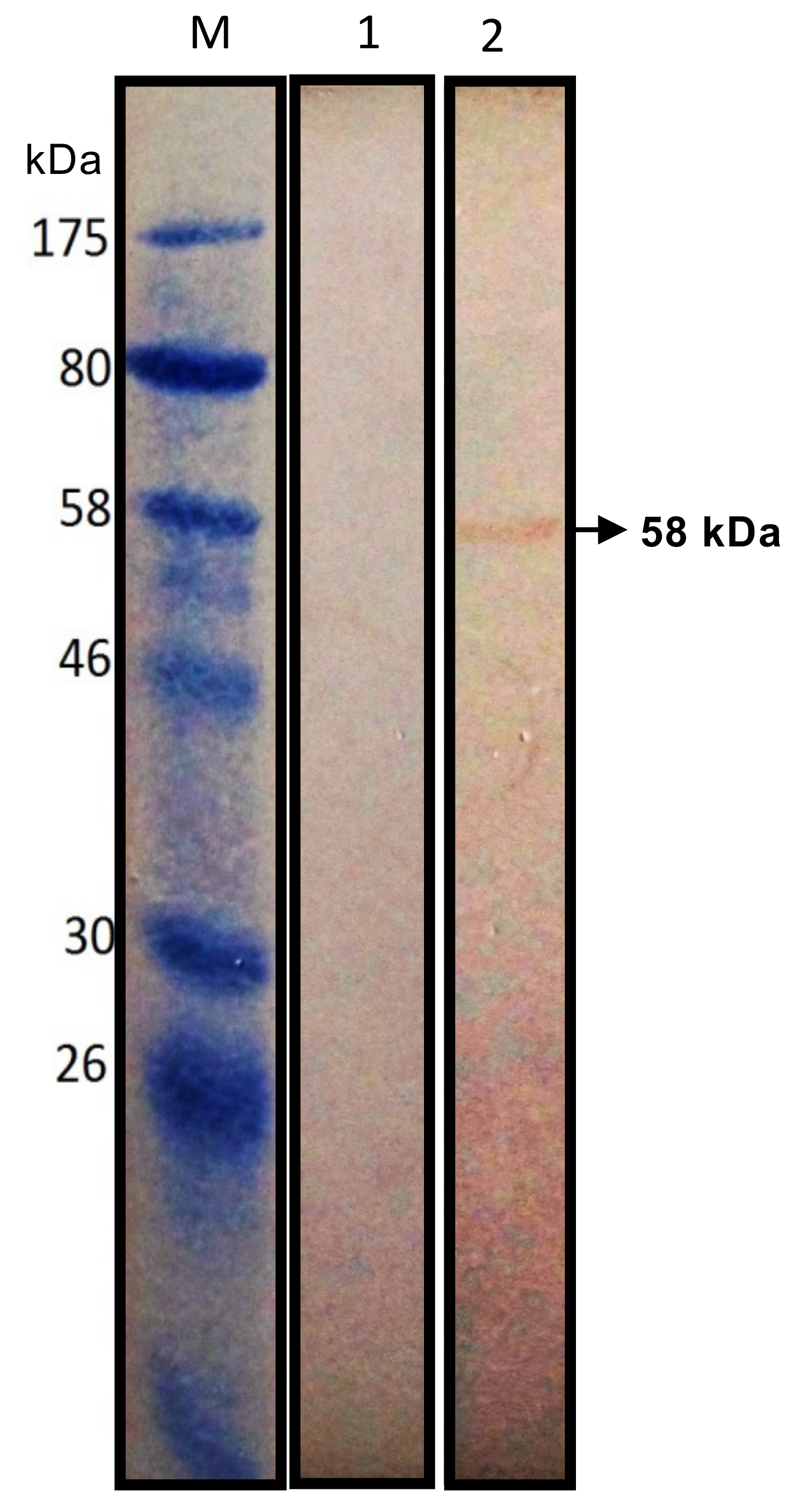
2.6. Western Blotting Analysis of Tecrem Expression on KF-1 and CFS Cells

KF-1 and CFS cells suspensions (1 × 10⁶ cells/mL in MEM) were prepared as above, harvested by centrifugation, and solubilized in SDS-PAGE sample buffer by boiling at 100 °C for 3 min. The samples were separated on 12% gel under reducing conditions and electroblotted onto a PVDF membrane (Immobilon P, Millipore, USA). The membrane was blocked with 5% skim milk-PBS and treated with anti-SCR1-2 pAb (1 µg/mL) and anti-SCR 3-4 pAb (1 µg/mL) as primary antibodies, then with peroxidase-conjugated second antibody. Color development was performed with EzWestBlue reagent (Atto, Japan) as the substrate.

2.7. Adhesion Assays of KF-1 Cells

Passaged KF-1 cells were harvested and suspended at 2 × 10⁵ cells/mL. The suspension (100 µL/well) was seeded to a 96-well plate precoated with 2 or 10 µg/mL anti-cTecrem mAb in PBS, and to a noncoated plate as a control. On the other hand, in the second cell adhesion assay, the KF-1 cell suspension was mixed with different concentrations (10, 100, 1000 ng/mL) of anti-cTecrem mAb, nonimmune mouse IgG, anti-SCR1-2 pAb, anti-SCR3-4 pAb, and nonimmune rabbit IgG, before seeding onto noncoated 96-well plates. The plates were then incubated at 20 °C for 0.5, 1, 2, 3, and 4 h, under the culture conditions described above. After respective incubation time, the plates were stained with 1% Crystal Violet in 30% formalin and observed under the microscope, and the number of adhered cells per unit area was counted manually. All the assays were performed in quadruples.

2.8. Wound Healing Assay

A published method [12] was closely followed with slight modification. KF-1 cells were seeded to a 24-well plate at 5 × 10⁴ cells/well and cultured as above until they formed a monolayer. Then a gap was made on the monolayer by scratching with a sterile 200 µL pipettor tip. The tip was held at an angle of about 30°, while scratching to keep the gap width constant. Then the monolayer with a gap was cultured as above for 48 h in the presence of various concentrations of anti-cTecrem mAb, nonimmune mouse IgG, anti-SCR1-2 pAb, anti-SCR3-4 pAb, and nonimmune rabbit IgG (each in triplicates). The gap width was recorded under a phase-contrast microscope at different time points (0, 12, 24, and 48 h), and the percentage of the wound closure was calculated based on the gap area measured using ImageJ software.

2.9. Immunohistochemistry Assay

CFS cells were harvested from the monolayer culture using trypsin and seeded onto a 96-well plate at 1 × 10⁴ cells/well and incubated at 20 °C until the cells formed a monolayer. The wells were then treated with anti-cTecrem mAb, anti-SCR1-2 pAb, and anti-SCR3-4 pAb at 25 °C for 48 h. The cells were washed with PBS, fixed with 200 µL of methanol. After washing twice with PBS, the wells were blocked by incubating with 1% BSA in 0.05% Tween 20-PBS for 1 h at room temperature. The cells were incubated with a 5 µg/mL concentration of rabbit anti-ZO1 polyclonal antibody (#61-7300, ThermoFisher Scientific, USA) for 1 h at room temperature, followed by 3x PBST and 2x PBS washes. After secondary antibody (anti-rabbit IgG-FITC) treatment and washings with PBST and PBS, the cells were counterstained with 1 µg/mL DAPI for 10 min and observed under the fluorescent microscope. The images were taken with a Leica DFC300F camera, and the fluorescent intensity of the cells were measured using ImageJ software.

3. Results

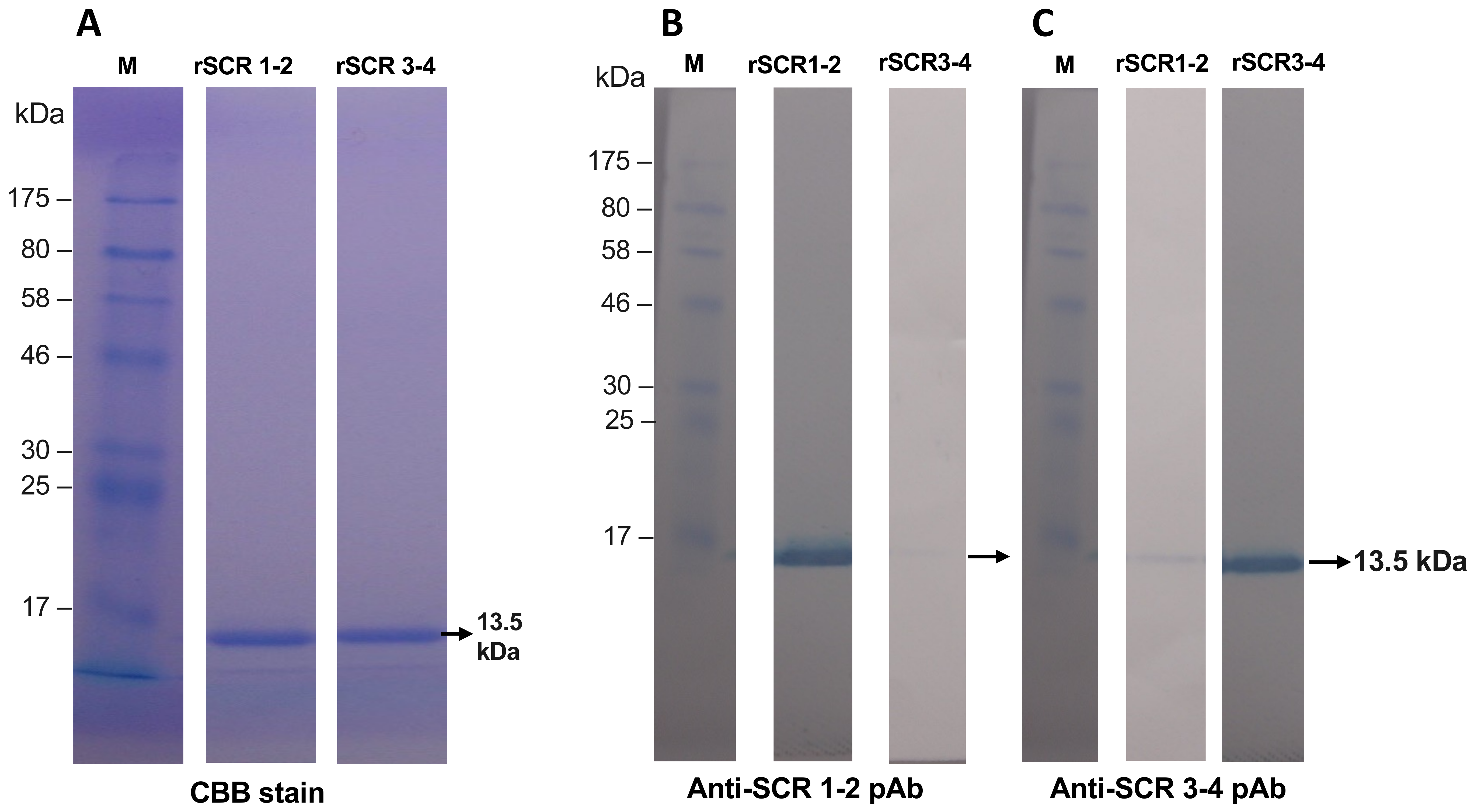
3.1. Preparation of Recombinant SCR 1-2 and SCR 3-4 and Its Polyclonal Antibodies

Primers were separately designed for the SCR 1-2 and SCR 3-4 regions of cTecrem, and the recombinant proteins were expressed in a prokaryotic system. The recombinant proteins, rSCR1-2 and rSCR3-4, were purified using Ni-NTA columns followed by size exclusion chromatography. Both rSCR1-2 and rSCR3-4 showed a single band of 13.5 kDa, agreeing well with the theoretical molecular masses of SCR1-2 and SCR3-4 domains calculated from their amino acid sequences (Figure 1A). The purified rSCR1-2 and rSCR3-4 were cleaved with Factor Xa to remove the 6xHis tag for subsequent immunization of rabbits. Polyclonal anti-SCR1-2 and anti-SCR3-4 IgG were affinity-purified from the antisera using immobilized rSCR1-2 and rSCR3-4 coupled to NHS-activated HiTtrap columns. The western blot results showed that the purified antibodies are specific to their own antigen with only slight cross-reactivity (Figure 1B,C).
Table 1 and CFS cells were also examined by western blot using KF-1 and CFS cell lysate as antigens. As shown in Figure 2, anti-SCR1-2 detected a major band of 58 kDa and two minor bands of 49 kDa and 40 kDa from KF-1 lysate, showing a weak 75 kDa band in the CFS lysate. On the other hand, anti-SCR3-4 detected 58 kDa and 49 kDa bands with less intensity from the KF-1 lysate, showing no band in the CFS lysate. The results suggest that anti-SCR1-2 recognizes both cTecrem and ginbuna Tecrem (gTecrem), while anti-SCR3-4 is specific for cTecrem. The observed molecular mass (58 kDa) of the major band of cTecrem agrees with that observed in immunoprecipitation using 1F12 mAb and KF-1 lysate (Figure 3), suggesting that natural cTecrem on KF-1 cell is a 58 kDa protein. The 49 kDa and 40 kDa minor bands may represent degradation products or less glycosylated isoforms of cTecrem.
Reactivity of anti-SCR1-2 and anti-SCR3-4 against native cTecrem protein on KF-1 were examined by FCM analysis. The results showed that anti-SCR1-2 detects cTecrem at a higher sensitivity than that of anti-SCR3-4 (Figure 4).
Using rSCR1-2 and rSCR3-4 as antigens, the epitope of 1F12 mAb was localized by ELISA. As shown in Figure 5, 1F12 mAb showed reactivity only to rSCR1-2, suggesting that the epitope of 1F12 mAb resides in SCR1-2 domains of cTecrem.

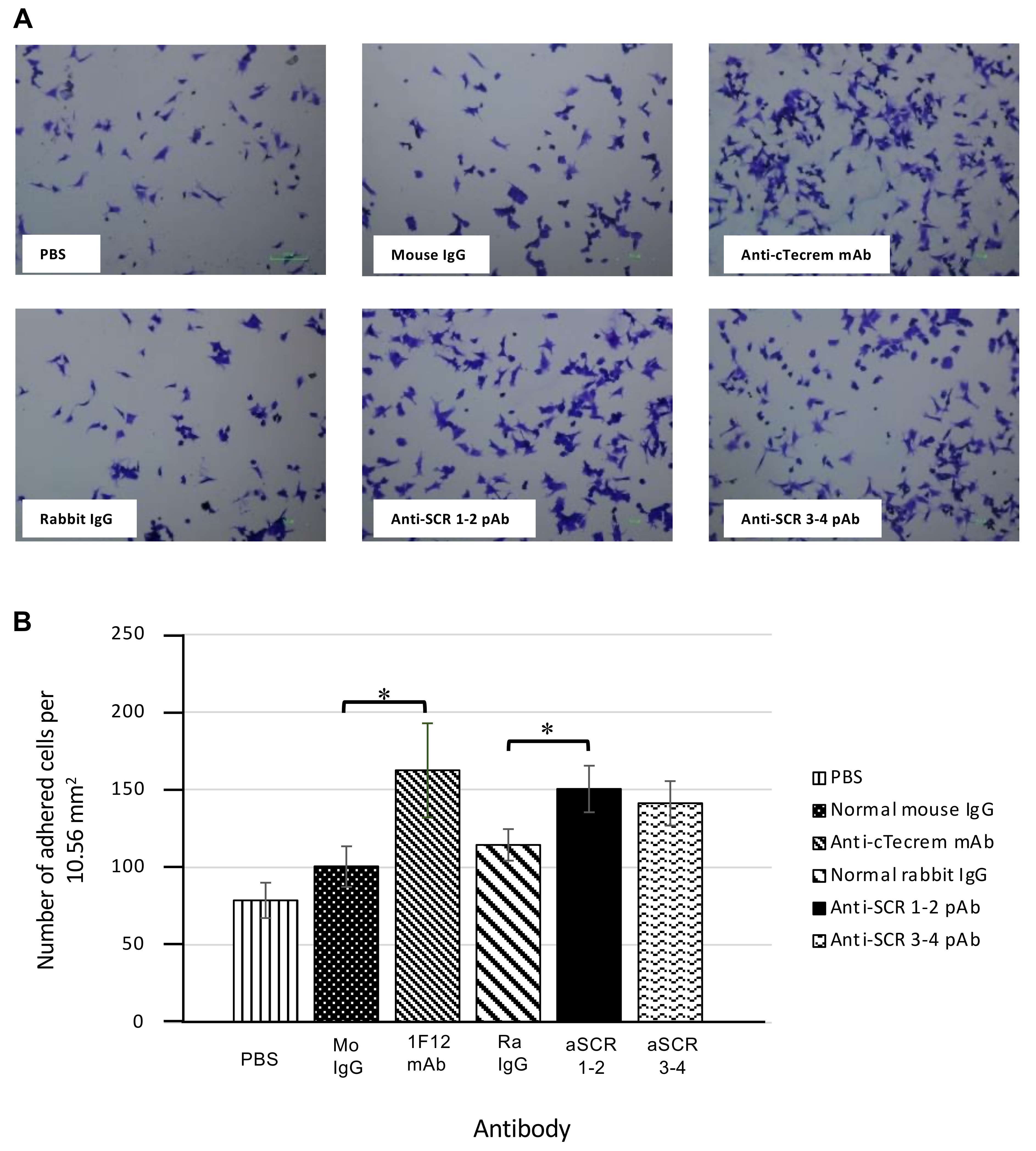
3.2. Activation of cTecrem Results in An Increase in the Adhesion of KF-1 Cells on to the Cell Culture Plate

To explore functions of cTecrem expression on KF-1 cells in their adhesion, the time course of surface cTecrem level was measured by ELISA using 1F12 mAb after seeding of KF-1 cells to a 96-well plate. As shown in Figure 6, during the first 2 h after seeding, cTecrem level kept below detectable level, while KF-1 cells have already settled at the plate bottom. The cTecrem level increased from 4 h postseeding, when KF-1 started to develop cell-cell contacts. This result indicates a functional association between cTecrem expression and intercellular adherence of KF-1 cells.
A possible association between cTecrem and cell adhesion of KF-1 was further examined by two lines of experiments. In the first assay, wells of ELISA plate were coated with 1F12 mAb, isotype control mouse IgG, or PBS prior to the cell seeding. Then the number of adhered cells was counted at different time points. Figure 7 shows that the number of adhered cells in the 1F12 mAb-coated wells was significantly higher than those in other wells treated with isotype control and PBS, at first 1 h postseeding. This result suggests cTecrem would enhance the stage of adhesion of KF-1 cells.
In the second cell adhesion assay, KF-1 cells were preincubated with 1F12 mAb, anti-SCR1-2, anti-SCR3-4, isotype control mouse IgG, or nonimmune rabbit IgG. Negative control KF-1 cells were incubated with PBS alone. Then the treated cells were seeded onto the plate, and the number of adhered cells were counted at 30 min postseeding. As shown in Figure 8, the numbers of adherent cells treated with 1F12 mAb and anti-SCR1-2 were significantly higher than that of the negative control cells. Anti-SCR3-4 also enhanced the cell adhesion compared to the isotype controls, though it is unclear whether cTecrem directly mediates interactions of KF-1 with the plate surface or it activates other adhesion molecules.

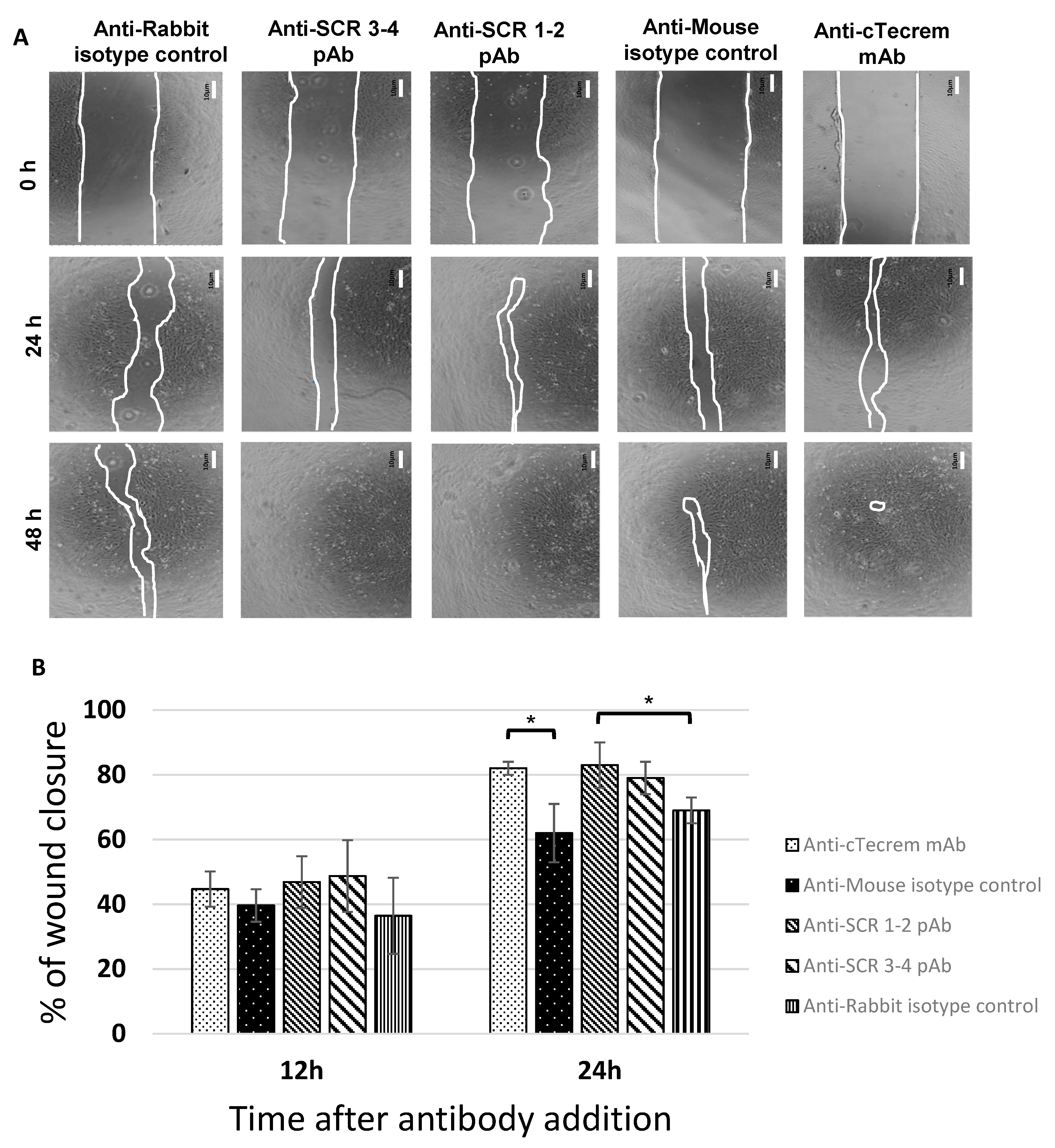
3.3. Tecrem Activation Promotes Epithelial Cell Growth as well as Wound Healing

To investigate if cTecrem plays a role in the proliferation of epithelial cells and wound repair, a gap-closure assay was performed using KF-1 monolayer culture. KF-1 cells were cultured on 24-well plate until they formed a complete monolayer. Then gaps were artificially created by scratching the monolayer using a 200 µL micropipette tip held at an angle of 30º to keep the scratch width limited. The monolayer was then overlaid by MEM-10 with 1F12 mAb, anti-SCR1-2, anti-SCR3-4, or nonimmune controls IgG. The degree of the gap closure was then monitored under a microscope after 12 h, 24 h, and 48 h, and the percentage of wound closure was calculated. This agrees with the wound healing assay data in human CD46 [12]. The gap closure of the KF-1 monolayer sheet was accelerated in the presence of 1F12 mAb, anti-SCR1-2, and anti-SCR3-4, but not the control antibodies (Figure 9).

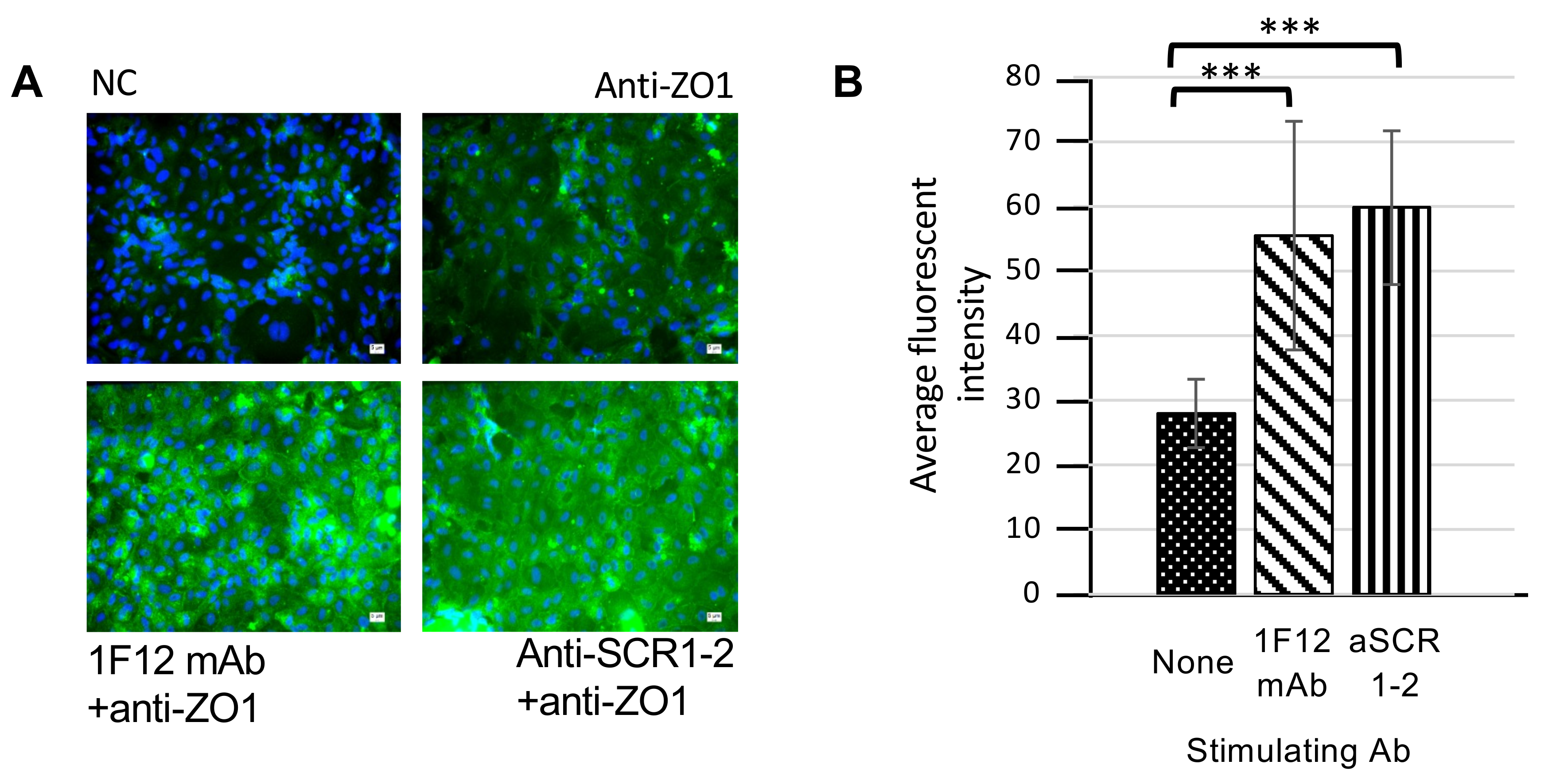
3.4. Enhancement of Tight Junction Protein Expression by cTecrem Stimulation in CFS Cells

Tight junction proteins play a major role in the epithelial formation, development, and maintenance of tissue integrity in vertebrates [16]. To investigate more on the cTecrem-associated epithelial wound healing, we hypothesized that cTecrem may functionally be associated with tight junction formation in the epithelial sheet, focusing on ZO1 (Zonula occludens 1), or tight junction protein 1, which has been recognized to play a major role in the epithelial cell proliferation [16]. An immunohistochemical analysis was performed using anti-ZO1 antibody and CFS, which was employed instead of KF-1, because CFS cell shows morphological stability upon fixation for the histochemistry and anti-cTecrem (1F12 mAb and anti-SCR1-2) can recognize gTecrem, which are expressed on CFS.
As shown in Figure 10A, the fluorescent signal of ZO1 expressed in CFS stimulated with anti-cTecrem was much brighter, showing faster proliferation, than that in nonstimulated control cells. A quantitative evaluation of ZO1 expression also showed significant elevation of ZO1 level by the anti-cTecrem treatment in the stimulated cells (Figure 10B). These results indicate that cTecrem has a regulatory role in the formation of tight junctions.

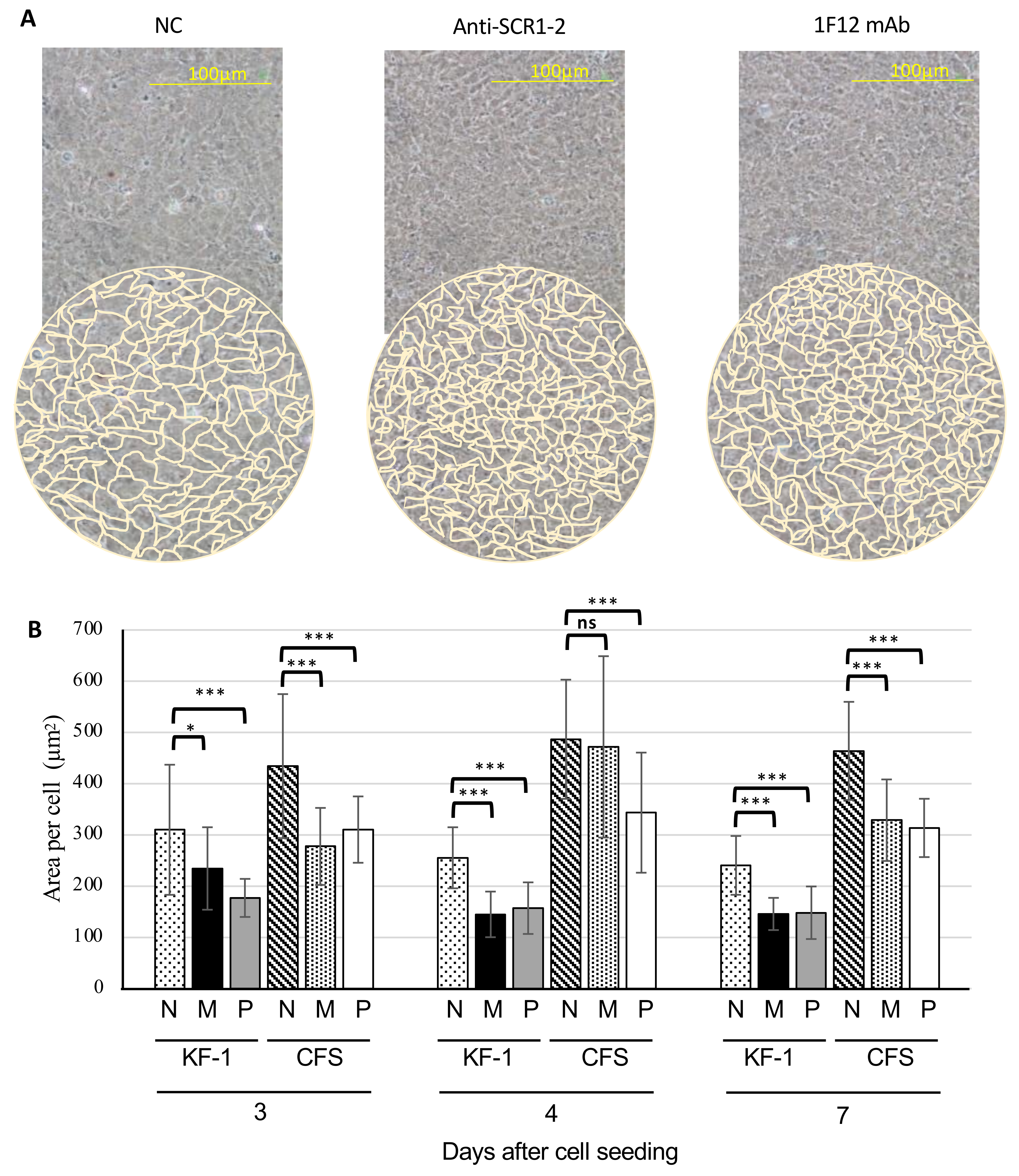
3.5. Morphological Changes in the cTecrem-Activated KF-1 and CFS Cell Lines

To investigate the effects of cTecrem on epithelial cell sheet formation, KF-1 and CFS cells were grown on 6-well plates with or without the anti-cTecrem antibodies for seven days. Then the cellular morphology in the monolayer was observed under a microscope, and cell densities of the monolayer were calculated using ImageJ software based on the microscopic images. As shown in Figure 11A, epithelial cells treated with anti-cTecrem antibodies (1F12 mAb or anti-SCR1-2) formed a sheet with a higher cell density. A quantitative evaluation of the cell densities was made by comparing the average area occupied by 50 cells, as shown in Figure 11B. The average area of KF-1 and CFS cells activated with anti-cTecrem antibodies was significantly lesser than that of the normal cells at day 3 and at day 7. The compactness of the cTecrem-stimulated cells implies that cTecrem mediates intracellular signals to reinforce cell-cell interactions to construct a more robust epithelial barrier.

4. Discussion

In the present study, we have raised polyclonal antibodies specific to two N-terminal domains (SCR1-2) and two C-terminal domains (SCR3-4) of the extracellular regions of cTecrem protein, using recombinant SCR1-2 and SCR3-4 proteins expressed in a prokaryotic system. These recombinant proteins were also utilized to localize an epitope of monoclonal anti-cTecrem (1F12), which had previously been established.
The domain-specific reactivities of the polyclonal anti-SCR1-2 and anti-SCR3-4 were confirmed by western blot. Both antibodies detect a 58 kDa polypeptide in KF-1 cell lysate, revealing the molecular entity of the natural Tecrem protein for the first time. The size of natural cTecrem is slightly smaller than that observed for recombinant cTecrem expressed on CHO cells (66 kDa), likely due to a different degree of glycosylation. In addition, anti-SCR1-2 was found to be cross-reactive to gTecrem, detecting a 75 kDa polypeptide in the lysate of ginbuna epithelial cell line, CFS. Ginbuna crucian carp has been reported to possess three Tecrem isoforms, gTecrem-1, gTecrem-2, and gTecrem-3. Among them, gTecrem-1, with highest mRNA expression level, contains seven SCR modules in its extracellular region, while cTecrem has four SCR modules [7]. This difference in the number of SCR modules likely reflects the difference in the observed polypeptide sizes, 58 kDa cTecrem and 75 kDa gTecrem.
It is interesting to note that anti-SCR1-2 recognized both cTecrem and gTecrem upon western blot analysis, but anti-SCR3-4 was specific only for cTecrem. These results agree well with that 1F12 mAb with an epitope within SCR1-2 also recognizes cTecrem and gTecrem, suggesting that the N-terminal two SCR modules are structurally and functionally well conserved, though the exact epitope of 1F12 mAb is yet to be mapped. The amino acid sequence identity of SCR 1-2 modules between cTecrem and gTecrem-1 is 13% higher than that of SCR 3-4 modules [7]. This could be the reason why anti-SCR 1-2 antibody bound to gTecrem better than anti-SCR 3-4.
It should also be noted that polyclonal anti-rSCR1-2 and anti-rSCR3-4 detected minor bands with smaller sizes (49 kDa and 40 kDa) than that of a major band of 58 kDa in KF-1 cell lysate (Figure 3). The minor bands may possibly represent degradation products generated during the sample preparation for the western blot. It seems more likely, however, that the minor bands represent isoforms of cTecrem generated by alternative splicing, as predicted in our previous study. Zebrafish Tecrem mRNA can be processed into four distinct variants differing in the serine/threonine/proline-rich region (STP-region) at the C-terminus of its extracellular domain, probably resulting in different degree of O-glycosylation withing the STP-region [6]. This could also happen in cTecrem mRNA.
As shown in Figure 7 and Figure 8, treatment of KF-1 cells with monoclonal and polyclonal antibodies specific for SCR1-2 and SCR3-4 enhanced cellular adherence to the surface of cell culture plates. Although it seems possible that Tecrem protein can directly mediate cell-to-plastic surface adherence, Tecrem may indirectly regulate the expression and function of other adhesion molecules through intracellular signal transduction. It has been reported that mammalian CD46, when activated, causes changes in the cellular constellation in epithelial sheets and the formation of tight junctions via SPAK and E-cadherine [5,12]. To examine the effect of Tecrem in epithelial cell proliferation, we proceed with the wound healing assay. The results clearly showed that binding of the antibodies to cTecrem enhances KF-1 proliferation resulting in faster closure of the gaps generated in the KF-1 monolayer (Figure 9), suggesting a wound healing function of cTecrem.
The immunohistochemical analysis revealed enhancement of proliferation of KF-1 cells with increased expression of a tight junction protein, ZO1, by stimulation of cTecrem, implying a possible mechanism of Tecrem-mediated reinforcement of epithelial cell layer (Figure 10). In the case of mammalian ZO1 protein, there are many reports that suggest that ZO1 protein is produced in a minimal amount when the cell proliferate [16,17,18]. ZO1 protein in mammals has been suggested to have a role in tumor suppression, as supported by the correlation between the lesser accumulation of ZO1 proteins and the poor differentiation of tumors [17,19]. The upregulation of the ZO1 protein happens when the cells form a monolayer after getting confluent in mammals [16,20,21,22]. A study done in rainbow trout gill epithelial cells also increases the ZO1 transcript in gill-derived cells during their monolayer formation [23]. This study also suggests that the ZO1 protein plays a role in the cell-cell contacts and establishing the gill epithelium integrity. ZONAB (ZO1 associated nucleic acid binding protein) identified in several organisms is also found to have a role in regulating epithelial cell proliferation in mammals. Since ZONAB is conserved in zebrafish and found to be up-regulated at the time of cell proliferation, and is found to be down-regulated after the formation of the epithelial monolayer [24]. It would be interesting to clone and characterize carp ortholog of zebrafish ZONAB to clarify the cTecrem-mediated epithelial cell activation. The time course of ZO1 mRNA level in the zebrafish flask-cultured epithelial cells agrees with our present results [23,25].
Morphological changes of the cell monolayer upon the activation of cTecrem were also in line with the results of the immunohistostaining. The higher cell density observed in the cTecrem-stimulated KF-1, and CFS indicates that the Tecrem can contribute to establishing a more robust epithelial cell layer with better integrity. This could be crucial to develop methods to keep the mucosal tissue of fish intact and to heal any damage on the epithelial sheet to minimize the risk of microbial invasion through the mucosal surface.

Author Contributions

Conceptualization, H.P., T.S. and M.N.; Methodology, H.P., S.M., T.N. and M.N.; Laboratory works, H.P., S.M. and T.N.; Data validation and curation, H.P., T.S. and M.N.; Writing—Original Draft Preparation, H.P.; Writing—Review & Editing, M.N., T.S., S.M., T.N. and H.P.; Supervision, M.N. and T.S.; Funding Acquisition, M.N. All authors have read and agreed to the published version of the manuscript.

Funding

This research was supported by JSPS KAKENHI Grant Number JP16H04982 to MN.

Institutional Review Board Statement

Not applicable.

Informed Consent Statement

Not applicable.

Conflicts of Interest

The authors declare no conflict of interest.

References

  1. Parra, D.; Reyes-Lopez, F.E.; Tort, L. Mucosal immunity and B cells in teleosts: Effect of vaccination and stress. Front. Immunol. 2015, 6, 354. [Google Scholar] [CrossRef] [PubMed] [Green Version]
  2. Merle, N.S.; Church, S.E.; Fremeaux-Bacchi, V.; Roumenina, L.T. Complement system part I – molecular mechanisms of activation and regulation. Front. Immunol. 2015, 6, 262. [Google Scholar] [CrossRef] [Green Version]
  3. Nakao, M.; Tsujikura, M.; Ichiki, S.; Vo, T.K.; Somamoto, T. The complement system in teleost fish: Progress of post-homolog-hunting researches. Dev. Comp. Immunol. 2011, 35, 1296–1308. [Google Scholar] [CrossRef]
  4. Liszewski, M.K.; Farries, T.C.; Lublin, D.M.; Rooney, I.A.; Atkinson, J.P. Control of the Complement System. Adv. Immunol. 1996, 61, 201–283. [Google Scholar] [CrossRef]
  5. Yamamoto, H.; Fara, A.F.; Dasgupta, P.; Kemper, C. CD46: The ‘multitasker’ of complement proteins. Int. J. Biochem. Cell. Biol. 2013, 45, 2808–2820. [Google Scholar] [CrossRef]
  6. Tsujikura, M.; Nagasawa, T.; Ichiki, S.; Nakamura, R.; Somamoto, T.; Nakao, M. A CD46-like Molecule Functional in Teleost Fish Represents an Ancestral Form of Membrane-Bound Regulators of Complement Activation. J. Immunol. 2015, 194, 262–272. [Google Scholar] [CrossRef]
  7. Nur, I.; Harada, H.; Tsujikura, M.; Somamoto, T.; Nakao, M. Molecular characterization and expression analysis of three membrane-bound complement regulatory protein isoforms in the ginbuna crucian carp Carassius auratus langsdorfii. Fish Shellfish. Immunol. 2013, 35, 1333–1337. [Google Scholar] [CrossRef]
  8. Nur, I.; Abdelkhalek, N.K.; Motobe, S.; Nakamura, R.; Tsujikura, M.; Somamoto, T.; Nakao, M. Functional analysis of membrane bound complement regulatory protein on T-cell immune response in Ginbuna Crucian Carp. Mol. Immunol. 2016, 70, 1–7. [Google Scholar] [CrossRef] [PubMed]
  9. Wu, J.; Li, H.; Zhang, S. Regulator of complement activation (RCA) group 2 gene cluster in zebrafish: Identification, expression, and evolution. Funct. Integr. Genomics 2012, 12, 367–377. [Google Scholar] [CrossRef]
  10. Li, M.F.; Sui, Z.H.; Sun, L. A teleost CD46 is involved in the regulation of complement activation and pathogen infection. Sci. Rep. 2017, 7, 15028. [Google Scholar] [CrossRef] [Green Version]
  11. West, E.E.; Kolev, M.; Kemper, C. Complement and the Regulation of T Cell Responses. Annu. Rev. Immunol. 2018, 36, 309–338. [Google Scholar] [CrossRef]
  12. Cardone, J.; Shouli, A.L.; Kemper, C. A novel role for CD46 in wound repair. Front. Immunol. 2011, 2, 28. [Google Scholar] [CrossRef] [Green Version]
  13. Lv, H.; Zhou, T.; Dong, C.; Kong, S.; Chen, L.; Pu, F.; Li, X.; Xu, P. Genome-wide identification, evolution, and mRNA expression of complement genes in common carp (Cyprinus carpio). Fish Shellfish. Immunol. 2020, 96, 190–200. [Google Scholar] [CrossRef] [PubMed]
  14. Gonzalez, S.F.; Buchmann, K.; Nielsen, M.E. Complement expression in common carp (Cyprinus carpio L.) during infection with Ichthyophthirius multifiliis. Dev. Comp. Immunol. 2007, 31, 576–586. [Google Scholar] [CrossRef]
  15. Brinchmann, M.F. Immune relevant molecules identified in the skin mucus of fish using -omics technologies. Mol. Biosyst. 2016, 12, 2056–2063. [Google Scholar] [CrossRef] [Green Version]
  16. Balda, M.S.; Matter, K. The tight junction protein ZO-1 and an interacting transcription factor regulate ErbB-2 expression. EMBO J. 2000, 19, 2024–2033. [Google Scholar] [CrossRef]
  17. Anderson, J.M.; Itallie, C.M.V. Physiology and Function of the Tight Junction. Cold Spring Harb. Perspect. Biol. 2009, 1, a002584. [Google Scholar] [CrossRef]
  18. Balda, M.S.; Matter, K. Review-Tight junctions and the regulation of gene expression. Biochim. Biophys. Acta 2009, 1788, 761–767. [Google Scholar] [CrossRef] [Green Version]
  19. Tsukita, S.; Furuse, M.; Itoh, M. Structural and signalling molecules come together at tight junctions. Curr. Opin. Cell Biol. 1999, 11, 628–633. [Google Scholar] [CrossRef]
  20. Georgiadis, A.; Tschernutter, M.; Bainbridge, J.W.B.; Balaggan, K.S.; Mowat, F.; West, E.L.; Munro, P.M.G.; Thrasher, A.J.; Matter, K.; Balda, M.S.; et al. The Tight Junction Associated Signalling Proteins ZO-1 and ZONAB Regulate Retinal Pigment Epithelium Homeostasis in Mice. PLoS ONE 2010, 5, e15730. [Google Scholar] [CrossRef] [Green Version]
  21. Mariscal, L.G.; Lechuga, S.; Garay, E. Role of tig.ht junctions in cell proliferation and cancer. Prog. Histochem. Cytochem. 2007, 42, 1–57. [Google Scholar] [CrossRef]
  22. Mariscal, L.G.; Tapia, R.; Chamorro, D. Crosstalk of tight junction components with signaling pathways. Biochim. Biophys. Acta 2008, 1778, 729–756. [Google Scholar] [CrossRef] [Green Version]
  23. Kolosov, D.; Chasiotis, H.; Kelly, S.P. Tight junction protein gene expression patterns and changes in transcript abundance during development of model fish gill epithelia. J. Exp. Biol. 2014, 217, 1667–1681. [Google Scholar] [CrossRef] [Green Version]
  24. Balda, M.S.; Garrett, M.S.; Matter, K. The ZO-1–associated Y-box factor ZONAB regulates epithelial cell proliferation and cell density. J. Cell. Biol. 2003, 160, 423–432. [Google Scholar] [CrossRef]
  25. Freshney, R.I. Culture of Animal Cells: A Manual of Basic Technique and Specialized Applications. Biol. Cult. Cells 2010, 11–23. [Google Scholar] [CrossRef]
Figure 1. Purification of recombinant SCR1-2 and SCR2-3 domains of cTecrem and productions of polyclonal antibodies (A) SDS-PAGE of rSCR1-2 and rSCR3-4 under reducing conditions on 12% gels. (B,C) Western blot analysis for the cross-reactivity of affinity-purified anti-rSCR1-2 and anti-SCR3-4 with rSCR1-2 and rSCR3-4 antigens.
Jmse 09 00687 g001
Figure 2. Reactivity of anti-SCR1-2 and anti-SCR3-4 with cell lysates of KF-1 and CFS. (A) Coomassie Blue-stained total proteins of KF-1 lysate (Lane 1) and CFS lysate (Lane 2) loaded for western blot. Lane M shows marker proteins with their molecular masses (kDa) on the left. (B) Western blot analysis of KF-1 (Lanes 2 and 4) and CFS cell lysates (Lanes 1 and 3) using anti-SCR1-2 (left panel) and anti-SCR3-4 (right panel). Lane M shows prestained marker proteins with their molecular masses (kDa) on the left. Molecular masses of detected bands (kDa) are inserted between the two panels.
Jmse 09 00687 g002
Figure 3. Detection of natural cTecrem expressed on KF-1 cells by immunoprecipitation with 1F12 mAb. The biotin-labeled KF-1 cell lysate was incubated with 1F12 mAb, and the immune complex was trapped by protein G-Sepharose beads, separated by SDS-PAGE, and developed with peroxidase-labeled streptavidin and 3,3-diaminobenzidine. Lane M, prestained marker proteins with their molecular masses on the left; lane 1, negative control without 1F12 mAb; test sample with 1F12 mAb.
Jmse 09 00687 g003
Figure 4. Flow cytometry analysis showing the expression of cTecrem on KF-1 cells using varying concentrations (0.01, 0.1, 1.0 µg/mL) of affinity-purified anti-SCR1-2 (A) and anti-SCR3-4 (B). Negative control was without the first antibodies. 1F12 mAb (1 µg/mL) was employed as a positive control.
Jmse 09 00687 g004
Figure 5. Reactivity of 1F12 mAb to rSCR1-2 and rSCR3-4 as antigens as measured by ELISA.
Jmse 09 00687 g005
Figure 6. (A) Expression of cTecrem detected during cell adhesion process of KF-1 to plastic culture media after seeding. The cTecrem level was measured by ELISA as adhered cells as antigen. (B) Phase-contrast microscopic images of the adhered KF-1 at 2 h and 6 h postseeding.
Jmse 09 00687 g006
Figure 7. Enhancement of early stage of the KF-1 cell adhesion onto plastic culture media in the presence of anti-cTecrem. (A) Microscopic images of the adhered KF-1 cells in the presence or absence of the antibodies shown above after 1 h and 2 h incubation. (Stained with Crystal Violet) (B) Quantitative representation of the cell adhesion, expressed in numbers of cells attached to a well of the culture plate with isotype control. Data shown are the mean ±SD obtained from triplicate experiments. Asterisks show significant differences at p < 0.05.
Jmse 09 00687 g007
Figure 8. Effect of anti-cTecrem on the early phase of KF1 cell adhesion. KF-1 cells were incubated in the presence of 1F12 mAb, anti-SCR1-2, anti-SCR3-4, isotype controls, or PBS, and cultured at different times. (A) The images were taken 30 min after seeding of antibody-incubated cells on the culture plate. The data shown are the representative images of three experiments. (B) The quantitative representation of the cell adhesion. Data shown are the mean ±SD from triplicate experiments. Asterisks show significant differences at p < 0.05 tested by ANOVA.
Jmse 09 00687 g008
Figure 9. Effect of anti-cTecrem treatment on the closure of the gap generated in the KF-1 monolayer sheet. After KF-1 cells formed a monolayer sheet in a 6-well plate, wounds were artificially created by scratching the monolayer using 200 µL pipette tips kept at a 30º angle; then, the KF-1 cell sheet was incubated with anti-cTecrem antibodies and its isotype controls. (A) The degree of the gap closure was observed under a phase-contrast microscope in triplicates. (B) Quantitative evaluation of wound healing assay. The area of the gap was measured using ImageJ software, and the percentage of the gap closure was calculated. Asterisks show significant differences at p < 0.05.
Jmse 09 00687 g009
Figure 10. Immunohistochemistry analysis showing the role of cTecrem in the formation of tight junctions. After CFS cells formed a monolayer on the culture plate, the monolayer was incubated with anti-cTecrem mAb, anti-SCR 1-2 pAb, and controls for 48 h at 25 °C. After the incubation, the monolayer was fixed with methanol and was analyzed using rabbit anti-ZO1 pAb as primary and rabbit anti-FITC pAb as secondary antibodies. (A) Fluorescent microscopic analysis of CFS monolayer treated with or without anti-cTecrem antibodies and anti-ZO1 pAb, and counterstained with DAPI. (B) Quantitative evaluation of immunohistochemical assay. The fluorescent intensity of each cell was measured using ImageJ software, and the graph was plotted by subtracting the background intensity from those values. Data shown are the mean ±SD from triplicate experiments. Asterisks show significant differences at p < 0.001.
Jmse 09 00687 g010
Figure 11. Morphological analysis of the KF-1 and CFS cell monolayers treated with anti-cTecrem antibodies. KF-1 and CFS monolayers were grown on 6-well plate that was incubated with and without 1F12 mAb and anti-SCR 1-2 pAb, and was further grown 7 days. The cells were analyzed for morphological changes under phase-contrast on the third, fourth, and seventh day. (A) Phase-contract microscopic images of the KF-1 cells treated with and without anti-cTecrem antibodies. (B) Quantitative evaluation of the morphological changes in anti-cTecrem antibody-treated KF-1 cells. N, M, and P represents untreated cells, cells treated with 1F12 mAb, and cells treated with anti-SCR 1-2 pAb, respectively. Area of randomly selected 50 cells per image was calculated using ImageJ software, and the graph was plotted using the mean area. Data shown are the mean ±SD from triplicate experiments using KF-1 cells. Similar results were observed using CFS cells as well (data not shown). Asterisks show significant differences. (*, p ≤ 0.05; ***, p ≤ 0.001; ns, nonsignificant).
Jmse 09 00687 g011
Table 1. Primers used in the present study.
NameNucleotide Sequence 1Amino Acids EncodedStrand
SCR1-2FgcctcgagGCGGAATGTACACAGCCTTCAECTQPSense
SCR1-2RcgctgcagttaCGATTCACAGAGAGGATCTCDPLCESAntisense
SCR3-4FgcctcgagGTAAAATGTTCAGCACCTCCAGCVKCSAPPSense
SCR3-4RcgctgcagttaGACAATGCATTGTGGTGGTTCAGEPPQCIVAntisense
1 Lower case letter sequences include XhoI and PstI sites and a stop codon.
Publisher’s Note: MDPI stays neutral with regard to jurisdictional claims in published maps and institutional affiliations.

