
Investigation of Adequate Calibration Methods for X-ray Fluorescence Core Scanning Element Count Data: A Case Study of a Marine Sediment Piston Core from the Gulf of Alaska

 , ,
, ,
Abstract
1. Introduction
2. Materials and Methods
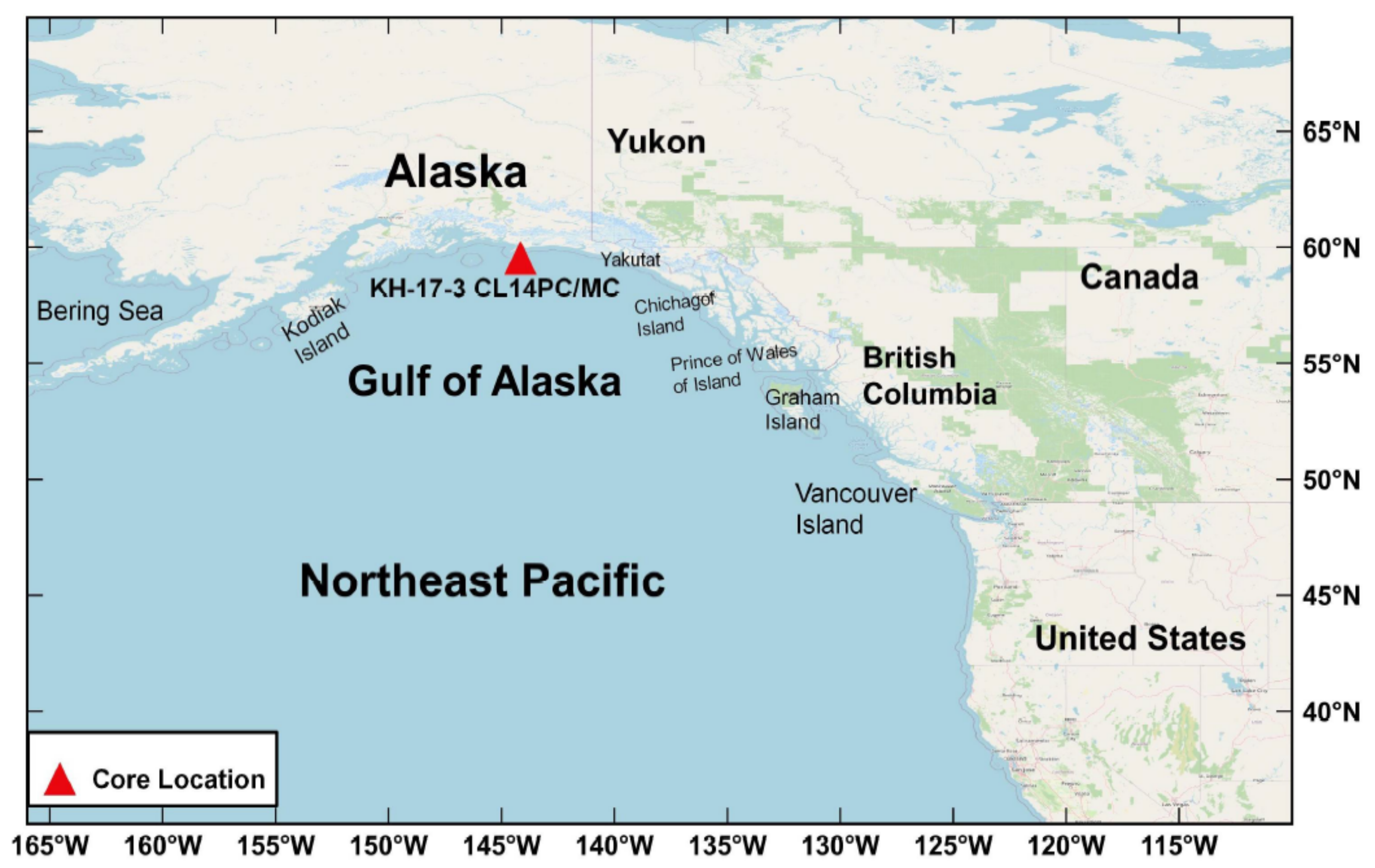
2.1. Sample Collection
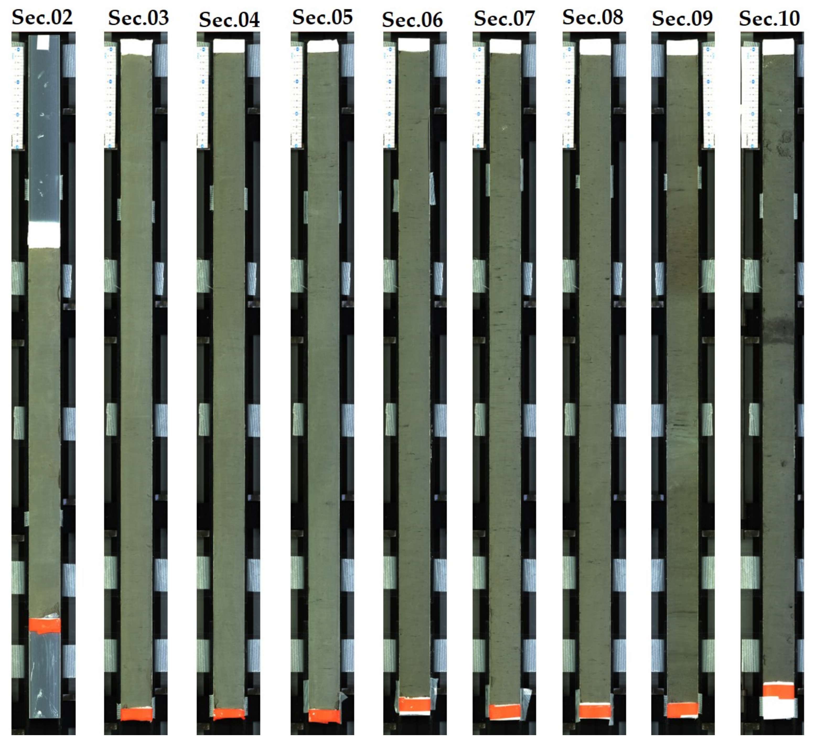
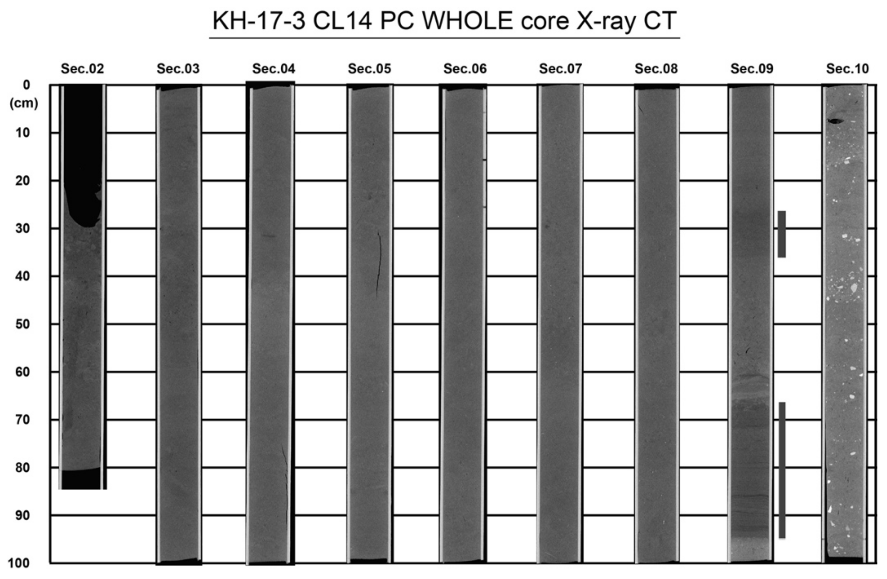
2.2. Core Description and Measurement
2.3. Sediment Analysis
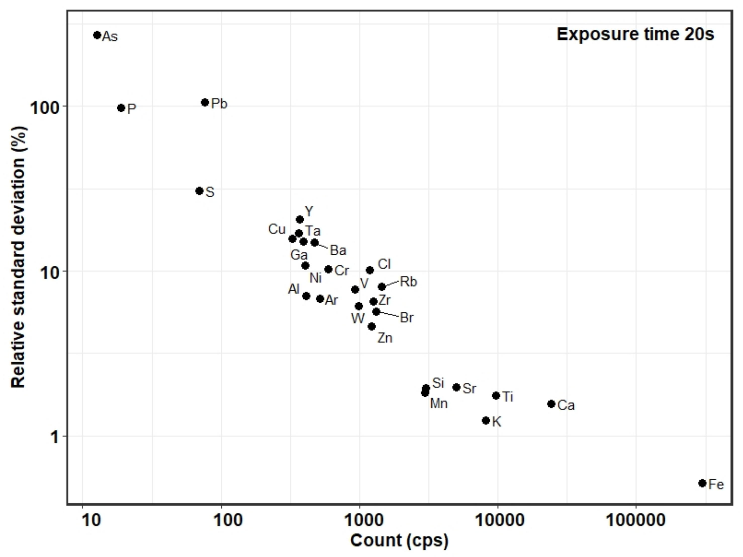
2.3.1. Optimization of Scanning Parameters for ITRAX-XRF Core Scanner
2.3.2. Measurement of Multi-Element Count through ITRAX-XRF Core Scanner
2.3.3. Preparation of Calibration Curve for WD-XRF Concentration Measurement
2.3.4. Validation of Calibration Method
2.3.5. Measurement of Element Concentration through WD-XRF Spectrometer
2.4. Total Organic Carbon Analysis
2.5. XRF Core Scanner Data Calibration
2.5.1. Calibration by Ratios
2.5.2. Log-Ratio Transformation
3. Results
3.1. Raw X-ray Fluorescence Core Scanner Data
3.2. Calibrated X-ray Fluorescence Core Scanner Data
4. Discussion
4.1. Sediment Physical Properties Affect Scanning Intensities
4.2. Element Intensities Calibration by Ratios
4.3. Element Intensities Calibration by Log-Ratios
5. Conclusions
Author Contributions
Funding
Data Availability Statement
Acknowledgments
Conflicts of Interest
Appendix A

| Category | Correlation Method | Correlation between ITRAX-XRF CS Intensities (cps) with WD-XRF Concentration ((wt%), n = 33), and TOC ((wt%), n = 100) | ||||||||
|---|---|---|---|---|---|---|---|---|---|---|
| Element | ||||||||||
| Sr | Fe | Mn | Ti | Ca | K | Br v TOC | Br/Cl v TOC | |||
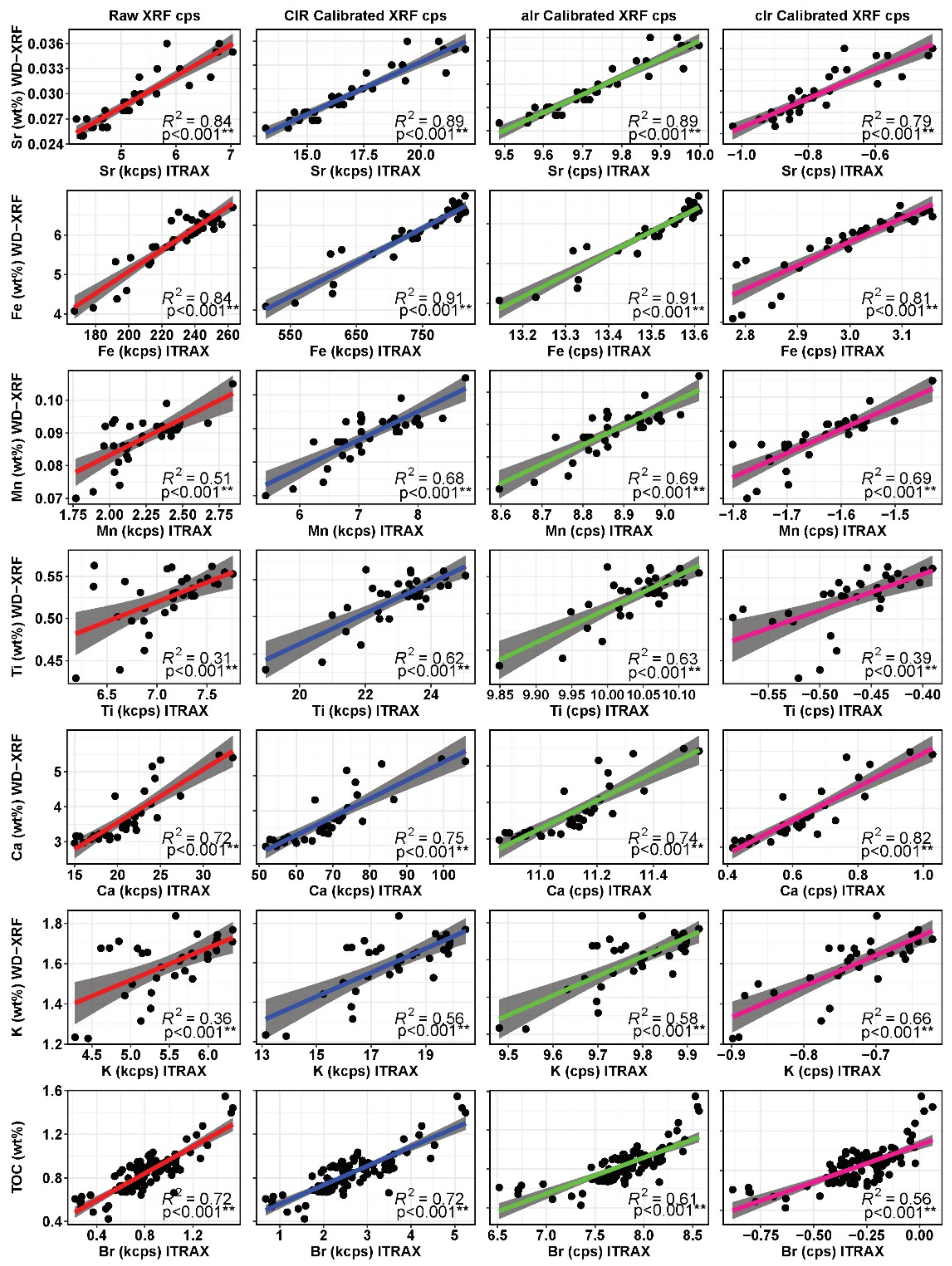
| Raw XRF CS | Pearson (R2) | 0.84 | 0.84 | 0.51 | 0.31 | 0.72 | 0.36 | 0.72 | 0.64 | |
| Kendall’s τ | 0.79 | 0.66 | 0.47 | 0.38 | 0.71 | 0.41 | 0.60 | 0.53 | ||
| Calibration by ratio | CIR a | Pearson (R2) | 0.89 | 0.91 | 0.68 | 0.62 | 0.75 | 0.56 | 0.72 | 0.64 |
| Kendall’s τ | 0.83 | 0.78 | 0.55 | 0.45 | 0.73 | 0.48 | 0.61 | 0.53 | ||
| ICR b | Pearson (R2) | 0.77 | 0.62 | 0.32 | 0.07 | 0.69 | 0.17 | 0.70 | 0.64 | |
| Kendall’s τ | 0.73 | 0.55 | 0.38 | 0.29 | 0.68 | 0.30 | 0.59 | 0.53 | ||
| coh c | Pearson (R2) | 0.74 | 0.64 | 0.33 | 0.05 | 0.72 | 0.18 | 0.70 | 0.64 | |
| Kendall’s τ | 0.75 | 0.61 | 0.37 | 0.30 | 0.70 | 0.32 | 0.60 | 0.52 | ||
| I + C d | Pearson (R2) | 0.69 | 0.45 | 0.22 | 0.00 | 0.68 | 0.07 | 0.68 | 0.64 | |
| Kendall’s τ | 0.67 | 0.53 | 0.29 | 0.18 | 0.66 | 0.24 | 0.59 | 0.53 | ||
| Total count (cps) | Pearson (R2) | 0.81 | 0.69 | 0.25 | 0.00 | 0.75 | 0.15 | 0.72 | 0.64 | |
| Kendall’s τ | 0.77 | 0.57 | 0.27 | 0.09 | 0.71 | 0.30 | 0.61 | 0.52 | ||
| Ti intensity (cps) | Pearson (R2) | 0.85 | 0.86 | 0.56 | 0.82 | 0.27 | 0.65 | 0.64 | ||
| Kendall’s τ | 0.84 | 0.80 | 0.56 | 0.75 | 0.32 | 0.57 | 0.52 | |||
| Ca intensity (cps) | Pearson (R2) | 0.00 | 0.64 | 0.51 | 0.46 | 0.63 | 0.52 | 0.64 | ||
| Kendall’s τ | −0.09 | 0.74 | 0.67 | 0.57 | 0.62 | 0.49 | 0.52 | |||
| Calibration by log-ratio | clr e of CIR calibrated data | Pearson (R2) | 0.79 | 0.81 | 0.69 | 0.39 | 0.82 | 0.66 | 0.56 | 0.29 |
| Kendall’s τ | 0.78 | 0.81 | 0.70 | 0.56 | 0.76 | 0.62 | 0.52 | 0.33 | ||
| clr of raw data | Pearson (R2) | 0.79 | 0.81 | 0.69 | 0.39 | 0.82 | 0.66 | 0.56 | 0.34 | |
| Kendall’s τ | 0.78 | 0.81 | 0.70 | 0.56 | 0.76 | 0.62 | 0.52 | 0.36 | ||
| alr f by CIR | Pearson (R2) | 0.89 | 0.91 | 0.69 | 0.63 | 0.74 | 0.58 | 0.61 | 0.60 | |
| Kendall’s τ | 0.82 | 0.79 | 0.58 | 0.45 | 0.74 | 0.48 | 0.61 | 0.55 | ||
| alr by coh after CIR calibration | Pearson (R2) | 0.82 | 0.82 | 0.52 | 0.30 | 0.74 | 0.40 | 0.58 | 0.52 | |
| Kendall’s τ | 0.79 | 0.75 | 0.52 | 0.49 | 0.72 | 0.42 | 0.61 | 0.50 | ||
| alr by coh | Pearson (R2) | 0.76 | 0.66 | 0.32 | 0.05 | 0.67 | 0.17 | 0.59 | 0.52 | |
| Kendall’s τ | 0.75 | 0.61 | 0.37 | 0.30 | 0.70 | 0.32 | 0.60 | 0.50 | ||
References
- Croudace, I.W.; Rindby, A.; Rothwell, R.G. ITRAX: Description and evaluation of a new multi-function X-ray core scanner. Geol. Soc. Spec. Publ. 2006, 267, 51–63. [Google Scholar] [CrossRef]
- Rothwell, R.G.; Croudace, I.W. Twenty Years of XRF Core Scanning Marine Sediments: What Do Geochemical Proxies Tell Us? In Micro-XRF Studies of Sediment Cores. Developments in Paleoenvironmental Research; Croudace, I., Rothwell, R., Eds.; Springer: Dordrecht, The Netherlands, 2015; Volume 17, ISBN 978-94-017-9848-8. [Google Scholar]
- Rothwell, R.G.; Rack, F.R. New techniques in sediment core analysis: An introduction. Geol. Soc. Spec. Publ. 2006, 267, 1–29. [Google Scholar] [CrossRef]
- Tjallingii, R.; Röhl, U.; Kölling, M.; Bickert, T. Influence of the water content on X-ray fluorescence corescanning measurements in soft marine sediments. Geochem. Geophys. Geosystems 2007, 8, 1–12. [Google Scholar] [CrossRef]
- Boyle, J.F.; Chiverrell, R.C.; Schillereff, D. Approaches to Water Content Correction and Calibration for µXRF Core Scanning: Comparing X-ray Scattering with Simple Regression of Elemental Concentrations. In Micro-XRF Studies of Sediment Cores. Developments in Paleoenvironmental Research; Croudace, I., Rothwell, R., Eds.; Springer: Dordrecht, The Netherlands, 2015; Volume 17, pp. 373–390. ISBN 9789401798495. [Google Scholar]
- Hennekam, R.; de Lange, G. X-ray fluorescence core scanning of wet marine sediments: Methods to improve quality and reproducibility of highresolution paleoenvironmental records. Limnol. Oceanogr. Methods 2012, 10, 991–1003. [Google Scholar] [CrossRef]
- MacLachlan, S.E.; Hunt, J.E.; Croudace, I.W. An Empirical Assessment of Variable Water Content and Grain-Size on X-ray Fluorescence Core-Scanning Measurements of Deep Sea Sediments. In Micro-XRF Studies of Sediment Cores. Developments in Paleoenvironmental Research; Croudace, I., Rothwell, R., Eds.; Springer: Dordrecht, The Netherlands, 2015; Volume 17, pp. 173–185. ISBN 9789401798495. [Google Scholar]
- Gregory, B.R.B.; Patterson, R.T.; Reinhardt, E.G.; Galloway, J.M.; Roe, H.M. An evaluation of methodologies for calibrating Itrax X-ray fluorescence counts with ICP-MS concentration data for discrete sediment samples. Chem. Geol. 2019, 521, 12–27. [Google Scholar] [CrossRef]
- Lee, A.S.; Huang, J.J.S.; Burr, G.; Kao, L.C.; Wei, K.Y.; Liou, S.Y.H. High resolution record of heavy metals from estuary sediments of Nankan River (Taiwan) assessed by rigorous multivariate statistical analysis. Quat. Int. 2019, 527, 44–51. [Google Scholar] [CrossRef]
- Woodward, C.A.; Gadd, P.S. The potential power and pitfalls of using the X-ray fluorescence molybdenum incoherent: Coherent scattering ratio as a proxy for sediment organic content. Quat. Int. 2019, 514, 30–43. [Google Scholar] [CrossRef]
- Agarwal, B.K. Scattering of X-rays. In X-Ray Spectroscopy. Springer Series in Optical Sciences; Springer: Berlin/Heidelberg, Germany, 1991; Volume 15, pp. 223–239. [Google Scholar]
- Weltje, G.J.; Tjallingii, R. Calibration of XRF core scanners for quantitative geochemical logging of sediment cores: Theory and application. Earth Planet. Sci. Lett. 2008, 274, 423–438. [Google Scholar] [CrossRef]
- Hunt, J.E.; Croudace, I.W.; MacLachlan, S.E. Use of Calibrated ITRAX XRF Data in Determining Turbidite Geochemistry and Provenance in Agadir Basin, Northwest African Passive Margin. In Micro-XRF Studies of Sediment Cores. Developments in Paleoenvironmental Research; Croudace, I.W., Rothwell, R., Eds.; Springer: Dordrecht, The Netherlands, 2015; Volume 17, pp. 127–146. ISBN 9789400720596. [Google Scholar]
- Kylander, M.E.; Ampel, L.; Wohlfarth, B.; Veres, D. High-resolution X-ray fluorescence core scanning analysis of Les Echets (France) sedimentary sequence: New insights from chemical proxies. J. Quat. Sci. 2011, 26, 109–117. [Google Scholar] [CrossRef]
- Chagué-Goff, C.; Chan, J.C.H.; Goff, J.; Gadd, P.; Oliva, F.; Peros, M.C.; Viau, A.E.; Reinhardt, E.G.; Nixon, F.C.; Morin, A. Late Holocene record of environmental changes, cyclones and tsunamis in a coastal lake, Mangaia, Cook Islands. Isl. Arc 2016, 25, 333–349. [Google Scholar] [CrossRef]
- Oliva, F.; Peros, M.C.; Viau, A.E.; Reinhardt, E.G.; Nixon, F.C.; Morin, A. A multi-proxy reconstruction of tropical cyclone variability during the past 800 years from Robinson Lake, Nova Scotia, Canada. Mar. Geol. 2018, 406, 84–97. [Google Scholar] [CrossRef]
- Lyle, M.; Oliarez Lyle, A.; Gorgas, T.; Holbourn, A.; Westerhold, T.; Hathorne, E.; Kimoto, K.; Yamamoto, S. Data report: Raw and normalized elemental data along the Site U1338 splice from X-ray fluorescence scanning. Proc. Integr. Ocean Drill. Progr. 2012, 320, 321. [Google Scholar] [CrossRef]
- Penkrot, M.; LeVay, L.J.; Jaeger, J.M. Data report: X-ray florescence scanning of sediment cores, Site U1419, Gulf of Alaska. Proc. Integr. Ocean Drill. Progr. 2017, 341, 1–7. [Google Scholar] [CrossRef]
- Lyle, M.; Kulhanek, D.K.; Bowen, M.G.; Hahn, A. Data report: X-ray fluorescence studies of Site U1457 sediments, Laxmi Basin, Arabian Sea. Proc. Int. Ocean Discov. Progr. 2018, 355. [Google Scholar] [CrossRef]
- Babin, D.P.; Franzese, A.M.; Hemming, S.R.; Hall, I.R.; LeVay, L.J.; Barker, S.; Tejeda, L.; Simon, M.H. Data report: X-ray fluorescence core scanning of IODP Site U1474 sediments, Natal Valley, Southwest Indian Ocean, Expedition 361. Proc. Int. Ocean Discov. Progr. 2020, 361. [Google Scholar] [CrossRef]
- Dunlea, A.G.; Murray, R.W.; Tada, R.; Alvarez-Zarikian, C.A.; Anderson, C.H.; Gilli, A.; Giosan, L.; Gorgas, T.; Hennekam, R.; Irino, T.; et al. Intercomparison of XRF Core Scanning Results From Seven Labs and Approaches to Practical Calibration. Geochem. Geophys. Geosystems 2020, 21, 1–13. [Google Scholar] [CrossRef]
- Boyd, B.L.; Anderson, J.B.; Wellner, J.S.; Fernández, R.A. The sedimentary record of glacial retreat, Marinelli Fjord, Patagonia: Regional correlations and climate ties. Mar. Geol. 2008, 255, 165–178. [Google Scholar] [CrossRef]
- Smith, N.T.; Shreeve, J.; Kuras, O. Multi-sensor core logging (MSCL) and X-ray computed tomography imaging of borehole core to aid 3D geological modelling of poorly exposed unconsolidated superficial sediments underlying complex industrial sites: An example from Sellafield nuclear site, UK. J. Appl. Geophys. 2020, 178, 104084. [Google Scholar] [CrossRef]
- Miller, J.N.; Miller, J.C.; Miller, R.D. Statistics and Chemometrics for Analytical Chemistry, 7th ed.; Pearson Education Limited: Harlow, UK, 2018. [Google Scholar]
- Plaza-Morlote, M.; Rey, D.; Santos, J.F.; Ribeiro, S.; Heslop, D.; Bernabeu, A.; Mohamed, K.J.; Rubio, B.; Martíns, V. Southernmost evidence of large European Ice Sheet-derived freshwater discharges during the Heinrich Stadials of the Last Glacial Period (Galician Interior Basin, Northwest Iberian Continental Margin). Earth Planet. Sci. Lett. 2017, 457, 213–226. [Google Scholar] [CrossRef]
- Rodríguez-Germade, I.; Rubio, B.; Rey, D.; Vilas, F.; López-Rodríguez, C.F.; Comas, M.C.; Martínez-Ruiz, F. Optimization of Itrax Core Scanner Measurement Conditions for Sediments from Submarine Mud Volcanoes. In Micro-XRF Studies of Sediment Cores. Developments in Paleoenvironmental Research; Croudace, I.W., Rothwell, R.G., Eds.; Springer: Dordrecht, The Netherlands, 2015; Volume 17, pp. 103–126. ISBN 9789401798495. [Google Scholar]
- Takahashi, G. Sample preparation for X-ray fluorescence analysis III. Pressed and loose powder methods. Rigaku J. 2015, 31, 25–30. [Google Scholar]
- Currie, L.A. Nomenclature in evaluation of analytical methods including detection and quantification capabilities (IUPAC Recommendations 1995). Pure Appl. Chem. 1995, 67, 1699–1723. [Google Scholar] [CrossRef]
- Gazulla, M.F.; Rodrigo, M.; Vicente, S.; Orduña, M. Methodology for the determination of minor and trace elements in petroleum cokes by wavelength-dispersive X-ray fluorescence (WD-XRF). X-ray Spectrom. 2010, 39, 321–327. [Google Scholar] [CrossRef]
- Linsinger, T. Application Note 1, Comparison of a Measurement Result with the Certified Value; ERM: Geel, Belgium, 2010; pp. 1–2. [Google Scholar]
- Terashima, S.; Imai, N.; Itoh, S.; Ando, A.; Mita, N. 1993 compilation of analytical data for major elements in seventeen GSJ geochemical reference samples, “Igneous rock series”. Bull. Geol. Soc. Jpn. 1994, 45, 305–381. [Google Scholar]
- Guevara, M.; Verma, S.P.; Velasco-Tapia, F.; Lozano-Santa Cruz, R.; Girón, P. Comparison of Linear Regression Models for Quantitative Geochemical Analysis: An Example Using X-Ray Fluorescence Spectrometry. Geostand. Geoanalytical Res. 2005, 29, 271–284. [Google Scholar] [CrossRef]
- Imai, N.; Terashima, S.; Itoh, S.; Ando, A. 1994 Compilation of Analytical Data for Minor and Trace Elements in Seventeen Gsj Geochemical Reference Samples, “Igneous Rock Series”. Geostand. Newsl. 1995, 19, 135–213. [Google Scholar] [CrossRef]
- Mondal, M.N.; Horikawa, K.; Seki, O.; Nejigaki, K.; Minami, H.; Murayama, M.; Okazaki, Y.; Noda, M.; Wakaki, S.; Shin, K.-C. Glacial meltwater injection into the northern Gulf of Alaska from Cordilleran Ice Sheet insights from alkenone proxy records. Status (Unpublished; Manuscript in Preparation). Unpublished work.
- Comas Cufí, M.; Thió-Henestrosa, S. CoDaPack 2.0: A stand-alone, multi-platform compositional software. In Proceedings of the 4th International Workshop on Compositional Data Analysis, CoDaWork’11, Girona, Spain, 10–13 May 2011; pp. 1–10. [Google Scholar]
- Ziegler, M.; Jilbert, T.; De Lange, G.J.; Lourens, L.J.; Reichart, G.J. Bromine counts from XRF scanning as an estimate of the marine organic carbon content of sediment cores. Geochem. Geophys. Geosystems 2008, 9, 1–6. [Google Scholar] [CrossRef]
- Cartapanis, O.; Tachikawa, K.; Bard, E. Latitudinal variations in intermediate depth ventilation and biological production over northeastern Pacific Oxygen Minimum Zones during the last 60 ka. Quat. Sci. Rev. 2012, 53, 24–38. [Google Scholar] [CrossRef]
- Seki, A.; Tada, R.; Kurokawa, S.; Murayama, M. High-resolution Quaternary record of marine organic carbon content in the hemipelagic sediments of the Japan Sea from bromine counts measured by XRF core scanner. Prog. Earth Planet. Sci. 2019, 6, 1–12. [Google Scholar] [CrossRef]
- Aitchison, J.; Egozcue, J.J. Compositional data analysis: Where are we and where should we be heading? Math. Geol. 2005, 37, 829–850. [Google Scholar] [CrossRef]
- Thomson, J.; Croudace, I.W.; Rothwell, R.G. A geochemical application of the ITRAX scanner to a sediment core containing eastern Mediterranean sapropel units. Geol. Soc. Lond. Spec. Publ. 2006, 267, 65–77. [Google Scholar] [CrossRef]
- Addison, J.A.; Finney, B.P.; Jaeger, J.M.; Stoner, J.S.; Norris, R.D.; Hangsterfer, A. Integrating satellite observations and modern climate measurements with the recent sedimentary record: An example from Southeast Alaska. J. Geophys. Res. Ocean. 2013, 118, 3444–3461. [Google Scholar] [CrossRef]


| Element | Analyte Line | Peak Measurement Angle (2θ) | Time (s) | Detector | PHA | Crystal | Voltage (kV) | Current (mA) |
|---|---|---|---|---|---|---|---|---|
| Sr | Kα | 25.130 | 20 | SC 1 | 100–300 | LiF (200) | 50 | 4.00 |
| Fe | Kα | 57.470 | 20 | SC | 100–350 | LiF (200) | 50 | 4.00 |
| Mn | Kα | 62.952 | 20 | SC | 100–350 | LiF (200) | 50 | 4.00 |
| Ti | Kα | 86.110 | 20 | SC | 50–300 | LiF (200) | 50 | 4.00 |
| Ca | Kα | 45.176 | 40 | PC 2 | 100–300 | PET | 50 | 4.00 |
| K | Kα | 50.150 | 40 | PC | 100–300 | PET | 50 | 4.00 |
| Element | LD (wt%) | LQ (wt%) |
|---|---|---|
| K | 0.01 | 0.02 |
| Ca | 0.03 | 0.10 |
| Ti | 0.01 | 0.02 |
| Mn | 0.003 | 0.01 |
| Fe | 0.03 | 0.10 |
| Sr | 0.0 | 0.0 |
| Element (wt%) | Certified Value a (Mean ± SD) | Value Measured by WD-XRF (Mean ± SD) | Δm | UΔ |
|---|---|---|---|---|
| K2O | 1.41 ± 0.06 | 1.32 ± 0.003 | −0.090 | 0.120 |
| CaO | 6.24 ± 0.16 | 6.56 ± 0.014 | 0.318 | 0.321 |
| TiO2 | 0.70 ± 0.06 | 0.69 ± 0.004 | −0.015 | 0.120 |
| MnO | 0.104 ± 0.01 | 0.105 ± 0.001 | 0.001 | 0.02 |
| T-Fe2O3 | 6.60 ± 0.18 | 6.41 ± 0.012 | −0.194 | 0.361 |
| Sr b | 0.0287 ± 0.0018 | 0.029 ± 0.0 | 0.0003 | 0.004 |
Publisher’s Note: MDPI stays neutral with regard to jurisdictional claims in published maps and institutional affiliations. |
© 2021 by the authors. Licensee MDPI, Basel, Switzerland. This article is an open access article distributed under the terms and conditions of the Creative Commons Attribution (CC BY) license (https://creativecommons.org/licenses/by/4.0/).
Share and Cite
Mondal, M.N.; Horikawa, K.; Seki, O.; Nejigaki, K.; Minami, H.; Murayama, M.; Okazaki, Y. Investigation of Adequate Calibration Methods for X-ray Fluorescence Core Scanning Element Count Data: A Case Study of a Marine Sediment Piston Core from the Gulf of Alaska. J. Mar. Sci. Eng. 2021, 9, 540. https://doi.org/10.3390/jmse9050540
Mondal MN, Horikawa K, Seki O, Nejigaki K, Minami H, Murayama M, Okazaki Y. Investigation of Adequate Calibration Methods for X-ray Fluorescence Core Scanning Element Count Data: A Case Study of a Marine Sediment Piston Core from the Gulf of Alaska. Journal of Marine Science and Engineering. 2021; 9(5):540. https://doi.org/10.3390/jmse9050540
Chicago/Turabian StyleMondal, Md Nurunnabi, Keiji Horikawa, Osamu Seki, Katsuya Nejigaki, Hideki Minami, Masafumi Murayama, and Yusuke Okazaki. 2021. "Investigation of Adequate Calibration Methods for X-ray Fluorescence Core Scanning Element Count Data: A Case Study of a Marine Sediment Piston Core from the Gulf of Alaska" Journal of Marine Science and Engineering 9, no. 5: 540. https://doi.org/10.3390/jmse9050540
APA StyleMondal, M. N., Horikawa, K., Seki, O., Nejigaki, K., Minami, H., Murayama, M., & Okazaki, Y. (2021). Investigation of Adequate Calibration Methods for X-ray Fluorescence Core Scanning Element Count Data: A Case Study of a Marine Sediment Piston Core from the Gulf of Alaska. Journal of Marine Science and Engineering, 9(5), 540. https://doi.org/10.3390/jmse9050540