Article

Analysis of Manganese Bioaccumulated in Mediterranean Blue Mussel (Mytilus galloprovincialis) from the Bay of Mali Ston (Adriatic Sea, Croatia) during Diarrhetic Shellfish Poisoning Toxicity

1
Department of Chemistry, Faculty of Science, University of Split, Ruđera Boškovića 33, 21000 Split, Croatia
2
Laboratory of Chemical Oceanography and Sedimentology of the Sea, Institute of Oceanography and Fisheries, Šetalište I. Meštrovića 63, P.O. Box 500, 21000 Split, Croatia
*
Author to whom correspondence should be addressed.
J. Mar. Sci. Eng. 2021, 9(5), 451; https://doi.org/10.3390/jmse9050451
Submission received: 15 March 2021 / Revised: 12 April 2021 / Accepted: 19 April 2021 / Published: 21 April 2021
(This article belongs to the Section Chemical Oceanography)

Abstract

:
Diarrhetic Shellfish Poisoning (DSP) toxicity was revealed in the Mediterranean blue mussel (Mytilus galloprovincialis) from the Bay of Mali Ston, in the south part of the Eastern Adriatic Sea, through the Croatian National Monitoring Programme in the period from January until June of 2011. A survey of DSP toxicity within the frame of regular controls carried out through the mouse bioassay (MBA, at the time the official method for DSP toxins) demonstrated that in some incidents, positive MBA, which manifested by the atypical symptomatology of the animals, dominated. Additional studies were done to explain the atypical results of the conducted biological tests at the time. In the current study, the bioaccumulated manganese concentration in the soft tissues of mussels was measured to investigate its influence on the MBA results. In both DSP negative and DSP positive samples, which were prepared for the analysis according to the modified US EPA 3052 method, the concentration of the bioaccumulated manganese was performed on the atomic absorption spectrometer using flame atomic absorption spectroscopy technique. The analysis revealed higher concentration of manganese in 87% of DSP positive samples and the expressed per wet weight ranged from 0.15 to 5.38 mg kg−1. The mean concentration of manganese for all DSP positive samples was 1.78 mg kg−1, while for DSP negative samples, it was 48% lower (0.93 mg kg−1). The highest concentration of manganese in DSP positive samples was measured in February 2011. Since the low concentrations of lipophilic biotoxins gymnodimines (GYMs) and spirolides (SPXs) were also detected in the analysed DSP positive samples in the parallel studies, the results obtained in this study suggest future investigations of the connection between the concentration of manganese and lipophilic biotoxins.

1. Introduction

Due to their characteristics such as wide geographic distribution, sessile lifestyles, ease of collection, and accumulation of various environmental contaminants, shellfish are often used as biological pollution indicators of coastal areas. Shellfish feed on plankton by filtering seawater and can accumulate many ingredients such as toxins produced by toxic plankton, trace metals, organic contaminants, or dangerous microorganisms, often without any changes in their flesh’s appearance, thus becoming the cause of the potential consumer poisoning [1].
Due to the concerns over shellfish bioaccumulation and possible toxic effects on humans consuming these organisms, monitoring programmes have been widely established. Through monitoring programmes, various studies dealing with the accumulation of trace metals and marine biotoxins in shellfish have been implemented and carried out parallelly. Some of those studies pointed out the influence of trace metals and algal toxins, gymnodimines (GYMs), and spirolides (SPXs) on the false-positive results of the mouse bioassay (MBA) [2,3,4].
Trace metals can enter the sea from a variety of natural (by breaking rocks, through soil erosion) and anthropogenic sources (through mines or quarries, or through domestic, industrial, or agricultural discharges) and be bioaccumulated in aquatic organisms [5,6].
Manganese is present in the seawater in low concentrations, while it is the twelfth most widespread element of the Earth’s crust. A rough estimate of Mn’s average concentration in the Earth’s crust is about 1000 mg kg−1 [7]. Various forms of manganese appear in the seawater, among which the biologically available form is dissolved Mn, which is absorbed by organisms. In the oxidised form, it has a role in removing several trace metals like Fe, Co, Ni, and Zn from the seawater [8]. Mn is an essential biological element used by phytoplankton in photosynthesis and other biological functions [9]. After the phytoplankton life cycle ends, incorporated Mn sinks toward the sea bottom together with the dead material [10].
Mn enters the marine environment through lithogenic dust deposition [11] and lateral advection from reducing sediments [12]. When none of oxygen or nitrates is present anymore, dissolved Mn diffuses from anoxic sediments through the sediment microorganism action [13,14]. It can also enter the marine environment through rivers [15]. Much of the fluvial Mn sediments, and part of it, is transferred in the marine environment by diffusion and sediment resuspension [16]. Commonly, smaller particles of sediment (0.5 μm–4 μm) contain plenty of Mn, which can, because of their small size, reach the open sea [8]. Mn can enter the sea by the sea ice melting as well as through hydrothermal vents [17].
Seawater is continuously filled with trace metals because of the nearness of the coastal roads and land runoff inputs, so the trace metal contamination of mussels is also due to land-based sources of contamination.
During the last decade, several studies on trace metal contamination in blue mussels from the Croatian area of the Eastern Adriatic Sea were conducted [18,19], and the results showed that the levels of trace metals were within the range of metal concentrations found in other low to moderately polluted Adriatic and Mediterranean areas [20,21,22,23,24,25]. The trace metal contamination of mussels occurs because of their strong affinity to organic ligands so that the ions can be easily transported through the cell membranes. Trace metal ions can bind with the membranes’ transport proteins, thus creating a concentration gradient of free metal ions through the cell membranes [26].
Particular trace metal in the seawater impacts algae and algal bloom and increases biotoxins production. Trainer et al. [27] reported that Pseudo-nitzschia spp. as producers of domoic acid could have a unique ability to accumulate traces of metals such as Fe and Cu, giving them a competitive advantage over other phytoplankton.
Toxic and potential toxic phytoplankton species produce marine biotoxins that can cause gastrointestinal syndrome recognised as DSP after consuming contaminated mussels. DSP’s main toxins are okadaic acid (OA) and dinophysistoxins, DTX1 and DTX2 [28]. These compounds are lipophilic toxins and potent phosphatase inhibitors that can cause intestinal tract inflammation and diarrhoea in humans [29]. These toxins’ production varies in different genera of dinoflagellates like Dinophysis and Prorocentrum species [1]. As a part of the Croatian National Monitoring Programme in 2011, MBA, the official method for DSP toxins at the time, showed positive results in the Mediterranean blue mussel (Mytilus galloprovincialis) from the breeding area in the Bay of Mali Ston [30] with consequent closure of the shellfish farms in the bay, which affected the Croatian mussel industry. Several studies have been carried out on phytoplankton toxins within the framework of monitoring programmes and the accumulation of trace metals in bivalve molluscs. Studies showed that MBA gave false-positive results because of the trace metals content in the mussels [2,3,31,32,33,34].
This paper aims to reveal whether the positive MBA results in the Bay of Mali Ston in 2011 have been caused by other compounds that were co-extracted from the mussel samples. The results of weekly analyses for DSP toxins in mussel samples from the Bay of Mali Ston did not meet the food control requirements provided by the EU Directive (Commission Regulation EC No. 853/2004). Performed tests showed rapid-acting toxicity in experimental animals through the symptoms that were atypical for DSP toxins, which manifested in neurological signs, convulsions, cramps, and death within a few minutes to half an hour after the injection. Some animals fully recovered and behaved normally. This study aimed to investigate the connection of the Mn concentrations in the examined samples of mussels from the Bay of Mali Ston in 2011 during DSP toxicity with positive MBA results, which is why the Mn concentration in both DSP positive and DSP negative mussel samples was measured.

2. Materials and Methods
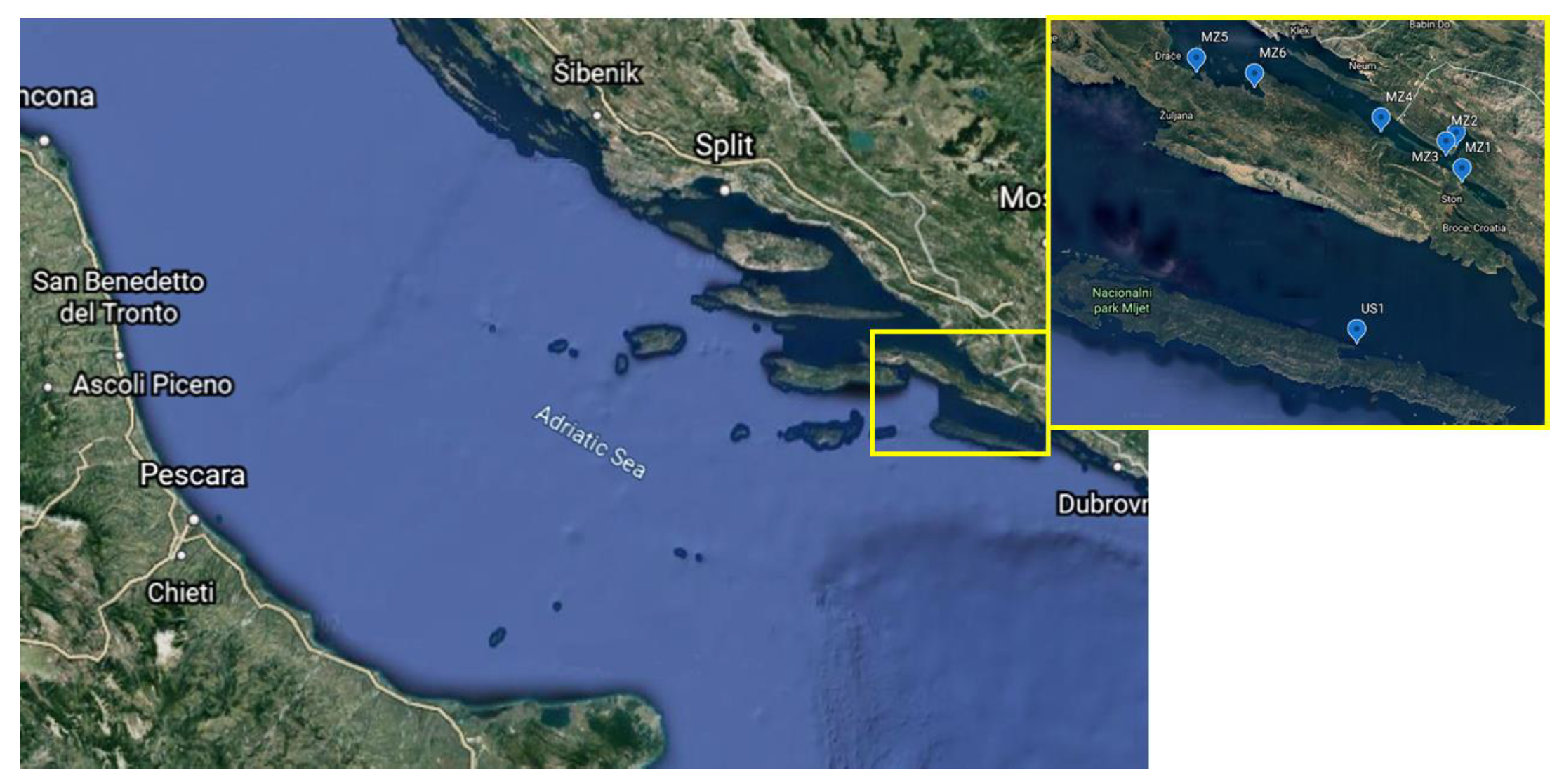
2.1. Study Area

Seven sampling stations were chosen for this study, of which six were in the Bay of Mali Ston and one on Mljet island. The Bay of Mali Ston is located on the south of the Croatian part of the Adriatic coast between the mainland and Pelješac Peninsula in the Neretva Channel’s vicinity. The bay’s ecological conditions mainly depend on the effects of the Neretva River, nearby land, and, to a lesser extent, on the open sea. Neretva’s flow through Croatia is 22 km long, and the mouth of the river is the only actual delta in Croatia, which covers an area of 12,000 hectares. After extensive land reclamation and cultivation of that agricultural-rich area, the wetland delta was turned into a rich agricultural area. The Bay of Mali Ston is 28 km long, and at its widest part, it is 6.1 km wide. The altitude ranges from 0 to 400 m. Due to the large indentation of the outer and the inner part of the bay, the coastline’s total length is about 100 km. In more than 80% of the bay, the depth is between 20 and 29 m. The outer and the middle part of the bay are occasionally under stronger influence, while the bay’s inner part is under the weaker influence of the river Neretva’s water, especially during higher river water level and stronger westerly winds.
The bay’s hydrophysical and ecological relations are mainly influenced by strong underwater freshwater sources located in the inner part of the bay. The input of organic matter from the land is significant for ecological and production relations, and rainwater and numerous underwater springs. Due to its favourable primary production and hydrographic characteristics, mussels have been farmed in the bay since ancient times. Until today, the Bay of Mali Ston has been one of the essential areas for shellfish farming in Croatia. Mljet is located in the archipelago of South Dalmatia. It stretches parallel to the eastern half of the Pelješac Peninsula, from which the 8 km wide Mljet Channel separates it. Strong waves, sea currents, and winds have shaped today’s structure of the Mljet’s shores, which abound in bays, caves, and rocks. In winter, the sea is colder only in deep bays and in colder periods. The average water temperature in Mali Ston in winter reaches 15 °C. In summer, the sea is warm, and along the coast, in the bays, it is 2–3 °C warmer than on the open sea. In summer, the average water temperature rises to 24 °C.

2.2. Mussel Sampling

Samples of Mediterranean blue mussels (Mytilus galloprovincialis) were collected for the analysis within the Croatian National Monitoring Programme activities on the seven stations from January to June 2011, as shown in Figure 1. Geographical longitudes and latitudes of sampling stations are given in Table 1. Samples were collected once a week or more frequently (but not less than every two days) when MBA showed positive DSP toxicity testing results. To get the representative sample, 4 kg of mussels were collected. Mussels were not depurated prior to analysis. The mussels’ entire soft tissues were separated from the mussel shell together with byssus fibre and weighed before and after the freeze-drying process to determine the water content in soft tissues. Mean water content (%) in soft tissues was (mean ± SD) 86.4 ± 1.6.

2.3. Reagents and Standard Solutions

The reagents used in this study included HNO3 (68%, trace analysis grade, Fisher Scientific UK, Loughborough, UK), HClO4 (65%, trace metal analysis, Fisher Scientific UK), and H2O2 (30%, Merck, Darmstadt, Germany). The standard solutions were prepared from the stock standard solution of ultrapure grade by Merck. The stock solution’s preparation and the dilution of the samples were carried out with deionised water with the Mill-Q Water Purification System (Merck Millipore, Molsheim, France), which was also used to clean the glass, plastic, and polytetrafluoroethylene (PTFE) vessels. All glass and plasticware used for tissue analysis were cleaned with a 5% HNO3 solution for 48 h and washed with deionised water.

2.4. Extraction of Manganese in Tissue Samples

About 10 g of the homogenised frozen soft tissue was lyophilised with a Lio 5P freeze dryer (Martin Christ, Osterode am Harz, Germany) and ground to a fine powder in an agate mortar. To achieve complete digestion of the tissue, the microwave-assisted acid digestion for trace metal analysis was performed according to the modified US EPA 3052 method. Approximately 0.25 g of freeze-dried and pulverised mussel soft tissue was weighed into the PTFE microwave digestion vessel. A mixture of concentrated acids, HNO3, and HClO4 (2:1) was added to samples in PTFE vessels. The vessels were transferred into a microwave digestion system. A digestion step at 190 °C was followed by an acid evaporation step on a hotplate. After acid evaporation, a mixture of HNO3 and H2O2 (1:1) was added to samples in PTFE vessels. Vessels were transferred to a microwave digestion system for the final digestion step. After cooling down to room temperature, samples were diluted with Mill-Q water to the final volume of 15 mL.

2.5. Atomic Absorption Spectroscopy (AAS) Analysis

Analysis of Mn concentration in digested mussels’ soft tissues was performed on the atomic absorption spectrometer (Analyst 800, PerkinElmer, Shelton, CT, USA), using flame atomic spectroscopy technique.
The quality and accuracy of the applied method were ensured by periodical analysis of blank samples and spiked samples and by analysis of certified reference material of marine biota (SRM 2976, freeze-dried mussel’s tissue, NIST, National Institute of Standards and Technology, Gaithersburg, MD, USA). Measured concentrations of Mn in CRM samples were within the 95% confidence interval of the assigned certified value. The recovered concentration (mean % recovery ± SD) was 104.6% ± 4.6%. The detection limit of the applied analytical method, corresponding to the mean procedural blank value and three times the standard deviation of the blank measures, was 0.91 mg kg−1 for Mn.

2.6. Data Analysis

To process data in this study, statistical methods (e.g., mean, minimum, and maximum concentration values, and standard error) were used and the diagrams were made with Microsoft Excel 2016 and STATISTICA (StatSoft 14, TIBCO Software, Palo Alto, California, US). The one-way ANOVA in IBM SPSS Statistics 25 was used to support the conclusions about differences between the mean Mn concentrations in DSP positive and DSP negative samples, among sampling sites and sampling months.

3. Results

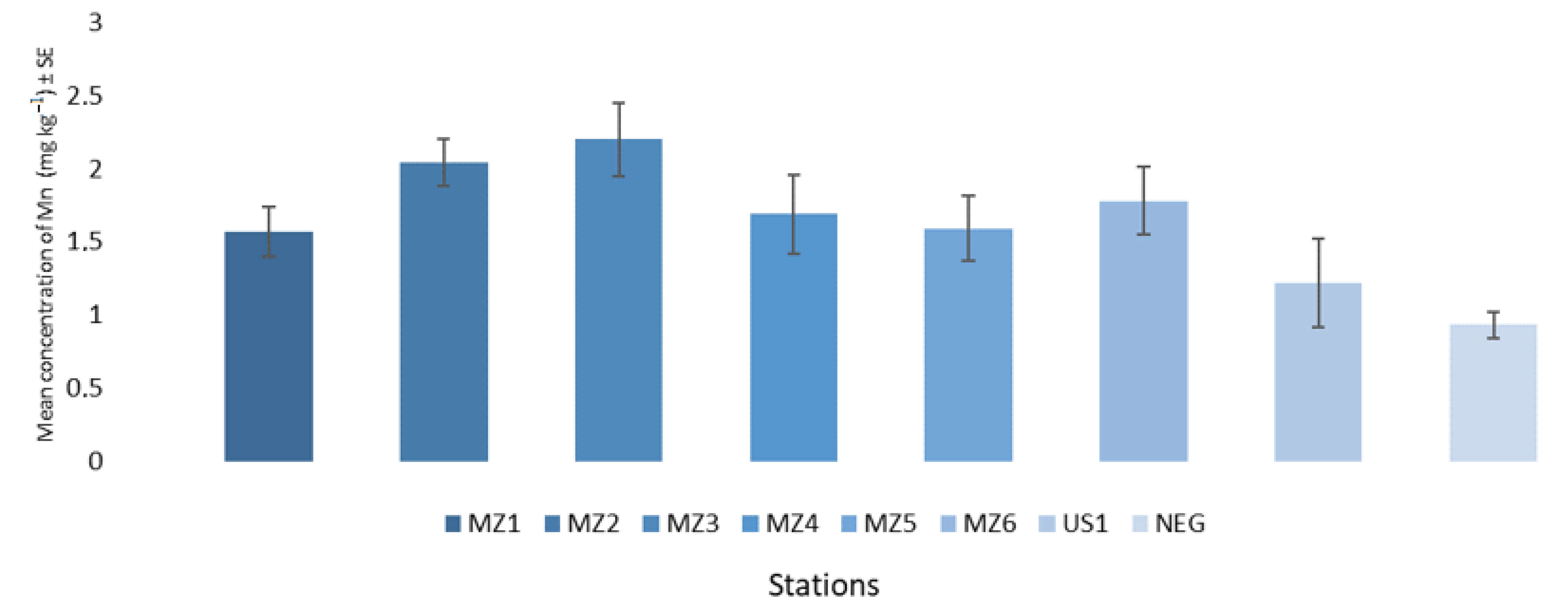
3.1. Spatial Distribution of Manganese during DSP Toxicity

Concentrations of Mn were analysed in 109 samples (16 DSP negative and 93 DSP positive samples) of blue mussels (M. galloprovincialis) gathered from the farming area in the Bay of Mali Ston located in the southern Adriatic Sea weekly or more frequently (but not less than 48 h) if the results of mouse bioassay were positive. Results of analysed Mn concentrations are presented in Figure 2. Mn concentrations in DSP positive samples were in the range of 0.15 mg kg−1 to 5.38 mg kg−1. Mean values measured at all control stations, and 87% of all DSP positive results were in a higher range than the mean value of Mn concentrations in DSP negative samples. (Figure 2). The mean Mn concentration for all DSP positive samples was 1.78 mg kg−1, while for the DSP negative samples, it was a 48% lower (0.93 mg kg−1) value. The highest value of Mn concentration of all DSP positive samples was 5.38 mg kg−1, and it was measured at the control point MZ6. The lowest value of Mn concentration of all DSP positive samples was 0.15 mg kg−1, and it was measured at the control point MZ1. When looking at the mean values of Mn concentrations between seven stations, the data showed variations. The results of the mean Mn concentrations ordered from the highest to the lowest value are as follows: MZ3 > MZ2 > MZ6 > MZ4 > MZ5 > MZ1 > US1 (Table 2).

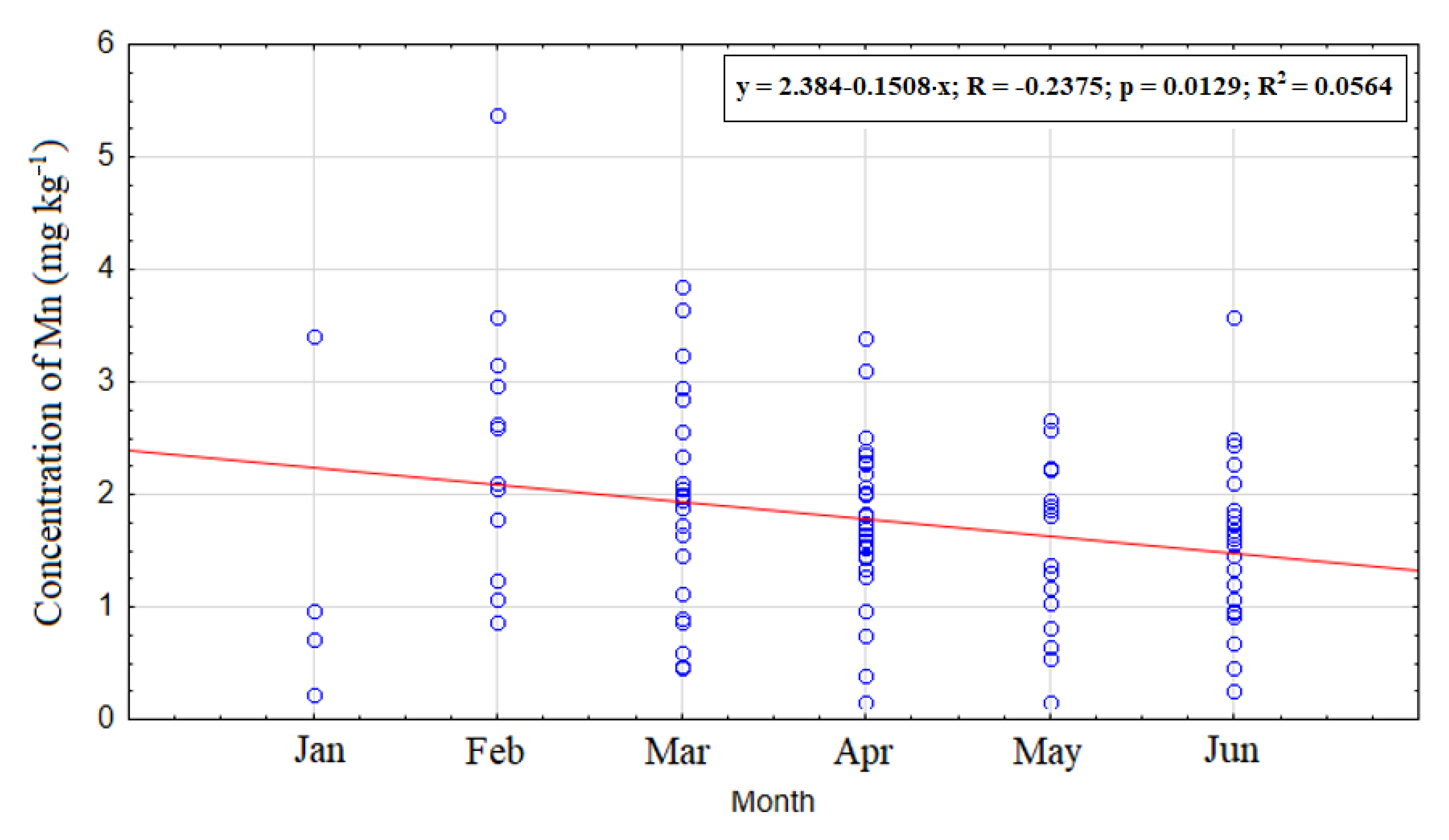
3.2. Temporal Distribution of Manganese during the Period of DSP Shellfish Toxicity

The present study investigated the temporal distribution of Mn concentrations in DSP positive mussel samples from January to June 2011. In the examined DSP positive samples of mussels, the highest concentrations of Mn were recorded in the coldest part of the year (late winter and early spring), which decreased with the arrival of warmer weather (summer and early fall) (Figure 3). A trend of decreasing Mn concentrations was found in the analysed samples in the studied period.

4. Discussion

The results of toxin analyses revealed that the Amnesic Shellfish Poisoning (ASP) toxins (domoic and epi-domoic acid) lay below the limit of detection (LoD) and that all samples were negative on Paralytic Shellfish Poisoning (PSP) MBA testing. Conducted MBA gave positive results on DSP toxicity checking. The weekly analyses for DSP toxins did not meet the food control requirements provided by the European Regulative (853/2004 EC). Performed tests showed the rapid death of mice through symptoms that were atypical for DSP toxins. To identify specific lipophilic toxins (OA-okadaic acid; DTXs-dynophysistoxins; AZA-1, AZA-2, and AZA-3-azaspiracids; YTXs-yessotoxins; GYMs-gymnodimines; and SPXs-spirolides), shell extracts in methanol were analysed using a tandem method of liquid chromatography and mass spectrometry (LC-MS/MS) characterised by high specificity and sensitivity. Analyses detected only low concentrations of GYMs and SPXs (in the range 5–15 μg kg−1), and none of the lipophilic toxins regulated by the EU Directive [32]. The GYMs and SPXs are classified into a heterogeneous group of marine bio compounds called cyclic imines [35]. GYMs and SPXs are globally distributed phycotoxins discovered in the early 1990s and detected in the Croatian mussels for the first time back in 2006 [36]. There is no clear evidence of their toxicity to humans, although the research on the Caco-2 cells shows that SPXs could be absorbed in the human intestinal epithelium [37]. GYMs and SPXs are highly toxic to rodents and constitute a source of false positives in lipophilic toxin detection by the mouse bioassay [35]. The presence of GYMs and SPXs could have caused positives in MBA of mussel samples from the Bay of Mali Ston in 2011 [30].
Since other compounds in shellfish tissue, such as metals, can also cause false-positive MBA, this research focused on measuring Mn’s concentration in both DSP positive and DSP negative samples to investigate Mn content’s connection with the positive diarrhetic shellfish poisoning.
The toxicity occurrence started in January 2011 and lasted until mid-summer, with the maximum number of positive samples in June.
Accumulated Mn concentrations measured in the soft tissue of DSP positive samples varied between sites. The positions of the sampling points can explain variations among mean values of Mn concentrations. The top-ranking value of the mean Mn concentration was measured at the control point MZ3. This control station is strongly indented into the mainland, and in such areas, sediment is a source of trace metals. Since sea currents at this site are weaker, the influence of wind and waves and the dispersion of suspended particles is reduced, increasing the concentration of Mn. This station is also the closest to the Split–Dubrovnik road and is exposed to the significant impact of heavy road traffic. The next point where high mean Mn concentration was measured was MZ2, which is also drawn deep into the mainland. Due to weaker sea currents, wind, and waves, there is no scattering of suspended particles, which is why the sediment can increase Mn concentration. MZ2 is the closest to MZ3 and with the second-highest mean value of Mn concentration measured. Control points MZ6 and MZ5 are next when looking rank of the mean values of Mn concentration. These stations are located in the bay’s outer part and are more strongly influenced by the Neretva river, which flows through agricultural areas carrying drainage of agricultural land. These points are exposed to a higher load of eroded material from the land enriched with Mn since they are located opposite Neretva’s mouth. Levels of Mn measured at these stations are strongly defined by their positions. MZ1 is indented into the mainland but further away from the Split–Dubrovnik road and its impact. The mean value of the Mn concentration at MZ4 can also be related to river Neretva’s influence. The lowest mean value of the concentration of Mn in mussels was detected at the control station US1 on the island of Mljet, which is furthest from the mainland and least exposed to the influence of river Neretva. A low impact of terrigenous particles, which are, in general, rich with Mn, was recorded at this sampling station. Mn in the Bay of Mali Ston can originate from sediment, the Earth’s crust, or the river Neretva’s discharges. A study by Maanan [38] pointed to quite elevated Mn concentrations in mussel tissue in stations close to the mouth of estuaries, while the study of Maanan et al. [39] indicated Mn as an excellent tracer of continental inputs into aquatic systems produced by breaking up rock and soils of surrounding watersheds.
When comparing the mean values of Mn concentrations in DSP positive and DSP negative samples by one-way ANOVA, the significance level was nearly 0.000, which is below 0.05, and confirms that the mean Mn concentration values of all DSP positive samples as well as 87% of all DSP positive samples had higher Mn concentrations than the mean value of all DSP negative samples.
The comparison of Mn concentrations in DSP positive samples among sampling sites showed the significance value of 0.182, which is higher than 0.05, so there was no statistically important difference between Mn concentrations among sampling sites.
When w wt contents of Mn measured in the soft tissue of mussels from the Bay of Mali Ston are converted in dry weight (d wt), they lie in the range from 0.5 to 18.7 mg kg−1 (Table 3).
When comparing concentrations of Mn measured in the soft tissue of mussels from the Bay of Mali Ston with the literature data on the concentrations of Mn measured in the soft tissue of mussels from the seas of other parts of the world, it can be seen that the concentrations of Mn in mussels from the Bay of Mali Ston are relatively low (Table 3).
Phytoplankton make the autotrophic component of sea plankton. As photosynthetic organisms, marine phytoplankton species are the primary producers and the basis of most food chains in these ecosystems. Various factors of either natural or anthropogenic origin can affect phytoplankton communities, especially in more closed parts of the sea, such as bays or canals. Some phytoplankton species are producers of marine biotoxins, and the presence of certain trace metals in the sea may be reflected in enhanced biotoxin production by increasing biomass and/or increasing toxin production per cell. Rhodes et al. [4] reported a 25% increase in the dinoflagellate Karenia selliformis, a GYMs producing microalgae, biomass with the addition of Se to batch cultures. Further increase in biomass was achieved with the dual addition of Mn and Se.
In the Bay of Mali Ston, the Neretva river’s influence, which flows through an agricultural area, and its inputs of agricultural land and organic matter are of high importance. Enrichment of estuarine waters with dissolved organic material (DOM) can affect phytoplankton communities. Paerl [46], Lewetus [47], Gilbert [48], and Mountfort [49] described the effect of organic acid additions in enhancing growth and GYMs production in axenic cultures of Karenia selliformis. Acetate enhanced GYMs by stimulating growth rate (μ, 0.23 days−1), and the large concentration of GYMs per cell (16 pg cell−1 cf. 9.8 pg cell−1 for the control) suggests a role of acetate in the GYMs biosynthesis. Amending culture with Mn2+ additions resulted in slightly decreased growth in control cultures and it increased the GYMs.
As the comparison of Mn concentrations measured in DSP positive and DSP negative samples of mussels from the Bay of Mali Ston revealed higher Mn concentrations in 87% of DSP positive samples, it could be assumed that there is a connection between the measured Mn levels and the occurrence of biotoxins GYMs and SPXs. Mn showed the same connection with the occurrence of phycotoxins GYMs and SPXs in DSP positive samples of blue mussels from the Bay of Mali Ston during DSP toxicity episodes in 2011, just as did Zn, Cu, Cd, and Ni, which showed higher levels in more than 70% of DSP positive samples as it was reported in the study of Ujević et al. [50].
The possible explanations for the role of Mn in the occurrence of GYMs and SPXs in analysed DSP positive samples from the Bay of Mali Ston may be that the increase of Mn content in the seawater could have either affected metabolic pathways in phytoplankton cells and enhanced the production of marine biotoxins [4], or that when bioaccumulated in shellfish tissue, it could have facilitated the transport of biotoxins and their accumulation in shellfish cells [50].
Temporal analysis of Mn concentrations measured in DSP positive samples showed a seasonal pattern with maximum values observed during the cold period of the year (end of winter, beginning of spring) and minimum values during the warmer period (summer, early fall) [51]. Seasonal differences of Mn concentrations in analysed DSP positive samples can be described with the sinusoid curve [42]. In the studied case, one can assume that mussels during the warm period bioaccumulate to a lesser degree than during the colder period. Mn as a trace metal essential for life showed the same seasonal evolution as other trace metals, which are more involved in biochemical activities and required by biota for various biochemical processes [45]. Mn concentrations of all DSP positive samples showed a similar decreasing trend from January to June, which is in accordance with the seasonal pattern demonstrated in concentrations of Cd, Pb, Zn, Cr, Cu, and Ni investigated in the first six months of 2011 during DSP toxicity of mussels from the Bay of Mali Ston by Ujević et al. [50].
When comparing Mn concentrations in DSP positive samples among sampling months by one-way ANOVA, the significance value was 0.007, which is below 0.05, and confirms a statistically important difference between the mean values of Mn concentration among sampling sites.
The bioavailability of Mn in the Bay of Mali Ston is highly dependent on seasonal and meteorological characteristics and the fluvial discharges of the Neretva. Depending on the metabolic rate of mussels, Mn can be accumulated in the soft tissue, especially in combination with the shallowness of the coastal area in the bay and concentration, as well as the physical and chemical forms of Mn present in the seawater and the intense influence of nutrients, winds, and waves. The results of temporal analyses of Mn concentrations demonstrated the same trend that was noticed by authors who studied seasonal changes in metal contents in the soft tissue of mussels, genus Mytilus, in moderate climates of the Northern Hemisphere [51,52,53,54].

5. Conclusions

Analysis of Mn concentration in DSP positive and DSP negative mussels revealed a higher concentration of Mn in the significant number (87%) of DSP positive samples compared to the mean value of Mn concentrations of DSP negative samples. This study suggests that a higher concentration of Mn in DSP positive samples may be connected to the occurrence of GYMs and SPXs and it calls out for future studies to get detailed insights towards understanding physiological interactions of Mn and lipophilic biotoxins. As we witness climate changes and the input of nutrient pollutants on a global level, this type of research is particularly important, especially given that the input of some micronutrients, such as trace metals (Cu, Zn, Ni, Co, Mn) and their concentrations in the habitats of some microalgae, are crucial for their toxicity.

Author Contributions

Conceptualization, N.V. and I.A.; methodology, J.L.; validation, J.L.; formal analysis, N.V. and J.L.; investigation, N.V.; resources, N.V.; data curation, I.A.; writing—original draft preparation, N.V.; writing—review and editing, I.A.; visualization, N.V.; project administration, N.V.; funding acquisition, N.V. and I.A. All authors have read and agreed to the published version of the manuscript.

Funding

This study was supported by the Ministry of Science and Education of the Republic of Croatia as a part of Multiannual Finnancing intended for institutions.

Institutional Review Board Statement

The work described in this article has been conducted in accordance with The Code of Ethics of the World Medical Association (Declaration of Helsinki) for animal experiments and approved by the National Ethics Committee for Protection of Animals used for Scientific Purposes in accordance with the national regulation on the Protection of Animals used for Scientific Purposes (NN 55/2013).

Informed Consent Statement

Not applicable.

Data Availability Statement

The data presented in this study are available on request from the corresponding author and with permission of the Ministry of Agriculture of the Republic of Croatia.

Conflicts of Interest

The authors declare no conflict of interest.

References

  1. Martinčić, D.; Kwokal, Ž.; Peharec, Ž.; Marguš, D.; Branica, M. Distribution of Zn, Pb, Cd and Cu between seawater and transplanted mussels (Mytilus galloprovincialis). Sci. Total Environ. 1992, 119, 211–230. [Google Scholar] [CrossRef]
  2. McCulloch, A.W.; Body, R.K.; de Freitas, A.S.; Foxall, R.A.; Jamieson, W.D.; Laycock, M.V.; Quilliam, M.A.; Wright, J.L.; Boyko, V.J.; McLaren, J.W. Zinc from oyster tissue as causative factor in mouse deaths in official bioassay for paralytic shellfish poison. J. Assoc. Off. Anal Chem. 1989, 72, 384–386. [Google Scholar] [CrossRef]
  3. Park, D.L.; Adams, W.N.; Graham, S.L.; Jackson, R.C. Variability of mouse bioassay for determination of paralytic shellfish poisoning. J. Assoc. Off. Anal. Chem. 1986, 69, 547–550. [Google Scholar] [CrossRef]
  4. Rhodes, L.; Selwood, A.; McNabb, P.; Briggs, L.; Adamson, J.; van Ginkel, R.; Laczka, O.F. Trace metal effects on the production of biotoxins by microalgae. Afr. J. Mar. Sci. 2006, 28, 393–397. [Google Scholar] [CrossRef]
  5. Libes, S.M. An Introduction to Marine Biogeochemistry; John Willey and Sons Inc.: New York, NY, USA, 1992. [Google Scholar]
  6. El-Moselhy, K.M.; Gabal, M.N. Trace metals in water, sediments and marine organisms from the northern part of Gulf of Suez, Red Sea. J. Mar. Syst. 2004, 46, 39–46. [Google Scholar] [CrossRef]
  7. Wedepohl, K. The composition of the continental crust. Geochim. Cosmochi. Acta 1995, 59, 1217–1232. [Google Scholar] [CrossRef]
  8. Van Hulten, M.; Dutay, J.-C.; Middag, R.; de Baar, H.; Roy-Barman, M.; Gehlen, M.; Tagliabue, A.; Sterl, A. Manganese in the world ocean: A first global model. Biogeosci. Discuss. 2016, 1–38. [Google Scholar] [CrossRef]
  9. Raven, J.A. Predictions of Mn and Fe use efficiencies of phototropic growth as a function of light availability for growth and C assimilation pathway. N. Phytol. 1990, 116, 1–18. [Google Scholar] [CrossRef]
  10. Sarmiento, J.; Gruber, N. Ocean Biogeochemical Dynamics; Princeton University Press: Princeton, NJ, USA, 2006. [Google Scholar]
  11. Baker, A.; Landing, W.; Bucciarelli, E.; Fietz, S.; Hayes, C.; Kadko, D.; Morton, P.; Rogan, N.; Sarthou, G.; Shelley, R.; et al. Trace element and isotope deposition across the air-sea interface: Progress and research needs. Philos. Trans. R. Soc. A 2016, 374. [Google Scholar] [CrossRef] [PubMed]
  12. Homoky, W.B.; Weber, T.; Berelson, W.M.; Conway, T.M.; Henderson, G.M.; Vun Hulten, M.M.P.; Jeandel, C.; Severmann, S.; Tagliabue, A. Quantifying trace element and isotope fluxes at the ocean-sediment boundary. Philos. Trans. R. Soc. A 2016, 374. [Google Scholar] [CrossRef] [Green Version]
  13. Middag, R.; de Baar, H.; Laan, P.; Huhn, O. The effects of continental margins and water mass circulation on the distribution of dissolved aluminium and manganese in Drake Passage. J. Geophys. Res. 2012, 117. [Google Scholar] [CrossRef] [Green Version]
  14. Pakhomova, S.; Hall, P.; Kononets, M.; Rozanov, A.; Tengberg, A.; Verenshinin, A. Fluxes of iron and manganese across the sediment-water interface under various redox conditions. Mar. Chem. 2007, 107, 319–331. [Google Scholar] [CrossRef]
  15. Aguilar-Islas, A.; Burland, K. Dissolved manganese and silicic acid in the Columbia River plume. Mar. Chem. 2006, 101, 233–247. [Google Scholar] [CrossRef]
  16. Jeandel, C. Overview of the mechanisms that could explain the “Boundary Exchange” at the land-ocean contact. Philos. Trans. R. Soc. A 2016, 374. [Google Scholar] [CrossRef] [PubMed] [Green Version]
  17. Middag, R.; de Baar, H.; Laan, P.; Klunder, M. Fluvial and hydrothermal input of manganese into the Arctic Ocean. Geochim. Cosmochim. Acta 2011, 75, 2393–2408. [Google Scholar] [CrossRef]
  18. Bogdanović, T.; Ujević, I.; Sedak, M.; Listeš, E.; Šimat, V.; Petričević, S.; Poljak, V. As, Cd, Hg and Pb in four edible shellfish species from breeding and harvesting areas along the eastern Adriatic Coast, Croatia. Food Chem. 2014, 146, 197–203. [Google Scholar] [CrossRef] [PubMed]
  19. Kljaković-Gašpić, Z.; Odžak, N.; Ujević, I.; Zvonarić, T.; Barić, A. Biomonitoring of trace metals (Cu, Cd, Cr, Hg, Pb, Zn) in eastern Adriatic using the Mediterranean blue mussel (2001–2005). Fresenius Environ. Bull. 2006, 15, 1041–1048. [Google Scholar]
  20. Cullaj, A.; Lazo, P.; Duka, S. Heavy metals and metallonine levels in mussel samples from the Albanian sea coast. MAP/MED Pol. In Biological Effects Monitoring Programme—Achievements and Future Orientations, Proceedings of the Workshop, Alessandria, Italy, 20–21 December 2006; MAP Technical Reports Serial No. 166; UNEP/MAP: Athens, Greece, 2007; pp. 141–151. [Google Scholar]
  21. Cardellicchio, N.; Buccolieri, A.; di Leo, A.; Giandomenico, S.; Spada, L. Levels of metals in reared mussels from Taranto Gulf (Ionian Sea, Southern Italy). Food Chem. 2008, 107, 890–896. [Google Scholar] [CrossRef]
  22. Ščančar, J.; Zuliani, T.; Turk, T.; Milačić, R. Organotin compounds and selected metals in the marine environment of Northern Adriatic Sea. Environ. Monit. Assess. 2007, 127, 271–282. [Google Scholar] [CrossRef] [PubMed]
  23. Stanković, S.; Jović, M.; Milanov, R.; Joksimović, D. Trace elements concentrations (Zn, Cu, Pb, Cd, As and Hg) in the Mediterranean mussel (Mytilus galloprovincialis) and evaluation of mussel quality and possible human health risk from cultivated and wild sites of the southeastern Adriatic Sea, Montenegro. J. Serb. Chem. Soc. 2011, 76, 1725–1737. [Google Scholar] [CrossRef]
  24. Marković, J.; Joksimović, D.; Stanković, S. Trace element concentrations in wild mussels from the coastal area of the southeastern Adriatic, Montenegro. Arch Biol. Sci. 2012, 64, 265–275. [Google Scholar] [CrossRef]
  25. Ramšak, A.; Ščančar, J.; Horvat, M. Evaluation of metallothioneins in blue mussels (Mytilus galloprovincialis) as a biomarker of mercury and cadmium exposure in the Slovenian waters (Gulf of Trieste): A long-term field study. Acta Adriat. 2012, 53, 71–86. [Google Scholar]
  26. Ali, H.; Khan, E.; Ilahi, I. Environmental chemistry and ecotoxicology of hazardous heavy metals: Environmental persistence, toxicity and bioaccumulation. J. Chem. 2019, 6730305. [Google Scholar] [CrossRef] [Green Version]
  27. Trainer, V.L.; Hickey, B.M.; Bates, S.S. Toxic Diatoms; Elsevier Science Publishers: New York, NY, USA, 2008; pp. 219–237. [Google Scholar]
  28. Yasumoto, T.; Murata, M. Marine toxins. Chem. Rev. 1993, 5, 1897–1901. [Google Scholar] [CrossRef]
  29. Rainbow, P.S. Heavy metal levels in the marine invertebrates. In Heavy Metal Levels in the Marine Environment; Furness, R.W., Rainbow, P.S., Eds.; CRC Press: Boca Raton, FL, USA, 1990; pp. 67–79. [Google Scholar]
  30. Ujević, I.; Nazlić, N.; Ninčević-Gladan, Ž.; Marasović, I. Gymnodimine and spirolide in shellfish during DSP toxicity in central and southern Adriatic Sea. In Proceedings of the 16th International Conference on Harmful Algae, Abstract Book Wellington, Welington, New Zeland, 27–31 October 2014. [Google Scholar]
  31. Suzuki, T.; Yoshizawa, R.; Kawamura, T.; Yamasaki, M. Interference of free fatty acids from the hepatopancreas of mussels with the mouse bioassay for shellfish toxins. Lipids 1996, 31, 641–645. [Google Scholar] [CrossRef] [PubMed]
  32. Takagi, T.; Hayashi, K.; Itabashi, Y. Toxic effect of free unsaturated fatty acids in the mouse bioassay of diarrhetic shellfish toxin by intraperitoneal injection. Bull. Jpn. Soc. Sci. Fish 1984, 50, 1413–1418. [Google Scholar] [CrossRef] [Green Version]
  33. Stabell, O.B.; Yndestad, M.; Heidenreich, B. Paralytic shellfish toxins seem absent in extracts of diarrhetic shellfish toxins. Environ. Toxicol. Chem. 1991, 10, 331–334. [Google Scholar] [CrossRef]
  34. Lawrence, J.F.; Chadha, R.K.; Ratnayabe, W.M.; Truelove, J.F. An incident of elevated levels of unsaturated free fatty acids in mussels from Nova Scotia and their toxic effect in mice after intraperitoneal injection. Nat. Toxins 1994, 2, 318–321. [Google Scholar] [CrossRef]
  35. Vilarino, N.; Fonfria, E.S.; Molgo, J.; Romulo, A.; Botana, L.M. Detection of gymnodimine-A and 13-desmethyl spirolide phycotoxins by fluorescence polarization. Anal. Chem. 2009, 81, 2708–2714. [Google Scholar] [CrossRef] [PubMed]
  36. Ninčević Gladan, Ž.; Ujević, I.; Milandri, A.; Marasović, I.; Ceredi, A.; Pigozzi, S.; Arapov, J.; Skejić, S. Lipophilic toxin profile in Mytilus galloprovincialis during episodes of diarrhetic shellfish poisoning (DSP) in the NE Adriatic Sea in 2006. Molecules 2011, 16, 888–899. [Google Scholar] [CrossRef] [PubMed]
  37. Espina, B.; Otero, P.; Louzao, M.C.; Alfonso, A.; Botana, L.M. 13-Desmethyl spirolide-c and 13,19-didesmethyl spirolide-c trans-epithelial permeabilities: Human intestinal permeability modelling. Toxycology 2011, 287, 69–75. [Google Scholar] [CrossRef] [PubMed]
  38. Maanan, M. Heavy metals concentrations in marine molluscs from the Moroccan coastal region. Environ. Pollut. 2007, 153, 176–183. [Google Scholar] [CrossRef]
  39. Maanan, M.; Zourarah, B.; Carruesco, C.; Aajjane, A.; Naud, J. Distribution of heavy metals in Sidi Moussa lagoon sediments (Atlantic Moroccan Coast). J. Afr. Earth Sci. 2004, 39, 473–483. [Google Scholar] [CrossRef]
  40. Joksimović, D.; Tomić, I.; Stanković, A.R.; Jović, M.; Stanković, S. Trace metal concentrations in Mediterranean blue mussel and surface sediments and evaluation of the mussel quality and possible risks of high human consumption. Food Chem. 2011, 127, 632–637. [Google Scholar] [CrossRef] [PubMed]
  41. Perošević, A.; Pezo, L.; Joksimović, D.; Đurović, D.; Milašević, I.; Radomirović, M.; Stanković, S. The impacts of seawater physicochemical parameters and sedimental metal contents on trace metal concentrations in mussels—A chemometric approach. Environ. Sci. Pollut. Res. 2018, 25, 28248–28263. [Google Scholar] [CrossRef] [PubMed]
  42. Fattorini, D.; Notti, A.; Di Mento, R.; Cicero, A.M. Seasonal, spatial and inter-annual variations of trace metals in mussels from the Adriatic Sea: Aregional gradient of arsenic, and implications for monitoring the impact of off-shore activities. Chemosphere 2008, 72, 1524–1533. [Google Scholar] [CrossRef]
  43. Romeo, M.; Frasila, C.; Gnassia-Berelli, M.; Damiens, G.; Micu, D.; Mustata, G. Biomonitoring of trace metals in the Black Sea (Romania) using mussels Mytilus galloprovincialis. Water Res. 2005, 39, 596–604. [Google Scholar] [CrossRef] [PubMed]
  44. Topcuoglu, S.; Kiribsoglu, C.; Gungor, N. Heavy metals in organisms and sediments from Turkish coast of the Black Sea, 1997–1998. Environ. Int. 2002, 27, 521–526. [Google Scholar] [CrossRef]
  45. Catsiki, V.A.; Florou, H. Study on the behavior of the heavy metals Cu, Cr, Ni, Zn, Fe, Mn and 137Cs in an estuarine ecosystem using Mytilus galloprovincialis as a bioindicator species: The case of Thermaikos gulf, Greece. J. Environ. Radioact. 2005, 86, 31–44. [Google Scholar] [CrossRef]
  46. Paerl, H.W. Nuisance phytoplankton blooms in coastal, estaurine, and inland waters. Limnol. Oceanogr. 1988, 33, 823–843. [Google Scholar] [CrossRef]
  47. Lewetus, A.J.; Willis, B.M.; Hayes, K.C.; Burkholder, J.M.; Glasgow, H.B.; Gilbert, P.M.; Burke, M.K. Mixotrophy and nitrogen uptake by Pfiesteria piscicida (Dinophyceae). J. Phycol. 1999, 35, 1430–1437. [Google Scholar] [CrossRef]
  48. Gilbert, P.M.; Magnien, R.; Lomas, M.W.; Alexander, J.; Tan, C.; Haramoto, E.; Trice, M.; Kana, T.M. Harmful algal blooms in the Chesapeake and coastal bays of Maryland, USA: Comparison of 1997, 1998 and 1999 events. Estuaries 2001, 24, 875–883. [Google Scholar] [CrossRef]
  49. Mountfort, D.; Beuzenberg, V.; MacKenzie, L.; Rhodes, L. Enhancement of growth and gymnodimine production by the marine dinoflagellatae, Karenia selliformis. Harmful Algae 2006, 5, 658–664. [Google Scholar] [CrossRef]
  50. Ujević, I.; Vuletić, N.; Lušić, J.; Nazlić, N.; Kušpilić, G. Bioaccumulation of trace metals in mussel (Mytilus galloprovincialis) from Mali Ston Bay during DSP toxicity episodes. Molecules 2015, 20, 13031–13040. [Google Scholar] [CrossRef]
  51. Regoli, F.; Orlando, E. Seasonal variation of trace metal concentration in the digestive gland of the Mediterranean mussel Mytilus galloprovincialis: Comparison between polluted and non-polluted site. Arch. Environ. Contam. Toxicol. 1994, 27, 36–43. [Google Scholar] [CrossRef]
  52. Borchardt, T.; Burchert, S.; Krabe, L.; Zeitner, R. Enhanced heavy metal concentrations in Mytilus edulis from the central North Sea. Sci. Mar. 1989, 53, 725–728. [Google Scholar]
  53. Cossa, D. A review of the use of Mytilus sas quantitative indicators of cadmium and mercury concentration in coastal waters. Oceanol. Acta 1989, 12, 417–432. [Google Scholar]
  54. Odžak, N.; Zvonarić, T.; Kljaković-Gašpić, Z.; Barić, A. Biomonitoring of copper, cadmium, lead, zinc and chromium in the Kaštela Bay using transplanted mussels. Fresenius Environ. Bull. 2001, 10, 37–41. [Google Scholar]
Figure 1. The Bay of Mali Ston map with locations of sampling stations.
Jmse 09 00451 g001
Figure 2. The mean values of Mn concentrations ± SE in blue mussels collected at seven stations from January to June 2011. NEG-DSP negative samples, while others are DSP positive samples. Concentrations of Mn are expressed per wet weight (w wt). The mean values of Mn concentrations with standard errors (SE) in DSP positive samples for all stations, and the mean Mn concentration with SE in DSP negative samples are shown on the graph.
Jmse 09 00451 g002
Figure 3. Temporal distribution of manganese in DSP positive mussel samples from January to June 2011.
Jmse 09 00451 g003
Table 1. Longitudes and latitudes of the sampling stations.
Station NameStation Mark on the MapLongitude (N) and Latitude (E) of the Sampling Stations
Mali Ston CoveMZ142°50.69′:17°42.69′
Banja CoveMZ242°52.16′:17°40.98′
Bistrina CoveMZ342°52.38′:17°42.17′
Kanal Usko CoveMZ442°52.94′:17°38.27′
Sutvid CoveMZ542°54.99′:17°28.57′
Brijesta CoveMZ642°54.35′:17°31.01′
Sobra CoveUS142°44.73′:17°36.80′
Table 2. The ranges, mean values, and standard errors of Mn concentrations measured on each control station in the DSP positive samples.
Station NameStation Mark on the MapThe Concentration Range of Mn (mg kg−1)Mean Value of Mn Concentration (mg kg−1)Standard Error (SE)
Mali Ston CoveMZ10.15–2.291.57 ± 0.600.17
Banja CoveMZ20.86–2.862.04 ± 0.550.16
Bistrina CoveMZ30.39–3.852.20 ± 1.000.25
Kanal Usko CoveMZ40.46–3.111.69 ± 0.870.27
Sutvid CoveMZ50.16–3.411.59 ± 0,940.22
Brijesta CoveMZ60.54–5.381.78 ± 0.980.23
Sobra CoveUS10.22–2.351.22 ± 0.730.30
Table 3. Comparison of Mn concentrations in Mytilus galloprovincialis obtained in this study with literature data from other regions all over the world (mg kg−1 d wt).
RegionConcentration of MnReference
Adriatic Sea, Bay of Mali Ston0.5–18.7Present study
Adriatic Sea, Montenegro7.3–85[40]
Adriatic Sea, Montenegro6.4–22.2[41]
Adriatic Sea, Albania12.3–139.3[20]
Safi coastal waters, Morocco7.2–27.5[38]
El Jadida coast, Morocco8.7–34.8[39]
Portonovo, Italy4.4–16.4[42]
Black Sea, Romania12.7–13.6[43]
Black Sea, Turkey5.7–22.8[44]
Aegean Sea, Greece7.2–25.3[45]
Publisher’s Note: MDPI stays neutral with regard to jurisdictional claims in published maps and institutional affiliations.

