Article

DNA Sequence Analyses Reveal Two New Species of Caloglossa (Delesseriaceae, Rhodophyta) from the Skin of West Indian Manatees

by
D. Wilson Freshwater
1,*,
Cathryn E. Miller
1,
Thomas A. Frankovich
2 and
Michael J. Wynne
3
1
Center for Marine Science, University of North Carolina at Wilmington, 5600 Marvin K. Moss Lane, Wilmington, NC 28409, USA
2
Institute of Environment, Florida International University, 11200 SW 8th Street, Miami, FL 33199, USA
3
Research Museums Center, University of Michigan Herbarium, 3600 Varsity Drive #1234, Ann Arbor, MI 48108-2288, USA
*
Author to whom correspondence should be addressed.
J. Mar. Sci. Eng. 2021, 9(2), 163; https://doi.org/10.3390/jmse9020163
Submission received: 31 December 2020 / Revised: 29 January 2021 / Accepted: 2 February 2021 / Published: 6 February 2021
(This article belongs to the Special Issue Diversity, Taxonomy and Conservation of Marine Algae)

Abstract

:
Epizoic macroalgae collected from the skin of West Indian manatees included specimens of the red algal family Delesseriaceae. Morphological and rbcL sequence analyses indicated that these specimens represented two novel species of Caloglossa. One species, described here as Caloglossa kamiyana Freshwater, Cath.E. Miller & Frankovich sp. nov., had been previously studied and recognized as part of the C. ogasawaraensis species complex. The rbcL sequence divergence between C. kamiyana and other taxa within the complex ranged from 4.6–5.3%, and tetrasporangial mother cells are cut off from the lateral pericentral cells by oblique divisions instead of transverse divisions as in C. ogasawaraensis. The second species was resolved as a closely related sister species to C. fluviatilis, with a minimum interspecific sequence divergence of 2.0%. It was morphologically indistinguishable from C. fluviatilis except for one potential character—mostly one, instead of multiple rhizoids, developing from rhizoid-bearing pericentral and marginal wing cells. It is herein described as Caloglossa manaticola Freshwater, Cath.E. Miller & Frankovich sp. nov.

1. Introduction

Charismatic marine megafauna are generally unrecognized substrates for sessile invertebrates and algae. While epizoic barnacles on whales and sea turtles have been the subject of numerous studies [1,2,3], micro- and macroalgae epizoic on marine megafauna have received only limited attention. Six diatom genera are known to include “ceticolous species”, that have been observed only on the skin of whales and porpoises [4,5,6,7]. Contemporary studies in one of these genera, Tursiocola Hustedt, revealed that it also included multiple species only known as epibionts of manatees [8,9].
Manatees are marine mammals within the Sirenia family Trichechidae. The West Indian manatee, Trichechus manatus (L.), is a threatened species that ranges throughout much of the tropical Western Atlantic. They have limited cold tolerance [10] and may migrate seasonally between fresh and marine habitats [11]. While cetaceans and pinnipeds have anti-microbial mechanisms [12] and many cetaceans have smooth skin to inhibit fouling by epibionts [13], West Indian manatees do not seem to have evolved similar defenses [14], although, their skin exfoliates after shifting between salt- and freshwater, which may help to reduce biofouling [15].
Studies of epizoic macroalgae on marine megafauna have been almost exclusively restricted to those growing on sea turtles [16,17], but a novel red algal species, Melanothamnus maniticola Woodworth, Frankovich & Freshwater, has been described from West Indian manatees [18]. This species, visible in many West Indian manatee images, is believed to be obligately epizoic and to have evolved a unique holdfast system specific for manatee skin. Collections of epizoic macroalgae from manatees in Crystal River, Florida, USA, included specimens of the red algal family Delesseriaceae in addition to M. maniticola. DNA sequence analyses of these specimens revealed that they represented two novel species of Caloglossa that are described herein.

2. Materials and Methods

Three samples of the red algal family Delesseriaceae were collected from the skin of three Trichechus manatus individuals captured on 6–7 December 2018 in Crystal River, FL, USA, during health assessment studies of manatee populations. Samples were removed from the skin using forceps and placed into vials containing water from the collection site. Subsamples were dried in silica gel desiccant for later morphological and molecular analyses. Portions of the dried samples were rehydrated, stained with 1% acidified aniline blue, and mounted with 25% Karo solution to make permanent whole mount voucher slides [19]. Slides were observed and measurements taken using an Olympus SZH dissecting microscope (Olympus America Inc., Center Valley, PA, USA) and a Nikon Labophot-2 compound microscope (Nikon Inc., Melville, NY, USA). Images were captured using a Zeiss Axio Imager.Z1 compound microscope fitted with an AxioCam MRc 5 camera system (Carl Zeiss Microimaging Inc., Thornwood, NY, USA).
DNA was extracted from the samples using a MyTaqExtract-PCR Kit (Bioline, Taunton, MA, USA) and modified protocol described in Taylor et al. [20]. The resulting extractions were run through a OneStepTM PCR Inhibitor Removal Kit (Zymo Research, Irvine, CA, USA) following the manufacturer’s protocol. Amplifications of rbcL were conducted in PCR reactions containing 10–50 ng of template DNA, 0.2 µM of each amplification primer and MyTaq HS Red Mix (Bioline) following the manufacturer’s protocol and thermocycling program of Taylor et al. [20]. Oligonucleotide primers used for the rbcL amplification and sequencing reactions were F57, R893, R1144, F753, and RrbcSstart [21,22]. Amplified products were enzymatically purified using illustra ExoProStar (GE Healthcare, Chicago, IL, USA) and sequenced using BDX64 enhancing buffer (Molecular Cloning Lab [MCLAB], San Francisco, CA, USA) and BigDye v.3 (Life Technologies Corp., Grand Island, NY, USA) following the MCLAB BDX64 protocol, and analyzed on an ABI 3500 Genetic Analyzer (Life Technologies Corp.). Sequences were assembled using Sequencher version 5.4.1 (GeneCodes Corporation, Ann Arbor, MI, USA).
The newly generated rbcL sequences were aligned using MUSCLE [23] as implemented in Geneious (v. R8; Biomatters, Auckland, New Zealand). Three rbcL sequences from the manatee Caloglossa samples were aligned with 49 Caloglossa sequences plus Apoglossum ruscifolium as an outgroup to determine the phylogenetic relationships of the collected samples. A maximum likelihood (ML) tree was generated using Randomized Axelerated Maximum Likelihood (RAxML v. 7.2.8,) [24], applying the rapid hill-climbing algorithm for 10 random starting trees, with the GTR CAT I model and data partitioned by codon position. Support for nodes was determined through 1000 replications of the rapid bootstrapping algorithm using the same model and data partitioning. Bayesian inference analyses were performed with MrBayes [25] using the GTR + gamma + invariable sites model and two simultaneous runs with 4 (3 heated and 1 cold) Monte-Carlo Markov chains for 4,000,000 generations, sampling every 3000 generations, and with a burn-in value of 1,000,000 generations.

3. Results

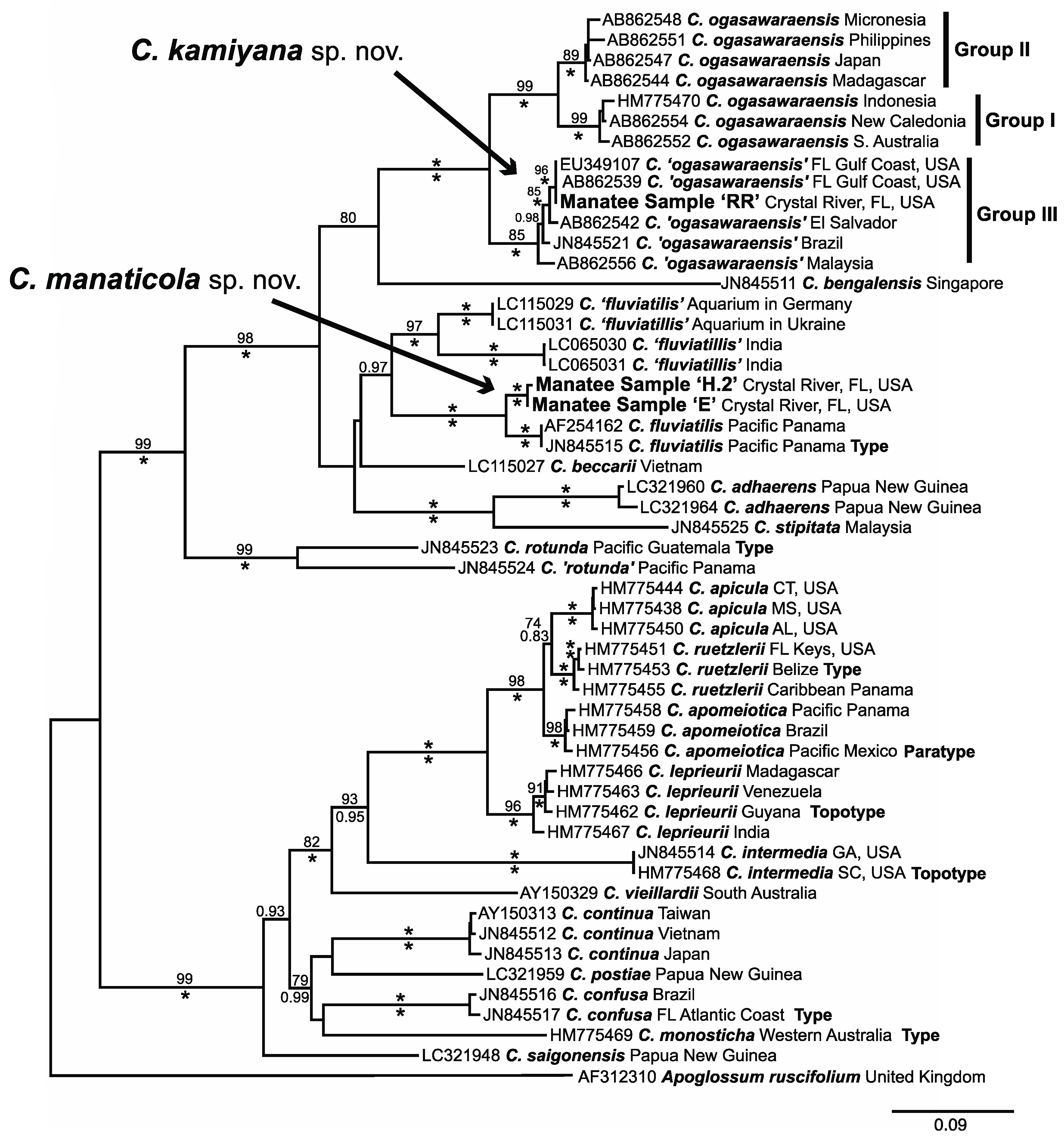
3.1. Molecular Analyses

Trees that resulted from maximum likelihood and Bayesian analyses were closely congruent, and only the maximum likelihood tree is presented here (Figure 1). Samples collected from manatees were resolved in two topological positions. Manatee Caloglossa sample RR was positioned within a fully-supported clade of specimens that were identified as Caloglossa ogasawaraensis. This clade was further subdivided into three well- to fully supported clades that have some geographic basis and were designated by Kamiya and West [26] as Groups I, II, and III (Figure 1). Sample RR was resolved in the Group III clade of predominately Western Hemisphere specimens. The inter-group rbcL sequence divergence range between Group III specimens and specimens in Groups I and II was 4.6–5.3% (Table 1). All intragroup divergences were less than 1.4%.
Manatee Caloglossa samples E and H.2 were resolved with full support as the sister taxa to Caloglossa fluviatilis from Pacific Panama (Figure 1). These two taxa formed a clade with two other taxa represented by specimens from India and aquarium specimens of unknown geographic origin, that were identified most recently as C. fluviatilis [27]. This relationship, however, had only limited support in the ML bootstrap analysis (69%), and was also not fully supported in the Bayesian analysis (posterior probability of 0.97). There was a minimum sequence divergence of 5.9% between the Panama C. fluviatilis/manatee sample clade specimens and those from the aquaria and India. The sequence divergence range between manatee samples E/H.2 and Caloglossa fluviatilis was 2.0–2.4% (Table 2).

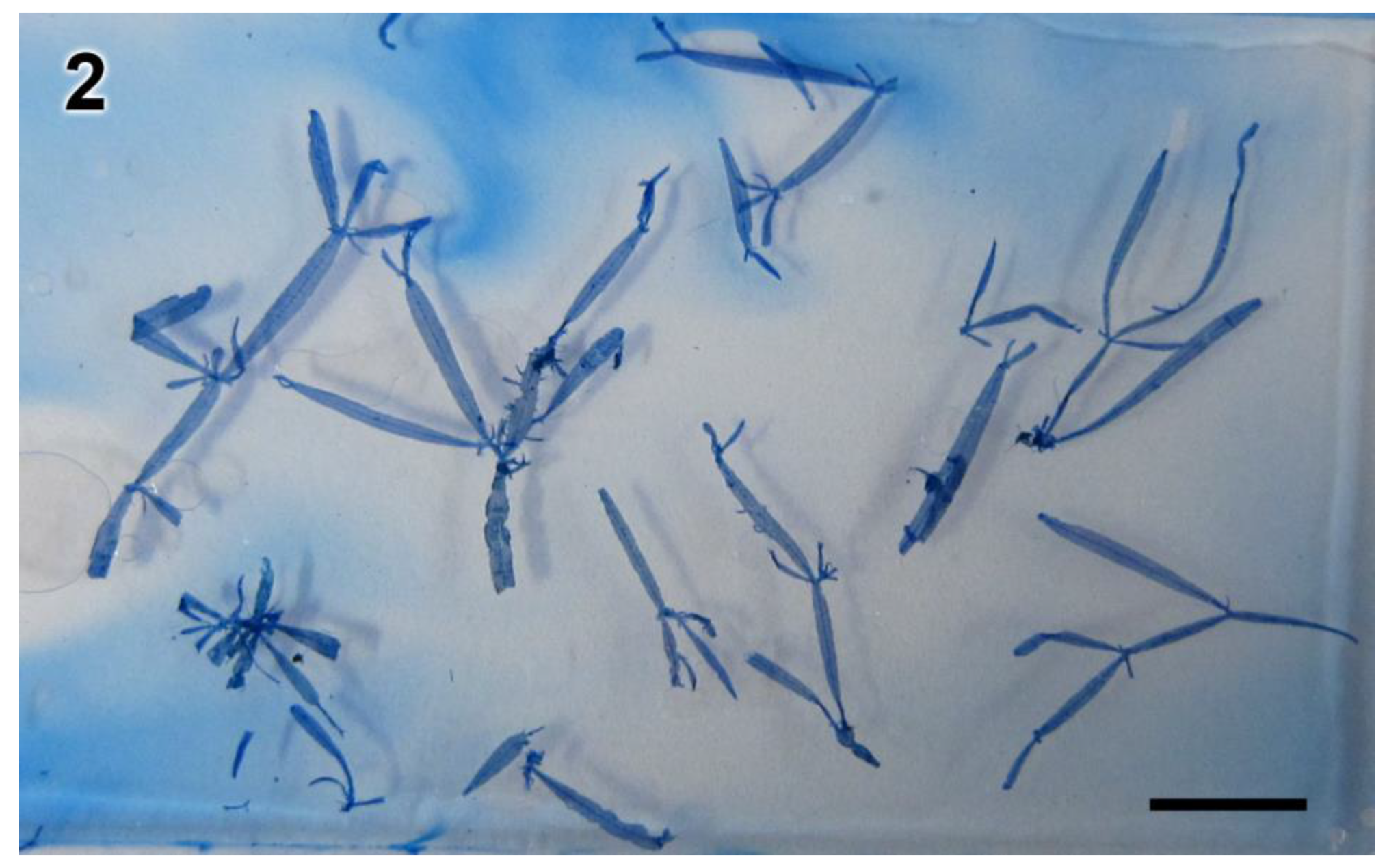
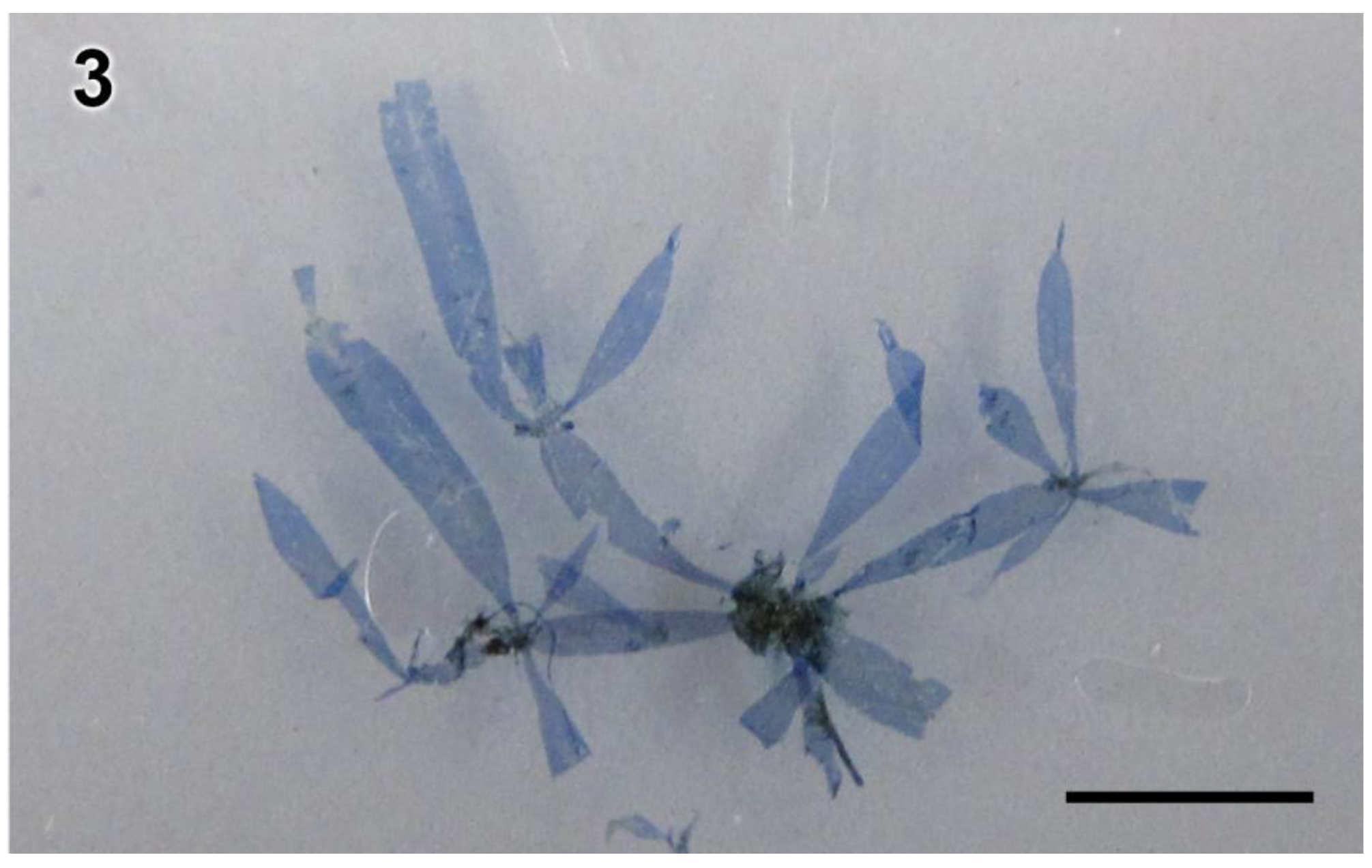
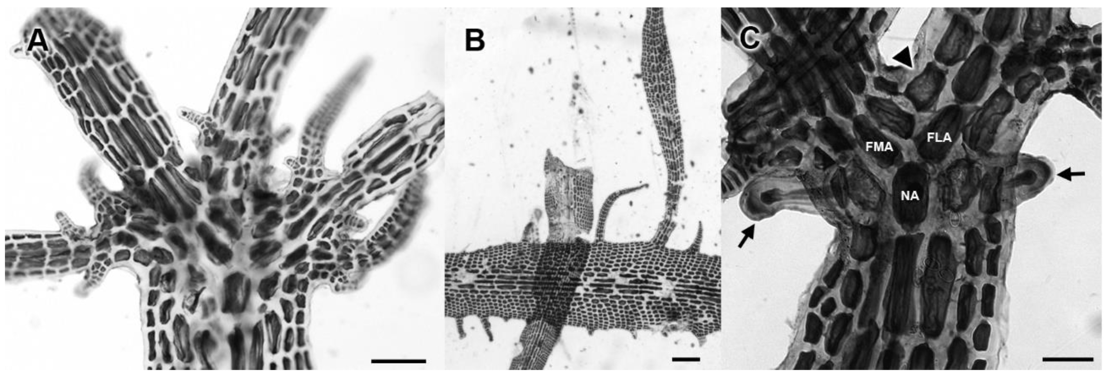
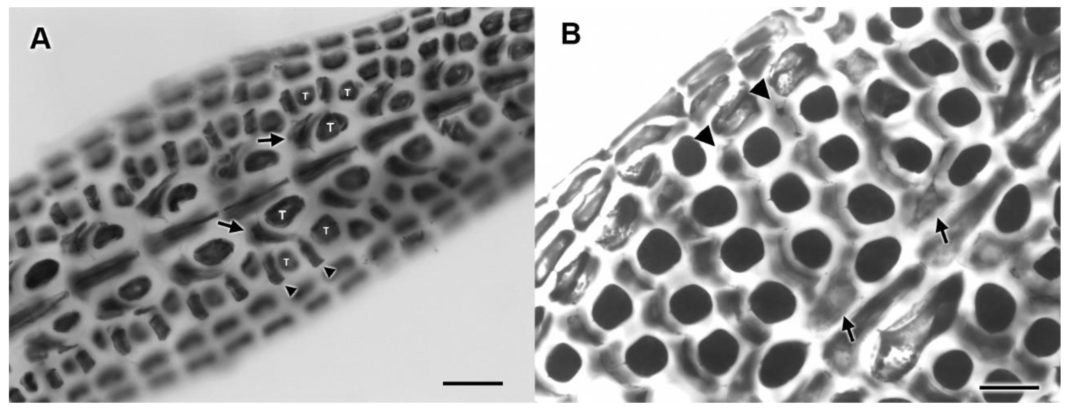
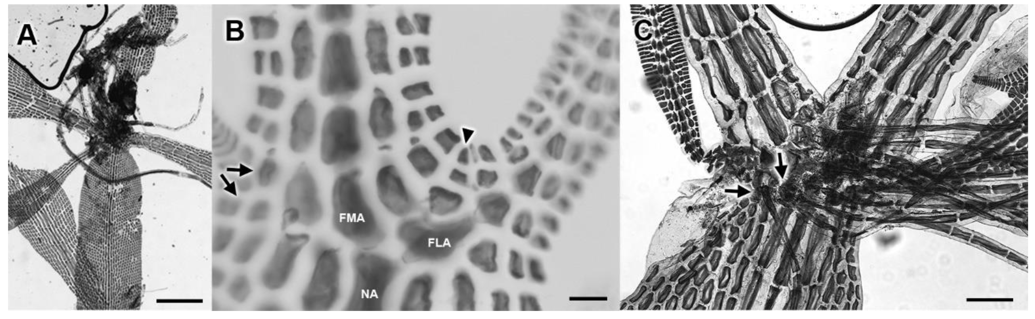
3.2. Morphological Analyses

Manatee Caloglossa sample RR had strap-like or lanceolate blades with slightly constricted nodes (Figure 2), and exhibited all other morphological characteristics described for specimens within the C. ogasawaraensis Group III of Kamiya and West [26] as described below. Manatee Caloglossa samples E and H.2 had relatively wide lanceolate blades with strongly constricted nodes, similar to blades of C. fluviatilis (Figure 3), and shared states with this species for morphological characters generally used to differentiate among Caloglossa species (see below).

3.3. New Species

Based on their topological placement in phylogenetic trees and rbcL sequence divergences from their most closely related sister species, the two species found growing epizoically on West Indian Manatees are proposed as new species.
Caloglossa kamiyana Freshwater, Cath.E. Miller & Frankovich sp. nov. (Figure 2, Figure 4 and Figure 5)
Diagnosis: morphological characters as for C. ogasawaraensis described in Okamura [28], Tanaka and Kamiya [29], and Kamiya and West [26], but with tetrasporangial mother cells cut off from lateral pericentral cells by oblique divisions; DNA sequences of rbcL GenBank no. MW508891.
Holotype: WNC 22781; growing on manatee, Crystal River, Citrus County, FL, USA; leg. T. Frankovich and M. Ashworth; 7 December 2018.
Isotypes: MICH and WNC 2019-s038; 2019-s042.
Other specimens examined: WNC 2019-s049; 2019-s051 thru -s057; 2019-s059; 2020-s083–s084; growing on manatee, Crystal River, FL, USA; leg. T. Frankovich and M. Ashworth; 6 December 2018.
Distribution: this species has been reported and molecularly confirmed from South Carolina, USA, to Brazil in the western Atlantic, El Salvador and Guatemala in the eastern Pacific, and Malaysia in the western Pacific.
Etymology: the species epithet honors Mitsunobu Kamiya whose detailed studies of Caloglossa have greatly enhanced the understanding of this genus, and who along with John West first described the phylogenetic groups within C. ogasawaraensis sensu lato that included the species.
Description: thalli were composed of narrow, subdichotomously divided, linear to lanceolate blades that were slightly constricted at the nodes, 1.0–6.0 mm long and 0.5–0.5 mm wide (Figure 2). Adventitious branches were frequent and endogenous branches were absent (Figure 4A,B). Nodal axial cells produced one or two cell rows towards both sides of the blades (Figure 4C). The first axial cells of the main axes formed one or two cell rows opposite the lateral axis (Figure 4C). The first axial cell of the lateral axis formed an adaxial cell row (Figure 4C). Rhizoids were produced on the ventral side of nodes with a single rhizoid originating from ventral and lateral pericentral cells of the node and those immediately above and below it (“Type E” distribution pattern of Kamiya et al. [30]). There was no corticated pad at the base of the rhizoid filaments.
Elongated tetrasporangial sori developed on both sides of the central axis of adventitious laterals that originated from the margins of blades (Figure 4B and Figure 5A). Tetrasporangial mother cells were cut off by oblique divisions from the lateral pericentral cells, which also cut off cover cells towards both the ventral and dorsal surface of the blade (Figure 5A,B). Tetrasporangial mother cells and cover cells were also cut off from the second-order and third-order row cells derived from the lateral pericentral cell (Figure 5A,B). Tetrasporangial mother cells were cut off from these cells by transverse divisions. Tetrasporangia were decussately divided and observed to develop from up to four second-order row wing cells, and from up to three third-order row wing cells. Other reproductive structures were not observed.
Caloglossa manaticola Freshwater, Cath.E. Miller & Frankovich sp. nov. (Figure 3 and Figure 6)
Diagnosis: morphological characters as for C. fluviatilis described in Krayesky et al. [31] with the exception of rhizoid-bearing cells having mostly one instead of multiple rhizoids; DNA sequences of rbcL GenBank no. MW508892.
Holotype: WNC 22782; growing on manatee, Crystal River, Citrus County, FL, USA; leg. T. Frankovich and M. Ashworth; 6 December 2018.
Isotypes: MICH and WNC 2019-s039; 2019-s048; 2019-s060; 2020-s085 thru –s088. Sequence of rbcL from isotype GenBank no. MW508893.
Distribution: this species is currently known only from the Gulf of Mexico coast of Florida, USA, but is probably more widely distributed.
Etymology: the species epithet refers to the epizoic habitat where the specimens were sampled: from Latin “–cola”, dwelling on; and Latin “Manatus”, manatee, (dwelling on manatees).
Description: thalli were composed of subdichotomously divided main axes that were strongly constricted at nodes (Figure 3). Endogenous branches were absent, but adventitious branches were frequent and created a palmate pattern of branches at many nodes (Figure 6A). Blades were 3.0–6.0 mm long and 0.5–1.25 mm wide; stipitate and lanceolate to elongated clavate. Nodal axial cells produced one cell row towards both sides of the blades. The first axial cells of the main axes formed two to three cell rows opposite the lateral axes (Figure 6B). The first axial cell of the lateral axis formed an adaxial cell row (Figure 6B). Rhizoids were produced on the ventral side of nodes with mostly single, but sometimes multiple, rhizoids originating from nodal periaxial and adjacent marginal wing cells (Type F distribution pattern of Kamiya et al. [30] (Figure 6C). There was no corticated pad at the base of the rhizoid filaments. Reproductive structures were not observed.

4. Discussion

Kamiya et al. [32] found that results of RuBisCO spacer sequence analyses agreed with morphological type and mating group designations among populations of Caloglossa leprieurii. This was followed by a series of studies using RuBisCO spacer and other DNA sequence data to describe species and populations and explore the significance of reproductive compatibility and morphological characters [30,33,34,35]. These studies established that sequence divergence was related to reproductive compatibility and some morphological characters, but not necessarily geographic distance. The genetic distinctness of specimens, however, is not always reflected in morphological differences as was demonstrated in a detailed study of specimens identified as C. ogasawaraensis from around the world [26], and cryptic diversity such as this is increasingly being discovered within red algae [36,37,38]. Previous studies combining molecular, culture, and morphological analyses provide a guide for classifying species diversity in Caloglossa based on DNA sequence analyses. For example, the closely related species, C. apicula, C. apomeiotica, and C. ruetzleri, were previously identified as C. leprieurii, but rbcL sequence analyses demonstrated that they were distinct, and this was further supported by subtle morphological differences [39]. Despite their close relationships, the minimum interspecific divergence between them was 2.4%, while their maximum intraspecific divergence was 1.1% (Table 3).
Krayesky et al. [31] suggested, based on DNA sequence analyses, that western Atlantic specimens identified as C. ogasawaraensis represented a different species from western Pacific specimens. Kamiya and West [26] expanded upon these DNA sequence analyses and designated three “groups” within C. ogasawaraensis: two in the western Pacific, and one restricted to the western Atlantic and eastern Pacific except for a single Malaysian specimen. They also conducted detailed morphological analyses of field and culture specimens from all three C. ogasawaraensis groups. Kamiya and West [26] found that only the width of rhizoid cells was different between Group III field specimens and field specimens in Groups I and II, and cultured Group III specimens differed from specimens of the other groups in the cell-wall thickness of rhizoids only when grown at a salinity of 32. They also found that, in general, rhizoid cell widths increased with increasing salinity or after attaching to substrata, and they cautioned that this variability made it difficult to determine if differences in this character existed in field specimens.
Phylogenetic analyses and sequence divergences of multiple loci [26,27,40] as well as those for rbcL here (Figure 1, Table 1) clearly indicate that the taxon previously identified as C. ogasawaraensis Group III represents a distinct species that we here describe as C. kamiyana. We also believe that, based on these sequence data, C. ogasawaraensis Group I and Group II specimens warrant treatment as different species, but an understanding of which of these species includes the type of C. ogasawaraensis is required before the correct classification can be made. Okamura [28] described C. ogasawaraensis based on specimens collected from Ogasawara-jima by R. Yatabe in March 1879. Seto [41] designated a specimen in the Imperial University of Tokyo as the lectotype, and his image of this specimen shows that there is sufficient material to use current methods for DNA extraction and sequencing of types [42,43] without damaging it.
Male, female and tetrasporophyte reproductive structures of C. ogasawaraensis have been described from Japanese specimens [29]. This study is the first to describe tetrasporangia development from Western Hemisphere specimens, formerly identified as C. ogasawaraensis and now included in C. kamiyana. Our observations closely matched those of Tanaka and Kamiya [29], except for the angle of lateral pericentral cell division giving rise to the tetrasporanigial mother cell. They described and illustrated the tetrasporangial mother cells as being cut off by transverse divisions of both lateral pericentral cells and wing cells. While tetrasporangial mother cells were observed to be cut off by transverse divisions of wing cells, tetrasporangial mother cells were cut off by oblique divisions of lateral pericentral cells in this study.
Caloglossa fluviatilis was described from specimens collected at the Pedro Miguel locks near the Pacific terminus of the Panama Canal [31]. A second collection from the same location had been previously identified as C. beccarii [44] and these were the only records of the new species. Sequence data from both of these are publicly available and were included in our analyses. Although the rbcL sequence divergence between C. fluviatilis and C. manaticola is lower than that between other closely related Caloglossa species, it is greater than intraspecific divergences between specimens covering similar or larger geographic distributions such as C. leprieurii and the newly described C. kamiyana.
Caloglossa manaticola was morphologically indistinguishable from C. fluviatilis except for one potential character. Species with rhizoid distribution type F such as C. fluviatilis are reported to have multiple rhizoids developed from individual blade cells [30]. Our interpretation was that only a single rhizoid developed from the majority of rhizoid bearing blade cells in C. manaticola, but two were occasionally observed. However, this was difficult to determine in our field collected material, and any morphological distinction based on this character remains uncertain.
West et al. [45] reported specimens from freshwater rivers in India with similar blade morphology to C. ogasawaraensis as C. beccari. A subsequent study of C. beccari, C. fluviatilis and C. stipitata placed these Indian specimens as well as ones of unknown provenance from European aquaria in C. fluviatilis, based upon their forming a clade in phylogenetic analyses and sharing a suite of morphological character states [27]. While the Indian and European aquarium taxa are resolved in a clade with C. fluviatilis and C. manaticola (Figure 1), this clade received only weak maximum-likelihood bootstrap support and was not fully supported by Bayesian posterior probabilities (Figure 1). The level of rbcL sequence divergence from C. fluviatilis and C. manaticola was also high (Table 2) and greater than any intraspecific divergence values within Caloglossa. Morphological comparisons of the Indian rivers and European aquariums specimens with C. fluviatilis also indicated differences for many of the characters cited by Kamiya et al. [27] as distinguishing C. beccarii and C. stipitata from their expanded concept of C. fluviatilis. We therefore consider these specimens to represent two species that are distinct from C. fluviatilis and C. manaticola.
Historical reports of Caloglossa in the warm-temperate and tropical Western Atlantic included a single species, C. leprieurii [46,47]. Research by Kamiya, Krayesky and colleagues has expanded the number of species recognized in this region [31,34,39]. Five species, C. apicula, C. confusa, C. intermedia, C. ogasawaraensis, and C. ruetzleri, are currently reported from Florida waters [31,39]. Florida specimens previously identified as C. ogasawaraensis have been reclassified as the new species C. kamiyana in this study. Caloglossa kamiyana is widely distributed, with sequence data confirming its occurrence in the western Atlantic, and eastern and western Pacific. Caloglossa manaticola is currently known only from specimens collected from manatees in freshwater of the Crystal River on the Florida Gulf Coast, and it is most closely related to other Caloglossa species found in freshwater habitats [27]. No characteristics of this species would suggest that C. manaticola is obligately epizoic on manatees, but manatees may be a vector for the distribution of this species and others depending upon their salinity tolerances.
Manatees migrate between fresh and marine waters [11]; therefore, persistent epizoic algal species must be able to tolerate a wide range in salinity. Majewska and Goosen [15] hypothesized that putative obligately-epizoic diatoms are limited more by the availability of the animal substratum rather than any of the abiotic environmental factors. Tursiocola ziemanii, an abundant diatom on manatee skin often reaching near 100% relative cell abundance, has been observed to numerically dominate the diatom skin flora on manatees from fresh [15] and marine waters [8], and on the manatees sampled during this study. The epizoic diatoms that are dominant on cetaceans, sea turtles, and manatees belong to species and often genera that are exclusive to the epizoic habit [5,6,8,9], and therefore, whether they are considered freshwater, brackish, or marine diatoms, is dependent on the habitat of the host animal. Algae that are growing epizoically but also known from other benthic environments may be easier to assign to known salinity environments. The epizoic Caloglossa species observed in this study were found in various associations with other red algal species, including Melanothamnus maniticola, a presumed marine epizoic alga, Bostrychia radicans (Montagne) Montagne, a brackish species, and Compsopogon caeruleus (Balbis ex C.Agardh) Montagne, a freshwater species, suggesting a wide salinity tolerance for Caloglossa kamiyana and Caloglossa manaticola.
Manatees are generally restricted to migrating along coastlines, and genetic studies indicate that there is interchange, albeit small, within the Gulf of Mexico and the Caribbean, even among riverine populations [48,49,50]. A wider distribution of C. manaticola is likely to be discovered with further study of Caloglossa diversity in the Gulf of Mexico and the Caribbean as well.

Author Contributions

Conceptualization, T.A.F., M.J.W. and D.W.F.; methodology, D.W.F.; formal analysis, C.E.M. and D.W.F.; investigation, C.E.M., D.W.F., M.J.W. and T.A.F.; resources, D.W.F.; data curation, D.W.F.; writing—original draft preparation, D.W.F. and C.E.M.; writing—review and editing, D.W.F., M.J.W. and T.A.F.; visualization, D.W.F. and C.E.M.; project administration, T.A.F. and D.W.F.; funding acquisition, D.W.F. All authors have read and agreed to the published version of the manuscript.

Funding

This research was funded by the Center for Marine Science DNA-Algal Trust.

Institutional Review Board Statement

Not applicable.

Informed Consent Statement

Not applicable.

Data Availability Statement

No new data was presented in this study.

Acknowledgments

We thank Bob Bonde and Cathy Beck of the United States Geological Survey (USGS) for allowing us to sample manatees captured during the annual Crystal River manatee health assessments under research permit MA-791721, and Matt Ashworth (University of Texas) for help with the collection of samples. We also thank the large team of numerous veterinarians, biologists, and volunteers who assisted in the manatee captures and surveys. Molecular work on this project was funded by the Center for Marine Science DNA-Algal Trust. This is contribution number 236 from the Coastlines and Oceans Division of the Institute of Environment at Florida International University.

Conflicts of Interest

The authors declare no conflict of interest. The funders had no role in the design of the study; in the collection, analyses, or interpretation of data; in the writing of the manuscript, or in the decision to publish the results.

References

  1. Cheang, C.C.; Tsang, L.M.; Chu, K.H.; Chen, I.-J.; Chan, B.K.K. Host-specific phenotypic plasticity of the turtle barnacle Chelonibia testudinaria: A widespread generalist rather than a specialist. PLoS ONE 2013, 8, e57592. [Google Scholar] [CrossRef] [PubMed] [Green Version]
  2. Ewers-Saucedo, C.; Chan, B.K.K.; Zardus, J.D.; Wares, J.P. Parallel patterns of host-specific morphology and genetic admixture in sister lineages of a commensal barnacle. Biol. Bull. 2017, 232, 171–185. [Google Scholar] [CrossRef]
  3. Siciliano, S.; Cardoso, J.; Francisco, A.; De Souza, S.P.; Hauser-Davis, R.A.; Iwasa-Arai, T. Epizoic barnacle (Xenobalanus globicipitis) infestations in several cetacean species in south-eastern Brazil. Mar. Biol. Res. 2020, 16, 356–368. [Google Scholar] [CrossRef]
  4. Denys, L. Morphology and taxonomy of epizoic diatoms (Epiphalaina and Tursiocola) on a sperm whale (Physeter macrocephalus) stranded on the coast of Belgium. Diatom Res. 1997, 12, 1–18. [Google Scholar] [CrossRef]
  5. Holmes, R.W. The morphology of diatoms epizoic on cetaceans and their transfer from Cocconeis to two new genera, Benettella and Epipellis. Br. Phycol. J. 1985, 20, 43–57. [Google Scholar] [CrossRef]
  6. Holmes, R.W.; Nagasawa, S.; Takano, H. The morphology and geographic distribution of epidermal diatoms of the Dall’s porpoise (Phocoenoides dalli True) in the Northern Pacific Ocean. Bull. Natl. Sci. Mus. Tokyo Ser. B 1993, 19, 1–18. [Google Scholar]
  7. Nemoto, T. On the diatoms of the skin film of the whales in the Northern Pacific. Sci. Rep. Whales Res. Inst. Tokyo 1956, 11, 99–132. [Google Scholar]
  8. Frankovich, T.A.; Sullivan, M.J.; Stacy, N.I. Three new species of Tursiocola (Bacillariophyta) from the skin of the West Indian manatee. Phytotaxa 2015, 204, 33–48. [Google Scholar] [CrossRef] [Green Version]
  9. Frankovich, T.A.; Ashworth, M.P.; Sullivan, M.J.; Theriot, E.C.; Stacy, N.I. Epizoic and apochlorotic Tursiocola species (Bacillariophyta) from the skin of Florida manatees (Trichechus manatus latirostris). Protist 2018, 169, 539–568. [Google Scholar] [CrossRef]
  10. Irvine, A. Manatee metabolism and its influence on distribution in Florida. Biol. Conserv. 1983, 25, 315–334. [Google Scholar] [CrossRef]
  11. Deutsch, C.; Reid, J.; Bonde, R.K.; Easton, D.; Kochman, H.; O’Shea, T. Seasonal movements, migratory behavior and site fidelity of West Indian Manatee along the Atlantic coast of the United States. Wildl. Monogr. 2003, 151, 1–77. [Google Scholar]
  12. Meyer, W.; Seegers, U. A preliminary approach to epidermal antimicrobial defense in the Delphinidae. Mar. Biol. 2004, 144, 841–844. [Google Scholar] [CrossRef]
  13. Baum, C.; Meyer, W.; Stelzer, R.; Feischer, L.G.; Siebers, D. Average nanorough skin surface of the pilot whale (Globicephala melas, Delphinidae): Considerations of the self-cleaning abilities based on nanoroughness. Mar. Biol. 2002, 140, 653–657. [Google Scholar] [CrossRef] [Green Version]
  14. Bledsoe, E.L.; Harr, K.E.; Cichra, M.F.; Philips, E.J.; Bonde, R.K.; Lowe, M. A comparison of biofouling communities associated with free-ranging and captive Florida manatees (Trichechus manatus latirostris). Mar. Mammal Sci. 2006, 22, 997–1003. [Google Scholar] [CrossRef]
  15. Majewska, R.; Goosen, W.E. For better, for worse: Manatee-associated Tursiocola (Bacillariophyta) remain faithful to their host. J. Phycol. 2020, 56, 1019–1027. [Google Scholar] [CrossRef] [PubMed]
  16. Sentíes, A.; Espinoza-Avalos, J.; Zurita, J.C. Epizoic algae of nesting sea turtles Caretta caretta (L.) and Chelona mydas (L.) from the Mexican Caribbean. Bull. Mar. Sci. 1999, 64, 185–188. [Google Scholar]
  17. Serio, D.; Furnari, G.; Moro, I.; Sciuto, K. Molecular and morphological characterisation of Melanothamnus testudinis sp. nov. (Rhodophyta, Rhodomelaceae) and its distinction from Polysiphonia carettia. Phycologia 2020, 59, 281–291. [Google Scholar] [CrossRef]
  18. Woodworth, K.A.; Frankovich, T.A.; Freshwater, D.W. Melanothamnus maniticola sp. nov. (Ceramiales, Rhodophyta): An epizoic species evolved for living on the West Indian manatee. J. Phycol. 2019, 55, 1239–1245. [Google Scholar] [CrossRef] [PubMed]
  19. Millar, A.J.K.; Wynne, M.J. Patulophycus eclipes gen. et sp. nov. (Delesseriaceae, Rhodophyta) from the southwestern Pacific. Syst. Bot. 1992, 17, 409–416. [Google Scholar] [CrossRef]
  20. Taylor, R.L.; Bailey, J.C.; Freshwater, D.W. Systematics of Cladophora spp. (Chlorophyta) from North Carolina, USA, based upon morphology and DNA sequence data with a description of Cladophora subtilissima sp. nov. J. Phycol. 2017, 53, 541–556. [Google Scholar] [CrossRef]
  21. Freshwater, D.W.; Rueness, J. Phylogenetic relationships of some European Gelidium (Gelidiales, Rhodophyta) species, based on rbcL nucleotide sequence analysis. Phycologia 1994, 33, 187–194. [Google Scholar] [CrossRef]
  22. Stuercke, B.; Freshwater, D.W. Consistency of morphological characters used to delimit Polysiphonia sensu lato species (Ceramiales, Florideophyceae): Analyses of North Carolina specimens. Phycologia 2008, 47, 541–559. [Google Scholar] [CrossRef]
  23. Edgar, R.C. Muscle: Multiple sequence alignment with high accuracy and high throughput. Nucleic Acids Res. 2004, 32, 1792–1797. [Google Scholar] [CrossRef] [Green Version]
  24. Stamatakis, R.; Ludwig, T.; Meier, H. RAxML-III: A fast program for maximum likelihood-based inference of large phylogenetic trees. Bioinformatics 2005, 21, 456–463. [Google Scholar] [CrossRef] [Green Version]
  25. Huelsenbeck, J.P.; Ronquist, F. MRBAYES: Bayesian inference of phylogenetic trees. Bioinformatics 2001, 17, 754–755. [Google Scholar] [CrossRef] [Green Version]
  26. Kamiya, M.; West, J.A. Cryptic diversity in the euryhaline red alga Caloglossa ogasawaraensis (Delesseriacea, Ceramiales). Phycologia 2014, 53, 374–382. [Google Scholar] [CrossRef]
  27. Kamiya, M.; West, J.A.; Karsten, U.; Ganesan, E.K. Molecular and morphological delineation of Caloglossa beccarii and related species (Delesseriaceae, Rhodophyta). Phycologia 2016, 55, 640–649. [Google Scholar] [CrossRef]
  28. Okamura, K. On the algae from Ogasawara-jima (Bonin Islands). Bot. Mag. Tokyo 1897, 11, 1–17. [Google Scholar] [CrossRef] [Green Version]
  29. Tanaka, J.; Kamiya, M. Reproductive structure of Caloglossa ogasawaraensis Okamura (Ceramiales, Rhodophyceae) in nature and culture. Jpn. J. Phycol. 1993, 41, 113–121. [Google Scholar]
  30. Kamiya, M.; Zuccarello, G.C.; West, J.A. Evolutionary relationships of the genus Caloglossa (Delesseriaceae, Rhodophyta) inferred from large-subunit ribosomal RNA gene sequences, morphological evidence and reproductive compatability, with description of a new species from Guatemala. Phycologia 2003, 42, 478–497. [Google Scholar] [CrossRef]
  31. Krayesky, D.M.; Norris, J.N.; West, J.A.; Kamiya, M.; Viguerie, M.; Wysor, B.S.; Fredericq, S. Two new species of Caloglossa (Delesseriaceae, Rhodophyta) from the Americas, C. confusa and C. fluviatilis. Phycologia 2012, 51, 513–530. [Google Scholar] [CrossRef] [Green Version]
  32. Kamiya, M.; West, J.A.; King, R.J.; Zuccarello, G.C.; Tanaka, J.; Hara, Y. Evolutionary divergence in the red algae Caloglossa leprieurii and C. apomeiotica. J. Phycol. 1998, 34, 361–370. [Google Scholar] [CrossRef]
  33. Kamiya, M.; Tanaka, J.; King, R.J.; West, J.A.; Zuccarello, G.C.; Kawai, H. Reproductive and genetic distinction between broad and narrow entities of Caloglossa continua (Delesseriaceae, Rhodophyta). Phycologia 1999, 38, 356–367. [Google Scholar] [CrossRef]
  34. Kamiya, M.; West, J.A.; Zuccarello, G.C.; Kawai, H. Caloglossa intermedia sp. nov. (Rhodophyta) from the western Atlantic coast: Molecular and morphological analyses with special reference to C. leprieurii and C. monosticha. J. Phycol. 2000, 36, 411–420. [Google Scholar] [CrossRef]
  35. Zuccarello, G.C.; Bartlett, J.; Yeates, P.H. Differentiation of Caloglossa leprieurii (Rhodophyta) populations in northern and eastern Australia using plastid haplotypes. Eur. J. Phycol. 2000, 35, 357–363. [Google Scholar] [CrossRef]
  36. Gabrielson, P.W.; Hughey, J.R.; Diaz-Pulido, G. Genomics reveals abundant speciation in the coral reef building alga Porolithon onkodes (Corallinales, Rhodophyta). J. Phycol. 2018, 54, 429–434. [Google Scholar] [CrossRef] [PubMed]
  37. Jesus, P.B.; Costa, A.L.; Castro Nunes, J.M.; Manghisi, A.; Genovese, G.; Morabito, M.; Schnadelbach, A.S. Species delimitation methods reveal cryptic diversity in the Hypnea cornuta complex (Cystocloniaceae, Rhodophyta). Eur. J. Phycol. 2019, 54, 135–153. [Google Scholar] [CrossRef]
  38. Hughey, J.R.; Leister, G.L.; Gabrielson, P.W.; Hommersand, M.H. Sarcopeltis gen. nov. (Gigartinaceae, Rhodophyta), with S. skottsbergii comb. nov. from southern South America and S. antarctica sp. nov. from the Antarctic Peninsula. Phytotaxa 2020, 468, 75–88. [Google Scholar] [CrossRef]
  39. Krayesky, D.M.; Norris, J.; West, J.A.; Fredercq, S. The Caloglossa leprieurii-complex (Delesseriaceae, Rhodphyta) in the Americas: The elucidation of overlooked species based on molecular and morphological evidence. Cryptogam. Algol. 2011, 32, 37–62. [Google Scholar] [CrossRef]
  40. Kamiya, M.; West, J.A.; Zuccarello, G.C. High species richness and genetic diversity of the genus Caloglossa (Delesseriaceae, Rhodophyta) in New Ireland, Papua New Guinea. Phycol. Res. 2018, 66, 218–230. [Google Scholar] [CrossRef]
  41. Seto, R. Typification of Caloglossa ogasawaraensis Okamura (Ceramiales, Rhodophyta). Jpn. J. Phycol. 1985, 33, 317–319. [Google Scholar]
  42. Hughey, J.; Gabrielson, P.W. Comment on “Acquiring DNA sequence data from dried archival red algae (Florideophyceae) for the purpose of applying available names to contemporary genetic species: A critical assessment”. Botany 2012, 90, 1191–1194. [Google Scholar] [CrossRef] [Green Version]
  43. Hughey, J.R.; Gabrielson, P.W.; Rohmer, L.; Tortolani, J.; Silva, M.; Miller, K.A.; Young, J.D.; Martell, C.; Ruediger, E. Minimally destructive sampling of type specimens of Pyropia (Bangiales, Rhodophyta) recovers complete plastid and mitochondrial genomes. Sci. Rep. 2014, 4, 5113. [Google Scholar] [CrossRef] [Green Version]
  44. Lin, S.-M.; Fredericq, S.; Hommersand, M.H. Systematics of the Delesseriaceae (Ceramiales, Rhodophyta) based on large subunit rDNA and rbcL sequences, including the Phycodryoideae, subfam. nov. J. Phycol. 2001, 37, 881–899. [Google Scholar] [CrossRef] [Green Version]
  45. West, J.A.; Kamiya, M.; Ganesan, E.K.; Louiseaux-de Goër, S.; Jose, L. Caloglossa beccarii (Delesseriaceae, Rhodophyta) from freshwater rivers in Kerala, India, a critical new record. Algae 2015, 30, 207–216. [Google Scholar] [CrossRef] [Green Version]
  46. Børgesen, F. The marine algae of the Danish West Indies. Part 3. Rhodophyceae (4). Dansk Bot. Ark. 1918, 3, 241–304. [Google Scholar]
  47. Taylor, W.R. Marine Algae of the Eastern Tropical and Subtopical Coasts of the Americas; The University of Michigan Press: Ann Arbor, MI, USA, 1960; p. 870. [Google Scholar]
  48. Vianna, J.A.; Bonde, R.K.; Caballero, S.; Giraldo, J.P.; Lima, R.P.; Clark, A.; Marmontel, M.; Morales-Vela, B.; Joséde Souza, M.; Parr, L.; et al. Phylogeography, phylogeny and hybridization in trichechid sirenians: Implications for manatee conservation. Mol. Ecol. 2006, 15, 433–447. [Google Scholar] [CrossRef]
  49. Hunter, M.E.; Auil-Gomez, N.E.; Tucker, K.P.; Bonde, R.K.; Powell, J.; McGuire, P.M. Low genetic variation and evidence of limited dispersal in the regionally important Belize manatee. Anim. Conserv. 2010, 13, 592–602. [Google Scholar] [CrossRef]
  50. Santizábal, P.; Mignucci-Giannoni, A.A.; Duchene, S.; Caicedo-Herrera, D.; Perea-Sicchar, C.M.; García-Dávila, C.R.; Trujillo, F.; Caballero, S.J. Phylogeography and sex-biased dispersal across riverine manatee populations (Trichechus inunguis and Trichechus manatus) in South America. PLoS ONE 2012, 7, e52468. [Google Scholar] [CrossRef] [Green Version]
Figure 1. Maximum likelihood tree resulting from analysis of 52 Caloglossa specimen rbcL sequences and outgroup Apoglossum ruscifolium. Maximum likelihood bootstrap support and Bayesian posterior probabilities are shown for branches when >70% and >0.80, respectively; *, full support. Type, sequence from type collection or culture of type collection.
Jmse 09 00163 g001
Figure 2. Caloglossa kamiyana sp. nov., holotype specimen from sample “RR”, collected from a West Indian manatee in Crystal River, FL, USA, scale = 4 mm.
Jmse 09 00163 g002
Figure 3. Caloglossa manaticola sp. nov., holotype specimen from sample “E”, collected from a West Indian manatee in Crystal River, FL, USA, scale = 4 mm.
Jmse 09 00163 g003
Figure 4. Caloglossa kamiyana sp. nov. (A) Proliferous development of adventitious branches at a node, scale = 100 µm. (B) Blade with marginal adventitious branches, one well developed and beginning to produce tetrasporangia, scale = 200 µm. (C) Cell arrangement at node showing the nodal axial cell (NA) with a single row of cells developing from it towards both margins; the first main axial cell (FMA) with two rows of cells developing from it opposite the lateral axis; the first lateral axial cell (FLA) with an adaxial cell row (arrowhead), and two rhizoid initials (arrows) developing from the lateral pericentral cells of the nodal axial cell, scale = 50 µm.
Jmse 09 00163 g004
Figure 5. Caloglossa kamiyana sp. nov. (A) Tetrasporangial mother cells (T) cut off by oblique divisions of lateral pericentral cells (arrows) and transverse divisions of second- and third-order row cells (arrowheads), scale = 50 µm. (B) Tetrasporangia cut off from lateral pericentral cells (arrows), second- and third-order row cells (arrowheads), scale = 50 µm.
Jmse 09 00163 g005
Figure 6. Caloglossa manaticola sp. nov. (A) Adventitious branches at node resulting in palmate pattern, scale = 500 µm. (B) Cell arrangement at node showing the nodal axial cell (NA) with a single row of cells developing from it towards both margins; the first main axial cell (FMA) with two rows of cells developing from it opposite the lateral axis (arrows), and the first lateral axial cell (FLA) with an adaxial cell row (arrowhead), scale = 20 µm. (C) Rhizoids originating from nodal periaxial and marginal wing cells with examples of a single rhizoid developing from a wing cell and two rhizoids developing from a lateral pericentral cell of the nodal axial cell indicated (arrows), scale = 100 µm.
Jmse 09 00163 g006
Table 1. Pairwise rbcL sequence divergences between Caloglossa ogasawaraensis “groups” designated by Kamiya and West [26].
Group IGroup IIGroup III: C. kamiyana sp. nov.
Group I
(n = 13)
0.0–0.9%
Group II
(n = 10)
2.9–3.6%0.0–1.2%
Group III
= C. kamiyana sp. nov.
(n = 8)
4.9–5.3%4.6–5.3%0.0–1.4%
Table 2. Pairwise rbcL sequence divergences between Caloglossa fluviatilis; Manatee Caloglossa samples “E” and “H.2”, C. ‘fluviatilis’ from European aquaria, and C. ‘fluviatilis’ from India.
C. fluviatilisManatee SamplesC. ‘fluviatilis’ European AquariaC. ‘fluviatilis’ India
C. fluviatilis
(n = 2)
0.0%
Manatee samples
(n = 2)
2.0–2.4%0.5%
C. ‘fluviatilis’
European aquaria
(n = 2)
6.0–6.1%5.9–6.1%0.0%
C. ‘fluviatilis’
India
(n = 2)
6.9%6.5%4.8%0.0%
Table 3. Pairwise rbcL sequence divergences between the closely related species Caloglossa apicula, C. apomeiotica, and C. ruetzleri.
C. apiculaC. apomeioticaC. ruetzleri
C. apicula
(n = 14)
0.0–0.4%
C. apomeiotica
(n = 4)
2.7–3.3%0.0–0.7%
C. ruetzleri
(n = 6)
2.8–3.6%2.4–3.3%0.0–1.1%
Publisher’s Note: MDPI stays neutral with regard to jurisdictional claims in published maps and institutional affiliations.

