Article

Role of Hepcidins from Black Rockfish (Sebastes schlegelii) in Iron-Metabolic Function and Bacterial Defense

1
Department of Chemistry and Chemistry Institute of Functional Materials, Pusan National University, Busan 609-735, Korea
2
Biotechnology Research Division, National Institute of Fisheries Science, Busan 46083, Korea
*
Author to whom correspondence should be addressed.
J. Mar. Sci. Eng. 2020, 8(7), 493; https://doi.org/10.3390/jmse8070493
Submission received: 12 June 2020 / Revised: 1 July 2020 / Accepted: 2 July 2020 / Published: 5 July 2020
(This article belongs to the Section Marine Biology)

Abstract

:
Hepcidin, an antimicrobial peptide produced by the liver, also controls the iron balance and regeneration in vertebrates. Two types of hepcidin (Hamp1 and Hamp2) have been found in the bodies of black rockfish (Sebastes schlegelii). The full-length cDNA of hepcidin was cloned to enable a study of the antibacterial roles of these two hepcidins (Hamp) in black rockfish. The antimicrobial function of recombinant hepcidins was tested both in vitro and in vivo by the synthesis in Escherichia coli of recombinant hepcidin (approximately 11 kDa) from black rockfish. The recombinant hepcidins inhibited the growth of two bacterial species, Streptococcus iniae FP5228 and Pseudomonas aeruginosa, at various concentrations, in vitro after 6 h post-incubation, respectively. During infection, the production of ferroportin was reduced, suggesting the preservation of iron to prevent microbial proliferation. In vivo administration of Hamp1, but not Hamp2, synthetic peptides induced a substantial reduction in the expression of ferroportin, suggesting that in black rockfish with two forms of hepcidin, ferroportin production is regulated by the iron-regulator Hamp1, and not by the dedicated antimicrobial Hamp2. The findings of this study suggest the various antimicrobial roles of these two types of hepcidin.

1. Introduction

Innate immunity is the first line of defense against infections for fish [1]. One of the major components of innate immunity is a fish’s rapid physiological reaction to injury or infection lasting 1–2 days. The proteins involved in the reaction are hepcidin, ferritin, and serum amyloid [2]. Hepcidin is a cationic, limited size (20–25 amino acids), antimicrobial, cysteine-rich peptide formed in the liver, which also regulates iron [3]. Infection and inflammation can induce hepcidin production [4,5]. This multipurpose protein inhibits ferroprotein (FPN), which brings iron out of the cell [6]. Hepcidin also prevents the absorption of iron in the small intestine, transfers iron across the placenta, and helps remove recycled iron from human and mice macrophages [7]. There is currently increased interest in understanding the function of hepcidin-like peptides in the natural immunity of fish [8]. This small peptide has demonstrated antimicrobial activity against bacterial, viral, fungal, and parasitic infections in different species of fish [9,10,11,12]. The amino acid sequence of hepcidin is highly evolutionarily conserved, with six to eight cysteine residues at specific positions, forming unique disulfide bridges essential for its antimicrobial properties [13]. The existence of the hepcidin gene has been recorded in several species of fish [14,15,16]. The expression of this gene has been observed in the kidneys, liver, spleen, skin, and intestines to a lesser extent [17]. Specific hepcidins are expressed in tissues such as the gills, heart, skin, cardiac stomach, intestines, spleen, liver, and head kidneys [18]. In many fish species, hepcidin production increases in response to bacterial infections. Significant hepcidin production was detected in various tissues of zebrafish and rock bream in response to bacterial, viral, parasitic, or lipopolysaccharide (LPS) challenges [19,20]. Substantially increased transcript levels for hepcidin have been observed in the spleen, liver, and kidney tissues of infected Lates calcarifer during parasitic infections with Cryptocaryon irritans [21]. These results indicate that hepcidins have an inductive response to antigenic stimulus and differing temporal transmission, indicating their strong capacity for customizing a unique inborn immunity [22]. Comprehensive analysis of synthetic hepcidin’s antimicrobial function has been performed in various fish species, whereas relatively little similar work has been done in higher vertebrates [23]. In an earlier study, we described olive flounder hepcidin, which exhibited a wide range of antibacterial activity against Edwardsiella tarda, Pseudomonas aeruginosa, Staphylococcus aureus, Escherichia coli, and Aeromonas hydrophila [24]. Hepcidin expression was also significantly modulated during infection with A. hydrophila [25]. Two hepcidin transcripts of an economically important species of melanops, black rockfish (Sebastes schlegelii), which constitutes a significant proportion of global aquacultural production, have been identified in the present study.
The liver primarily participates in digestion, metabolism, detoxification, and bile secretion. In addition, it plays an important role in immunological reactions [10]. The hepatic mononuclear phagocyte network contains several immunologically active cells that function in the defense against pathogens transmitted to the liver through the portal network [26]. Hence, the liver is a multifunctional organ. In humans, the peptide hepcidin is predominantly generated by hepatocytes; it is then transferred into the serum and contributes to iron regulation and immunity [27]. The peptide was first identified in 2000 as a human liver-expressed antimicrobial protein and was known as LEAP-1. It is also called hepcidin, because it is extracted from liver tissue and has antibacterial properties [28]. Hepcidin is abundant in cysteine and is also present in human urine. It was first isolated from hybrid striped bass and purified in fish in 2002 [29].
Iron loading can induce in vivo expression of hepcidin in mice [30] and humans [31]. Ferroportin 1 (FPN1), transferrin receptor 1 (TFR1), and ferritin (FTN) are three essential genes that regulate iron balance. Hepcidin controls the iron balance by internalizing and destroying basolateral membrane (BLM) cell FPN1 to suppress iron production, thus controlling the extracellular iron content and the systemic iron balance [32]. The production of hepcidin is also highly increased during infection [33], and several reports [34,35,36] on infection pathogenesis support hepcidin’s regulating role in the inflammatory response. For example, in mammals, hepcidin induces anti-inflammatory and pro-inflammatory activity of inflammatory cytokines, such as cytokine signaling suppressor 3 (SOCS3) and alpha tumor necrosis factor (TNF-α) [34,37,38]. In fish such as Pseudosciaena crocea, Danio rerio, Ctenopharyngodon idellus, Sparus aurata, Dicentrarchus labrax, Trachidermus fasciatus, Brachymystax lenok, and Salmo trutta, the hepcidin gene has been cloned and is active in the immune response [39]. Iron overload in tissues can lead to increased oxidizing stress and tissue damage [40].
Human bodies regulate serum and cell iron levels through various proteins, and hepcidin (a small size peptide) has been identified as the principal iron metabolism regulator [41]. The functions of systemic hepcidin have been extensively researched, but now, an increasing number of studies are throwing light on the role of local hepcidin in cardiac homeostasis. Indeed, a number of studies support local function of hepcidin in the heart, lungs, stomach, and prostate [42,43]. Hepcidin is the principal control protein in the metabolism of systemic iron. It is developed in the liver and binds to ferroportin-1 (FPN-1), an exporter of cellular iron, causing the complex to be internalized and degraded [44,45]. Thus, hepcidin prevents the efflux of cellular iron from enterocytes, macrophages, and hepatocytes. As hepcidin synthesis is primarily regulated at the transcriptional stage, the expression level of hepcidin antimicrobial peptide messenger RNA (mRNA) is a good indicator of the quantity of hepcidin peptide produced. The myocardium contains a measurable amount of hepcidin [46]. Inhibition of hepcidin peptide in patients suffering from anemia or chronic inflammation could be a potentially promising therapeutic technique.
Hamp1 cDNA (accession no. EU555379) consisted of 479 bases and Hamp2 cDNA (accession no. EU555380) contained an ORF for the predicted polypeptide of 88 amino acids. All known hepcidin genes have three exons and two introns. The conserved cysteines form 3 or 4 intramolecular disulfide bonds that stabilize a hairpin-like structure, important for the proper binding of the hepcidin to its receptor, the cellular iron exporter, ferroportin (FPN). The length of mature peptide varies between species. The hepcidin gene is commonly distributed in different fish, indicating that, in the innate immune system, this antimicrobial peptide is a very significant part of it [47]. The results in our previous study suggested that Hamp1 and 2 may play an crucial role in the immune response of black rockfish to pathogenic bacterial infection, and two hepcidins may have different functions [48]. Nevertheless, the process by which hepcidin affects the immune system and iron balance in black rockfish remains unknown. We studied the molecular characteristics and expression patterns of hepcidin in black rockfish and examined the mechanisms for controlling transcription in our research. Our findings provide useful information about hepcidin function in fish.

2. Materials and Methods

2.1. Experimental Strains

Escherichia coli (E. coli) strain DH5α was used as the host for plasmid propagation. As a prokaryotic expression host, E. coli strain M15 (pREP4) was added. In vector construction, E. coli strain JM109 was used as the host strain. The vector pQE-30 plasmid and E. coli M15 (pREP4) strain (Qiagen, Seoul, Korea) were applied for cloning and expression. The expression of foreign genes could be induced effectively.

2.2. Plasmid Construction

The plasmid containing the genes of black rockfish hepcidin was donated by the National Fisheries Research and Development Institute, South Korea. Because of the oxidative environment, periplasm space was chosen as the place of expression. A small transmembrane protein YkgR was added. This leading protein could be found inside the inner membrane and was associated with the Nin-Cout position. The hepcidin encoding DNA fragment was amplified by PCR with the primers (Table 1). In addition, His-tag for the purification of affinity chromatography and Factor Xα cleavage site (Ile-Glu-Gly-Arg) for cleavage were added between the hepcidin gene and the YkgR gene. Digested with KpnI and EcoRI, the purified PCR sample was ligated to the plasmid vector pQE-30 utilizing KpnI-EcoRI restriction sites. We transformed the ligation product into E. coli strain JM109, and Sequence Corporation (COSMO, Seoul, Korea) confirmed the sequence (Figure 1).

2.3. Expression of Peptides in E. coli

A single positive transformant colony was grown at 37 °C in 1.0-L culture in a 5-L shaking flask with 50 μg/L kanamycin and 100 μg/L ampicillin until OD600 reached 0.6–~0.8. Thereafter, a final concentration of 0.4 mM IPTG (isopropyl β-D-1-thiogalactopyranoside) was supplementary, and the mixture was shaken for another 4 h at 30 °C at 90 rpm.

2.4. Purification of the Peptides Using an Affinity Column

Cells were centrifuged at 6000 rpm at 4 °C for 5 min, and sonication on ice was performed to release the proteins inside (6 cycles × 30 s, with 2-min intervals between cycles) by using lysis buffer (20 mM Tris-HCl buffer, pH 7.4; 0.2 mM PMSF; 1% Triton X-100). The supernatant fraction was extracted from the lysate at 4 °C, by centrifugation at 13,000 rpm for 30 min. The filtered product was then loaded through 0.45-μm filters onto a pre-packaged Ni2+ column (GE Healthcare, Chicago, IL, USA). It was then pre-balanced with a buffer (20 mM Tris-HCl, 200 mM NaCl, 10 mM imidazole) and cleaned with a buffer in the column (20 mM Tris-HCl, 200 mM NaCl, 100 mM imidazole). Subsequently, an elution buffer was used to extract the purified peptide (20 mM Tris-HCl, 200 mM NaCl, 500 mM imidazole), and dialysis was applied to remove the salts from the final peptide elution buffer. The final peptide yield was measured by a 280-nm absorbance test.

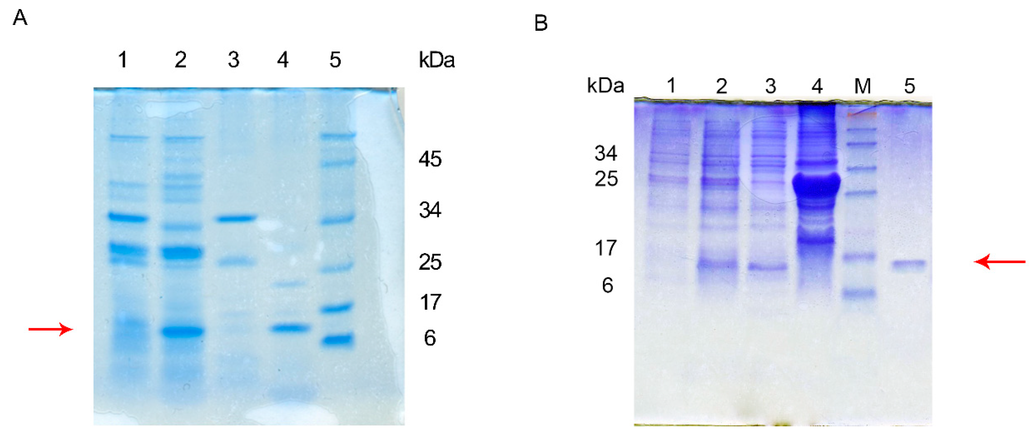
2.5. Tricine-SDS-PAGE Gel Electrophoresis

A previous study reported that electrophoresis of the Tricine-SDS-PAGE gel performs well, particularly in proteins/peptides of small molecules [49]. To achieve a high resolution, a 15% tricine-SDS-PAGE gel was prepared. A 20-μL supernatant sample was combined with 20 μL sample buffer and heated at 95 °C for 10 min in a heat blocker. The SDS-PAGE gels were stained for 30 min with Coomassie Bright Blue R-250.

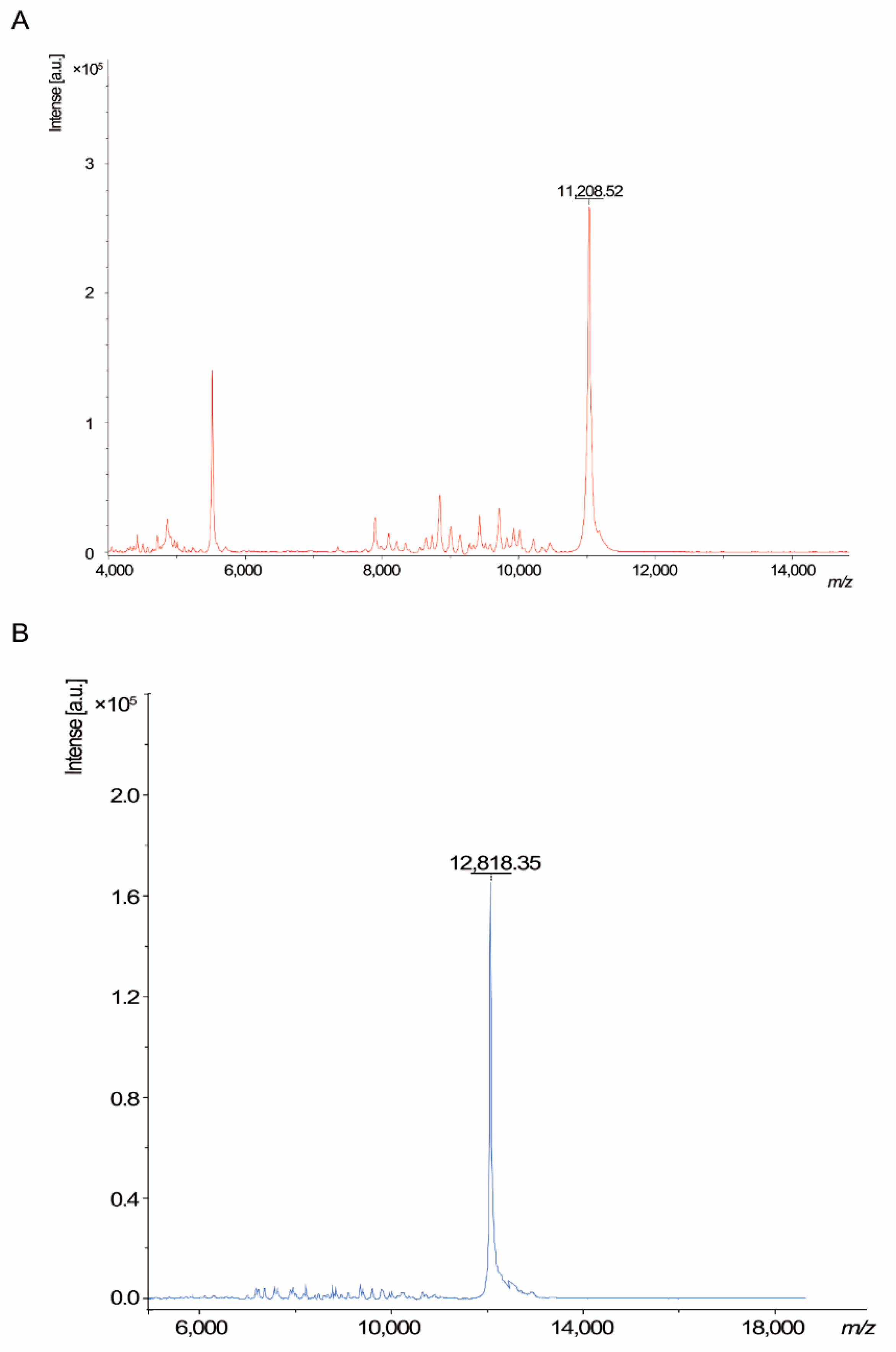
2.6. Mass Spectroscopy Analysis and Amino Acid Sequence

Mass spectroscopy was performed using the methods mentioned elsewhere to evaluate the exact molecular weight and structure [50]. For the analysis of peptide samples, MALDI-TOF MS mass spectrometry (Ultraflextreme, Bruker, Shimadzu, Kyoto, Japan) was employed, and α-cyano-4-hydroxycinnamic acid (Sigma-Aldrich, Seoul, Korea) was used as a matrix. The data were subjected to LC-MS/MS sequencing of amino acids to evaluate the definite changes that occurred during the expression process.
In-gel digestion was performed according to the aforementioned process [51]. Following reduction and alkylation of protein cysteines, gels were digested with trypsin (1.2 μg) for 16 h at 37 °C. The digested peptides were removed by extraction solution. The sample solution (10 μL) comprising 0.1% formic acid and distilled water was used to resolve digested peptides. Ultimate 3000 UPLC network (Dionex Corp, Sunnyvale, CA, USA) attached to a Q Exactive Plus mass spectrometer (ThermoFisher Scientific, Waltham, MA, USA) fitted with a nanoelectrospray ion supply (Dionex Corp) was used to isolate the peptide samples (5 μL). Peptides were eluted from the column and loaded onto a 15-cm Acclaim® PepMap RSLC equal to 75 μm i.d. C18 reverse-phase (ThermoFisher Scientific, MA, USA) column with 300 nL/min flow rate. Peptides were eluted in 0.1% formic acid for 120 min by a gradient of 0–~65% acetonitrile. All MS and MS/MS spectra were obtained using the Q Exactive Plus orbitrap mass spectrometer in top10 mode.
MALDI-TOF mass spectrometry was conducted under specific conditions. Samples were combined on a MALDI sample plate with a solution of α-cyano-4-hydroxycinnamic acid mixture; the plate was then air-dried and analyzed using the spectrometer.

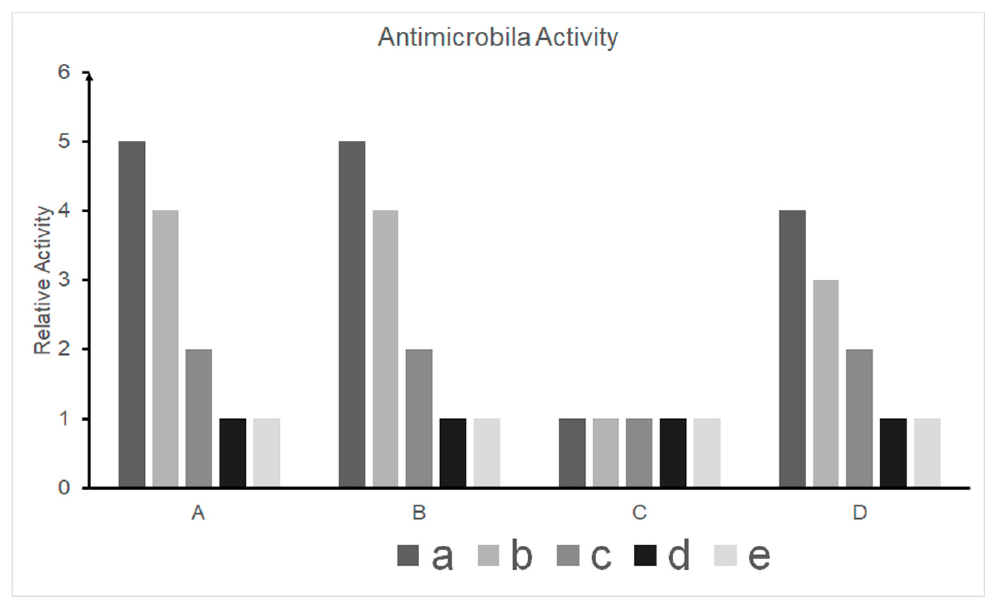
2.7. Antimicrobial Activity Testing

Recombinant hepcidin of black rockfish was developed both in vitro and in vivo, and its antimicrobial activity was assessed [48]. Briefly, it is described as follows: A template growth-inhibition assay was used to assess the antimicrobial activity spectra. Of the eight types of microbes, Pseudomonas aeruginosa and Streptococcus iniae FP5228 were used due to their aggressive action against bioactive peptide hepcidin.

2.8. RT-PCR

Primers were engineered to amplify target tissue and degree of gene expression appropriately (sequences and applications given in Table 1). The isolated RNA was used as a basis for the quantitative real-time PCR (RT-qPCR) study, which came from the total collected RNA before TRIzol (Invitrogen, Seoul, Korea) use. RT-qPCR was conducted in duplicate according to the instructions from the previous study team. Every 20-μL reaction based on the TOPrealTM One-Step RT qPCR Kit (Enzynomics Inc., Daejeon, Korea) developed a 20-μL reaction with the final concentration of 0.25 μM including SYBR Green. Both primers were at a concentration of 10 pmol/μL each and an extract of 50 ng/μL of RNA. The reaction was conducted at 50 °C for 30 min, 95 °C for 10 min, 95 °C for 40 periods of 5 s, and 60 °C for 30 s (with data collection at the endpoint). For the two independent tests of relative Ct values, the 2−ΔΔCt approach was used. Beta-actin levels were used as a guide for the normalization of target gene expression.

2.9. Production of Hamp1, Hamp2, and FPN1 mRNA Response to Excess Iron and Infection

Multiple experimental models were developed to assess Hamp1, Hamp2, and FPN1 transcriptional regulators under different circumstances. RNA was extracted and transformed to cDNA, and gene expression in the liver and intestines was analyzed as described above. The fish were treated intraperitoneally with 200 μL iron dextran (Sigma-Aldrich, Seoul, Korea), diluted in sterile PBS at a final concentration of 10 mg/mL to cause iron overload. The control fish were treated with 200 μL sterile PBS. At treatment 1, 4, 7, 10, and 14 d, four fish from each of the test groups were anesthetized, and blood was collected from the caudal arteries to establish hematological parameters. The fish were then euthanized with an anesthetic injection and dissected. The tissues were excised, frozen in liquid nitrogen, and maintained at −80 °C for further use.
The bacteria Pseudomonas aeruginosa and Streptococcus iniae FP5228 were used in the infection experiments. Both species were cultivated to the mid-logarithmic stage in appropriate growth media, absorbance was measured at 600 nm, and 5.0 × 10⁵ colony forming units (CFU) mL−1 of the bacteria were resuspended. The fish were anesthetized and treated with 200 μL (1.0 × 10⁵ CFU) of bacterial suspension for the experimental infections. The fish in the control group were treated with 200 μL of sterile growth medium. After 24, 48, 72, and 96 h post-infection, four fish from each group were anesthetized, and blood was taken from the caudal vessels for determination of hematological parameters. The fish were then euthanized by using an anesthetic injection and dissected; their tissues were excised, frozen in liquid nitrogen, snapped, and stored at −80 °C for further use. During the experimental infections, mortality was measured every 12 h, and CFU counts were conducted at each experimental time point.

2.10. Analysis of Hepcidin-Related Gene Expression

Ninety days after hatching, 20 healthy and uniformly sized fish were picked and divided into four small groups, each group having five fish. Group 1 was injected with PBS (pH 7.4) and used as the control. Five fish from each group were used for isolating tissue and extracting RNA 24 h after the injection.

2.11. Ethical Statement

Pusan National University-Institutional Animal Care and Use Committee (PNU-IACUC) reviewed and approved the procedures used in this study. The fish were euthanized. The fish organs and tissues were destroyed by an anesthetic injection of MS-222 (Sigma-Aldrich, Seoul, Korea), instantly frozen in liquid nitrogen, and preserved at −80 °C until RNA was collected.

3. Results

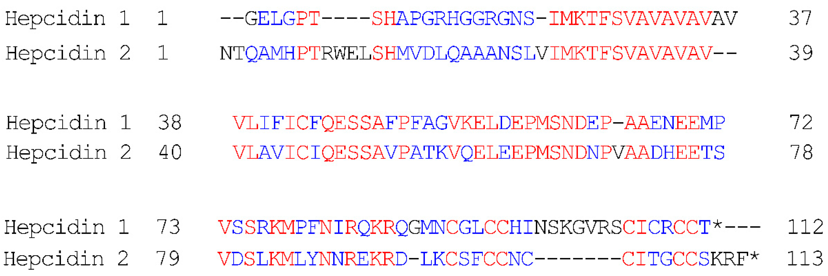
3.1. Construction of a Recombinant Peptide Expression System and Expression

A DNA sequencing technology firm (COSMO, Incheon, Korea) tested for hepcidins. The fragment comprising the concerned gene was cloned into a vector built to produce the peptide of hepcidin. We also picked up several E. coli M15 colonies, and a test system was established by tricine-SDS-PAGE for a small-scale expression trial. We picked a single transformant for expression into the flask, and the degree of expression was assessed with tricine-SDS-PAGE. Figure 2A Lane 2, and Figure 2B Lane 2 shows an obvious band at ~11 kDa after induction of IPTG for 4 h.
We initiated large-scale expression in a 5-L shaking flask, after checking the target peptide expression, and the cultured medium was processed through centrifugation at the end of expression. The cells were disrupted with sonication, to release their components. The recombinant peptide was obtained at levels as high as 25 mg/L at the initial expression; the culture containing it was subjected to the Ni2+column chromatography during the purification stage, and the bound peptides were eluted. For further expression level identification, the fractions containing target expressed peptides were collected and loaded on to the tricine-SDS-PAGE. Using this procedure, a large number of purified peptides were obtained; they were frozen in liquid N2, and maintained for further use at −20 °C.

3.2. Characterization of Recombinant Peptide

The purified recombinant peptide was subjected to MALDI-TOF MS to assess the molecular weight. The recombinant Hamp1 MALDI-TOF spectrum (Figure 3A) displayed the predicted peak at m/z 11208.52, corresponding to [M + H]+, which was similar to the theoretical monoisotopic mass of the hepcidin with three disulfide bonds, 11208. The recombinant Hamp2 MALDI-TOF spectrum (Figure 3B) displayed the predicted peak at m/z 12818.35 corresponding to [M + H]+, which was similar to the theoretical monoisotopic mass of hepcidin 12818 with four disulfide bonds.

3.3. Antimicrobial Activity Test

A radial diffusion assay was used to check the recombinant hepcidin’s antimicrobial function. Increasing the concentration of recombinant hepcidin (Hamp1 and Hamp2) inhibited the development of Streptococcus iniae FP5228 (Figure 4A,B). The recombinant hepcidin was effective in suppressing the growth of the strains. Conversely, no inhibition region was found in the black rockfish peptide Hamp2 (Figure 4C), and this peptide showed antimicrobial activity against Pseudomonas aeruginosa (Figure 4D).

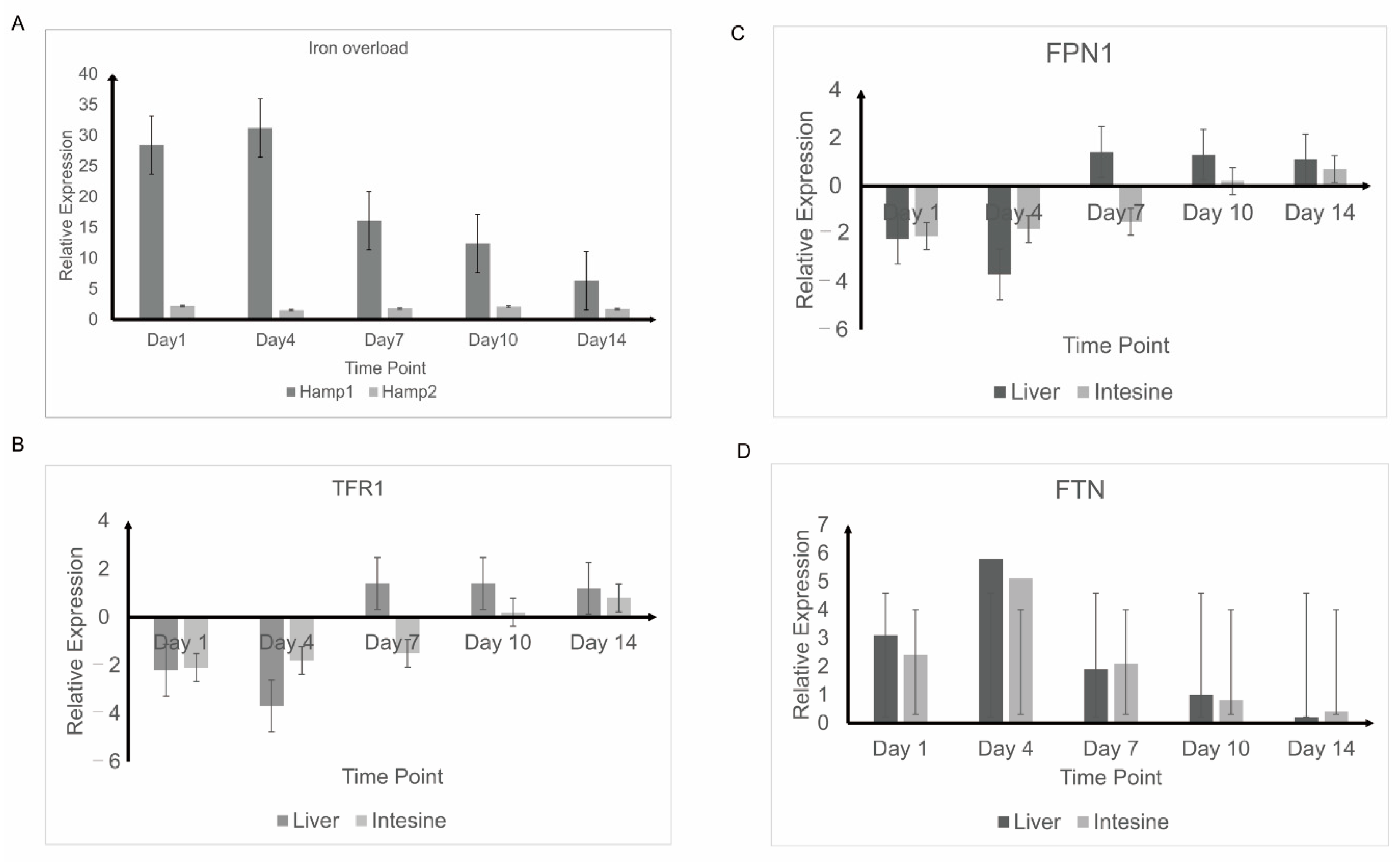
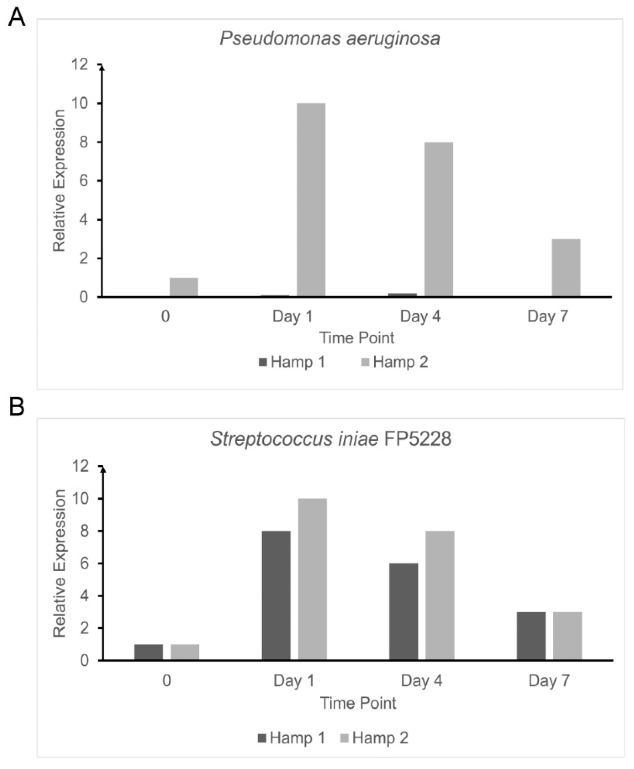
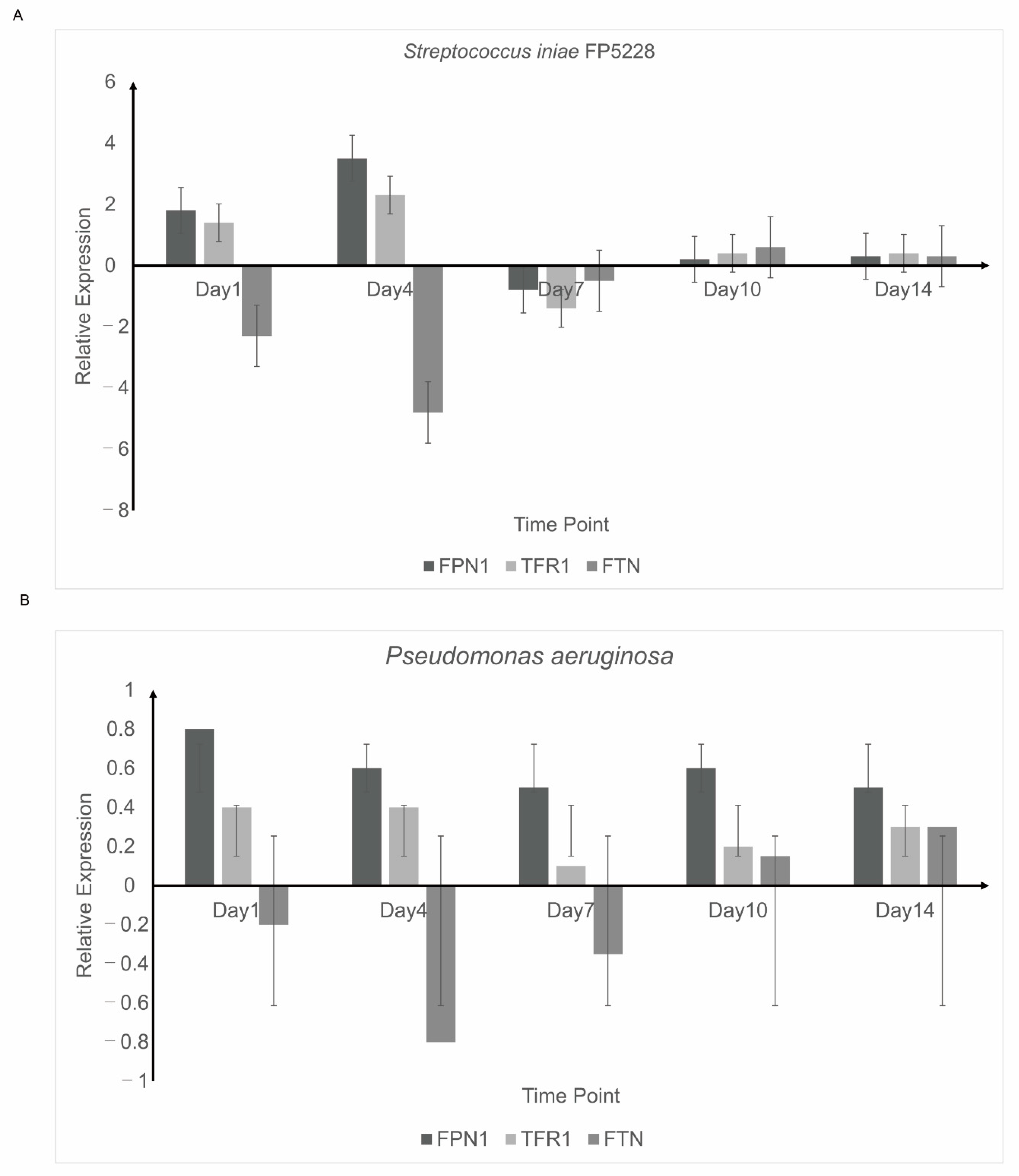
3.4. Response to Experimental Iron Overload and Infection

Black rockfish Hamp1, Hamp2, and FPN1 mRNA expression rates were measured under various experimental conditions expected to affect them. These conditions included iron overload, anemia, and several bacterial infections. In the iron overload experiment, Hamp1 and Hamp2 expressions were detected in the liver (Figure 5A), and FPN1 expression was found both in the liver and intestine (Figure 5B), 1, 4, 7, 10 and 14 d after overload with 2 mg of injected iron dextran, black rockfish Hamp1, Hamp2, and FPN1 mRNA were evaluated. Hamp1 was dramatically overexpressed throughout the course of the trial, beginning to rise as early as day 1, hitting maximum overexpression on day 4, and gradually declining by day 14, although not yet returning to control rates. By contrast, the form of Hamp2 did not alter substantially at any point. In the liver, the expression of FPN1 decreased significantly until day 7, returning to normal levels at day 10. Important reductions in expression were reported in the intestine between days 1 and 4, with recovery to control stages on day 7. Hamp2 was substantially overexpressed throughout the infection trial, with its levels beginning to increase at 24 h (Figure 6). Hamp1, however, did not change dramatically over the course of the experiment, a trend that was repeated for the iron overload (Figure 6B).

3.5. Detection of Iron Balance-Related Genes

The mRNA levels of black rockfish hepcidins and iron balance-related genes (FPN1, TFR1, and FTN) were measured in the liver 24 h post-injection with Streptococcus iniae FP5228 and Pseudomonas aeruginosa (Figure 7). In the Pseudomonas aeruginosa group, the relative expression levels of FPN1 and TFR1 decreased significantly to 8.7- and 4.5-fold, respectively. Conversely, the relative expression level of FTN was upregulated 2-fold. In the Streptococcus iniae FP5228 injection group, the relative expression level of FPN1, TFR1, and FTN did not vary greatly (Figure 7B).

4. Discussion

Hepcidin, a cysteine-rich peptide with a small molecular weight, plays a crucial role in iron metabolism as the primary regulator. Iron is essential for the development of hemoglobin, the main component of red blood cells responsible for oxygen attachment and transportation, and many other cellular processes [22]. Hepcidin is mainly generated in liver hepatocytes, but it has also been reported in other cells and tissues [52]. As a regulator of iron metabolism, hepcidin synthesis is regulated by multiple factors via multiple mechanisms, caused by high iron levels and infection/inflammation, and suppressed by low iron, anemia, and hypoxia. If the quantity of iron is sufficient, then hepcidin, an iron exporter, will bind to ferroportin, causing it to internalize and decay. Throughout our research, we observed that this is accompanied by iron overload and bacterial infection (Figure 5, Figure 6 and Figure 7). Hepcidin inhibits iron release from macrophages, hepatocytes, and enterocytes and subsequently, results in lower iron absorption. Inflammation often reduces iron use for pathogens and causes erythropoiesis, which could result in inflammatory anemia. In contrast, when iron or oxygen levels are low, the output of hepcidin is either attenuated or blocked, the efflux of cellular iron is upregulated, and intestinal absorption increases.
One of the effects of anemia is a decrease in the amount of oxygen that the blood can hold, which ultimately contributes to hypoxia. Iron deficiency is typically due to inadequate intake of iron (low dietary iron), iron absorption defects, failure in storage or transfer, or severe blood loss. In hypoxia, the normal cellular functions need less oxygen, resulting in decreased energy supply, cell proliferation, regeneration, decreased muscle function, and, in serious situations, a major loss of oxygen for the brain, which may cause death [53].
An unusual aspect of fish hepcidins is that they exhibit two kinds of hepcidin (commonly known as Hamp1 and Hamp2), while mammals (the only known exception being the mouse) have only a single gene [29], which functions both as an antimicrobial peptide and as an iron regulator. Although the complete range of functions of the two hepcidins in fish remains unclear, one appears to be associated with regulation of iron, and the other with an antimicrobial function. By analyzing the expression of genes believed to be involved in hematopoiesis and iron homeostasis, against the commercial background of black rockfish, we intend to explain the molecular pathways of reaction to infection, focusing on the functions of the different hepcidin genes.
Hamp1 is found predominantly in the liver of vertebrates and consists of mammalian sequences. The Hamp2 series, however, is contained in several tissues at low concentration [54]. In black rockfish, Hamp1 is upregulated and decreases in reaction to iron overload and infection during anemia and hypoxic conditions, while Hamp2 does not respond to either iron overload or anemia, but is strongly upregulated during bacterial infection and hypoxia [55,56]. One form of hepcidin similar to Hamp1 in black rockfish has been cloned in the present research. The black rockfish gene hepcidin is similar in structure to that of other vertebrates and possesses three exons and two introns; however, because of the small introns, the black rockfish gene hepcidin is smaller than the human gene hepcidin. These small introns may represent the size of the genome or the different gene expression regulatory mechanisms [57].
To obtain a better understanding of the associations between hepcidin and ferroportin in a fish with two forms of hepcidin, the rates of expression were tested in several experimental models including in vivo models of iron overload (Figure 5), anemia, infection with multiple pathogens, and hepcidin administration. Variations in mRNA levels could be measured to detect the hepcidin expression level (Figure 6).
Interestingly, we found two groups of peptides playing different antibacterial roles. In our research, we observed that with certain bacteria under external pressure, Hamp1 and Hamp2 had different rates of resistance to peptide concentrations, and different gene expression levels. The differences in the genes correlated with iron-balance expression also became apparent during regulation of iron levels in black rockfish. Both peptides have a role in the defense against foreign pathogens, with the difference being that they react differently to different pathogenic agents. We also compared their sequences of genes and amino acids, and the results showed that most sequences of amino acids are conserved (Figure 1). The differences in the two sequences could be due to a complex and changing environment during the evolution process, resulting in two peptides present in the same fish that complement each other to give protection against external attacks. In the context of the evolution of living organisms, we hypothesized that the functional scientific importance of peptides found in fishes might provide us with new research angles to explore.
Linked in-depth studies can help discover the unique characteristics of the expression of hepcidin in the liver of black rockfish. Research on hepcidin in mammals has focused mainly on iron balance control [58] and participation in immune responses. Hepcidin prevents bacterial infection by destroying microbial cell membranes and inhibiting cell wall formation, cell respiration, and the entry of nucleic acids or proteins into cells [59], and it can also help with the secretion of inflammatory cytokines [53]. Because hepcidin can prevent pathogenic bacteria from invading fish and has a wide spectrum of antibacterial activity, hepcidin treatment can significantly improve the survival rate of C. idellus infected with Flavobacterium columnare [60] and T. fasciatus infected with Vibrio anguillarum [61]. Pseudosciaena crocea hepcidin shows high resistance to many pathogens, such as Aeromonas hydrophila, Vibrio parahaemolyticus, Vibrio alginolyticus, and Vibrio harvryi [62]. The mRNA expression levels of hepcidin in B. lenok [63] and S. trutta [64] significantly increased after they were infected with Aeromonas salmonicida and A. hydrophila. Platichthys stellatus. V. parahaemolyticus and Edwardsella tarda can be inhibited by hepcidin [61].
These findings open up significant potential applications of hepcidins, prophylactically or therapeutically obtained from fish. The research results are important not only for black rockfish and other closely related species, but also for mammals, because different types of hepcidin can be administered according to specific needs. Hamp1 could be used to regulate iron balance and cure iron deficiencies, while Hamp2 could be administered to prevent or fight a number of infections without interfering with the iron balance. Fish and mammals are, of course, different, but there are similarities between them, and this is why studying hepcidin in fish can yield valuable information for use with mammals.
The mechanism by which hepcidin controls the iron level in humans primarily includes internalizing and breaking down FPN1 to inhibit the development of intracellular iron, balancing the concentration of extracellular iron and the iron function [54,56]. The membrane protein FPN1 is known primarily for introducing iron into vertebrate cells [57]. Transferrin (TF) is a protein with a strong iron affinity, which brings iron to parts of the body where it is stored. Transferrin receptor protein 1 is a TF cell surface receptor and is the dominant protein regulating iron absorption by endocytosis in most cells [59]. The FPN1 rapidly moves intracellular iron around the BLM when iron demands are high in the body. If so, FTN can involve intracellular iron, consisting of an apoprotein shell of 24 light and heavy chain subunits containing a core of up to 4500 iron atoms [65]. Therefore, when the body’s iron content is high, the expression of black rockfish hepcidin (Hamp 1) (Figure 5A) in the liver increases (indicating its key role in maintaining iron levels). The FPN1 expression in the liver (Figure 5B) is then suppressed, showing that the iron output is blocked. The expression of TFR1 is also reduced in the liver, which indicates a decreased intake of cell iron (Figure 5C). The rise in FTN expression in the liver suggests that the cells in FTN contain a sufficient quantity of iron (Figure 5D). These results demonstrate that black rockfish and human hepcidin can have identical biological pathways. Iron metabolism is inextricably linked to immunity. When the body becomes compromised or inflamed, the serum hepcidin levels increase and the iron content decreases [66]. By inhibiting the release of iron from macrophages, hepcidin reduces the amount of iron available for microbial growth and survival [26].
A substantial rise in Hamp1 (not Hamp2) by the liver was followed by a decrease in FPN1 expression in both the liver and intestines in addition to the experimental iron overload. The hepcidin reaction is consistent with what we reported earlier, and further findings agree with the idea that Hamp1 is engaged in iron homeostasis while Hamp2 has little to no involvement in that process. The significant rise in levels of Hamp1 potentially contributes to an improvement in the production of Hamp1 as a means of preserving iron homeostasis. These changes have an inhibitory effect on FPN1 production, which reduces iron release from liver hepatocytes and intestinal enterocytes, and decreases iron mobilization and absorption. Domestication influences the level of Hamp1 in the liver, which is small in rapidly growing strains and strong in slowly growing strains. Domesticated rainbow trout can improve innate immunity and alter the iron balance with more iron in hemoglobin synthesis [29]. Fast expression of the black rockfish hepcidin gene through short-term domestication in this study could be associated with the enhanced immune stress development of black rockfish after injection with LPS (data not shown). The expression of FPN1 in the liver also increased, while the expression of TFR1 in the liver decreased, indicating that iron-balanced genes were developed differentially by the invading pathogens (Figure 7). These findings revealed that when pathogens invaded the host, the degree of cytokine production and the genes associated with the iron balance were disrupted. Under specific biochemical conditions, the black rockfish hepcidin has modified significantly, suggesting that this gene may be considered as a biomarker for black rockfish liver.
Our study also reveals that the two forms of hepcidin play different roles in anemia reactions, with specific transcriptional responses and impacts on hematological parameters and liver iron quantity. When enhanced erythropoiesis was needed, Hamp1 appeared to be the main regulator of iron homeostasis. By contrast, Hamp2 appeared to have a possible secondary function connected with severe anemia caused by bacteria. More studies will be needed to explain the full extent of these functions.

5. Conclusions

In conclusion, we have shown two distinct functional types of hepcidin in black rockfish. We have also shown that iron levels are mediated by Hamp1 and not by the dedicated antimicrobial peptide Hamp2, which plays an important role in immunity. Our results provide a fresh insight into the potential medicinal application of fish-derived hepcidin.

Author Contributions

Formal analysis: Y.M. and J.-S.P.; Funding acquisition: J.-S.P.; Investigation: Y.M.; Methodology: Y.M.; Project administration: Y.M.; Resources: C.-J.L., S.-S.K., D.N.-J.K., B.-H.N., Y.-O.K., and C.-M.A.; Supervision: J.-S.P. All authors have read and agreed to the published version of the manuscript.

Funding

The APC was funded by the National Institute of Fisheries Science, Korea, grant number R2020035.

Acknowledgments

This research was supported by a grant from the National Institute of Fisheries Science (R2020035), Korea.

Conflicts of Interest

The authors have no potential conflict of interest to declare.

References

  1. Al-Hakeim, H.K.; Al-Khakani, M.M.; Al-Kindi, M.A. Correlation of hepcidin level with insulin resistance and endocrine glands function in major thalassemia. Adv. Clin. Exp. Med. 2015, 24, 69–78. [Google Scholar] [CrossRef] [PubMed] [Green Version]
  2. Aschemeyer, S.; Qiao, B.; Stefanova, D.; Valore, E.V.; Sek, A.C.; Ruwe, T.A.; Vieth, K.R.; Jung, G.; Casu, C.; Rivella, S.; et al. Structure-function analysis of ferroportin defines the binding site and an alternative mechanism of action of hepcidin. Blood 2018, 131, 899–910. [Google Scholar] [CrossRef] [PubMed]
  3. Chen, T.; Xu, P.C.; Hu, S.Y.; Gao, S.; Jia, J.Y.; Yan, T.K. High serum hepcidin is associated with the occurrence of anemia in anti-myeloperoxidase antibody-associated vasculitis with normal kidney function: A cross-sectional study. Rheumatol. Int. 2019, 39, 851–857. [Google Scholar] [CrossRef]
  4. Detivaud, L.; Nemeth, E.; Boudjema, K.; Turlin, B.; Troadec, M.B.; Leroyer, P.; Ropert, M.; Jacquelinet, S.; Courselaud, B.; Ganz, T.; et al. Hepcidin levels in humans are correlated with hepatic iron stores, hemoglobin levels, and hepatic function. Blood 2005, 106, 746–748. [Google Scholar] [CrossRef] [PubMed] [Green Version]
  5. Ikuta, K. Function of hepcidin in iron metabolism. Jpn. J. Clin. Hematol. 2007, 48, 36–45. [Google Scholar]
  6. Lakhal-Littleton, S. Cardiomyocyte hepcidin: From intracellular iron homeostasis to physiological function. Vitam. Horm. 2019, 110, 189–200. [Google Scholar]
  7. Le Tertre, M.; Ka, C.; Guellec, J.; Gourlaouen, I.; Ferec, C.; Callebaut, I.; Le Gac, G. Deciphering the molecular basis of ferroportin resistance to hepcidin: Structure/function analysis of rare slc40a1 missense mutations found in suspected hemochromatosis type 4 patients. Transfus. Clin. Biol. J. Soc. Fr. Transfus. Sang. 2017, 24, 462–467. [Google Scholar] [CrossRef] [Green Version]
  8. Lee, S.W.; Kim, Y.H.; Chung, W.; Park, S.K.; Chae, D.W.; Ahn, C.; Kim, Y.S.; Sung, S.A. Serum hepcidin and iron indices affect anemia status differently according to the kidney function of non-dialysis chronic kidney disease patients: Korean cohort study for outcome in patients with chronic kidney disease (know-ckd). Kidney Blood Press. Res. 2017, 42, 1183–1192. [Google Scholar] [CrossRef]
  9. Liu, J.; Qian, L.; Guo, L.; Feng, Y. Studying hepcidin and related pathways in osteoblasts using a mouse model with insulin receptor substrate 1loss of function. Mol. Med. Rep. 2018, 17, 350–357. [Google Scholar]
  10. Malyszko, J.; Kowalewski, R.; Glowinski, J.; Malyszko, J.; Koc-Zorawska, E.; Glowinska, I.; Lebkowska, U.; Gacko, M. Prospective assessment of hepcidin in relation to delayed or immediate graft function in patients undergoing kidney transplantation. Transplant. Proc. 2016, 48, 1506–1510. [Google Scholar] [CrossRef]
  11. Malyszko, J.; Malyszko, J.S.; Hryszko, T.; Pawlak, K.; Mysliwiec, M. Is hepcidin a link between anemia, inflammation and liver function in hemodialyzed patients? Am. J. Nephrol. 2005, 25, 586–590. [Google Scholar] [CrossRef] [PubMed]
  12. Malyszko, J.; Malyszko, J.S.; Kozminski, P.; Mysliwiec, M. Type of renal replacement therapy and residual renal function may affect prohepcidin and hepcidin. Ren. Fail. 2009, 31, 876–883. [Google Scholar] [CrossRef] [PubMed]
  13. Malyszko, J.; Malyszko, J.S.; Pawlak, K.; Mysliwiec, M. Hepcidin, iron status, and renal function in chronic renal failure, kidney transplantation, and hemodialysis. Am. J. Hematol. 2006, 81, 832–837. [Google Scholar] [CrossRef] [PubMed]
  14. Mohammadi, E.; Tamaddoni, A.; Qujeq, D.; Nasseri, E.; Zayeri, F.; Zand, H.; Gholami, M.; Mir, S.M. An investigation of the effects of curcumin on iron overload, hepcidin level, and liver function in beta-thalassemia major patients: A double-blind randomized controlled clinical trial. Phytother. Res. PTR 2018, 32, 1828–1835. [Google Scholar] [CrossRef] [PubMed] [Green Version]
  15. Mu, Y.; Huo, J.; Guan, Y.; Fan, D.; Xiao, X.; Wei, J.; Li, Q.; Mu, P.; Ao, J.; Chen, X. An improved genome assembly for larimichthys crocea reveals hepcidin gene expansion with diversified regulation and function. Commun. Biol. 2018, 1, 195. [Google Scholar] [CrossRef] [PubMed]
  16. Nemeth, E.; Preza, G.C.; Jung, C.L.; Kaplan, J.; Waring, A.J.; Ganz, T. The n-terminus of hepcidin is essential for its interaction with ferroportin: Structure-function study. Blood 2006, 107, 328–333. [Google Scholar] [CrossRef] [Green Version]
  17. Praschberger, R.; Schranz, M.; Griffiths, W.J.; Baumgartner, N.; Hermann, M.; Lomas, D.J.; Pietrangelo, A.; Cox, T.M.; Vogel, W.; Zoller, H. Impact of d181v and a69t on the function of ferroportin as an iron export pump and hepcidin receptor. Biochim. Biophys. Acta 2014, 1842, 1406–1412. [Google Scholar] [CrossRef] [Green Version]
  18. Rajanbabu, V.; Chen, J.Y. Antiviral function of tilapia hepcidin 1-5 and its modulation of immune-related gene expressions against infectious pancreatic necrosis virus (ipnv) in chinook salmon embryo (chse)-214 cells. Fish Shellfish. Immunol. 2011, 30, 39–44. [Google Scholar] [CrossRef]
  19. Rodrigues, P.N.; Vazquez-Dorado, S.; Neves, J.V.; Wilson, J.M. Dual function of fish hepcidin: Response to experimental iron overload and bacterial infection in sea bass (dicentrarchus labrax). Dev. Comp. Immunol. 2006, 30, 1156–1167. [Google Scholar] [CrossRef]
  20. Van der Weerd, N.C.; Grooteman, M.P.; Bots, M.L.; van den Dorpel, M.A.; den Hoedt, C.H.; Mazairac, A.H.; Nube, M.J.; Penne, E.L.; Gaillard, C.A.; Wetzels, J.F.; et al. Hepcidin-25 in chronic hemodialysis patients is related to residual kidney function and not to treatment with erythropoiesis stimulating agents. PLoS ONE 2012, 7, e39783. [Google Scholar] [CrossRef] [Green Version]
  21. Xue, D.; Zhou, C.; Shi, Y.; Lu, H.; He, X. Hepcidin as a biomarker of impaired renal function in rat models for chronic allograft nephropathy. Med. Sci. Monit. Int. Med. J. Exp. Clin. Res. 2016, 22, 608–616. [Google Scholar] [CrossRef] [Green Version]
  22. Yang, C.G.; Liu, S.S.; Sun, B.; Wang, X.L.; Wang, N.; Chen, S.L. Iron-metabolic function and potential antibacterial role of hepcidin and its correlated genes (ferroportin 1 and transferrin receptor) in turbot (scophthalmus maximus). Fish Shellfish. Immunol. 2013, 34, 744–755. [Google Scholar] [CrossRef] [PubMed]
  23. Yazdani, Y.; Sadeghi, H.; Alimohammadian, M.; Andalib, A.; Moazen, F.; Rezaei, A. Expression of an innate immune element (mouse hepcidin-1) in baculovirus expression system and the comparison of its function with synthetic human hepcidin-25. Iran. J. Pharm. Res. IJPR 2011, 10, 559–568. [Google Scholar] [PubMed]
  24. Chen, J.; Shi, Y.H.; Li, M.Y. Changes in transferrin and hepcidin genes expression in the liver of the fish pseudosciaena crocea following exposure to cadmium. Arch. Toxicol. 2008, 82, 525–530. [Google Scholar] [CrossRef] [PubMed]
  25. Douglas, S.E.; Gallant, J.W.; Liebscher, R.S.; Dacanay, A.; Tsoi, S.C. Identification and expression analysis of hepcidin-like antimicrobial peptides in bony fish. Dev. Comp. Immunol. 2003, 27, 589–601. [Google Scholar] [CrossRef]
  26. Li, W.; Tao, Y.; Zhao, D.; Xu, B. connection of hepcidin genes from two fish species and their expression in pichia pastoris. Chin. J. Biotechnol. 2015, 31, 682–691. [Google Scholar]
  27. Shen, Y.; Zhao, Z.; Zhao, J.; Chen, X.; Cao, M.; Wu, M. Expression and functional analysis of hepcidin from mandarin fish (siniperca chuatsi). Int. J. Mol. Sci. 2019, 20, 5602. [Google Scholar] [CrossRef] [Green Version]
  28. Ribeiro, S.; Garrido, P.; Fernandes, J.; Rocha-Pereira, P.; Costa, E.; Belo, L.; Reis, F.; Santos-Silva, A. Liver iron is a major regulator of hepcidin gene expression via bmp/smad pathway in a rat model of chronic renal failure under treatment with high rhuepo doses. Biofactors 2016, 42, 296–306. [Google Scholar]
  29. Wang, K.J.; Bo, J.; Yang, M.; Hong, H.S.; Wang, X.H.; Chen, F.Y.; Yuan, J.J. Hepcidin gene expression induced in the developmental stages of fish upon exposure to benzo[a]pyrene (bap). Mar. Environ. Res. 2009, 67, 159–165. [Google Scholar] [CrossRef] [Green Version]
  30. Ribeiro, S.; Garrido, P.; Fernandes, J.; Rocha, S.; Nunes, S.; Rocha-Pereira, P.; Costa, E.; Belo, L.; Reis, F.; Santos-Silva, A. Liver iron regulates hepcidin expression. Studies in a rat model of chronic renal failure under recombinant human erythropoietin therapy. Febs J. 2015, 282, 98. [Google Scholar]
  31. Wang, K.J.; Cai, J.J.; Cai, L.; Qu, H.D.; Yang, M.; Zhang, M. Cloning and expression of a hepcidin gene from a marine fish (pseudosciaena crocea) and the antimicrobial activity of its synthetic peptide. Peptides 2009, 30, 638–646. [Google Scholar] [CrossRef] [PubMed]
  32. Aoki, C.; Rossaro, L.; Ramasamooj, R.; Bowlus, C.L. Hepcidin mrna expression in human liver correlates with serum ferritin. Gastroenterology 2003, 124, A713. [Google Scholar] [CrossRef]
  33. Gardenghi, S.; Casu, C.; Renaud, T.M.; Meloni, A.; Cooke, K.S.; Sasu, B.J.; Rivella, S. Investigating the role of cytokines and hepcidin in anemia of inflammation. Am. J. Hematol. 2013, 88, E124. [Google Scholar] [CrossRef]
  34. Besson-Fournier, C.; Gineste, A.; Latour, C.; Gourbeyre, O.; Meynard, D.; Aguilar-Martinez, P.; Oswald, E.; Martin, P.; Coppin, H.; Roth, M.P. Hepcidin upregulation by inflammation is not causally related to liver activation of smad1/5/8 signaling by activin b. Blood 2016, 128. [Google Scholar] [CrossRef]
  35. Harrison-Findik, D.D.; Gerjevic, L.; Eroglu, E. Inflammation-mediated activation of liver hepcidin expression is suppressed by alcohol: Role of stat3 protein. Hepatology 2009, 50, 867a. [Google Scholar]
  36. Rivera, S.; Ganz, T. Hepcidin is the principle mediator of anemia of inflammation. Chest 2005, 128, 149s–150s. [Google Scholar] [CrossRef]
  37. Gardenghi, S.; Renaud, T.M.; Meloni, A.; Ramos, P.; Casu, C.; Cooke, K.S.; Sasu, B.J.; Giardina, P.; Grady, R.W.; Rivella, S. Investigating the role of cytokines and hepcidin in anemia of inflammation. Blood 2011, 118, 482. [Google Scholar] [CrossRef]
  38. Kanamori, Y.; Sugiyama, M.; Hashimoto, O.; Murakami, M.; Matsui, T.; Funaba, M. Regulation of hepcidin expression by inflammation-induced activin b. Sci. Rep. 2016, 6, 38702. [Google Scholar] [CrossRef] [Green Version]
  39. Alvarez, C.A.; Santana, P.A.; Guzman, F.; Marshall, S.; Mercado, L. Detection of the hepcidin prepropeptide and mature peptide in liver of rainbow trout. Dev. Comp. Immunol. 2013, 41, 77–81. [Google Scholar] [CrossRef]
  40. Mercadel, L.; Metzger, M.; Haymann, J.P.; Thervet, E.; Boffa, J.J.; Flamant, M. The relation of hepcidin to iron disorders, inflammation and hemoglobin in chronic kidney disease (vol 9, e99781, 2014). PLoS ONE 2015, 10, e99781. [Google Scholar] [CrossRef] [Green Version]
  41. Kroot, J.J.C.; Tjalsma, H.; Fleming, R.E.; Swinkels, D.W. Hepcidin in human iron disorders: Diagnostic implications. Clin. Chem. 2011, 57, 1650–1669. [Google Scholar] [CrossRef] [PubMed] [Green Version]
  42. Yamaji, S.; Sharp, P.; Ramesh, B.; Srai, S.K. Inhibition of iron transport across human intestinal epithelial cells by hepcidin. Blood 2004, 104, 2178–2180. [Google Scholar] [CrossRef] [PubMed]
  43. Zhao, B.; Li, R.Q.; Cheng, G.; Li, Z.Y.; Zhang, Z.P.; Li, J.; Zhang, G.Y.; Bi, C.W.; Hu, C.; Yang, L.B.; et al. Role of hepcidin and iron metabolism in the onset of prostate cancer. Oncol. Lett. 2018, 15, 9953–9958. [Google Scholar] [CrossRef] [PubMed] [Green Version]
  44. Vyoral, D.; Petrak, J. Hepcidin: A direct link between iron metabolism and immunity. Int. J. Biochem. Cell B 2005, 37, 1768–1773. [Google Scholar] [CrossRef] [PubMed]
  45. Weizer-Stern, O.; Adamsky, K.; Margalit, O.; Ashur-Fabian, O.; Givol, D.; Amariglio, N.; Rechavi, G. Hepcidin, a key regulator of iron metabolism, is transcriptionally activated by p53. Brit. J. Haematol. 2007, 138, 253–262. [Google Scholar] [CrossRef] [PubMed]
  46. Weber, C.S.; Neto, L.B.D.; Biolo, A.; Goldraich, L.A.; Clausell, N. Anemia in heart failure: Iron metabolism dictates hepcidin levels in stable outpatients. Eur. Heart J. 2011, 32, 291. [Google Scholar]
  47. Cuesta, A.; Meseguer, J.; Esteban, M.A. The antimicrobial peptide hepcidin exerts an important role in the innate immunity against bacteria in the bony fish gilthead seabream. Mol. Immunol. 2008, 45, 2333–2342. [Google Scholar] [CrossRef]
  48. Kim, Y.O.; Park, E.M.; Nam, B.H.; Kong, H.J.; Kim, W.J.; Lee, S.J. Identification and molecular characterization of two hepcidin genes from black rockfish (sebastes schlegelii). Mol. Cell. Biochem. 2008, 315, 131–136. [Google Scholar] [CrossRef]
  49. Haider, S.R.; Reid, H.J.; Sharp, B.L. Modification of tricine-sds-page for online and offline analysis of phosphoproteins by icp-ms. Anal. Bioanal. Chem. 2010, 397, 655–664. [Google Scholar] [CrossRef] [Green Version]
  50. Chang, Z.; Lu, M.; Ma, Y.; Kwag, D.G.; Kim, S.H.; Park, J.M.; Nam, B.H.; Kim, Y.O.; An, C.M.; Li, H.; et al. Production of disulfide bond-rich peptides by fusion expression using small transmembrane proteins of escherichia coli. Amino Acids 2015, 47, 579–587. [Google Scholar] [CrossRef]
  51. Choi, C.W.; Park, E.C.; Yun, S.H.; Lee, S.Y.; Lee, Y.G.; Hong, Y.; Park, K.R.; Kim, S.H.; Kim, G.H.; Kim, S.I. Proteomic characterization of the outer membrane vesicle of pseudomonas putida kt2440. J. Proteome Res. 2014, 13, 4298–4309. [Google Scholar] [CrossRef]
  52. Cardaropoli, S.; Todros, T.; Nuzzo, A.M.; Rolfo, A. Maternal serum levels and placental expression of hepcidin in preeclampsia. Pregnancy Hypertens. 2018, 11, 47–53. [Google Scholar] [CrossRef] [PubMed]
  53. Toshiyama, R.; Konno, M.; Eguchi, H.; Asai, A.; Noda, T.; Koseki, J.; Asukai, K.; Ohashi, T.; Matsushita, K.; Iwagami, Y.; et al. Association of iron metabolic enzyme hepcidin expression levels with the prognosis of patients with pancreatic cancer. Oncol. Lett. 2018, 15, 8125–8133. [Google Scholar] [CrossRef] [PubMed]
  54. Cui, Q.; Chen, F.Y.; Zhang, M.; Peng, H.; Wang, K.J. Transcriptomic analysis revealing hepcidin expression in oryzias melastigma regulated through the jak-stat signaling pathway upon exposure to bap. Aquat. Toxicol. 2019, 206, 134–141. [Google Scholar] [CrossRef]
  55. Fillebeen, C.; Charlebois, E.; Wagner, J.; Katsarou, A.; Mui, J.; Vali, H.; Garcia-Santos, D.; Ponka, P.; Presley, J.; Pantopoulos, K. Transferrin receptor 1 controls systemic iron homeostasis by fine-tuning hepcidin expression to hepatocellular iron load. Blood 2019, 133, 344–355. [Google Scholar] [CrossRef] [PubMed] [Green Version]
  56. Han, L.; Liu, Y.; Lu, M.; Wang, H.; Tang, F. Retinoic acid modulates iron metabolism imbalance in anemia of inflammation induced by lps via reversely regulating hepcidin and ferroportin expression. Biochem. Biophys. Res. Commun. 2018, 507, 280–285. [Google Scholar] [CrossRef] [PubMed]
  57. Huang, S.N.; Ruan, H.Z.; Chen, M.Y.; Zhou, G.; Qian, Z.M. Aspirin increases ferroportin 1 expression by inhibiting hepcidin via the jak/stat3 pathway in interleukin 6-treated pc-12 cells. Neurosci. Lett. 2018, 662, 1–5. [Google Scholar] [CrossRef] [PubMed]
  58. Samba-Mondonga, M.; Calve, A.; Mallette, F.A.; Santos, M.M. Myd88 regulates the expression of smad4 and the iron regulatory hormone hepcidin. Front. Cell Dev. Biol. 2018, 6, 105. [Google Scholar] [CrossRef]
  59. Sharp, P.A.; Clarkson, R.; Hussain, A.; Weeks, R.J.; Morison, I.M. DNA methylation of hepatic iron sensing genes and the regulation of hepcidin expression. PLoS ONE 2018, 13, e0197863. [Google Scholar] [CrossRef] [Green Version]
  60. Wang, L.; Liu, X.; You, L.H.; Ci, Y.Z.; Chang, S.; Yu, P.; Gao, G.; Chang, Y.Z. Hepcidin and iron regulatory proteins coordinately regulate ferroportin 1 expression in the brain of mice. J. Cell. Physiol. 2019, 234, 7600–7607. [Google Scholar] [CrossRef]
  61. Wei, X.; Sarath Babu, V.; Lin, L.; Hu, Y.; Zhang, Y.; Liu, X.; Su, J.; Li, J.; Zhao, L.; Yuan, G. Hepcidin protects grass carp (ctenopharyngodon idellus) against flavobacterium columnare infection via regulating iron distribution and immune gene expression. Fish Shellfish. Immunol. 2018, 75, 274–283. [Google Scholar] [CrossRef] [PubMed]
  62. Wu, J.; Yang, L.; Zhang, X.; Li, Y.; Wang, J.; Zhang, S.; Liu, H.; Huang, H.; Wang, Y.; Yuan, L.; et al. Mc-lr induces dysregulation of iron homeostasis by inhibiting hepcidin expression: A preliminary study. Chemosphere 2018, 212, 572–584. [Google Scholar] [CrossRef] [PubMed]
  63. Xu, G.; Huang, T.; Gu, W.; Zhang, Y.; Yao, Z.; Zhao, C.; Wang, B. Characterization, expression, and functional analysis of the hepcidin gene from brachymystax lenok. Dev. Comp. Immunol. 2018, 89, 131–140. [Google Scholar] [CrossRef] [PubMed]
  64. Zuo, E.; Lu, Y.; Yan, M.; Pan, X.; Cheng, X. Increased expression of hepcidin and associated upregulation of jak/stat3 signaling in human gastric cancer. Oncol. Lett. 2018, 15, 2236–2244. [Google Scholar] [CrossRef] [PubMed] [Green Version]
  65. Ma, J.; Zhang, F.L.; Zhou, G.; Bao, Y.X.; Shen, Y.; Qian, Z.M. Different characteristics of hepcidin expression in il-6+/+ and il-6-/- neurons and astrocytes treated with lipopolysaccharides. Neurochem. Res. 2018, 43, 1624–1630. [Google Scholar] [CrossRef]
  66. Mao, P.; Wortham, A.M.; Enns, C.A.; Zhang, A.S. The catalytic, stem, and transmembrane portions of matriptase-2 are required for suppressing the expression of the iron-regulatory hormone hepcidin. J. Biol. Chem. 2019, 294, 2060–2073. [Google Scholar] [CrossRef] [Green Version]
Figure 1. Translated amino acid sequence of target hepcidin genes and BLAST (Basic Local Alignment Search Tool) alignment results. Those identical deduced amino acid residues were marked in red, indicating conversed amino acid sequence, and those different deduced amino acid residues were marked in blue.
Jmse 08 00493 g001
Figure 2. Recombinant expression and purification of hepcidins. (A) Lane 1, before induction; Lane 2, after induction; Lane 3, flow-through faction using the FPLC (Fast protein liquid chromatography) purification system. Lane 4, purified Hamp1. (B) Lane 1, before induction; Lane 2, after induction; Lane 3, supernatant fraction after sonication; Lane 4, pellet fraction after sonication; Lane 5, purified Hamp2.
Jmse 08 00493 g002
Figure 3. MALDI-TOF MASS result. (A) MALDI-TOF MASS result of Hamp1 from black rockfish. (B) MALDI-TOF MASS result of Hamp2 from black rockfish.
Jmse 08 00493 g003
Figure 4. Antimicrobial activity of Hamp1 and Hamp2. a. Biological activity testing of Hamp1 against Streptococcus iniae FP5228; from a to d, the concentration of hepcidin was 0.60 mg/L, 0.48 mg/L, 0.36 mg/L, and 0.24 mg/L, respectively. With decreasing concentration, the antimicrobial effect was reduced. b. Biological activity test of Hamp2 against Streptococcus iniae FP5228; from a to d, the concentration of hepcidin is 0.56mg/L, 0.42mg/L, 0.30mg/L, and 0.20mg/L, respectively. The antimicrobial effect decreased with decreasing concentration. c. Hamp1 antimicrobial activity against Pseudomonas aeruginosa; from a to d, the concentration of Hamp1 was 0.20 mg/L, 0.40 mg/L, 0.80 mg/L, and 1.00 mg/L, respectively. No bioactive effect variation with increasing concentration of Hamp1 was observed. d. Hamp2 peptide from E. coli showed antibacterial activity against Pseudomonas aeruginosa; the inhibition zone shrank with decreasing peptide concentration. From d to a, the concentration was 0.20 mg/L, 0.40 mg/L, 0.80 mg/L, and 1.00 mg/L, respectively. e. the negative control: buffer used for dissolving the peptide.
Jmse 08 00493 g004
Figure 5. Black rockfish (A) hepcidin (Hamp1 and Hamp2) expression in the liver after iron overload and (B) Ferroportin 1 (FPN1) expression in the liver and intestine under experimental iron overload. (C) Transferrin receptor 1 (TFR1), expression in the liver and intestine under experimental iron overload. (D) Ferritin (FTN) expression in the liver and intestine under experimental iron overload. Fish were injected with 2 mg of iron dextran (n = 4). Values are expressed as mean fold change ± SD. β-Actin was used as the housekeeping gene.
Jmse 08 00493 g005
Figure 6. Hepcidin (Hamp1 and Hamp2) mRNA expression in the liver after bacterial infection (A) hepcidin (Hamp1 and Hamp2) mRNA expression in the liver after infection with Pseudomonas aeruginosa. (B) Hepcidin (Hamp1 and Hamp2) mRNA expression in the liver after infection with Streptococcus iniae FP5228.
Jmse 08 00493 g006
Figure 7. FPN1, TFR1, and FTN mRNA expression in the liver after bacterial infection. (A) FPN1, TFR1, and FTN mRNA expression in the liver after infection with Streptococcus iniae FP5228. (B) FPN1, TFR1, and FTN mRNA expression in the liver after infection with Pseudomonas aeruginosa.
Jmse 08 00493 g007
Table 1. Primers used in our study.
PrimerApplicationSequence (5′ → 3′)
Hamp1-FHepcidin I amplificationGGCCGGAATTCGATTGTTCTACTATTCAAGGT
Hamp1-RHepcidin I amplificationCCGGAATTCTACGTAAGCTTCAGCCTCTCT
Hamp2-FHepcidin II amplificationTCTTCTACTGTTTGTTGTATTACTAAGCCATAAGCG
Hamp2-RHepcidin II amplificationCGCTTATGGCTTAGTAATACAACAAACAGTAGAAGA
Hamp1RT-FHepcidin I RT-PCRATGAAGACATTCAGTGTTGCG
Hamp1RT-RHepcidin I RT-PCRTCAGAATCTTTTTGAGCAGCA
Hamp2RT-FHepcidin II RT-PCRATGAAGACATTCAGTGTTGCA
Hamp2RT-RHepcidin II RT-PCRTCATGTGCAGCACCTAATGCA
Beta-actin-FcontrolAGGCTCAGAGCAAGAGAG
Beta-actin-RcontrolCGGTGAGCGGACGGGTGC

