Article

Amphipathic Alpha-Helical Peptides AH1 and AH3 Facilitate Immunogenicity of Enhanced Green Fluorescence Protein in Rainbow Trout (Oncorhynchus mykiss)

1
Department of Marine Biotechnology, Institute of Marine and Environmental Technology, University of Maryland, Baltimore County, 701 E. Pratt Street, Baltimore, MD 21202, USA
2
Graduate Institute of Life Sciences, College of Biomedical Sciences, National Defense Medical University, Taipei 10051, Taiwan
*
Author to whom correspondence should be addressed.
J. Mar. Sci. Eng. 2025, 13(8), 1497; https://doi.org/10.3390/jmse13081497
Submission received: 9 June 2025 / Revised: 3 July 2025 / Accepted: 1 August 2025 / Published: 4 August 2025
(This article belongs to the Section Marine Aquaculture)

Abstract

Vaccination is the most effective method to counteract infectious diseases in farmed fish. It secures aquaculture production and safeguards the wild stock and aquatic ecosystem from catastrophic contagious diseases. In vaccine development, recombinant subunit vaccines are favorable candidates since they can be economically produced in large quantities without growing many pathogens, as in inactivated or attenuated vaccine production. However, recombinant subunit vaccines are often weak or deficient in immunogenicity, resulting in inadequate defenses against infections. Technologies that can increase the immunogenicity of recombinant subunit vaccines are in desperate need. Enhanced green fluorescence protein (EGFP) has a low antigenicity and is susceptible to folding changes and losing fluorescence after fusing with other proteins. Using these valuable features of EGFP, we comprehend two amphipathic alpha-helical peptides, AH1 and AH3, derived from Hepatitis C virus and Influenza A virus, respectively, that can induce high immune responses of their fused EGFP in fish without affecting their folding. AH3-EGFP has the most elevated cell binding, significantly 62% and 36% higher than EGFP and AH1-EGFP, respectively. Immunizations with AH1-EGFP or AH3-EGFP significantly induced higher anti-EGFP antibody levels 300–500-fold higher than EGFP immunization after the boost injection in rainbow trout. Our results suggest that AH1 and AH3 effectively increase the immunogenicity of EGFP without influencing its structure. Further validation of their value in other recombinant proteins is necessary to demonstrate their broader utility in enhancing the immunogenicity of subunit vaccines. We also suggest that EGFP and its variants are promising candidates for initially screening proper immunogenicity-enhancing peptides or proteins to advance recombinant subunit vaccine development.

1. Introduction

As the shift in dependence from fishery harvests to artificially propagated aquatic species continues, aquaculture expansion is necessary to maximize production to meet the fast-growing seafood demands and minimize ecological impacts [1]. Healthy farmed fish are essential to achieving cost-effective and sustainable aquaculture development. The increase in aquaculture operations consequently provides many breeding grounds for pathogen cultivation. Infectious diseases are critical economic and ecological threats in fish farming. If not controlled and managed well, they are detrimental to the aquaculture business and the aquatic ecosystem [2].
Vaccination is the most effective approach to mitigate infectious diseases and warrant aquaculture production [3,4]. The introduction of vaccines in finfish aquaculture has reduced the use of antibiotics and other chemicals, contributing to environmental and economic sustainability in aquaculture [5]. As such, vaccination has become a common practice in high-value finfish farming. However, unlike bacterial vaccines that can stimulate effective immunity to protect fish from target bacterial pathogens, current commercial fish viral vaccines do not adequately shield fish against viral diseases [6,7,8]. Moreover, the development of fish viral vaccines is lagging far behind and cannot meet the rising demands. Subsequently, outbreaks of viral infections have left devastating losses to fish farmers and jeopardized the sustainability of aquaculture [9,10]. As a result, fish viral diseases are escalating due to more intensive aquaculture methods and becoming incalculable threats to the expanding finfish aquaculture and its surrounding ecosystems.
The traditional viral vaccines mainly rely on inactivated or attenuated viral pathogens. Inactivated vaccines are relatively safe but have the disadvantage of being less immunogenic in stimulating protective immunity [5]. In addition, inactivated vaccines require the large-scale growth of live viruses to reach high concentrations, presenting production and hazardous containment challenges [11]. On the other hand, attenuated vaccines comprise weakened viruses that could induce a strong and long-lasting immune response to counteract viral infections. However, they are less stable and have a relatively shorter shelf life [12]. They can also regain infectivity when attenuated viruses replicate in the hosts [13] and present the risk of introducing pathogenic strains into the aquatic environment, infecting wild stocks [14]. As such, attenuated viral vaccines pose an enormous financial burden on vaccine developers. Extensive research, clinical testing, and risk assessments require huge investments. Another issue with live vaccines is the regulatory hurdles in vaccine registration domestically and globally due to considerably different legislation on the control and administration of vaccines [15].
Recombinant subunit vaccines that use one or multiple defined protein antigens to elicit immune responses against target pathogens are promising candidates in vaccine development [16]. As recombinant subunit vaccines can be produced in various heterologous expression systems such as bacteria, yeast, insect cells, and mammalian cells, this allows for the production of large quantities without growing and handling a large number of pathogens [16]. However, one of the main challenges in developing recombinant subunit vaccines is their weak immunogenicity [17]. Consequently, efforts have been made toward improving recombinant subunit vaccines, including adding adjuvants into vaccine formulation or fusing immunogens to vaccine candidates to increase their immunogenicity [18,19]. However, the use of traditional adjuvants is often accompanied by undesirable side effects, such as inflammatory reactions, granulomatous lesions, growth impairment, and tissue and organ adhesions [19]. Furthermore, several immunogens have been developed to improve vaccine immunogenicity after linking them to their candidates. For example, Hepatitis B core antigen HBcAg [20] and Pertussis toxin CyaA from bacteria [21] were used to increase the antigenicity of their attached proteins through different mechanisms. Still, the insertion or fusion of a heterologous protein often interferes with the proper folding of the target pathogen proteins due to the large size of these immunogens. Hence, small peptides that can facilitate high immunogenicity without affecting the structures of their fused pathogen proteins are highly desired.
Amphipathic alpha-helical peptides possess hydrophobic amino acids on the one side and hydrophilic amino acids on the opposite side of the helix, which further their interaction with the phospholipid membrane [22]. When fused with antigens, this amphipathic property may enhance the interaction of fusion antigens with antigen-presenting cells and induce a higher immune response. Our previous study found that amphipathic alpha-helical peptides can also help assemble their fused proteins into thermally stable protein nanoparticles that stimulate lasting humoral immunity in mice [23]. In this study, we investigated the applications of these immunogenicity-enhancing amphipathic alpha-helical peptides in fish vaccine development with two amphipathic alpha-helical peptides derived from either Hepatitis C virus isolate H77, NS3 protein (AH1 peptide), or Influenza A virus, H5N1 (AH3 peptide). After fusion to enhanced green fluorescence protein (EGFP), immunizations with AH1-EGFP or AH3-EGFP significantly induced much higher anti-EGFP antibody levels than immunizations with EGFP in rainbow trout.

2. Materials and Methods

2.1. Fish, Rearing Conditions, and Animal Ethics

Female rainbow trout (33–37 g) obtained from the USDA/ARS National Center for Cool and Cold Water Aquaculture, West Virginia, USA, and Atlantic salmon (28–35 g) acquired from the National Cold Water Marine Aquaculture Center, Maine, USA, were housed in 90-gallon tank systems supplied with recirculated fresh water at 13–14 °C and fed with commercial pelleted food (Bio-Oregon, Longview, WA, USA), at 2–4% body weight/day at our Aquaculture Research Center at 12 h-D/12 h-L light cycle. The fish care and experiment procedures were approved by the University’s Institutional Animal Care and Use Committee and adhered to the National Research Council’s Guide for Care and Use of Laboratory Animals.

2.2. Recombinant Protein Expression and Purification

pET28a(+) vector was used to express EGFP, AH1-EGFP, and AH3-EGFP (Figure 1). Each construct was co-transformed with pLemo plasmid (NEB, Ipswich, MA, USA) into endotoxin-free ClearColi BL21 (DE3) (Lucigen, Middleton, WI, USA). The expression of each protein was achieved with a culture in PA-5052 medium [23] at 37 °C for 3 h and then IPTG (0.4 mM) induction at 25 °C overnight. After centrifugation, the recombinant proteins were extracted using BugBuster® Protein Extraction Reagent (Millipore, Burlington, MA, USA) and purified using Ni-NTA Superflow resin (Qiagen, Germantown, MD, USA) following the manufacturers’ instructions. The purified proteins were desalted and the buffer was exchanged to phosphate-buffered saline (PBS) using the Econo-Pac 10DG column (Bio-Rad, Hercules, CA, USA). The concentration of protein was measured using the Pierce™ BCA Protein Assay Kit (Thermo Fisher, Waltham, MA, USA). The expression and size of each protein were analyzed using SDS-PAGE. An LED light at 450 nm was used to examine the fluorescence of each protein. The excitation and emission spectra of each protein were measured using SpectraMax M5 (Molecular Devices, San Jose, CA, USA).

2.3. Cell Binding of EGFP, AH1-EGFP, and AH3-EGFP

HEK 293 cells were cultured in DMEM medium with 4.5 g/L glucose and 10% FBS (Corning, Corning, NY, USA) in a CO2 incubator at 37 °C. Twenty-four hours before the assay, cells were seeded into a 96-well plate at 30,000 cells/well density. The next day, 30 µg (0.3 mg/mL medium) of soluble EGFP, AH1-EGFP, or AH3-EGFP was added into each well, 5 wells/each protein, following the procedures modified from a published method [24]. After a 6 h incubation, the cells were washed with medium 6 times (5 min/time) before fluorescent images were taken using a Revolve microscope (Echo, Lake Zurich, IL, USA).

2.4. Recombinant Protein Immunization, Blood Collection, and ELISA Analysis

EGFP, AH1-EGFP, and AH3-EGFP were diluted to 0.74 µg/µL with PBS and then mixed and emulsified with MontanideTM ISA 763A VG adjuvant (Seppic, Paris, France) at a 27:73 ratio in volume (27 µL protein and 73 µL adjuvant to a final 20 µg protein/100 µL). Rainbow trout (N = 5) were anesthetized with 50 mg/L of tricaine-methanesulphonate (Sigma-Aldrich, St. Louis, MO, USA) and intraperitoneally injected with 1 µg of protein/g of body weight on days 0 and 22. In the pilot trials, no significant increase in antibody titers was detected until 3 weeks after the first vaccine injection; therefore, the booster was introduced on day 22. A total of 1 ml of blood from caudal vasculature was collected from each fish before the first immunization (day 0 as controls, pre-immunization) and the second immunization (day 22, prime). The boost and post-boost blood were collected on days 43 and 77. Plasma was collected by centrifuging the blood sample at 2000 g for 10 min at 4 °C and storing it at −80 °C.
For the ELISA, EGFP was diluted with 100 mM of bicarbonate/carbonate coating buffer (pH 9.6) at 10 μg/mL for coating on 96-well high-binding ELISA plates (Costar, Arlington, VA, USA). The plates were incubated at 4 °C overnight, washed 3 times with wash buffer (0.05% Tween20 in PBS pH 7.4), and blocked with 200 μL of blocking buffer containing 5% skim milk. Plasma was diluted in blocking buffer from 1:1000 to 1:16,348,000 by sequential 4-fold dilution. After overnight incubation at 4 °C, the plates were washed 3 times with wash buffer and then incubated with the secondary antibody rabbit anti-salmonid Ig (Bio-Rad, USA) at a 1:5000 dilution for 90 min at room temperature. After 3 washes, HRP conjugated goat anti-rabbit antibody at 1:10,000 dilution (Sigma-Aldrich, USA) was added into each well for a 2 h incubation at room temperature. After 3 washes, chromogenic development was performed using the Pierce™ TMB Substrate Kit (Thermo Fisher, USA) and read using SpectraMax M5.

2.5. Statistical Analysis

Data obtained from the cell binding were presented as the mean and standard deviation (SD). One-way analysis of variance (ANOVA) was used to determine differences in the cell binding of EGFP, AH1-EGFP, and AH3-EGFP, followed by Tukey’s test for pairwise comparisons of the means using the IBM SPSS version 27 program. The significance was accepted at p < 0.05. Plasma antibody titers were transformed and calculated using a standard curve derived from a series of titrations of the highest anti-EGFP plasma and presented as geometric mean and geometric SD. The differences between groups were determined using Mann–Whitney U tests. The significance was accepted at p < 0.05.

3. Result

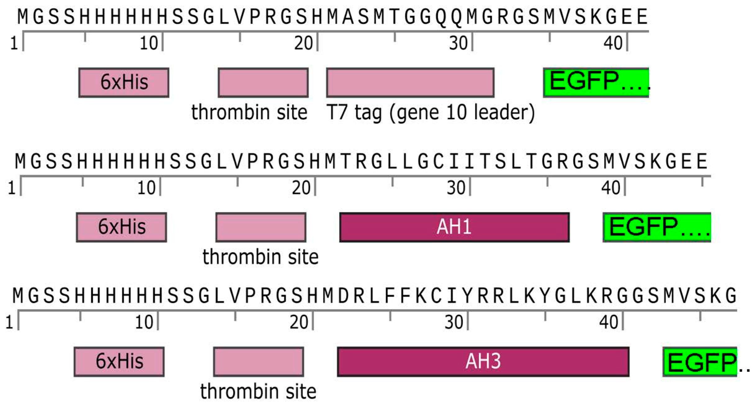
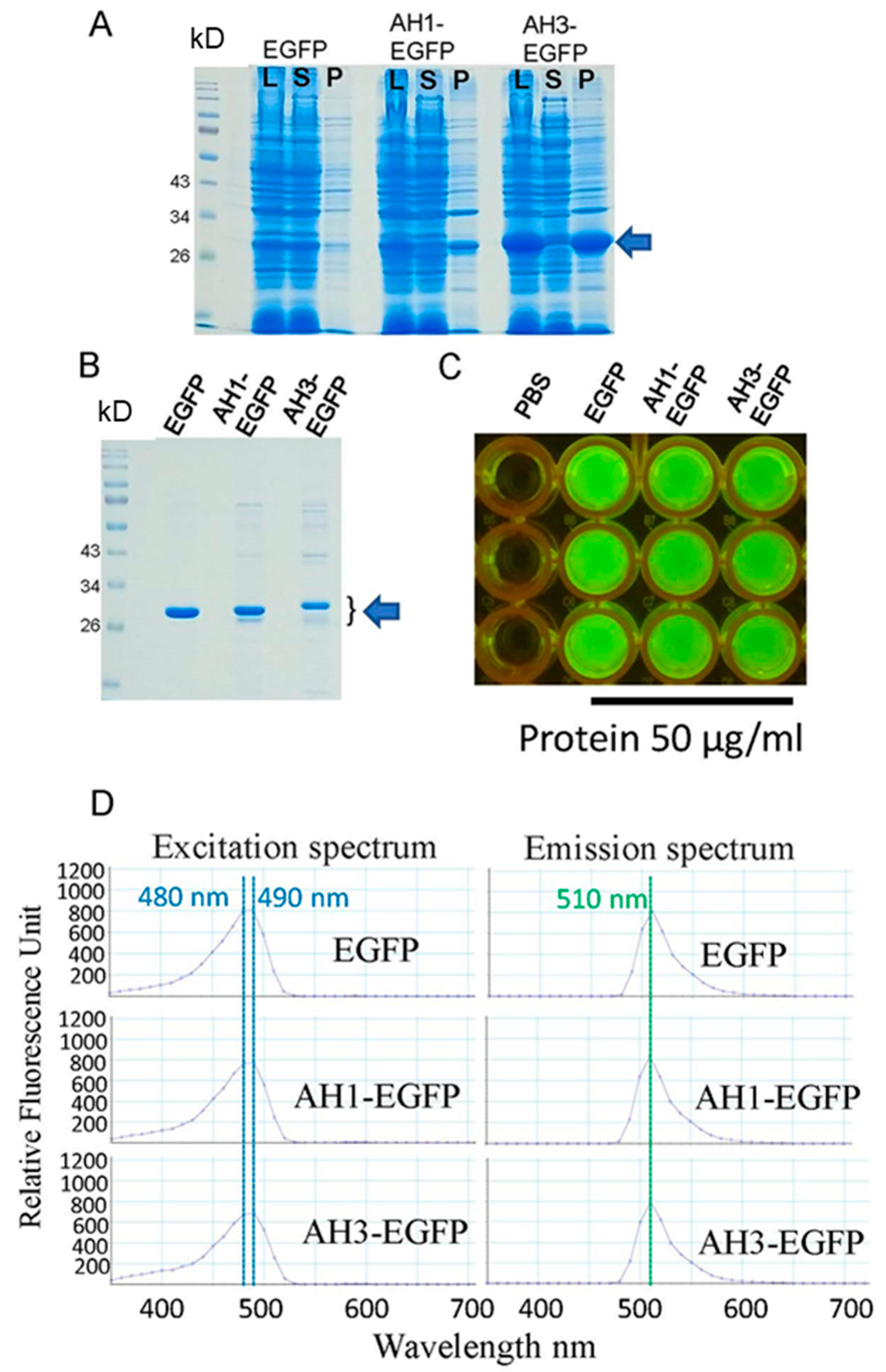
3.1. The Addition of AH1 or AH3 Peptide to the N-Terminal of EGFP Changed the Solubilities of Fusion Proteins but Did Not Affect Their Green Fluorescence

Using pET28a(+) that introduces six histidines (His tag) to the N-terminal of recombinant proteins, EGFP, AH1-EGFP, and AH3-EGFP were expressed, extracted, and purified. Adding AH1 or AH3 to the N-terminal of EGFP altered the solubility of the fusion proteins. Unlike EGFP, which was primarily found in the supernatant after cell lysis and centrifugation, higher amounts of AH1-EGFP and AH3-EGFP were found to be insoluble, staying in the pellet (Figure 2A). Interestingly, AH3 promoted the total fusion protein expression. Still, most AH3-EGFP was insoluble (Figure 2A). Low solubilities of AH1-EGFP and AH3-EGFP also reduced the final production of soluble fusion proteins. For the total 4 mL elution, the concentration is 6.62 mg/mL for EGFP, 3.82 mg/mL for AH1-EGFP, and 1.92 mg/mL for AH3-EGFP. The size of each purified protein is close to its predicted molecular weight (Figure 2B). To investigate the influence of AH1 or AH3 addition on EGFP structure or folding that may then affect the green fluorescence of EGFP, LED light at 450 nm and SpectraMax M5 were used to examine the fluorescence of EGFP, AH1-EGFP, and AH3-EGFP. All three proteins were found to be brightly fluorescent (Figure 2C) and had highly similar excitation and emission spectra with the same excitation peak at 480–490 nm and emission peak at 510 nm (Figure 2D). Additionally, the two-time serial dilution of each protein to the concentrations of 25 and 12.5 μg/mL also revealed a similar excitation peak at 480–490 nm and a similar emission peak at 510 nm, except for the reduction in the amplitude of the peaks.

3.2. AH1 and AH3 Peptides Enhance the Binding of Their Fusion EGFPs to HEK 293 Cells

Amphipathic alpha-helical peptides form hydrophilic and hydrophobic faces, which are often involved in phospholipid membrane interaction [25,26,27]. The AH3 peptide was also taken from the Influenza A H5N1 M2 protein, a member of integral membrane proteins embedded in the membrane bilayer [28]. The above information indicates that AH1-EGFP and AH3-EGFP could bind to cells effectively. Soluble EGFP, AH1-EGFP, and AH3-EGFP were diluted to 0.3 mg/mL in a culture medium and incubated with HEK 293 cells. The results presented a higher fluorescent intensity noticed in the HEK 293 cells incubated with AH3-EGFP (Figure 3A). Using ImageJ2 (https://imagej.nih.gov/ij/, accessed on 22 November 2021), the measurement of 5 fluorescence images taken from each incubation, AH3-EGFP incubation showed significantly higher fluorescence intensity than EGFP and AH1-EGFP incubation (62% and 36% more, respectively; raw data in Supplementary Table S1). AH1-EGFP incubation also has a higher fluorescence intensity than EGFP incubation (Figure 3B). Notably, larger fluorescent aggregates were found in AH3-EGFP incubation (Figure 3(A5,A6)).

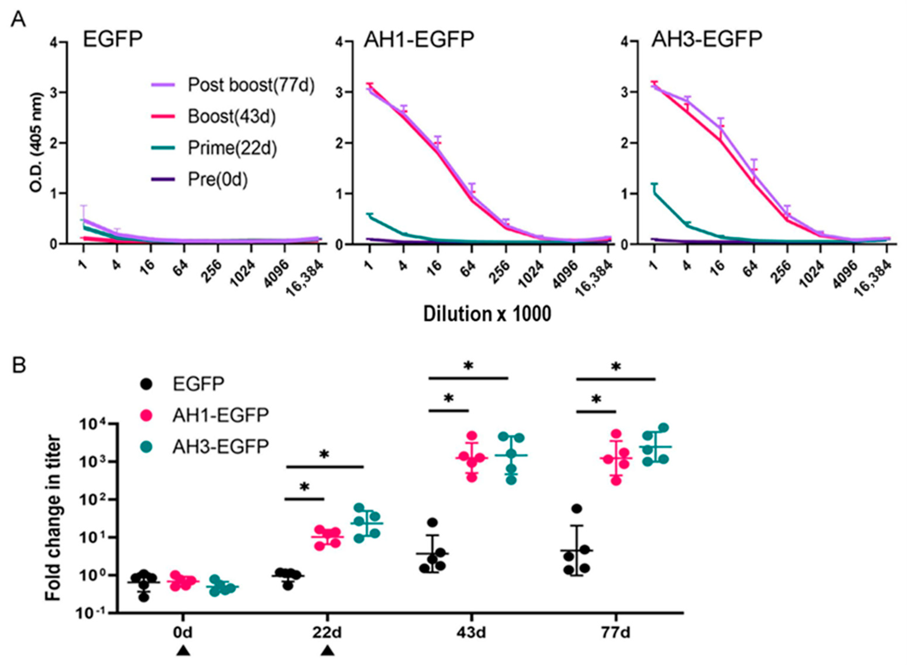
3.3. AH1 or AH3 Peptide Enhances the Immunogenicity of EGFP

Purified EGFP, AH1-EGFP, or AH3-EGFP was used to immunize rainbow trout by intraperitoneal injection with 1 µg of protein/g of body weight on days 0 and 22 to study the influence of AH1 or AH3 on the immunogenicity of EGFP. Plasma collected on days 0, 22, 43, and 77 were investigated by ELISA to evaluate the anti-EGFP antibody titers. Our results (Figure 4A) revealed that EGFP is a poor antigen and only gained high immunogenicity after fusing with AH1 or AH3 peptide. Before immunization, antibody titers against EGFP were low at relative levels, much less than one optical density (O.D.) in all three groups. However, in prime (day 22), boost (day 43), and post-boost (day 77) plasma, antibody titers against EGFP were elevated in the AH1-EGFP and AH3-EGFP immunized groups compared to the EGFP immunized group. One of the high-titer anti-EGFP plasmas was used as a standard (sequentially titrated) for calculating the relative levels of anti-EGFP antibody titers among the three groups. The results revealed that the fusion of AH1 or AH3 to the N-terminal of EGFP significantly induced higher anti-EGFP antibody levels to 10–20-fold on day 22 (prime plasma) and 300–500-fold on days 43 and 77 (boost and post-boost plasma) compared with the EGFP group (Figure 4B). No significant differences in anti-EGFP antibody titers were found between AH1-EGFP and AH3-EGFP among prime, boost, or post-boost plasma (Figure 4B). In Atlantic salmon, we investigated another amphipathic alpha-helical peptide derived from G-protein coupled receptor protein kinase 5 (GRK5), GRK5-EGFP, together with AH1-EGFP and EGFP. The results revealed that GRK5 was much less effective than AH1 in inducing anti-EGFP antibody titers in Atlantic salmon. We also found that one immunization of AH1-EGFP could significantly induce a much higher (1000-fold) anti-EGFP antibody titer than EGFP immunization when the prime plasma was collected much later, 50 days after the first immunization (Figure 5).

4. Discussion

Effective recombinant subunit vaccines can provide more economically and environmentally sustainable strategies that avert the risk of growing large amounts of pathogens in inactivated vaccine methods and possibly introducing infectious pathogens into the aquatic ecosystem in attenuated vaccine approaches. We identified two amphipathic alpha-helical peptides, AH1 and AH3, effectively stimulating a high immune response to their fusion EGFP in rainbow trout. Thus, these two peptides are valuable candidates for fostering recombinant subunit vaccine development in fish.
GFP variants fold properly and are brightly fluorescent when expressed alone but often misfold and lose fluorescence when expressed as fusions with other proteins [29,30,31]. The fusion of a puromycin-resistant gene or a neomycin-resistance gene directly to the N-terminal of EGFP caused the loss of the green fluorescence of EGFP (unpublished results). Yet, this characteristic of GFP variants makes them helpful for searching peptides that can boost the immunogenicity of recombinant subunit vaccine candidates without affecting their structures. Candidate peptides that cause the loss of GFP fluorescence may also alter the folding of their recombinant vaccine counterparts after fusion, resulting in the ineffectiveness of vaccines. Therefore, GFP variants serve as a practical tool for immunogenic peptide screening by monitoring the changes in GFP fluorescence after fusing candidate peptides. Unlike other fusion proteins that affected the fluorescence of GFPs [32], AH1 and AH3 did not influence EGFP fluorescence. Both the excitation and emission spectra of AH1-EGFP and AH3-EGFP are highly comparable to the excitation and emission spectra of EGFP. Thus, AH1 and AH3 can enhance the immunogenicity of vaccine candidates without affecting their structures, which is a crucial requirement of recombinant subunit vaccines mimicking the antigen presentation of pathogens. Although GRK5-EGFP is also brightly fluorescent (Figure 5), unlike AH1-EGFP, it did not induce a higher immune response in Atlantic salmon. Since GRK5-GFP could induce higher antibody titers in mice [24], we do not have a clear explanation for its ineffectiveness in the Atlantic salmon immune response. The validation of each potential amphipathic alpha-helical peptide in each targeted species should be incorporated into fish vaccine development. Our approach provides a promising tool for initially screening immunogenicity-enhancing peptides or proteins from a large pool of candidates. The promising candidates can be further assessed for immunogenicity enhancement using the target vaccine subunit.
Amphipathic alpha-helical peptides are often identified in proteins related to phospholipid membrane interaction, leading to cell uptake through endocytic mechanisms [33] or non-endocytic mechanisms [34,35]. In addition, several antimicrobial peptides also possess amphipathic properties and function by forming membrane pores or causing membrane disruption [36]. However, the membrane-disrupting nature of amphipathic alpha-helical peptides may have more or less toxic side effects, causing hemolysis and inducing various types of inflammation [37]. To secure clinical applications, it is necessary to develop a rigorous evaluation process to assess the effectiveness and safety of these immunogenicity-enhancing candidates. We also found that AH1-EGFP and AH3-EGFP can bind to HEK 293 cells more effectively than EGFP. The addition of AH1 or AH3 to EGFP may lead to the better interaction of AH1-EGFP and AH3-EGFP with antigen-presenting cells like dendritic cells [38] and macrophages [39], resulting in a higher immunogenicity induction. More studies examining how AH1 and AH3 peptides influence the immunogenicity of other proteins will help solidify our findings.
One noticeable difference among EGFP, AH1-EGFP, and AH3-EGFP is that both AH1 and AH3 caused a reduction in the solubility of their fusion EGFPs. We also found that the His-tag of soluble AH3-EGFP has a much lower binding affinity to Ni-NTA resin. Even after the salt reduction and several reapplications of the flow-through back to the Ni-NTA column, a large amount of soluble AH3-EGFP stayed in the flow-through, different from the His-tagged EGFP that binds to Ni-NTA resin effectively. This impediment also contributed to the low yield of soluble AH3-EGFP production. As AH1 and AH3 also increased the impurity of Ni-NTA purified fusion proteins, additional methods such as reversed-phase HPLC that can improve protein purity will help understand whether impurity plays the role of an adjuvant in immunogenicity enhancement or not. Since AH3 and His tag are close to each other at the N-terminal of the fusion protein, and AH3 also plays a central role in promoting nanoparticle formation [36], moving His tag to the C-terminal of EGFP to keep these two elements separated from each other may improve the production of AH3-EGFP. However, further investigation into the solubility and antigenicity change after this rearrangement is needed. The modification of AH3 has also been undertaken to make it more effective in inducing its fusion protein immune responses. The results obtained from mouse studies revealed that the two-point mutations of AH3 in I8L and K13E stimulated long-lasting immune responses in a single immunization [23]. A further investigation into the AH3 variants in fish vaccine development is also needed. As the modified AH3-superfolder (sf) GFP platform has been demonstrated to be effective in enhancing M2e peptide immunogenicity of human Influenza A viruses [23], the AH3-sfGFP platform is likely to be helpful for the development of fish viral vaccines.

Supplementary Materials

The following supporting information can be downloaded at: https://www.mdpi.com/article/10.3390/jmse13081497/s1, Table S1: Raw data for Figure 3B.

Author Contributions

Conceptualization, T.-T.W. and K.C.P.; methodology, T.-T.W. and K.C.P.; validation, K.C.P. and T.-T.W.; formal analysis, K.C.P. and T.-T.W.; investigation, K.C.P. and T.-T.W.; resources, T.-T.W. data curation, T.-T.W.; writing—original draft preparation, T.-T.W.; writing—review and editing, T.-T.W. and K.C.P.; supervision, T.-T.W.; project administration, T.-T.W.; funding acquisition, T.-T.W. All authors have read and agreed to the published version of the manuscript.

Funding

This research was funded by the University of Maryland, Baltimore County STRT7TEN-1113 to T.-T.W.

Institutional Review Board Statement

All of the experimental protocols involved in this study were approved by the Institutional Animal Care and Use Committee at the University of Maryland School of Medicine, IACUC #0521013 (approved on 2 July 2021) and #00001072 (approved on 17 May 2024).

Data Availability Statement

All the data were presented in this manuscript.

Acknowledgments

The authors acknowledge Ming-Chung Kan from Vaxsia Biomedical Inc., Taipei, Taiwan, who gave us the expression constructs used in this research. We also thank the USDA/ARS National Center for Cool and Cold Water Aquaculture and National Cold Water Marine Aquaculture Center for providing us with the experimental fish, and the staff of the IMET Aquaculture Research Center for maintaining the fish.

Conflicts of Interest

The authors declare no conflicts of interest.

References

  1. Naylor, R.L.; Hardy, R.W.; Buschmann, A.H.; Bush, S.R.; Cao, L.; Klinger, D.H.; Little, D.C.; Lubchenco, J.; Shumway, S.E.; Troell, M. A 20-Year Retrospective Review of Global Aquaculture. Nature 2021, 591, 551–563. [Google Scholar] [CrossRef]
  2. Føre, M.; Frank, K.; Norton, T.; Svendsen, E.; Alfredsen, J.A.; Dempster, T.; Eguiraun, H.; Watson, W.; Stahl, A.; Sunde, L.M.; et al. Precision Fish Farming: A New Framework to Improve Production in Aquaculture. Biosyst. Eng. 2018, 173, 176–193. [Google Scholar] [CrossRef]
  3. Brudeseth, B.E.; Wiulsrød, R.; Fredriksen, B.N.; Lindmo, K.; Løkling, K.-E.; Bordevik, M.; Steine, N.; Klevan, A.; Gravningen, K. Status and Future Perspectives of Vaccines for Industrialised Finfish Farming. Fish. Shellfish. Immunol. 2013, 35, 1759–1768. [Google Scholar] [CrossRef] [PubMed]
  4. Horzinek, M.C.; Schijns, V.E.C.J.; Denis, M.; Desmettre, P.; Babiuk, L.A. General Description of Vaccines. In Veterinary Vaccinology; Pastoret, P.P., Blancou, J., Vannier, P., Verschueren, C., Eds.; Elsevier Press: Amsterdam, The Netherlands, 1997; pp. 132–152. [Google Scholar]
  5. Gudding, R.; Goodrich, T. The History of Fish Vaccination. Fish. Vaccin. 2014, 1, 1–11. [Google Scholar]
  6. Dhar, A.K.; Manna, S.K.; Thomas Allnutt, F.C. Viral Vaccines for Farmed Finfish. Virusdisease 2014, 25, 1–17. [Google Scholar] [CrossRef]
  7. Rathor, G.S.; Swain, B. Advancements in Fish Vaccination: Current Innovations and Future Horizons in Aquaculture Health Management. Appl. Sci. 2024, 14, 5672. [Google Scholar] [CrossRef]
  8. Du, Y.; Hu, X.; Miao, L.; Chen, J. Current status and development prospects of aquatic vaccines. Front. Immunol. 2022, 13, 1040336. [Google Scholar] [CrossRef]
  9. Kibenge, F.S.B.; Godoy, M.G.; Fast, M.; Workenhe, S.; Kibenge, M.J.T. Countermeasures Against Viral Diseases of Farmed Fish. Antivir. Res. 2012, 95, 257–281. [Google Scholar] [CrossRef]
  10. Valero, Y.; Cuesta, A. Reassortant viruses threatening fish aquaculture. Rev. Aquac. 2023, 15, 1720–1731. [Google Scholar] [CrossRef]
  11. Plant, K.P.; Lapatra, S.E. Advances in Fish Vaccine Delivery. Dev. Comp. Immunol. 2011, 35, 1256–1262. [Google Scholar] [CrossRef]
  12. Ebensen, T.; Link, C.; Guzmán, C.A. Classical and Novel Vaccination Strategies: A Comparison. In Novel Vaccination Strategies; Wiley-VCH: Weinheim, Germany, 2004. [Google Scholar]
  13. Goldman, M.; Lambert, P.H.; Kaufmann, S.H. Novel Vaccination Strategies; Blackwell Pub: New York, NY, USA, 2004. [Google Scholar]
  14. Munang’andu, H.M.; Mutoloki, S.; Evensen, Ø. An Overview of Challenges Limiting the Design of Protective Mucosal Vaccines for Finfish. Front. Immunol. 2015, 6, 542. [Google Scholar] [CrossRef]
  15. Loessner, H.; Schwantes, A.; Hamdorf, M.; Komor, U.; Leschner, S.; Weiss, S. Employing Live Microbes for Vaccine Delivery. In Development of Novel Vaccines: Skills, Knowledge and Translational Technologies; Springer: Berlin/Heidelberg, Germany, 2012; pp. 87–124. [Google Scholar] [CrossRef]
  16. Clark, T.G.; Cassidy-Hanley, D. Recombinant Subunit Vaccines: Potentials and Constraints. Dev. Biol. 2005, 121, 153–163. [Google Scholar]
  17. Pardoll, D.M. Spinning Molecular Immunology into Successful Immunotherapy. Nat. Rev. Immunol. 2002, 2, 227–238. [Google Scholar] [CrossRef]
  18. Di Pasquale, A.; Preiss, S.; Tavares Da Silva, F.; Garçon, N. Vaccine Adjuvants: From 1920 to 2015 and Beyond. Vaccines 2015, 3, 320–343. [Google Scholar] [CrossRef]
  19. Tammas, I.; Bitchava, K.; Gelasakis, A.I. Advances in Vaccine Adjuvants for Teleost Fish: Implications for Aquatic Welfare and the Potential of Nanoparticle-Based Formulations. Vaccines 2024, 12, 1347. [Google Scholar] [CrossRef] [PubMed]
  20. Schödel, F.; Peterson, D.; Hughes, J.; Wirtz, R.; Milich, D. Hybrid Hepatitis B Virus Core Antigen as a Vaccine Carrier Moiety: I. Presentation of Foreign Epitopes. J. Biotechnol. 1996, 44, 91–96. [Google Scholar] [CrossRef]
  21. Chenal, A.; Ladant, D. Bioengineering of Bordetella Pertussis Adenylate Cyclase Toxin for Antigen-Delivery and Immunotherapy. Toxins 2018, 10, 302. [Google Scholar] [CrossRef] [PubMed]
  22. Giménez-Andrés, M.; Čopič, A.; Antonny, B. The Many Faces of Amphipathic Helices. Biomolecules 2018, 8, 45. [Google Scholar] [CrossRef] [PubMed]
  23. Wong, T.-T.; Liou, G.-G.; Kan, M.-C. A Thermal-Stable Protein Nanoparticle that Stimulates Long Lasting Humoral Immune Response. Vaccines 2023, 11, 426. [Google Scholar] [CrossRef] [PubMed]
  24. Lyu, J.-H.; Liou, G.-G.; Wang, M.; Kan, M.-C.M. The Adjuvant Effects of Amphipathic Helical Peptides. bioRxiv 2023. [Google Scholar] [CrossRef]
  25. He, Y.; Weng, L.; Li, R.; Li, L.; Toyoda, T.; Zhong, J. The N-Terminal Helix α(0) of Hepatitis C Virus NS3 Protein Dictates the Subcellular Localization and Stability of NS3/NS4A Complex. Virology 2012, 422, 214–223. [Google Scholar] [CrossRef] [PubMed]
  26. Horner, S.M.; Park, H.S.; Gale, M., Jr. Control of Innate Immune Signaling and Membrane Targeting by the Hepatitis C Virus NS3/4A Protease Are Governed by the NS3 Helix A0. J. Virol. 2012, 86, 3112–3120. [Google Scholar] [CrossRef] [PubMed]
  27. Rossman, J.S.; Jing, X.; Leser, G.P.; Lamb, R.A. Influenza Virus M2 Protein Mediates ESCRT-Independent Membrane Scission. Cell 2010, 142, 902–913. [Google Scholar] [CrossRef] [PubMed]
  28. Roberts, K.L.; Leser, G.P.; Ma, C.; Lamb, R.A. The Amphipathic Helix of Influenza A Virus M2 Protein Is Required for Filamentous Bud Formation and Scission of Filamentous and Spherical Particles. J. Virol. 2013, 87, 9973–9982. [Google Scholar] [CrossRef]
  29. Waldo, G.S.; Standish, B.M.; Berendzen, J.; Terwilliger, T.C. Rapid Protein-Folding Assay Using Green Fluorescent Protein. Nat. Biotechnol. 1999, 17, 691–695. [Google Scholar] [CrossRef]
  30. Waldo, G.S. Genetic Screens and Directed Evolution for Protein Solubility. Curr. Opin. Chem. Biol. 2003, 7, 33–38. [Google Scholar] [CrossRef]
  31. Pédelacq, J.-D.; Piltch, E.; Liong, E.C.; Berendzen, J.; Kim, C.-Y.; Rho, B.-S.; Park, M.S.; Terwilliger, T.C.; Waldo, G.S. Engineering Soluble Proteins for Structural Genomics. Nat. Biotechnol. 2002, 20, 927–932. [Google Scholar] [CrossRef]
  32. Pédelacq, J.-D.; Cabantous, S.; Tran, T.; Terwilliger, T.C.; Waldo, G.S. Engineering and Characterization of a Superfolder Green Fluorescent Protein. Nat. Biotechnol. 2006, 24, 79–88. [Google Scholar] [CrossRef]
  33. Vlassov, V.V.; Balakireva, L.A.; Yakubov, L.A. Transport of Oligonucleotides Across Natural and Model Membranes. Biochim. Biophys. Acta 1994, 1197, 95–108. [Google Scholar] [CrossRef]
  34. Derossi, D.; Joliot, A.H.; Chassaing, G.; Prochiantz, A. The Third Helix of the Antennapedia Homeodomain Translocates Through Biological Membranes. J. Biol. Chem. 1994, 269, 10444–10450. [Google Scholar] [CrossRef]
  35. Fawell, S.; Seery, J.; Daikh, Y.; Moore, C.; Chen, L.L.; Pepinsky, B.; Barsoum, J. Tat-Mediated Delivery of Heterologous Proteins into Cells. Proc. Natl. Acad. Sci. USA 1994, 91, 664–668. [Google Scholar] [CrossRef]
  36. Tossi, A.; Sandri, L.; Giangaspero, A. Amphipathic, α-Helical Antimicrobial Peptides. Pept. Sci. 2000, 55, 4–30. [Google Scholar] [CrossRef]
  37. Duarte-Mata, D.I.; Salinas-Carmona, M.C. Antimicrobial peptides’ immune modulation role in intracellular bacterial infection. Front. Immunol. 2023, 14, 1119574. [Google Scholar] [CrossRef]
  38. Pavli, P.; Hume, D.A.; Van De Pol, E.; Doe, W.F. Dendritic Cells, the Major Antigen-Presenting Cells of the Human Colonic Lamina Propria. Immunology 1993, 78, 132–141. [Google Scholar]
  39. Unanue, E.R. Antigen-Presenting Function of the Macrophage. Annu. Rev. Immunol. 1984, 2, 395–428. [Google Scholar] [CrossRef]
Figure 1. Diagram of the constructs used to produce EGFP, AH1-EGFP, and AH3-EGFPs. In AH1-EGFP and AH3-EGFP constructs, the T7 tag was replaced with amphipathic alpha-helical peptide AH1 or AH3.
Jmse 13 01497 g001
Figure 2. Production and characterization of EGFP, AH1-EGFP, and AH3-EGFPs. (A) Total cell lysate (L) before centrifugation and supernatant (S) and pellet (P) after centrifugation were analyzed by SDS-PAGE using 12% gel and Coomassie blue staining. The addition of AH1 or AH3 to the N-terminal of EGFP decreased the solubility of their fusion proteins. AH3 enhances the total fusion protein expression; however, most AH3-EGFP was insoluble, staying in the pellet. (B) The soluble recombinant proteins were purified using Ni-NTA agarose and analyzed by 12% SDS-PAGE gel and Coomassie blue staining. A total of 1 μL of each purified protein (EGFP, 6.62 mg/mL; AH1-EGFP, 3.82 mg/mL; and AH3-EGFP, 1.92 mg/mL) was used for the SDS-PAGE analysis. The arrows indicate the size of the candidate proteins. (C) Purified proteins were diluted to 50 µg/mL with PBS and examined under LED light at 450 nm. All three proteins appeared to be brightly green. (D) The fusion of AH1 or AH3 to EGFP does not affect EGFP fluorescence (indicating correct folding and structure). All three proteins have highly similar excitation and emission spectra with the same excitation peak at 480–490 nm and emission peak at 510 nm.
Jmse 13 01497 g002
Figure 3. AH1 and AH3 promote the cell binding of their fusion proteins. (A) Photomicrographs show that levels of protein binding (the fluorescent intensity) to HEK 293 cells were low in the EGFP incubation group, medium in the AH1-EGFP incubation group, and high in the AH3-EGFP incubation group ((A1,A3,A5): fluorescent photomicrographs, (A2,A4,A6): mergers of fluorescent photomicrographs with their bright-field photomicrographs). (B) Quantitative data derived from 5 photomicrographs of each protein incubation show that the binding of AH3-EGFP to HEK 293 cells was more than 60% of the binding of EGFP to HEK 293 cells. Data labeled with different letters (a, b, c) indicate a significant difference (p < 0.05) from each other by Tukey’s tests. Scale bar = 50 µm.
Jmse 13 01497 g003
Figure 4. Fusing an AH1 or AH3 peptide to EGFP significantly enhanced the immunogenicity of EGFP. (A) The geometric mean titer and standard deviation of anti-EGFP antibody obtained from rainbow trout immunized with EGFP, AH1-EGFP, or AH3-EGFP during the experimental period. Plasma was collected on days 0 (pre), 22 (prime), 43 (boost), and 77 (post-boost) and investigated by ELISA. Antibody titers against EGFP were significantly elevated in the AH1-EGFP and AH3-EGFP immunized groups compared to the EGFP immunized group. (B) Relative levels of anti-EGFP antibody titers were calculated using the reference standards derived from one of the high titers of anti-EGFP plasma. AH1-EGFP or AH3-EGFP significantly induced higher anti-EGFP antibody levels by 10–20-fold on day 22 (prime) and 300–500-fold on days 43 and 77 (boost and post-boost) compared with the EGFP immunized group. * indicates a significant difference (p < 0.05) between the two groups by Mann–Whitney U tests.
Jmse 13 01497 g004
Figure 5. AH1, but not GRK5, significantly enhanced the immunogenicity of EGFP in Atlantic Salmon. (A) Diagram of the construct used to produce GRK5-EGFP. (B) Purified GRK5-EGFP was diluted to 50 µg/mL with PBS and examined under LED light at 450 nm. GRK5-EGFP appeared to be bright green. (C) The geometric mean titer and standard deviation of anti-EGFP antibody obtained from Atlantic salmon immunized with EGFP, AH1-EGFP, or GRK5-EGFP on days 0 and 50 (indicated by triangles). Plasma was collected on days 50 (prime) and 92 (boost) and investigated by ELISA. Relative levels of anti-EGFP antibody titer were calculated using the standards derived from one of the high titers of anti-EGFP plasma. AH1-EGFP immunization significantly induced higher anti-EGFP antibody levels by more than 1000-fold on day 50 (prime) than the EGFP immunization. GRK5-EGFP immunization was less effective compared to AH1-EGFP immunization. * indicates a significant (p < 0.05) difference between the two groups by Mann–Whitney U tests.
Jmse 13 01497 g005
Disclaimer/Publisher’s Note: The statements, opinions and data contained in all publications are solely those of the individual author(s) and contributor(s) and not of MDPI and/or the editor(s). MDPI and/or the editor(s) disclaim responsibility for any injury to people or property resulting from any ideas, methods, instructions or products referred to in the content.

