Article

Ontogenetic Growth Changes in Mercury and Stable Isotope Ratios of Carbon, Nitrogen, and Oxygen in Male and Female Dalli-Type Dall’s Porpoises (Phocoenoides dalli) Stranded in Hokkaido, Japan

1
School of Pharmaceutical Sciences, Health Sciences University of Hokkaido, Ishikari-Tobetsu 061-0293, Hokkaido, Japan
2
Kagawa School of Pharmaceutical Sciences, Tokushima Bunri University, Takamatsu-shi 760-8542, Kagawa, Japan
3
Department of Pharmaceutical Sciences, Daiichi University of Pharmacy, Minami-ku, Fukuoka 815-8511, Fukuoka, Japan
*
Author to whom correspondence should be addressed.
J. Mar. Sci. Eng. 2025, 13(5), 892; https://doi.org/10.3390/jmse13050892
Submission received: 31 January 2025 / Revised: 22 April 2025 / Accepted: 25 April 2025 / Published: 30 April 2025
(This article belongs to the Section Marine Aquaculture)

Abstract

:
We investigated the ontogenetic growth changes in total mercury (THg) concentrations, δ13C, δ15N, and δ18O values, and body length (BL) of dalli-type Dall’s porpoises. THg concentrations in the liver of mature porpoises stranded in Hokkaido, Japan, were markedly higher than those in the muscle. The THg concentrations in the livers of males and females increased sharply when their BLs exceeded approximately 1.9 m and 1.8 m, respectively, the BLs at which they might attain maturity. The asymptotes of the THg increases were close to their maximum BLs of 2.2 m and 2.0 m for males and females, respectively. The δ15N levels in muscles were higher in the calves than in the weaned porpoises, probably due to the consumption of 15N-enriched milk, whereas the δ13C values in the calves were variable and similar to those in the weaned porpoises. The δ18O values of male and female muscles increased with increasing BL. Positive correlations were found between the THg concentrations and either the δ13C values or the δ18O values in the weaned animals, but not with the δ15N values. These results imply a feeding shift towards deeper pelagic areas with growth, as the δ13C and δ18O values and the THg concentrations tend to be higher in these areas.

1. Introduction

Mercury (Hg) is a global pollutant emitted to the atmosphere from both natural and anthropogenic sources [1]. Top-level and long-lived predators such as odontocetes (toothed whales, dolphins, and porpoises), swordfish, sharks, and tuna are known to contain high levels of methylmercury (MeHg) as the Hg contamination in the predators tend to increase with an increase in trophic levels inferred from δ15N values [2,3,4,5]. Total mercury (THg) is generally accumulated in these predators in an age- and/or body-length (BL)-dependent manner, particularly in the liver of odontocetes [6,7,8] and in the liver and/or muscle of sharks [9,10,11]. Formation of mercuric selenide (HgSe) after demethylation of MeHg is thought to be the detoxification mechanism for MeHg, and the cause of the preferential accumulation of THg in the liver tissues [12,13,14]. The Hg poisoning has been suspected in some marine mammals, as high levels of THg have been found in them [15]. There is great concern about MeHg-induced adverse effects on fetal development and neurotoxicity due to the consumption of seafood [16,17], as MeHg readily crosses the placental and blood–brain barriers [18].
Dall’s porpoise (Phocoenoides dalli) is one of the small odontocetes living in the pelagic areas of the North Pacific Ocean and adjacent seas [19,20]. According to FRA [21], Dall’s porpoises are born at 0.8–1.0 m BL, grow to a BL of approximately 1.6 m in the first year, and weaning begins at 1–2 months and is completed within the first year. Males and females attain sexual maturity when they attain the BLs of around 1.9 m and 1.8 m, respectively, and the maximum BLs are approximately 2.2 m for males and 2.0 m for females [20,21].
Dall’s porpoises comprise two major distinct color morphs—dalli- and truei-types. Two populations of dalli-type off Japan are reported—one is the Sea of Japan/the Sea of Okhotsk population, which migrates from the southern Sea of Japan to the southern Sea of Okhotsk, and the other migrates in the North Pacific Ocean off Japan [19]. The truei-type also lives in the North Pacific Ocean off Japan, but they migrate further inland than the dalli-type (Figure 1).
Several cetacean species inhabiting the waters around Japan are the target of commercial whaling, where their products are sold for human consumption in Japan [22,23]. Dall’s porpoises are the most abundantly hunted odontocete species; quotas set by the Japanese government for 2023 to hunt dalli- and truei-types were 4137 and 4398 porpoises, respectively [24].
Red meat (muscle) products from several odontocete species sold in Japan were reported to contain high concentrations of THg, ranging from 0.52 to 81.0 μg/wet g [22]. All THg concentrations reported in the red meat products exceeded the provisional permitted level in fish and shellfish set by the Japanese government (0.4 μg/wet g); however, this restriction does not cover cetacean products. The contamination level of THg in the red meat products from Dall’s porpoises (1.23 ± 0.49 μg/wet g) was moderate among the products from several odontocete species [23]. Considering the neurotoxicity of MeHg to fetuses, the Japanese government advises pregnant women to limit the consumption of red meat from Dall’s porpoise to no more than 160 g per week [25]. However, there have been no detailed studies of THg contamination in Dall’s porpoises focusing on growth (BL) and sex to date, although a few studies have reported the THg level in truei-type mature porpoises [26,27].
Stable isotopes such as 13C, 15N, and 18O are naturally occurring biomarkers and useful tools for providing information on the feeding ecology, biology, and movement (migration) of marine animals. The δ15N values are mainly used as a proxy of trophic level [28,29]: positive correlations were reported between the δ15N values and THg concentrations or log THg concentrations in muscle or liver tissues [2,3,4,5,9]. On the other hand, the δ13C values are generally used to infer the habitat and feeding areas of marine animals because the δ13C values tend to be higher in the inshore zone than in the offshore zone and in the benthic zone than in the pelagic zone [30,31,32]. Recently, the combined analysis of δ13C and δ18O in the teeth, bones, and otoliths of marine animals has been widely used as a powerful proxy for vertical and horizontal spatial differences. The δ13C and δ18O values in those increase with increasing depth (decreasing temperature) [33,34,35,36]. Unlike the case of δ13C values, the δ18O values are lower in the inshore zone than in the offshore zone [37,38,39]. In addition to the trophic level, age, and BL, the vertical and horizontal factors could affect the THg accumulation in marine animals [40]. For instance, a positive correlation between the THg concentrations and either the δ13C values or the δ18O values was reported in marine mammals [41,42].
Several studies reported higher δ15N levels in calves than in mature animals due to the consumption of 15N-enriched milk from their mothers [28,29,43,44,45,46]. Previous studies found 15N-enrichment peaks during the lactation period in muscle samples from common minke whales (Balaenoptera acutorostrata) [47], humpback whales (Megaptera novaeangliae) [48], and killer whales (Orcinus orca) [49]. On the other hand, lactation-induced δ13C changes in calf muscle are generally variable with no particular trend because of the variability of 13C-depleted lipids in milk [43,46,50]. Decreases in δ18O values in muscle samples were reported during the lactation period of humpback whales [48] and killer and common minke whales [49]. The cause of decreases in δ18O values is likely to be the lower levels of 18O in milk than in weaning diets [49].
Liver tissue is more metabolically active than muscle tissue, and the δ15N levels are generally higher in the liver than in the muscle of mature animals [51,52,53,54,55], possibly due to the faster isotopic turnover rate of the liver. However, few studies have compared the δ15N values in calf liver with those in calf muscle. Furthermore, there are no studies that have investigated the ontogenetic growth changes (during and after lactation) of δ13C and δ15N values in the liver tissue of marine mammals, except for our previous study in killer whales (Orcinus orca) [49].
Our preliminary study reported ontogenetic growth changes in THg concentrations, and δ13C and δ15N values in muscle samples from dalli-type Dall’s porpoises, combining the samples from stranded porpoises along the coast of Hokkaido and incidental catches in the North Pacific [56]. However, the number of samples in the previous study was limited, and the ontogenetic growth changes in THg concentrations, and δ13C and δ15N values in organs other than muscle are unknown. Further detailed studies separating male and female porpoises are needed.
In the present study, we analyzed the THg concentrations, δ13C, and δ15N values in muscle and liver tissues during and after the lactation of Dall’s porpoises. We also analyzed the δ18O values in the muscle tissues of some of these porpoises. Based on these results, we estimated the BL at the onset of weaning using the δ15N values in muscle tissues and the BL with THg concentrations above 0.4 μg/wet g in muscle tissues and studied the ontogenetic growth change of δ13C and δ15N values in muscle and liver tissues and Δ13Cliver-muscle and Δ15Nliver-muscle values. We also investigated the ontogenetic growth change of δ18O values in the muscle tissues and investigated the relationship between the THg concentrations and either δ13C, δ15N, or δ18O values in the weaned animals (BL > 1.5 m).

2. Materials and Methods

2.1. Samples of Dalli-Type and Truei-Type Dall’s Porpoises

All samples from dalli-type Dall’s porpoises stranded along the coast of the Sea of Japan, the Sea of Okhotsk, and the Nemuro Strait in Hokkaido between 2010 and 2019 (see Figure 1) were provided by the Stranding Network of Hokkaido (SNH) with information on the stranded location, sex, and BL. Muscle samples from all individuals were analyzed for THg concentrations, and δ13C and δ15N values. In some individuals, the THg concentrations in kidney, lung, spleen, heart, and/or blood samples and δ18O values in the muscle samples were analyzed. Details of the samples provided from SNH are summarized in Supplementary Tables S1 and S2. Based on BL from FRA [21] and the present results, we classified the porpoises less than 1.5 m BL as calves and those larger than 1.5 m BL as weaned animals (immature and mature animals).
In addition, the muscle and liver samples from truei-type mature Dall’s porpoises, comprising two males and three females, were obtained directly from the fishermen who had hunted them off the coast of Miyagi prefecture in the North Pacific Ocean in 2002 (Figure 1). Although the exact BL of the animals was unknown (estimated to be approximately 175–190 m), their body weights after removing internal organs (carcass) were 135–163 kg. The muscle and liver samples were submitted to analyze the THg concentrations and the δ13C and δ15N values.
All samples from dalli-type and truei-type Dall’s porpoises were stored at −20 °C prior to analysis.

2.2. Chemical Analyses

THg concentrations in the muscle and other tissue samples were analyzed using a flameless atomic absorption spectrophotometer (HG-310; Hiranuma Sangyo Co., Ltd., Ibaraki, Japan). As previously reported [47], approximately 0.5 g of the sample was digested in a mixture of HNO3, H2SO4, and HClO4. DOLT-2 (National Research Council of Canada) was used as the analytical quality control for THg, where the recovery of THg was 94 ± 3% (n = 5). The THg concentrations were expressed on a wet-weight basis, and the determination limit of quantitation was approximately 0.01 µg Hg/wet g. The THg data in this study were the mean of at least two measurements of the same samples.
The δ13C, δ15N, and δ18O values were analyzed in the dried samples after removing lipids using chloroform/methanol solution extraction [57]. The extraction of lipids was repeated three or more times until the color of the extraction solvent ran clear.
The analyses of δ13C and δ15N in the muscle and liver samples were performed using an isotope ratio mass spectrometer (IRMS) (Delta S, Finnigan MAT, Bremen, Germany, and EA1108, Fisons, Roano, Milan, Italy), as described previously [49]. Histidine (M5P8062, −10.7‰), alanine (M6K9292, −19.6‰), and glycine (LTG6322, −33.8‰) were used as the working standards for δ13C, and three types of alanine (M6K9292, M6R397410 and M0H912820) at 1.58, 9.97, and 20.6‰, respectively, were used as the working standards for δ15N.
The δ18O was analyzed in the remaining frozen muscle samples after the analyses of THg concentration and δ13C and δ15N values using IRMS (Delta V PLUS, Thermo Fisher Scientific, Tokyo, Japan), as described previously [49,58]. Two types of benzoic acid (IAEA-601 and -602) were used as the working standards for δ18O at 23.1 and 71.3‰, respectively.
Isotopic ratios are reported in the standard delta (δ) notation relative to the internal standard of Vienna Pee Dee Belemnite (VPDB) (δ13C), atmospheric nitrogen (δ15N), and standard mean ocean water (δ18O).

2.3. Statistical Analyses

The THg data, shown in Figure 2, were fitted to the Bézier curve using KaleidaGraph (Hulinks Inc. version 4.1, Tokyo, Japan). The BL at the δ15N peak among the calf muscle samples (Figure 3) was estimated by the curve fitting to a quadratic function as previously reported [46,49,56], using JMP (SAS Institute Japan Ltd., version 14.3, Tokyo, Japan). The data were analyzed using Student’s t-test and Pearson’s correlation coefficient.

3. Results

3.1. General Information on the Male and Female Dalli-Type Dall’s Porpoises

The mean BL of male dalli-type Dall’s porpoises provided by SNH (1.91 ± 0.34 m was slightly larger than that of females (1.57 ± 0.32 m (Table 1), in agreement with the previous findings [20,21]. There were more male samples (n = 37) than female samples (n = 17), reflecting the more frequent stranding of males.

3.2. THg Distribution in the Male and Female Dalli-Type Dall’s Porpoises

The distribution of THg in the dalli-type Dall’s porpoises is summarized in Table 1. The THg concentrations in the liver samples from males and females were 10.3 ± 11.1 (n = 35) and 5.24 ± 8.78 (n = 17) μg/wet g, respectively, and those in muscle samples were 0.95 ± 0.62 (n = 37) and 0.71 ± 0.60 (n = 17) μg/wet g, respectively. The THg concentrations in the liver tissue from both males and females were one order of magnitude higher than those in the kidney, muscle, and other tissues. The THg concentrations in male tissues were in the following order (the order of females was similar).
Liver ≫ Kidney > Muscle ≈ Spleen ≈ Lung ≈ Heart > Blood
No significant sex differences were observed for the THg concentrations in liver, kidney, muscle, and other tissues due to the large variation in THg concentrations.
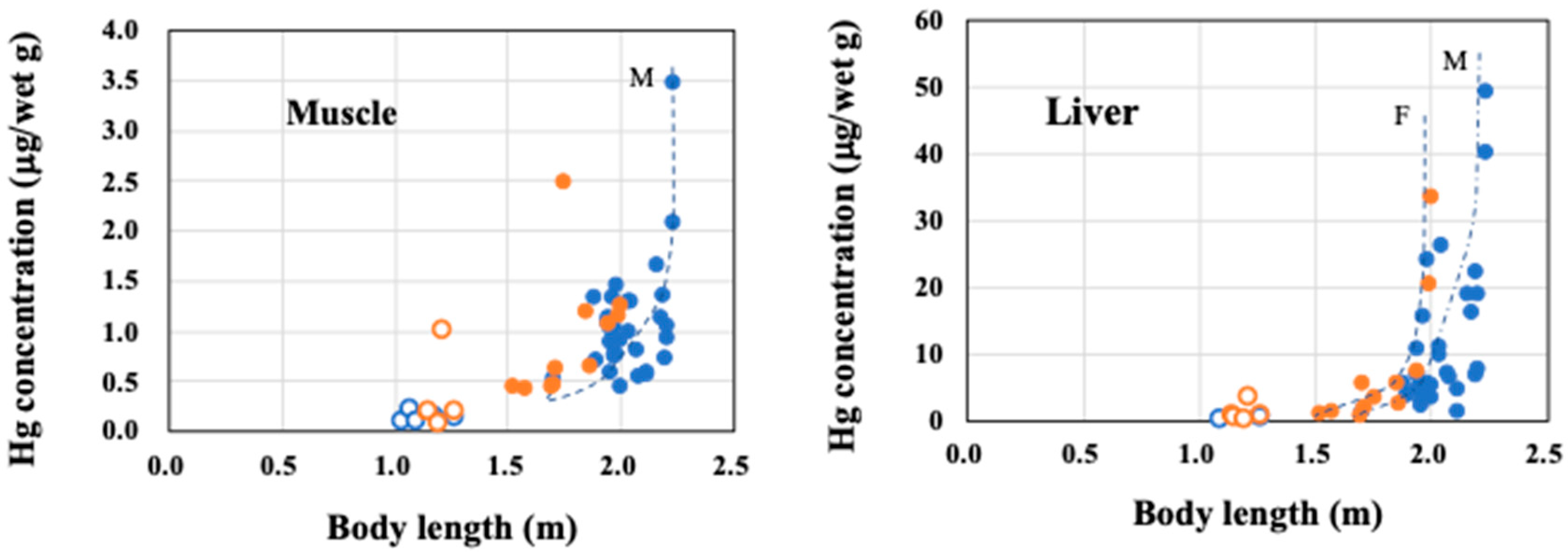
Ontogenetic growth changes in THg concentrations of the muscle and liver samples are presented in Figure 2. For BLs up to 1.5 m (calves), only a slight increase in THg concentrations was observed in the muscle and liver samples from both males and females, except for one calf at 1.2 m BL (SNH 12024-2). All the THg concentrations observed in the muscle and liver samples, excluding SNH 12024-2, were less than 0.4 and 1.0 μg/wet g, respectively. However, the THg concentrations in the muscle and liver samples of SNH 12024-2 were 1.01 μg/wet g and 3.49 μg/wet g, respectively. On the other hand, the THg concentrations in the combined muscle samples from the weaned males and females (<1.5 m BL) were 1.04 ± 0.58 μg/wet g (n = 43), and those in the combined liver samples were 10.3 ± 11.0 μg/wet g (n = 43).
The THg concentrations in the liver samples from both males and females increased sharply after reaching BLs of approximately 1.9 m and 1.8 m, respectively, and the asymptotes of the THg increase observed in males and females were approximately 2.2 m and 2.0 m, respectively (Figure 2). As in the case of the liver, a sharp but slight increase in THg concentrations was observed in male muscles, but such an increase was unclear in female muscles.

3.3. Ontogenetic Growth Changes of δ13C and δ15N Values in the Muscles and Livers of Males and Females

The δ15N values in the combined calf muscles from males and females (14.6 ± 0.6‰, n = 11) were significantly higher than those in the combined muscles of weaned animals (12.8 ± 0.5‰, n = 43) (p < 0.05), whereas the δ13C values in the combined calf muscles were similar to those in the combined muscles of weaned animals (−19.4 ± 0.6‰, n = 11 vs. −19.4 ± 0.4‰, n = 43) (Table 2).
Ontogenetic growth changes in the δ13C and δ15N values in the muscle and liver tissues of Dall’s porpoises are presented in Figure 3.
The calculated BL at the δ15N peak by the curve fitting was 1.15 m with the δ15N value of 14.8‰, although the fitting was not significant (p = 0.188). Meanwhile, the combined δ15N values of the calves (n = 11) and two small weaned animals at BLs of 1.52 m (SNH 1022-2) and 1.57 m (SNH 11013) were significantly fitted to a quadratic function (δ15N = −22.836 × BL2 + 55.171 × BL − 18.52, p < 0.01), resulting in the δ15N peak being at the BL of 1.22 m and the δ15N value of 14.8‰. The difference in δ15N value between the highest δ15N value (15.6‰) and the lowest δ15N value (13.6‰) among the calf samples was 2.0‰.
The δ13C and δ15N values in the muscle and liver samples from the weaned males and females varied widely and demonstrated no particular trend with growth (Figure 3). No significant sex differences were observed in the δ13C and δ15N values of muscle and liver samples from the calves and the weaned animals due to large variations.

3.4. Relationships Between the δ13C and δ15N Values for Males, Females, and Calves

The relationships between the δ13C and δ15N values in the calf samples (n = 11), the weaned male samples (n = 32), and the weaned female samples (n =11) are presented in Figure 4. A significant positive correlation between the δ13C and δ15N values was found in the calf samples, whereas no correlation was found in the weaned male samples and the weaned female samples.
No correlation (p > 0.10) was found between the δ13C and δ18O values and between the δ15N and δ18O values in the combined weaned animals (not shown in figure).

3.5. Comparison of δ13C and δ15N Values in Liver Samples with Those in Muscle Samples

The δ15N and δ13C values in the liver samples from the calves and the weaned animals were compared with the corresponding values in the muscle samples.
The δ15N level in calf liver samples was similar to that in calf muscle samples (14.6 ± 0.6‰, n = 4 vs. 14.6 ± 0.6‰, n =11) (Table 2). However, the δ15N level of the weaned animals was significantly higher in liver (13.9 ± 0.7‰, n = 29) than in muscle (12.8 ± 0.5‰, n = 43) (p < 0.05). On the other hand, no difference was observed in the δ13C levels between the muscle and the liver samples from the calves (−19.4 ± 0.6‰, n = 11 vs. −19.0 ± 0.5‰, n = 4) and from the weaned animals (−19.4 ± 0.4‰, n = 43 vs. −19.6 ± 0.4‰, n = 29).
The difference in δ15N values between the liver and muscle tissues (Δ15Nliver-muscle) and that in δ13C values between those tissues (Δ13Cliver-muscle) were studied (Figure 5).
The Δ15Nliver-muscle level in the weaned animals (1.07 ± 0.58, n =29) was significantly higher than that in the calves (0.11 ± 0.015, n = 4) (p < 0.05); the Δ15Nliver-muscle values and these variations tended to increase with increasing BL. On the other hand, the Δ13Cliver-muscle level in the calves was similar to that in the weaned animals (−0.120 ± 0.391, n = 4 vs. 0.014 ± 0.115, n = 29), and these variations increased with increasing BL.

3.6. Ontogenetic Growth Changes of δ18O Values in the Muscles of Males and Females

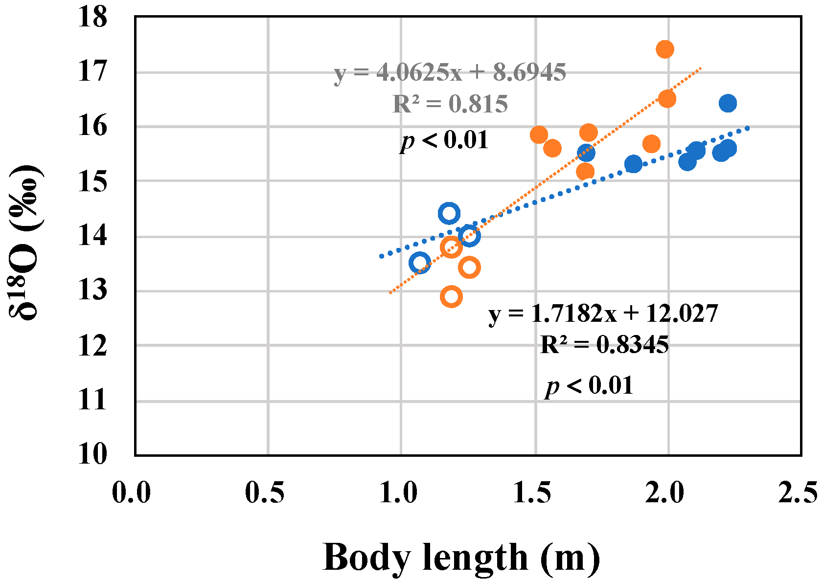
The ontogenetic growth change of δ18O values in muscle samples is presented in Figure 6. The δ18O values in the combined calf samples were significantly lower than those in the combined weaned animal samples (13.7 ± 0.5‰, n = 6 vs. 15.6 ± 0.8‰, n =14, p < 0.05).
The δ18O values in the male samples increased significantly with increasing BL (n = 10), as did the female muscle samples (n = 10).

3.7. Correlations of THg Concentrations in Muscle and Liver with Either δ13C, δ15N, or δ18O Values

Correlations of THg concentrations in muscle and liver samples from the weaned animals with either δ13C, δ15N, or δ18O values in their muscles are summarized in Table 3.
The δ13C values in the muscle samples from males and females significantly correlated with their log THg concentrations in liver samples (p < 0.05), and the δ13C values in the muscle samples from males tended to correlate with their log THg concentrations in muscle samples (p = 0.087). Similarly, the δ18O values in the muscle samples from females significantly correlated with their log THg concentrations in liver samples (p < 0.05), and those from males and females tended to correlate with their log THg concentrations in muscle samples (p = 0.083 and 0.082, respectively). No correlation was found between the δ15N values and the log THg concentrations in muscle or liver samples from the males and from the females.
Data not shown in Table 3, the δ13C values and δ15N values in liver samples did not correlate with their log THg concentrations in liver samples (p > 0.10).

3.8. Comparison of THg Concentrations and δ13C and δ15N Values of Dalli-Type with Those of Truei-Type of Dall’s Porpoises

The THg concentrations and δ13C and δ15N values in muscle and liver samples from the truie-type of mature Dall’s porpoises hunted in the North Pacific Ocean (n = 5) were compared with those from weaned dalli-type samples (Table 4).
The THg concentrations in the muscle and liver samples of the mature truie-type Dall’s porpoises (n = 5) were 1.10 ± 0.35 and 8.74 ± 2.54 μg/wet g. These were similar to those from the weaned dalli-type (1.04 ± 0.58 and 10.3 ± 11.0 μg/wet g, respectively, n = 43). Similarly, the δ15N values in muscle and liver samples from the truie-type were similar to those from the weaned dalli-type; 13.1 ± 0.2‰ vs. 12.8 ± 0.5‰ for muscle samples and 13.9 ± 0.7‰ vs. 13.9 ± 0.7‰ for liver samples. However, the δ13C level in muscle samples from the truie-type was slightly higher than that from the dalli-type (−18.4 ± 0.5‰ vs. −19.4 ± 0.4‰), and the level in liver samples from the former were significantly higher than that from the latter (−17.8 ± 0.5‰ vs. −19.6 ± 0.4‰, p < 0.05).
The δ13C and δ15N values in muscle samples from truei-type were close to those in red meat products from unknown type purchased in Iwate prefecture, respectively (−18.4 ± 0.5‰ vs. −18.8 ± 0.2‰, and 13.1 ± 0.2‰ vs. 13.2 ± 0.3‰).

4. Discussion

4.1. THg Concentrations in the Muscle and Liver of Dalli-Type Dall’s Porpoises with Growth

The higher THg concentration in liver compared to other tissues in dalli-type Dall’s porpoises (Table 1) is similar to the THg tissue distributions that were reported in true-type Dall’s porpoises by Fujise et al. [26] and Yang et al. [27]. However, these studies did not investigate the ontogenetic growth changes in THg concentrations in these tissues. The THg concentrations in liver tissue increased sharply when the BLs of male and female porpoises exceeded 1.9 m and 1.8 m, respectively, and the asymptotes for the sharp increases in the BLs for males and females were approximately 2.2 m and 2.0 m, respectively (Figure 2). These phenomena could be related to the different BLs at their maturation and at their maximum BLs [19,21]. The maturation (decrease in growth rate) and the formation of the HgSe complex are likely to be the causes of the sharp increase in THg in the liver tissues [12,14,23]. Similarly to Dall’s porpoise, BLs of striped dolphins (Stenella coeruleoalba) at maturation and maximum are larger in males than in females, and the BLs at the sharp increases and the asymptotes of the THg increase observed in livers were larger in males than in females [6]. A sharp but more minor THg increase was observed in male muscles at a similar BL found in the liver of Dall’s porpoises (Figure 2), whereas the sharp Hg increase in the female muscles was unclear due to the variation and the small sample size.
The THg concentrations in all muscle samples from the weaned animals (1.04 ± 0.58 μg/wet g, n = 43) exceed the THg guideline for fish and shellfish set by the Japanese government (0.4 μg/wet g). Similar levels of THg were reported in the red meat products from Dall’s porpoises sold for human consumption in Japan (1.26 ± 0.53 μg/wet g, n =17) [22]. Care should be taken not to consume large amounts of Dall’s porpoise meat, especially in pregnant women. Meanwhile, the THg concentrations in all the muscle samples from the calves (0.20 ± 0.12 μg/wet g, n = 10) did not exceed the THg concentration set by the Japanese guideline, except for one calf (1.01 μg/wet g, SNH 12024-2). The low level of THg in calves may be explained by the suckling of milk containing trace amounts of THg [60,61] and the growth dilution effect [60], while the high THg concentration found in the calf (SNH 12024-2) could be explained by the crossing of MeHg through the placenta [60]. High levels of THg contamination in fetuses due to the placental transport of MeHg have been studied in striped dolphins in detail [60]. Yang et al. [62] analyzed the THg concentrations in the liver samples from a pregnant mother-fetus pair of dalli-type Dall’s porpoises (BLs were 1.85 m and 0.55 m, respectively), and reported the THg concentrations of 3.16 and 0.818 μg/wet g, respectively. This hepatic THg concentration in the fetus (0.818 μg/wet g) was lower than that in the calf contaminated with the high level of THg (3.49 μg/wet g, SNH 12024-2), but was higher than that in the other calves (0.149 ± 0.052 μg/wet, n =10). On the other hand, the THg concentration in the maternal liver (3.16 μg/wet g, 1.85 m BL) was a compatible level with that in the liver samples from mature females at the BL of 1.85 m (5.46 μg/wet g, SNH 11021-2) and 1.86 m (2.55 μg/wet g, SNH 11024-1).

4.2. δ13C, δ15N, and δ18O Values During and After Lactation

The difference in δ15N values between the highest and the lowest values of calves was 2.0‰ (Figure 3), which corresponds approximately to one trophic level of δ15N. Similar differences in δ15N values during the lactation period have been observed in common minke whales [47], humpback whales [48], and killer whales [49].
The estimated δ15N peaks from the calf samples, as well as the combined calf and small immature samples, were approximately 1.2 m BL (Figure 2). The increase in δ15N values toward this peak could represent the extensive nursing of 15N-enriched milk, and the decrease in δ15N values from the peak could represent the onset of weaning [47,48,49,56,63]. The onset of weaning in Dall’s porpoises is likely to begin early, within approximately two months [20,21]. Considering that they are born at 0.8–1.0 m BL and grow to 1.6 m BL in the first year [21], the estimated BL at the onset of weaning (approximately 1.2 m) is likely to be consistent with the BL at about two months after birth. A significant positive correlation was found between the δ13C and δ15N values in calf muscles (Figure 3, p < 0.05). This correlation may imply similar changes in δ13C and δ15N values during the lactation period. However, the 13C-enriched peak could not be observed in the present study, possibly due to the variation of δ13C values and small sample size.
No particular trends were found in the δ13C and δ15N changes in muscle and liver samples from the weaned males and the weaned females, owing to the large variations (Figure 3).
The δ18O values in muscles increased with growth from calves to mature animals (Figure 6). We previously reported lower δ18O values in calves than in immature animals of Hubb’s beaked whale muscles [42], and an 18O-depleted peak during the lactation period of killer whale muscles [49]. It seems likely that the δ18O levels in milk are lower than those in weaning foods. Further analysis of the δ18O in neonates, milk, and weaning food consumed by calves is essential.
This present study found that the hepatic δ13C values in truei-type porpoises obtained from Miyagi prefecture were significantly higher than those in the dalli-type porpoises stranded in Hokkaido prefecture; however, the sample number of truei-type was small (Table 4). In agreement, we have previously reported the decreasing trend of δ13C values in toothed whale species and tuna species caught off Japan from south to north [59,64]. The δ13C and δ15N values in the red meat products from porpoises (type unknown), purchased in Iwate prefecture, were close to those in truei-type porpoises hunted in Miyagi prefecture (Table 4), and Iwate prefecture is the neighboring prefecture of Miyagi prefecture (Figure 1). The red meat products purchased in Iwate prefecture may have originated from the truei-type Dall’s porpoises.
Ohizumi and Miyazaki [65] reported the variations of δ13C and δ15N values in the muscle samples from dalli- and truei-type Dall’s porpoises caught off several areas from the Sea of Japan, the Sea of Okhotsk, and the North Pacific Ocean, although they were measured in the muscle samples from which lipids were not removed. The presence of lipids could decrease the δ13C and δ15N values in the bulk samples [66]. Removal of lipids is necessary for a detailed study of regional differences.

4.3. Comparison of δ13C and δ15N Values in Liver Samples with Those in Muscle Samples

The δ15N values in liver tissues of animals are generally higher than those in muscle tissues [51,52,53,54,55]. Higher values in liver tissue are believed to reflect the higher metabolic turnover rates in liver tissue than in muscle tissue. In agreement, the δ15N values were higher in liver samples than in muscle samples of the weaned animals, whereas the δ15N values in calf livers were similar to those in calf muscles (Figure 3). Thus, the Δ15Nliver-muscle values were significantly lower in the calves than in the weaned animals, and these values tended to increase with increasing BL (Figure 5). A possible reason for the Δ15Nliver-muscle trend may be that the turnover rates of 15N in the liver and the muscle tissues from calves (rapidly growing animals) are likely to be similar, and the difference in turnover rate of 15N between these tissues may be gradually increased with growth.
No differences were observed in the δ13C values between the liver and muscle samples from the calves and the weaned animals. Thus, the Δ13Cliver-muscle values in calves were similar to those in the weaned animals (Figure 5). However, it is noted that the variation in these values increased with increasing BL. This increase in the variation may be due to the variation in prey items and the growth of the inhabiting areas. We recently reported that the Δ15Nliver-muscle and Δ13Cliver-muscle values in killer whale calves were significantly lower than those in mature whales, respectively [49]. Further research is needed on the changes ofΔ15Nliver-muscle and Δ13Cliver-muscle values in other cetacean species due to the growth.

4.4. Correlations of THg Concentrations with Wither δ13C, δ15N, or δ18O Values

The δ18O values in marine animals tend to increase with increasing depth [33,35,38,39]. Furthermore, the δ13C values also increase with increasing depth [30,31,32], although many factors influence the δ13C values. Marine animals inhabiting the mesopelagic zone (200–1000 m) tend to be contaminated with a higher level of MeHg than the epipelagic zone (0–200 m) due to higher MeHg concentration in the mesopelagic zone [67,68]. Thus, the positive correlations between the δ18O values and the log THg concentrations and between the δ13C values and the log THg concentrations observed in the weaned animals (Table 3) may be attributed to the feeding shift towards deeper pelagic areas with growth.
In agreement with the present results (Table 3), we reported positive correlations between the δ13C values and the log THg concentrations (p < 0.01) and between the δ18O values and the log THg concentrations (p < 0.10) among combined muscle samples from six deep-sea whale species [49], and Das et al. [68] reported a significant positive correlation between the δ13C values in muscle and the log Hg concentrations in liver of harbor porpoises (Phocoena phocoena).
We found no correlation between the δ15N values and THg concentrations in muscle or liver samples (Table 3), although several studies reported positive correlations between those [2,3,4,5,9]. No correlation between the δ15N values in muscle and log THg concentrations in liver was reported in harbor porpoises [41], and a negative correlation was found between the combined samples of deep-sea whale species [49]. Horizontal and vertical factors and physiological factors may influence the correlation between the THg concentrations and δ15N values in whale species.
Recently, mercury stable isotopes (119Hg, 200Hg, 201Hg, and 202Hg) in deep pelagic species have been used to study the vertical distribution and applied to their trophic ecology along with the δ13C and δ15N values [69,70]. The relationship between the δ18O values and mercury stable isotopes is of great interest.

5. Conclusions

We found the sharp increases in hepatic THg concentrations from 1.9 m and 1.8 m BLs in male and female porpoises, respectively, due to the different BLs of their maturation. We also found that the different BLs at the asymptotes of THg increase in relation to the different maximum BLs of males and females. The δ15N levels in muscles were higher in the calves than in the weaned porpoises, probably due to the consumption of 15N-enriched milk. Δ15Nliver-muscle values in the calves were small and increased with increasing BL, which may be due to an underdeveloped turnover rate of 15N in the calves. The shift in feeding area towards deeper pelagic waters with the growth of porpoises could be expected, as the δ18O values in muscles increased with increasing BL, and the THg concentrations were correlated with the δ13C values and the δ18O values.

Supplementary Materials

The following supporting information can be downloaded at https://www.mdpi.com/article/10.3390/jmse13050892/s1, Table S1: Sample information of male dalli-type Dall’s porpoises (n = 37), and their Hg concentrations and stable isotope ratios; Table S2: Sample information of female dalli-type Dall’s porpoises (n = 17), and their Hg concentrations and stable isotope ratios.

Author Contributions

T.E.: Conceptualization, investigation, data curation, methodology, writing—original draft, writing—review and editing; O.K.: methodology, data curation; M.T.: methodology, data curation; Y.K.: methodology, writing–review and editing; Y.F.: formal analysis, writing–review and editing; K.H.: funding acquisition, writing–review and editing. All authors have read and agreed to the published version of the manuscript.

Funding

This research was funded in part by the Japan Society for the Promotion of Science (JSPS) KAKENHI (grant number 20K12188 (K.H.) and 21K12262 (Y.F.).

Data Availability Statement

The raw data supporting the conclusions of this article will be made available by the authors on request.

Acknowledgments

We would like to thank the Stranding Network of Hokkaido (SNH) for providing samples and information on the stranded whales used in this study.

Conflicts of Interest

The authors declare that there are no conflicts of interest.

References

  1. Selin, N.E. Global biogeochemical cycling of mercury: A Review. Annu. Rev. Environ. Resour. 2009, 34, 43–63. [Google Scholar] [CrossRef]
  2. Atwell, L.; Hobson, K.A.; Welch, H.E. Biomagnification and bioaccumulation of mercury in an Arctic marine food web: Insights from stable nitrogen isotope analysis. Can. J. Fish. Aquat. Sci. 1998, 55, 1114–1121. [Google Scholar] [CrossRef]
  3. Power, M.; Klein, G.M.; Guiguer, K.R.R.A.; Kwan, M.K.H. Mercury accumulation in the fish community of a subArctic lake in relation to trophic position and carbon sources. J. App. Ecol. 2002, 39, 819–830. [Google Scholar] [CrossRef]
  4. Pethybridge, H.; Butler, E.C.V.; Cossa, D.; Daley, R. Trophic structure and biomagnification of mercury in an assemblage of deepwater chondrichthyans from southeastern Australia. Mar. Ecol. Prog. Ser. 2012, 451, 163–174. [Google Scholar] [CrossRef]
  5. Chouvelon, T.; Caurant, F.; Cherel, Y.; Simon-Bouhet, B.; Spitz, J.; Bustamante, P. Species- and size-related patterns in stable isotopes and mercury concentrations in fish help refine marine ecosystem indicators and provide evidence for distinct management units for hake in the Northeast Atlantic. ICES Mar. Sci. 2014, 71, 1073–1087. [Google Scholar] [CrossRef]
  6. Honda, K.; Tatsukawa, R.; Itano, K.; Miyazaki, N.; Fujiyama, T. Heavy metal concentrations in muscle, liver and kidney tissue of striped dolphin, Stenella coeruleoalba, and their variations with body length, weight, age, and sex. Agric. Biol. Chem. 1983, 47, 1219–1228. [Google Scholar] [CrossRef]
  7. Endo, T.; Kimura, O.; Hisamichi, Y.; Minoshima, Y.; Haraguchi, K. Age-dependent accumulation of heavy metals in a pod of killer whales (Orcinus orca) stranded in the northern area of Japan. Chemosphere 2007, 67, 51–59. [Google Scholar] [CrossRef]
  8. Endo, T.; Hisamichi, Y.; Kimura, O.; Haraguchi, K.; Baker, C.S. Contamination levels of mercury and cadmium in melon-headed whales (Peponocephala electra) from a mass stranding on the Japanese coast. Sci. Total Environ. 2008, 401, 73–80. [Google Scholar] [CrossRef]
  9. Endo, T.; Hisamichi, Y.; Haraguchi, K.; Kato, Y.; Ohta, C.; Koga, N. Hg, Zn and Cu levels in the muscle and liver of tiger sharks (Galeocerdo cuvier) from the coast of Ishigaki Island, Japan: Relationship between metal concentrations and body length. Mar. Pollut. Bull. 2008, 56, 1774–1780. [Google Scholar] [CrossRef]
  10. Endo, T.; Ohta, C.; Hisamichi, Y.; Kimura, O.; Ogasawara, H.; Koga, N.; Kato, Y.; Haraguchi, Y. Levels of mercury in muscle and liver of star-spotted dogfish (Mustelus manazo) from the northern region of Japan: A comparison with spiny dogfish (Squalus acanthias). Arch. Environ. Contam. Toxicol. 2013, 64, 467–474. [Google Scholar] [CrossRef]
  11. Le Bourg, B.; Kiszka, J.J.; Bustamante, P.; Heithaus, M.R.; Jaquemet, S.; Humber, F. Effect of body length, trophic position and habitat use on mercury concentrations of sharks from contrasted ecosystems in the southwestern Indian Ocean. Environ. Res. 2019, 169, 387–395. [Google Scholar] [CrossRef] [PubMed]
  12. Endo, T.; Haraguchi, K.; Sakata, M. Mercury and selenium concentrations in the internal organs of toothed whales and dolphins marketed for human consumption in Japan. Sci. Total Environ. 2002, 300, 15–22. [Google Scholar] [CrossRef] [PubMed]
  13. Endo, T.; Kimura, O.; Hisamichi, Y.; Minoshima, Y.; Haraguchi, K.; Kakumoto, C.; Kobayashi, M. Distribution of total mercury, methyl mercury and selenium in pod of killer whales (Orcinus orca) stranded in the northern area of Japan: Comparison of mature females with calves. Environ. Pollut. 2006, 144, 145–150. [Google Scholar] [CrossRef] [PubMed]
  14. Yang, J.; Kunito, T.; Tanabe, S.; Miyazaki, N. Mercury and its relation with selenium in the liver of Dall’s porpoises (Phocoenoides dalli) off the Sanriku coast of Japan. Environ. Pollut. 2007, 148, 669–673. [Google Scholar] [CrossRef]
  15. Dietz, R.; Letcher, R.J.; Desforges, J.P.; Eulaers, I.; Sonne, C.; Wilson, S.; Andersen-Ranberg, E.; Basu, N.; Barst, B.D.; Bustnes, J.O.; et al. Current state of knowledge on biological effects from contaminants on arctic wildlife and fish. Sci. Total Environ. 2019, 696, 133792. [Google Scholar] [CrossRef]
  16. WHO. Methylmercury. Environmental Health Criteria 101; WHO: Geneva, Switzerland, 1990. [Google Scholar]
  17. Lavoie, R.A.; Jardine, T.D.; Chumchal, M.M.; Kidd, K.A.; Campbell, L.M. Biomagnification of mercury in aquatic food webs: A worldwide meta-analysis. Environ. Sci. Technol. 2013, 47, 13385–13394. [Google Scholar] [CrossRef]
  18. Bridges, C.C.; Zalups, R.K. Molecular and ionic mimicry and the transport of toxic metals. Toxicol. Appl. Pharmacol. 2005, 204, 274–308. [Google Scholar] [CrossRef]
  19. Amano, M.; Hayano, A. Intermingling of dalli-type Dall’s porpoises into a wintering truei-type population off Japan: Implications from color patterns. Mar. Mamm. Sci. 2007, 23, 1–14. [Google Scholar] [CrossRef]
  20. Kasuya, T. Small Cetaceans of Japan. Exploitation and Biology; CRC Press: Boca Raton, FL, USA, 2017; pp. 219–244. [Google Scholar]
  21. FRA (Japan Fisheries Research and Education Agency). Dall’s Porpoise (Phocoenoides dalli). 2022. Available online: https://kokushi.fra.go.jp/R03/R03_49_PDA.pdf (accessed on 22 April 2025).
  22. Endo, T.; Hotta, Y.; Haraguchi, K.; Sakata, M. Mercury contamination in the red meat of whales and dolphins marketed for human consumption in Japan. Environ. Sci. Technol. 2003, 37, 2681–26815. [Google Scholar] [CrossRef]
  23. Endo, T.; Haraguchi, K.; Hotta, Y.; Hisamichi, Y.; Lavery, S.; Dalebout, M.L.; Baker, C.S. Total mercury, methyl mercury, and selenium levels in the red meat of small cetaceans sold for human consumption in Japan. Environ. Sci. Technol. 2005, 39, 5703–5708. [Google Scholar] [CrossRef]
  24. Fisheries Agency of Japanese Government. Small Cetacean Hunting Quotas for FY 2023. 2023. Available online: https://www.jfa.maff.go.jp/j/whale/attach/pdf/index-15.pdf (accessed on 22 April 2025).
  25. MHLW (Ministry of Health, Labour and Welfare). Advice for Pregnant Women on Fish Consumption and Mercury. 2005. Available online: https://www.mhlw.go.jp/english/wp/other/councils/mercury/index.html (accessed on 22 April 2025).
  26. Fujise, Y.; Honda, K.; Tatsukawa, R.; Mishima, S. Tissue distribution of heavy metals in Dall’s porpoise in the northwestern Pacific. Mar. Pollut. Bull. 1988, 19, 226–230. [Google Scholar] [CrossRef]
  27. Yang, J.; Kunito, T.; Tanabe, S.; Miyazaki, N. Mercury in tissues of Dall’s porpoise (Phocoenoides dalli) collected off Sanriku coast of Japan. Fish. Sci. 2002, 68, 256–259. [Google Scholar] [CrossRef] [PubMed]
  28. Kelly, J.F. Stable isotopes of carbon and nitrogen in the study of avian and mammalian trophic ecology. Can. J. Zool. 2000, 78, 1–27. [Google Scholar] [CrossRef]
  29. O’Brien, D.M. Stable isotope ratios as biomarkers of diet for health research. Ann. Rev. Nutr. 2015, 35, 565–595. [Google Scholar] [CrossRef]
  30. Cherel, Y.; Hobson, K.A. Geographical variation in carbon stable isotope signatures of marine predators: A tool to investigate their foraging areas in the Southern Ocean. Mar. Ecol. Prog. Ser. 2007, 329, 281–287. [Google Scholar] [CrossRef]
  31. Baylis, A.M.M.; Orben, R.A.; Arnould, J.P.Y.; Peters, K.; Knox, T.; Costa, D.P.; Staniland, I.J. Diving deeper into individual foraging specializations of a large marine predator, the southern sea lion. Oecologia 2015, 179, 1053–1065. [Google Scholar] [CrossRef]
  32. Chouvelon, T.; Spitz, J.; Caurant, F.; Mèndez-Fernandez, P.; Autier, J.; Lassus-Débat, A.; Chappuis, A.; Bustamante, P. Enhanced bioaccumulation of mercury in deep-sea fauna from the Bay of Biscay (north-east Atlantic) in relation to trophic positions identified by analysis of carbon nitrogen stable isotopes. Deep-Sea Res. Part I 2012, 65, 113–124. [Google Scholar] [CrossRef]
  33. Kalish, J.M. 13C and 18O isotopic disequilibria in fish otoliths: Metabolic and kinetic effects. Mar. Ecol. Prog. Ser. 1991, 75, 191–203. [Google Scholar] [CrossRef]
  34. Lukeneder, A.; Harzhauser, M.; Mullegger, S.; Piller, W.E. Stable isotopes (δ18O and δ13C) in Spirula spirula shells from three major oceans indicate developmental changes paralleling depth distributions. Mar. Boil. 2008, 154, 175–182. [Google Scholar] [CrossRef]
  35. Lin, H.Y.; Shiao, C.; Chen, Y.G.; Iizuka, Y. Ontogenetic vertical migration of grenadiers revealed by otolith microstructures stable isotopic composition. Deep-Sea Res. Part I 2012, 61, 123–130. [Google Scholar] [CrossRef]
  36. Méndez-Fernandez, P.; Pierce, G.J.; Bustamante, P.; Chouvelon, T.; Ferreira, M.; González, A.F.; López, A.; Read, F.L.; Santos, M.B.; Spitz, J.; et al. Ecological niche segregation among five toothed whale species off the NW Iberian Peninsula using ecological tracers as multiapproach. Mar. Biol. 2013, 160, 2825–2840. [Google Scholar] [CrossRef]
  37. Clementz, M.T.; Koch, P.L. Differentiating aquatic mammal habitat and foraging ecology with stable isotopes in tooth enamel. Oecologia 2001, 129, 461–472. [Google Scholar] [CrossRef] [PubMed]
  38. Matthews, C.J.D.; Longstaffe, F.J.; Ferguson, S.H. Dentine oxygen isotopes (δ18O) as a proxy for odontocete distributions and movements. Ecol. Evol. 2016, 6, 4643–4653. [Google Scholar] [CrossRef] [PubMed]
  39. Ciner, B.; Wang, Y.; Parker, W. Oxygen isotopic variations in modern cetacean teeth and bones: Implications for ecological, paleoecological, and paleoclimatic studies. Sci. Bull. 2016, 61, 92–104. [Google Scholar] [CrossRef]
  40. Le Croizier, S.G.; Schaal, G.; Point, D.; Le Loc’h, F.; Machu, E.; Fall, M.; Munaron, J.M.; Boyé, A.; Walter, P.; Laë, R.; et al. Stable isotope analyses revealed the influence of foraging habitat on mercury accumulation in tropical coastal marine fish. Sci. Total Environ. 2019, 650, 2129–2140. [Google Scholar] [CrossRef]
  41. Das, K.; Holsbeek, L.; Browning, J.; Siebert, U.; Birkun, A., Jr.; Bouquegneau, J.M. Trace metal and stable isotope measurements (δ13C and δ15N) in the harbour porpoise Phocoena phocoena relicta from the Black Sea. Environ. Pollut. 2004, 13, 197–204. [Google Scholar] [CrossRef]
  42. Endo, T.; Kimura, O.; Terasaki, M.; Kato, Y.; Fujii, Y.; Haraguchi, K. Comparison of carbon, nitrogen, and oxygen stable isotope ratios and mercury concentrations in muscle tissues of five beaked whale species and sperm whales stranded in Hokkaido, Japan. Isot. Environ. Health Stud. 2024, 60, 251–271. [Google Scholar] [CrossRef]
  43. Newsome, S.D.; Clementz, M.T.; Koch, P.L. Using stable isotope biogeochemistry to study marine mammal ecology. Mar. Mamm. Sci. 2010, 26, 509–572. [Google Scholar] [CrossRef]
  44. Valenzuela, L.O.; Sironi, M.; Rowntree, V.J. Interannual variation in the stable isotope differences between mothers and their calves in southern right whales (Eubalaena australis). Aquat. Mamm. 2010, 36, 138–147. [Google Scholar] [CrossRef]
  45. Drago, M.; Franco-Trecu, V.; Cardona, L.; Inchausti, P. Diet-to-female and female-to-pup isotopic discrimination in South American sea lions. Rapid Commun. Mass. Spectrom. 2015, 29, 1513–1520. [Google Scholar] [CrossRef]
  46. Borrell, A.; Gómez-Campos, E.; Aguilar, A. Influence of reproduction on stable-isotope ratios: Nitrogen and carbon isotope discrimination between mothers, fetuses, and milk in the fin whale, a capital breeder. Physiol. Biochem. Zool. 2016, 89, 41–50. [Google Scholar] [CrossRef] [PubMed]
  47. Endo, T.; Kimura, O.; Terasaki, M.; Kobayashi, M. Body length, stable carbon and nitrogen isotope ratios and mercury levels in common minke whales (Balaenoptera acutorostrata) stranded along the coast of Hokkaido, Japan. Aquat. Mamm. 2021, 47, 86–95. [Google Scholar] [CrossRef]
  48. Endo, T.; Terasaki, M.; Kimura, O. Stable isotope ratios of carbon, nitrogen, oxygen, and mercury concentrations in North Pacific baleen whales and the comparison of their calves with toothed whale calves. J. Veter Sci. Med. 2022, 10, 10. [Google Scholar] [CrossRef]
  49. Endo, T.; Kimura, O.; Terasaki, M.; Kato, Y.; Fujii, Y.; Haraguchi, K. Changes in carbon, nitrogen, and oxygen stable isotope ratios and mercury concentrations in killer whales (Orcinus orca) during and after lactation. J. Mar. Sci. Eng. 2024, 12, 623. [Google Scholar] [CrossRef]
  50. Oftedal, O.T. Lactation in whales and dolphins: Evidence of divergence between baleen- and toothed-species. J. Mammary Gland Biol. Neoplasia 1997, 2, 205–230. [Google Scholar] [CrossRef]
  51. Kurle, C.M.; Worthy, G.A.J. Stable nitrogen and carbon isotope ratios in multiple tissues of the northern fur seal Callorhinus ursinus: Implications for dietary and migratory reconstructions. Mar. Ecol. Prog. Ser. 2002, 236, 289–300. [Google Scholar] [CrossRef]
  52. Caut, S.; Angulo, E.; Courchamp, F. Variation in discrimination factors (Δ15N and Δ13C): The effect of diet isotopic values and applications for diet reconstruction. J. Appl. Ecol. 2009, 46, 443–453. [Google Scholar] [CrossRef]
  53. Borrell, A.; Abad-Oliva, N.; Gómez-Campos, E.; Giménez, J.; Aguilar, A. Discrimination of stable isotopes in fin whale tissues and application to diet assessment in cetaceans. Rapid Commun. Mass. Spectrom. 2012, 26, 1596–1602. [Google Scholar] [CrossRef]
  54. Vanderklift, M.A.; Ponsard, S. Sources of variation in consumer-diet δ15N enrichment: A meta-analysis. Oecologia 2003, 136, 169–182. [Google Scholar] [CrossRef]
  55. Camin, F.; Bontempo, L.; Perini, M.; Piasentier, E. Stable isotope ratio analysis for assessing the authenticity of food animal origin. Compr. Rev. Food Sci. Food Saf. 2016, 15, 868–877. [Google Scholar] [CrossRef]
  56. Endo, T.; Kobayashi, M. Typical changes in carbon and nitrogen stable isotope ratios and mercury concentration during the lactation of marine mammals. In Marine Mammals; Kaoud, H.A.E., Ed.; IntechOpen: London, UK, 2022; pp. 55–78. Available online: https://www.intechopen.com/predownload/81844 (accessed on 22 April 2025).
  57. Logan, J.M.; Lutcavage, M.E. A comparison of carbon and nitrogen stable isotope ratios of fish tissues following lipid extractions with non-polar and traditional chloroform/methanol solvent systems. Rapid Commun. Mass. Spectrom. 2008, 22, 1081–1086. [Google Scholar] [CrossRef] [PubMed]
  58. Endo, T.; Hotta, Y.; Hisamichi, Y.; Kimura, O.; Sato, R.; Haraguchi, K.; Funahasi, N.; Baker, C.S. Stable isotope ratios and mercury levels in red meat products from six baleen whales sold in Japanese markets. Ecotoxicol. Environ. Saf. 2012, 79, 35–41. [Google Scholar] [CrossRef] [PubMed]
  59. Endo, T.; Hisamichi, Y.; Kimura, O.; Haraguchi, K.; Lavery, S.; Dalebout, M.L.; Funahashi, N.; Baler, C.S. Stable isotope ratios of carbon and nitrogen and mercury concentrations in 13 toothed whale species taken from the western Pacific Ocean off Japan. Environ. Sci. Technol. 2010, 44, 2675–2681. [Google Scholar] [CrossRef] [PubMed]
  60. Itano, K.; Kawai, S.; Miyazaki, N.; Tatsukawa, R.; Fujiyama, T. Mercury and selenium levels at the fetal and suckling stages of striped dolphin, Stenella coeruleoalba. Agric. Biol. Chem. 1984, 48, 1691–1698. [Google Scholar] [CrossRef]
  61. Caon, G.; Secchi, E.R.; Capp, E.; Kucharski, L.C. Milk composition of franciscana dolphin (Pontoporia blainvillei) from Rio Grande do Sul, southern Brazil. J. Mar. Biol. Assoc. U. K. 2008, 88, 1099–1101. [Google Scholar] [CrossRef]
  62. Yang, J.; Kunito, T.; Anan, Y.; Tanabe, S.; Miyazaki, N. Total and subcellular distribution of trace elements in the lever of a mother–fetus pair of Dall’s porpoises (Phocoenoides dalli). Mar. Pollut. Bull. 2004, 48, 1122–1129. [Google Scholar] [CrossRef]
  63. Vales, D.G.; Cardona, L.; Garcia, N.A.; Zenteno, L.; Crespo, E.A. Ontogenetic dietary changes in male South American fur seals Arctocephalus australis in Patagonia. Mar. Ecol. Prog. Ser. 2015, 525, 245–260. [Google Scholar] [CrossRef]
  64. Hisamichi, Y.; Haraguchi, K.; Endo, T. Levels of mercury and organochlorine compounds and stable isotope ratios in three tuna species taken from different regions of Japan. Environ. Sci. Technol. 2010, 44, 5971–5978. [Google Scholar] [CrossRef]
  65. Ohizumi, H.; Myasaki, N. Differences in stable isotope ratios of Dall’s porpoises (Phocoenoides dalli) between coastal and oceanic areas of the North Pacific. Fish. Oceanogr. 2010, 19, 257–261. [Google Scholar] [CrossRef]
  66. Ryan, R.C.; McHugh, B.; Trueman, C.N.; Harrod, C.; Berrow, S.D.; O’Connor, I. Accounting for the effects of lipids in stable isotope (δ13C and δ15N values) analysis of skin and blubber of balaenopterid whales. Rapid Commun. Mass. Spectrom. 2012, 26, 2745–2754. [Google Scholar] [CrossRef]
  67. Choy, C.A.; Popp, B.N.; Kaneko, J.J.; Drazen, J.C. The influence of depth on mercury levels in pelagic fishes and their prey. Proc. Natl. Acad. Sci. USA 2009, 106, 13865–13869. [Google Scholar] [CrossRef] [PubMed]
  68. Logan, J.M.; Lutcavage, M.E. Assessment of trophic dynamics of cephalopods large pelagic fishes in the central North Atlantic Ocean using stable isotope analysis. Deep-Sea Res. Part II 2013, 95, 63–73. [Google Scholar] [CrossRef]
  69. Tsui, M.T.-K.; Blum, J.D.; Kwon, S.Y. Review of stable mercury isotopes in ecology and biogeochemistry. Sci. Total Environ. 2020, 716, 135386. [Google Scholar] [CrossRef] [PubMed]
  70. Médieu, A.; Spitz, J.; Point, D.; Sonke, J.E.; Loutrage, L.; Laffont, L.; Chouvelon, T. Mercury stable isotopes reveal the vertical distribution and trophic ecology of deep-pelagic organisms over the North-East Atlantic Ocean continental slope. Environ. Sci. Technol. 2024, 58, 18733–18743. [Google Scholar] [CrossRef]
Figure 1. Map showing Hokkaido and migration routes of dalli- and truei-type Dall’s porpoises around Japan.
Jmse 13 00892 g001
Figure 2. Increases in THg concentrations of muscle and liver from males and females with growth. Weaned males (), weaned females (), and calves (open circles).
Jmse 13 00892 g002
Figure 3. Changes in δ13C and δ15N values in muscle and liver from males and females with growth. Weaned males (), weaned females (), and calves (open circles).
Jmse 13 00892 g003
Figure 4. Relationships between δ13C and δ15N values in muscle from weaned males (), weaned females (), and calves (open circles).
Jmse 13 00892 g004
Figure 5. Effect of growth (body length) on δ13Cliver-muscle and δ15Nliver-muscle. Weaned males (), weaned females (), and calves (open circles).
Jmse 13 00892 g005
Figure 6. Effect of growth (body length) on δ18O values in muscles from males and females. Weaned males (), weaned females (), and calves (open circles).
Jmse 13 00892 g006
Table 1. Body length and total mercury (THg) distribution in male and female dalli-type Dall’s porpoises.
MaleFemale
Body length (m) 1.91 ± 0.34 (n = 37)1.57 ± 0.32 (n = 17)
THg concentrationsMuscle0.95 ± 0.62 (n = 37)0.71 ± 0.60 (n = 17)
(μg/wet g)Liver10.3 ± 11.1 (n = 35)5.24 ± 8.78 (n = 17)
Kidney2.08 ± 2.26 (n = 33)1.05 ± 0.82 (n = 15)
Lung0.60 ± 0.56 (n = 23)0.37 ± 0.34 (n = 14)
Spleen0.82 ± 1.08 (n = 33)0.42 ± 0.49 (n = 12)
Heart0.58 ± 0.29 (n = 25)0.41 ± 0.29 (n = 14)
Blood0.24 ± 0.11 (n = 29)0.20 ± 0.11 (n = 14)
Table 2. δ13C, δ15N, and δ18O values in muscle and liver of dalli-type Dall’s porpoises.
δ13C (‰)δ15N (‰)δ18O (‰)
MuscleAll animalsMale−19.4 ± 0.5 (n = 37)13.0 ± 0.8 (n = 37)15.1 ± 0.9 (n = 10)
Female−19.3 ± 0.3 (n = 17)13.4 ± 1.2 (n = 17)15.2 ± 1.4 (n = 10)
Total−19.4 ± 0.4 (n = 54)13.1 ± 0.9 (n = 54)15.2 ± 1.2 (n = 20)
CalvesMale−19.5 ± 0.8 (n = 5)14.4 ± 0.7 (n = 5)14.0 ± 0.4 ** (n = 3)
Female−19.2 ± 0.2 (n = 6)14.8 ± 0.5 (n = 6)13.3 ± 0.5 ** (n = 3)
Total−19.4 ± 0.6 (n = 11)14.6 ± 0.6 * (n = 11)13.7 ± 0.5 ** (n = 6)
WeanedMale−19.4 ± 0.4 (n = 32)12.8 ± 0.6 (n = 32)15.6 ± 0.9 (n = 7)
AnimalsFemale−19.4 ± 0.3 (n = 11)12.6 ± 0.5 (n = 11)15.6 ± 1.1 (n = 8)
Total−19.4 ± 0.4 (n = 43)12.8 ± 0.5 (n = 43)15.6 ± 0.8 (n = 15)
LiverAll animalsMale−19.5 ± 0.5 (n = 25)14.0 ± 0.8 (n = 25)ND
Female−19.4 ± 0.4 (n = 8)13.8 ± 0.7 (n = 8)ND
Total−19.5 ± 0.4 (n = 33)13.9 ± 0.8 (n = 33)ND
CalvesMale−19.6 and −19.3 (n = 2)13.9 and 14.4 (n = 2)ND
Female−18.6 and −18.5 (n = 2)14.7 and 5.3 (n = 2)ND
Total−19.0 ± 0.5 (n = 4)14.6 ± 0.6 (n = 4)ND
WeanedMale−19.6 ± 0.4 (n = 23)13.9 ± 0.7 (n = 23)ND
AnimalsFemale−19.6 ± 0.5 (n = 6)13.7 ± 0.7 (n = 6)ND
Total−19.6 ± 0.4 (n = 29)13.9 ± 0.7 (n = 29)ND
Data are represented by mean ± SD. Significantly higher than the weaned animals (Student’s t-test, p < 0.05) * and lower than the weaned animals (Student’s t-test, p < 0.01) **. Weaned animals (BL > 1.5 m). ND: Not determined.
Table 3. Correlations of Log THg concentrations in muscle and liver with either δ13C, δ15N, or δ18O values in muscle.
MuscleLiver
R2p ValueR2p Value
δ13CMale (n =32)0.3080.0870.5140.003 *
Female (n =11)0.1830.590 **0.6100.046 *
δ15NMale (n =32)0.1040.569 **0.0710.697 **
Female (n =11)0.3560.283 **0.0600.860 **
δ18OMale (n = 7)0.4840.0830.2830.219 **
Female (n =7)0.4840.0820.6400.031 *
A single asterisk (*) indicates the p-value below 0.05. Double asterisk (**) indicates p-value greater than 0.10.
Table 4. Comparison of THg concentrations, and δ13C and δ15N values of dalli-type weaned Dall’s porpoises stranded in Hokkaido with those of truei-type mature Dall’s porpoises hunted in the North Pacific Ocean.
Dalli-Type (n = 43)Truei-Type (n = 5)Type Unknown (n = 8) *
Stranded in Hokkaido
(Weaned Animals)
Hunted in the North Pacific Ocean
off Miyagi Prefecture (Mature Animals)
Hunted in the North Pacific Ocean
Area and Maturity Not Known
MuscleTHg (μg/wet g)1.04 ± 0.581.10 ± 0.351.27 ± 0.33
δ13C (‰)−19.4 ± 0.4−18.4 ± 0.5−18.8 ± 0.2
δ15N (‰)12.8 ± 0.513.1 ± 0.213.2 ± 0.3
LiverTHg (μg/wet g)10.3 ± 11.08.74 ± 2.54ND
δ13C (‰)−19.6 ± 0.4−17.8 ± 0.5 **ND
δ15N (‰)13.9 ± 0.713.9 ± 0.7ND
* Red meat products from Endo et al. [59]. ** Significantly higher than dalli-type weaned porpoises (Student’s t-test, p < 0.05). ND: not determined.
Disclaimer/Publisher’s Note: The statements, opinions and data contained in all publications are solely those of the individual author(s) and contributor(s) and not of MDPI and/or the editor(s). MDPI and/or the editor(s) disclaim responsibility for any injury to people or property resulting from any ideas, methods, instructions or products referred to in the content.