Article

Machine Learning-Based Classification of Malnutrition Using Histological Biomarkers of Fish Intestine: Preliminary Data

by
Joana Oliveira
1,2,
Marisa Barata
3,4,
Florbela Soares
3,4,
Pedro Pousão-Ferreira
3,4,
Aires Oliva-Teles
1,2,* and
Ana Couto
1,2
1
FCUP—Department of Biology, Faculty of Sciences, University of Porto, 4169-007 Porto, Portugal
2
CIIMAR—Interdisciplinary Centre of Marine and Environmental Research, University of Porto, 4050-208 Matosinhos, Portugal
3
IPMA—EPPO—Portuguese Institute for Sea and Atmosphere—Aquaculture Research Station, 8700-194 Olhão, Portugal
4
S2AQUA—Collaborative Laboratory, Association for a Sustainable and Smart Aquaculture, 8700-194 Olhão, Portugal
*
Author to whom correspondence should be addressed.
J. Mar. Sci. Eng. 2024, 12(12), 2177; https://doi.org/10.3390/jmse12122177
Submission received: 18 October 2024 / Revised: 19 November 2024 / Accepted: 26 November 2024 / Published: 28 November 2024
(This article belongs to the Special Issue New Challenges in Marine Aquaculture Research—2nd Edition)

Abstract

:
The gut is the first organ to contact food, and it is often the target of nutrition studies performed on aquaculture fish. Histological analysis reveals morphological changes in fish intestines caused by ingredients in formulated feeds. However, this type of analysis is mainly based on a semi-quantitative approach, often restricted to specialized researchers, and may provide inconsistent results between studies. This study addresses these limitations by combining semi-quantitative and quantitative features to characterize the anterior, intermediate, and distal sections of the intestine of meagre (Argyrosomus regius) subjected to different nutritional status. Collected data were used to build machine learning models, select the most accurate ones, and identify key features for predicting malnutrition. Logistic regression, support vector machines, and ensemble stacking performed best across all intestinal sections. Combining semi-quantitative and quantitative features yielded the best predictions, with villi number, density and area, and goblet cell count being the most crucial for the classification task. When considering the distal intestine alone, semi-quantitative features outperformed quantitative ones. The intermediate section of the intestine showed the best model accuracy, indicating higher sensitivity to nutritional changes. These results demonstrate the potential of machine learning models to streamline histomorphological analyses to evaluate nutritional status, making them more accessible and standard across users.

1. Introduction

In recent years, new diets for aquaculture fish have aimed to reduce the inclusion of high amounts of fisheries by-products, focusing instead on using alternative ingredients that will provide adequate growth and health while guaranteeing environmental and economic benefits [1,2]. Applying such feeds to carnivore species can be complicated as alternative ingredients rely mostly on vegetable protein and oil, which often negatively affect fish health [3,4,5,6,7]. Thus, fish nutrition studies have focused on observing the long-term effects of diets on fish health by evaluating physiological, immunological, and morphological responses. The gut is often a target in such studies, as the balance of intestinal health usually relies on the quality of feeds. Furthermore, the link between intestinal well-being and immunity in fish is well established in research and is frequently the focus of many studies [8,9,10,11]. Intestinal health in fish can be evaluated through various approaches, including assessment of gut microbiota composition [12,13], oxidative stress status [14,15], gene and protein expression [16,17], histomorphological analysis [18,19], immune response markers [20,21], and the measurement of nutrient absorption efficiency [22,23].
One of the most selected forms of evaluating intestine health is histological analysis, in which it is easier to observe intestine inflammation and other morphological alterations caused by feeds [24]. However, most histological evaluations are limited to semi-quantitative analysis and only focus on one portion of the intestine, most often the distal portion [25,26,27].
Semi-quantitative analysis typically applied to evaluate histomorphological features in fish intestines is laborious and highly dependent on specialized personnel. Results are often inconsistent and variable between studies, which hinders results comparison. Despite this, it allows for the observation of specific characteristics that thoroughly describe the histomorphology of the gut, often independently of the quality of the histological technique and tissue sample and accounting for the variability of sampling location. Furthermore, it typically requires no software or computational tools, making it relatively inexpensive and accessible [28].
Quantitative measurements in fish intestines have become increasingly used in histological studies as less specialized researchers can perform them, thus allowing for a more straightforward and consistent analysis. However, this type of analysis is still limited to few parameters, such as lamina propria width, villus height and width, goblet cell count, and measurements of the submucosa layer and mucosal tissue [18,29,30]. Additionally, most quantitative studies do not consider fish size differences and the variety of sizes between intestinal sections, which can affect morphological measurement comparisons. Consequently, it is essential to establish histomorphological biomarkers that are precise and practical to measure, allowing for a more straightforward evaluation of the effects of diets on aquaculture fish.
Despite fish intestine sections being less morphologically distinguishable than those of other vertebrates [31], different portions of the intestine have distinct histomorphological traits and physiological functions depending on species and feeding habits. The anterior portion of the intestine is known to play a role in digestion and nutrient absorption, while posterior portions are typically connected to immunological functions [32,33]. It is known that feeds can differentially modulate the intestine sections’ morphology and physiology [34,35], which highlights the importance of evaluating different intestinal sections in fish nutrition research. Moreover, establishing a standardized sampling practice for each species is essential, as variations in sampling location or the amount of intestinal content can hinder result comparisons between studies.
Machine learning is still scarcely applied in aquaculture but has increased in recent years. It has been used for example to help predict reproductive status in Chilean hake (Merluccius gayi gayi) [36], age and sex estimation in walleye (Sander vitreus) [37], ovarian development in channel catfish (Ictalurus puncatatus) [38], disease resistance in produced fish [39], and fish production levels in specific regions [40]. Machine learning algorithms produce models by “learning” patterns directly from the data without needing an explicitly defined function as a model [36]. These algorithms can help researchers extract information from datasets more efficiently, especially when these datasets are abundant [41].
The utilization of machine learning to help predict the nutritional status of fish is a new concept, absent to date in fish nutrition research, but can be an important tool for the diagnostic of fish health and welfare in response to formulated feeds. Combining machine learning with histological analysis not only ensures thorough results to train different algorithms but also allows the models to aid in interpreting the extensive findings from histomorphological examinations in an aquaculture setting.
The present study aimed to establish a model to classify fish’s nutritional status using meagre (Argyrosomus regius) juveniles as a biological model. Meagre is a carnivore species for which production has increased in recent years [42], largely due to its favorable traits such as fast growth, good feed conversion ratio, and good adaptation to captivity [43,44,45]. However, optimization of meagre production is still underway, including the need for an improved feed for this species capable of maximizing growth and health [46,47,48]. For that purpose, attention must be given to gut health and physiology [15,49] and gut microbiota diversity [26,50] to facilitate future nutritional studies in this species.
In this study, a robust dataset with reliable, controlled, and thoroughly annotated data was constructed to establish a preliminary model based on gut histomorphology that predicts the fish’s nutritional status when animals are submitted to diets that induce graded levels of malnutrition. This dataset consisted of features from various intestinal sections and was used to train machine learning models capable of classifying the nutritional status of the animals based on the histomorphological features. By evaluating features importance for the classification task, this study also aimed to obtain reliable biomarkers for the histological evaluation of fish intestines, focusing not only on semi-quantitative features but also on eight quantitative features that were sufficient to thoroughly describe intestinal morphology while being straightforward enough to be applied without extensive expertise in intestinal histology. This approach enables a more accessible assessment of fish gut health in aquaculture settings.

2. Materials and Methods

This study was directed by certified scientists (FELASA category C), and all procedures were conducted according to the European Union Directive 2010/63/EU recommendations on the protection of animals for scientific purposes.
The growth trial was performed at the Portuguese Institute for the Ocean and Atmosphere Aquaculture Research Station facilities (IPMA-EPPO, Olhão, Portugal) with meagre (A. regius) juveniles produced at the station.

2.1. Growth Trial

Three isoproteic (48%) and isolipidic (18%) diets were formulated. A fishmeal (FM)- and fish oil (FO)-based diet (55% FM; 11% FO) was used as a control (CTRL diet). Two other diets were also formulated: a challenge diet (CD diet) with 15% FM and 7% FO and an extreme challenge diet (ED) with 5% FM and 5% FO, containing increasing amounts of plant feedstuffs and vegetable oils. All dietary ingredients were finely ground, well mixed, and dry pelleted in a laboratory pellet mill (California Pellet Mill, CPM, Crawfordsville, IN, USA) through a 2 mm die. The pellets were then dried in an oven at 40 °C for 24 h and stored in airtight bags until they were used. The proximate composition of ingredients and diets is presented in Table 1.
The growth trial was conducted in 9 fiberglass tanks of 200 L water capacity in an open water system supplied with a continuous flow of filtered seawater (6 L min−1). At the start of the trial, 9 groups of 60 fish with an average weight of 4.6 ± 0.4 g were established, and the experimental diets were randomly assigned to triplicate tanks. During the trial, which lasted 7 weeks, fish were fed daily by hand 6 times a day (to avoid cannibalistic behavior) until visual satiation. Utmost care was taken to ensure that all feed supplied was consumed. During the trial, the water temperature averaged 23.6 ± 0.4 °C; salinity was 36 ± 1 g L−1; and dissolved oxygen was kept near saturation (5.7 ± 0.4 mg L−1).
At the end of the trial, 18 fish from each tank were sampled 4 h after the first meal, slightly anesthetized with 0.3 mL/L of ethylene glycol monophenyl ether, and weighted. The fish were then euthanized by decapitation, and the intestine was removed and divided into anterior, intermediate, and distal intestine (Figure 1). The samples were immediately fixed in phosphate-buffered formalin (4%, pH 7.4) for 24 h and subsequently transferred to ethanol at 70% until further processing.
The remaining fish were kept unfed for one day and then bulk-weighed after being slightly anesthetized with 0.3 mL/L of ethylene glycol monophenyl ether. Three fish from each tank were then randomly selected, and whole-fish, viscera, intestine, and liver weights of these fish were recorded for the determination of hepatosomatic (HSI), enterosomatic (ESI), and visceral (VI) indexes.
Final weight (g) was measured, and the weight gain (g) was calculated. The daily growth index (DGI) was calculated using the formula: DGI = [(final body weight1/3 − initial body weight1/3)/time (days)] × 100. Feed intake was calculated as grams of feed consumed per kilogram of average body weight per day (g kg−1 ABW day−1) using the formula: Feed intake (g kg−1 ABW day−1) = (total feed consumed/average body weight of fish)/number of days. Feed efficiency (FE) was determined as the ratio of wet weight gain to dry feed intake: FE = wet weight gain/dry feed intake.
The hepatosomatic index (HSI), viscerosomatic index (VSI), and enterosomatic index (ESI) were calculated using the following formulas: HSI = (liver weight/body weight) × 100; VSI = (viscera weight/body weight) × 100; ESI = (intestine weight/body weight) × 100.

2.2. Proximate Analysis of Experimental Diets

Chemical analysis of the experimental diets (Table 1) was performed as follows: dry matter was determined by drying samples in an oven at 105 °C until constant weight; ash by incineration in a muffle furnace at 450 °C for 16 h; protein content (N × 6.25) according to the Kjeldahl method, using a Velp distillation unit (Velp® Scientifica, Usmate Velate, Lombardy, Italy; model UDK 129 Kjeldahl Distillation Unit) and Tecator digestor unit (Tecator Systems, Höganäs, Sweden; model 1015); and lipids by petroleum ether extraction in a SoxTec extraction system (Tecator Systems, Höganäs, Sweden; extraction unit model 1043 and service unit model 1046). Dietary energy content was determined by direct combustion in an adiabatic bomb calorimeter (PARR model 6200, PARR Instruments, Moline, IL, USA).

2.3. Histological Analysis

The three intestinal sections were processed and sectioned using standard histological techniques. Briefly, sections were dehydrated through standard ethanol series to 100%, cleared in xylene, embedded in paraffin, transversely sectioned at 7 μm intervals, and stained with hematoxylin and eosin. Both quantitative (QT) and semi-quantitative (SQT) analyses (Figure 1) were performed using a ZEISS Axio Imager A2 microscope equipped with a ZEISS Axio Cam Mrc camera and with ZEISS software ZEN 3.5 (blue edition). For the SQT analysis, a scoring system as described by [25,51], with the range of tissue scores being set from 1 (normal) to 5 (highly modified), was used to evaluate the extent of the following structures: intestinal folds fusion, lamina propria and submucosa widening, goblet cells presence, internal and external muscularis width, leukocytes infiltration in the lamina propria and submucosa, and enterocytes architecture alterations, including supranuclear vacuolization. The overall value of histomorphological alterations was calculated by averaging scores of the individual features described above. For the QT analysis, the following measurements were manually taken with the aid of ZEISS software: total area and maximum total diameter, total lumen area and maximum lumen diameter, area occupied by both villi and lumen, and villi number. Villi-only area and density were calculated after measurements. Villi density was calculated as the ratio of villi number to villi area: villi density = villi number/villi-only area.
Figure 1. Meagre (Argyrosomus regius) intestine, characterized by being short, a typical feature of carnivore fish, and having a larger diameter in the anterior portion that reduces in the intermedium portion and increases once more in the distal region [52]. Selected sections in meagre intestine with the incision localization are presented. Semi-quantitative analysis method (A) and quantitative analysis method (B) of histological samples.
Jmse 12 02177 g001

2.4. Nutritional Status Classification Using Machine Learning Algorithms

2.4.1. Data Acquisition and Preprocessing

The machine learning workflow is represented in Figure 2. Data were acquired through the histological evaluation described in Section 2.3. Datasets were built to include all the intestinal sections (ALL; n = 486) or one of the three evaluated sections (AI, MI, DI; n = 162) and both types of histological features (ALL) or just one type of histological feature (QT, SQT) in a total of 12 datasets.
Because the data were collected from an experimental setup designed to induce malnutrition in fish, which was confirmed through growth and intestinal histomorphological evaluation, all samples were considered valid for the study.
Z-score normalization was applied to all data prior to training.

2.4.2. Algorithms and Models

The algorithms used were decision tree (DT), k-nearest neighbors (KNN), naïve Bayes (NB), logistic regression (LR), support vector machine (SVMs), and three ensemble learning methods: ensemble stacking (ES), random forest (RF) and adaptive boosting (AB).
DT and KNN are commonly used machine learning classification algorithms. DT is based on a type of algorithm that distributes input space into different regions, with each region having independent parameters; with KNN, each sample is regarded as most similar to the k nearest samples in the dataset, and if most of those k samples belong to a category, the sample also belongs to said category [53]. NBs are algorithms based on probability with conditional independence. SVM is a model applied for classification and regression tasks and helps separate different data groups [54]. LR is typically used for binary classification tasks when the aim is to predict one of two possible outcomes based on the values of datasets [54]. However, using a one-vs-all approach, it can also be used for multi-classification tasks. ES combines the predictions of multiple base models using a meta-model [55]. RF uses bagging and random feature selection to build multiple decision trees and aggregate their predictions. AB sequentially trains weak learners, adjusts for the errors of previous models, and combines their predictions using weighted voting [37].
For DT, KNN, NB, SVM, RF, and AB, default hyper-parameters from Scikit-Learn implementation were used. LR ‘max_iter’ was set to 1000 to ensure convergence, and ES used NB, DT, LR, and KNN as base classifiers and RF as the final estimator, with ‘passthrough = True’ to include original features in addition to base model predictions. All experiments were conducted in a Jupyter Notebook 6.5.4 with Python 3.11.7 and Scikit-Learn 1.2.2. The code used for data preprocessing, model training, and evaluation is publicly available at: https://github.com/AnaICouto/ML_Histol (accessed 19 November 2024).

2.4.3. Training Procedure

A 10-fold cross-validation was used to evaluate the performance of each algorithm. The dataset was divided into 10 subsets, training the model on 9 subsets and testing it on the remaining subset and repeating the training/testing 10 times, with each subset serving as the test set once. A fixed random state (42) was used to ensure reproducibility.

2.4.4. Evaluation Metrics

Accuracy (ratio of correctly predicted instances over the total number of instances) provides an overall measure of the model’s performance. Precision was calculated for each class to measure the proportion of true positive predictions out of all positive predictions made, and it was performed separately for each class to assess performance in the multi-class settings of the present study.
Predictions were made using the trained models, and the evaluation metrics were computed for each fold and recorded. Each model’s performance was aggregated and compared to determine the best classification accuracy and precision for the given datasets.

2.4.5. Learning Performance Evaluation

For SVM, AB, and LR, learning curves showing training vs cross-validation scores against different training examples (ScikitLearn 1.2.2; cv = 5, train_sizes = np.linspace (0.1, 1.0, 10)) were plotted for each dataset to evaluate sample size.

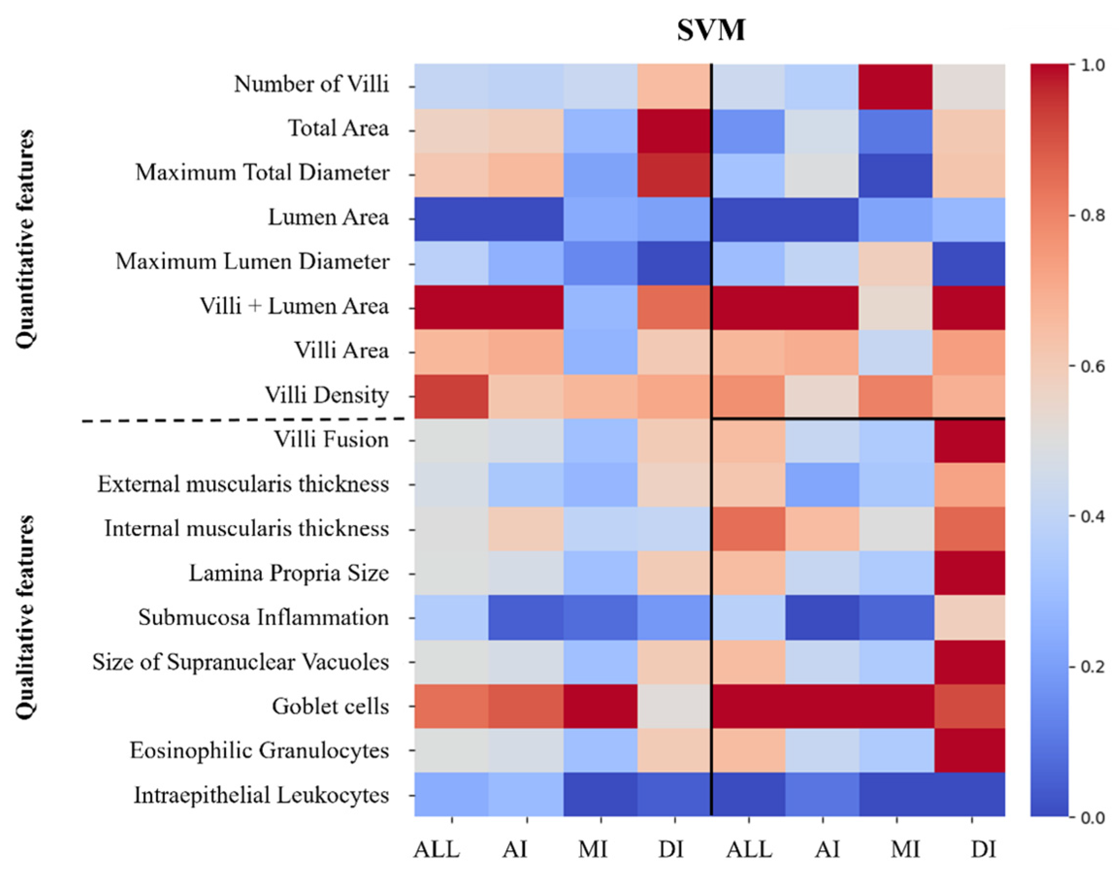
2.4.6. Feature Importance Extraction

For SVM and LR, with accessible feature importance attributes (coef_ or feature_importances_), these were extracted post-training. Feature importances were averaged across folds to determine their overall contribution to the model performance, and a dataset was constructed to store the importance values for each feature and algorithm. Feature importances were summarized and normalized to a 0–1 range for comparison purposes and used to produce heatmaps (Seaborn 0.12.2) to visualize the relative importance of each feature across the different intestinal section/feature types, providing insights into which features are most influential in each models’ predictions.

2.5. Statistical Analysis

Results were analyzed using the IBM SPSS statistics 27.0.1 software package (SPSS®, IBM, Chicago, IL, USA). Quantitative data were first tested for normality and homogeneity of variances (Shapiro–Wilk and Levene tests, respectively) and then analyzed by one-way ANOVA tests. Tukey’s multiple range test was used to identify differences between means. Semi-quantitative data were analyzed using nonparametric Kruskal–Wallis tests followed by multiple comparisons. Data on accuracy and precision were analyzed using a three-way ANOVA test with intestinal sections, features, and algorithms as factors. When significant interactions between two factors were observed, data were analyzed by a one-way ANOVA test to disclose each factor’s main effects. A probability level of 0.05 was used for the rejection of the null hypothesis in all tests.

3. Results

3.1. Growth Performance

Data on the growth performance and feed utilization of fish fed the experimental diets are presented in Table 2. The final weight, weight gain, daily growth index, and feed efficiency decreased significantly with the increase in the diet’s challenge level. Feed intake was higher in the ED group.
The biometric indexes of fish fed the different diets also presented significant differences (Table 3). The HSI, ESI, and VSI increased significantly with the diet’s challenge level.
The mortality rate was 1.7% in the CTRL group, 3.3% in the CD group, and 2.2% in the ED group with no significant difference in mortality rates between the control and treatment groups (p > 0.05). Mortality was observed across all treatment groups, with no clear correlation to diet composition.

3.2. Gut Morphology

The histological analysis showed differences in the intestinal sections of fish fed the different challenge diets (Figure 3). Results from the semi-quantitative analysis are presented in Table 4. In the anterior intestine, intraepithelial leukocytes and eosinophilic granulocytes showed higher scores in the challenged groups, while the number of goblet cells was higher in the CTRL group. Supranuclear vacuole size increased with the diet challenge. In the intermediate intestine, villi fusion and eosinophilic granulocytes showed higher scores in the CD group than the other groups, while goblet cells were decreased in the challenge groups. Lamina propria size, submucosa inflammation, supranuclear vacuoles, and intraepithelial leukocyte size were higher in the challenged groups. In the distal intestine, lamina propria size, submucosa inflammation, and eosinophilic granulocytes had higher scores in the CD group, followed by the ED and the CTRL groups, while all other features had higher scores in both challenged groups than the control.
In the quantitative analysis (Table 5) of the anterior intestine, all features measured decreased with the increase in diet challenge level, except villi density, which was increased. In the intermediate intestine, total area, total maximum diameter, lumen maximum diameter, villi area, and villi + lumen area presented higher values in the CD group, followed by the CTRL, with the lowest values being observed in the EC group. Villi density was highest, and number of villi was lowest in the ED group, followed by the CTRL. In the distal intestine, total area and total maximum diameter, villi number and area, and villi + lumen area were lower in the ED group than in the CTRL and CD groups. Lumen maximum diameter was highest in the CD group and lowest in the CTRL group, villi density was highest in ED group, and lumen area was higher in the CD and ED groups than in the CTRL group.

3.3. Nutritional Status Classification

Accuracy and precision results across algorithms, features, and intestinal sections are presented in Table 6. The three-way ANOVA analysis showed significant differences in the accuracy and precision of all treatments across different sections, features, and algorithms. Significant interactions were also found in the accuracy and precision of sections and features (Table 7). Within sections, accuracy was the highest in the intermediate section and lowest in the anterior section. The precision for the CD group followed the same pattern.
In contrast, precision in the CTRL group was higher in both intermediate and distal sections, and precision in the ED group was higher in the intermediate section. Accuracy was highest when considering all types of features and lowest when only considering quantitative features. The same results were observed for precision in the CD group, while in the CTRL group, the highest precision was observed when all features and semi-quantitative features were considered. The highest precision was obtained in the ED group when all features were used.
The top three algorithms with the highest accuracy were ensemble stacking, SVM, and logistic regression, while the algorithm with the lowest accuracy was decision trees. Ensemble stacking and logistic regression presented the highest values for precision in the CTRL group. In the CD group, logistic regression had the highest precision value, with decision trees presenting the lowest value. In the ED group, precision was higher in the SVM, KNN, logistic regression, ensemble stacking, and random forest algorithms, while the lowest value was also with decision trees. A summary of the algorithms with average accuracy above 70% across intestinal sections of meagre and precision across treatments are presented in Table S1. For the algorithms with the highest accuracy, the impact of all analyzed features per intestinal section is represented in Figure 4 (SVM) and Figure 5 (logistic regression). Representative learning curves for the better performing algorithms (SVM, ES, LR) and datasets (including all intestinal sections and features) are presented in Figure 6. The learning curves for all datasets can be found in Figures S1–S3. Overall, CV scores improve with more samples but remain lower than the high training scores observed.

4. Discussion

Fish nutrition studies are crucial for the future advancement of the aquaculture industry by providing new, more sustainable, and economical feeds. However, novel feeds usually incorporate alternative ingredients that may affect fish health, particularly fish gut health. Histological analysis remains one of the most used practices to evaluate fish gut alterations. Yet, improvements are necessary to this practice to make histomorphological changes in the fish gut easier to evaluate. Implementing recent technologies, such as image-based measurements and machine learning methods, can be a valuable strategy to make histological studies easier for non-skilled personnel to apply.
This study aimed to contribute to this endeavor by testing the efficiency of a combination of histological evaluation methods to train a machine learning model capable of identifying sub-optimal nutritional status.
As expected, in this study, the challenging diets negatively affected the overall zootechnical response of meagre. Although nutritionally complete, these diets contained plant feedstuffs at levels not typically used for the species due to their content of antinutritional factors, lower digestibility, and low palatability [4,7]. As such, they led to reduced final weight, weight gain, and daily growth index in groups fed these diets. In response to the dietary challenge, the animals in these groups likely maintained a higher feed intake as a compensatory mechanism to meet their basic metabolic requirements, despite slower growth. In contrast, the animals in the control group, which exhibited better growth, required less feed later in the trial as their growth stabilized.
As expected, the challenging diets also affected the biometric indexes measured (HSI, ESI, and VSI), indicating that physiological consequences of malnutrition occurred in the fish. It is known that high HSI can be an indicator of liver damage, as it is often associated with an increase in liver size from the hypertrophy and/or hyperplasia of hepatocytes [56,57]. High ESI can indicate possible inflammation and damage in the fish gut, as previously seen in Atlantic salmon (Salmo salar) fed plant protein [4,58,59]. Intestinal inflammation in this study is later confirmed by the data from the histological analysis. Typically, inflammation occurs as a response to injury, and it is the result of the activity of the innate immune system, with the migration of granulocytes and macrophages to the site [60]. However, gut inflammation can also be caused by plant ingredients present in the diet, for example, soybean meal, which contains saponins, membrane-disturbing molecules that increase the quantity of antigens that cross the epithelium, leading to inflammation [61]. A large number of studies have established a link between vegetable content in feeds, mainly soybean meal, and the onset of inflammation and other gut problems in various species of fish [35,62,63].
Histological analysis has long been used in aquaculture nutrition studies to evaluate gut inflammation. It is generally based on a semi-quantitative, score-based analysis, allowing for a relatively complete description of a particular intestinal section [51,58]. This type of analysis includes features that illustrate the gut health status in fish well; however, the need for specialized personnel often restrains it, and it is time-consuming and prone to subjectivity. A quantitative evaluation is typically limited to a few parameters, such as villi length and width, lamina propria width, and mucous cell number [29,64]. Although it is more straightforward to apply and only requires personnel with minimum histological training, it does not account for factors such as differences in fish size and between sections.
This work used both semi-quantitative and quantitative histological features to train machine learning algorithms and evaluate how accurately it could distinguish malnourished and healthy meagre. This strategy was applied to three sections of the intestine, and it was possible to observe differences between groups of fish that were fed different diets.
In the anterior intestine, the semi-quantitative analysis presented differences in four out of seven features evaluated, a low number compared with the other intestine sections. The increase in vacuole presence and size in challenged groups was likely linked with an abnormal accumulation of lipids due to a high level of n-6 and n-9 fatty acids fats in diets high in vegetable oils [65] and fatty acid absorption rate that exceeds the enterocyte’s capacity for lipoprotein synthesis [29,66] necessary for the transport of lipids from the intestine to the rest of the body [67]. On the other hand, the increase in immune cell numbers, such as granulocytes and intraepithelial leukocytes, indicates inflammation and innate immune system activation in both challenged groups [24]. The lower goblet cell number in the challenged groups is indicative of decreased mucus production and unhealthier status in those fish.
Quantitative data showed a decrease in size and villosity density in the challenged groups, which cannot be dissociated from the lower size of those fish. However, it can also be related to plant antinutritional factors in challenging diets. For instance, shorter villosities related to dietary soybean meal have been observed in other species, such as Atlantic salmon and largemouth bass (Micropterus nigricans) [18,63].
As the intermediate section of the intestine becomes more involved with immune function, a more evident inflammatory reaction is expected to occur with the increase in the challenge level of the diets. Accordingly, this section presented higher villi fusion, lamina propria size, and submucosa inflammation in the challenged groups than in the anterior section. Increased villi fusion is often considered a sign of intestine inflammation [33,68,69].
Quantitative data also indicated alterations in the size of various features measured in this section. The larger total area and diameter in the CTRL group could be linked to the bigger size of the fish. However, the CD group had the highest values for these features, likely due to a combination of factors. Although the fish in the CD group were smaller than those in the CTRL group, they presented a more inflamed intestine, contributing to increased measurements.
The distal intestine showed even more distinguishable semi-quantitative features between groups. In this section, the CD group also presented the highest value of some features, indicating increased inflammation compared with the CTRL group. It is important to note that in this section, unlike the others, goblet cells presented higher numbers in both challenged groups. Most nutrition studies focus on the distal intestine, which is typically the most affected by diet composition and where the inflammatory and immunological responses are more evident. This section also generally shows an increase in the number of goblet cells required for the lubrification of undigested feed [32]. Additionally, the increase in goblet cells and mucus production is likely linked to an activation of the immunological system in response to inflammation in challenged fish caused by antinutritional factors in the feed [70].
As for the quantitative analysis, higher values were also detected in the CD group for most features, except for villi density, which was higher in the ED group. The increase in villosity density in the distal intestine of the ED group may be related to the higher amount of fiber in this diet and the need to increase intestine surface area to improve nutrient absorption, as also seen in gilthead seabream (Sparus aurata) [35]. Nonetheless, the potential influence of fish size on villi density cannot be overlooked, as it may be a contributing factor in this group, where the fish are the smallest.
Overall, this study’s results showed that the response trend of the quantitative features analyzed seems to agree with those of the semi-quantitative features for all sections of the intestine.
Recently, some studies aimed to compare the effectiveness of histological techniques versus machine learning models to predict different fish characteristics, like age and reproductive status [36,37]. This study used a different approach, combining both methods to obtain an optimum way of detecting malnutrition in fish through intestinal health. For that purpose, various machine learning algorithms were tested to see if they could distinguish between fish fed with optimal, sub-optimal, and poor diets using information provided by semi-quantitative and quantitative histomorphological features. The application of machine learning stands out as it as it enables the generation of predictive models that improve and adapt over time, making histomorphological analysis faster and more accessible, even for individuals with limited experience in histology.
The results of this study indicate that machine learning is a promising tool for improving the evaluation of intestinal histomorphology and aiding in the diagnosis of malnutrition in aquaculture fish. Moreover, it was observed that a combination of semi-quantitative and quantitative data best detects malnourished fish due to the increase in data amount and a better description of intestinal physiology. However, if used individually, the semi-quantitative features provided better accuracies.
This is likely not only due to semi-quantitative features remaining the most descriptive and detailed method of characterizing the intestine histomorphology, but also because the quantitative analysis in this study is not based on an extensive number of features and can be biased by the fish size, which was very different between groups. Additionally, the semi-quantitative analysis is not as affected by tissue sample integrity, which is a major flaw in quantitative histological methods [30]. Authors such as [30,34] provide a more extensive list of quantitative features using advanced image analysis technology. However, such technology still needs to be optimized before the industry can use it.
In this study, the goal was to combine simple and ready-to-perform measurements in an aquaculture scenario. For this, we applied simple software to perform hand-made measurements that were later combined with machine learning algorithms to test how such algorithms would perform with information from traditional histological measurements performed by researchers. Despite this, an increase in the variety of quantitative measurements taken and careful attention to fish size can be key to enhancing the accuracy of the selected models in future studies. This could be achieved, for example, by normalizing the histological measurements to the fish’s weight or length or by applying statistical methods to adjust for size-related effects.
When considering all features from the entire intestine, SVM, ensemble stacking, and logistic regression presented accuracies above 75%. Considering the intestinal sections separately, the anterior intestine performed worse than the other sections in the algorithm methods, and only Logistic regression presented an accuracy above 70%. Therefore, results from this study suggest that the anterior portion is not optimal for evaluating malnutrition in meagre.
The intermediate intestine presented the best results among the different intestinal sections, with accuracies above 85% for various algorithms, and the highest accuracy being achieved at 90% with logistic regression. The accuracy level in this intestinal section indicates that it was more susceptible to nutritional changes than other sections in meagre. As in this study, in European seabass (Dicentrarchus labrax), the intermediate intestine was also where it was easier to observe differences between fish fed a control commercial diet and fish fed an algae blend diet, both through quantitative and semi-quantitative data [34]. The sensitivity of the intermediate section is likely because it presents a midpoint between different intestinal functions, those of digestion and absorption and those of immunity. Therefore, the adverse effects of inflammation and the adaptation of intestinal structures to improve digestion and absorption can be better represented in this section of the fish gut. However, it is important to note that results can vary within this section depending on whether it is obtained closer to the anterior or posterior portion of the intestine, which highlights the importance of establishing reference sampling points for different species and sizes. In accordance with the results of this study, the optimal sampling location for the intermediate intestine is suggested to be precisely at the midpoint of the intestine’s middle section.
For the distal intestine, the best-performing algorithms were KNN, logistic regression, and ensemble stacking, all with accuracy above 80%. The distal section of the intestine is consistently used in nutritional studies for the evaluation of the effects of diets, as it has many immune-relevant functions [35,62,70]. Despite presenting a lower accuracy than the intermediate section, the distal portion of the intestine remains an informative place to observe the effects of malnutrition on gut inflammation.
For precision, the intermediate and distal sections of the intestine provided similar values, higher than those of the anterior section, aligning with the accuracy results. Across treatments, independently of the section, precision was highest for identifying fish in the CTRL and ED groups, showing less consistent results in classifying the intermediate case, the CD group. The improvement in the precision of the CD group in the posterior sections of the intestine highlights their choice in malnutrition diagnosis.
For SVM and logistic regression, the importance of each feature used for the classification task was extracted and showed that the models provided similar results. Considering the quantitative features, most villi-related features were relevant to classifying nutritional status in all sections, but the number of villosities was particularly important in the intermediate intestine. Alterations in villosities are explored in most nutrition studies involving histological analysis and often present interesting results. Because the mucosal folds play a crucial role in nutrient digestion and absorption [71,72], any damage caused to villosities is detrimental to fish’s health, making them an important feature to focus on.
Total area and total maximum diameter were only relevant in the distal section. In contrast, in orange-spotted grouper, the intestine diameter was also important in distinguishing fish fed a soybean meal-heavy diet, but only in the anterior section [73]. In other studies, however, the total area and diameter of the intestine did not show promising results in malnutrition diagnosis [34,74]. This again indicates that in the present study, these features were likely influenced by fish size. The number of goblet cells was an important feature in discriminating between malnutrition conditions in all sections of the intestine; however, intraepithelial leukocytes and submucosa inflammation did not significantly help in any section.
It is clear from this analysis that the semi-quantitative features have a higher impact in the distal section. This was to be expected, as the semi-quantitative method applied [25,51] was based on a system initially designed to diagnose soybean meal-induced enteritis in this section.
Overall, the low CV scores observed in the learning curves across datasets and models indicate sample size was a limiting factor in the present study, and increasing the datasets size could improve the generalization capacity of all three models.

5. Conclusions

Combining semi-quantitative histological features with quantitative measures of intestinal structure can be an important strategy for predicting fish nutritional status using machine learning approaches. Furthermore, if considered independently, semi-quantitative features presented better accuracies than quantitative features. Further research is required to improve quantitative methods to better describe the fish’s gut health status, and fish size should be taken into consideration as it can influence results. Results of this study also showed that the logistic regression, SVM, and ensemble stacking algorithms performed best across all sections of the intestine. However, the intermediate section showed the best levels of accuracy among different algorithms, possibly indicating its higher sensitivity to nutritional changes. The present study, despite being preliminary, shows potential for the use of ML models to classify fish nutritional status based on gut histological features. Further exploration into parameter tuning and selection of the most representative features could improve the models using the present datasets. However, dataset size was a limiting factor, indicating the need to build larger datasets with data from the scientific community working in fish nutrition fields. To achieve that goal, standardization of sampling procedures, data acquisition, and adherence to fair data principles are recommended to produce a valid and useful dataset to develop ML applications in fish nutrition.

Supplementary Materials

The following supporting information can be downloaded at: https://www.mdpi.com/article/10.3390/jmse12122177/s1, Table S1: Algorithms with average accuracy above 70% across intestinal sections of meagre (Argyrosomus regius) and precision across treatments. Figure S1. Representative learning curves for the best-performing algorithms (logistic regression, ensemble stacking, and support vector machines), across each intestinal section (anterior, intermediate, and distal) and for all sections combined, with all features combined. Figure S2. Representative learning curves for the best-performing algorithms (logistic regression, ensemble stacking, and support vector machines), across each intestinal section (anterior, intermediate, and distal) and for all sections combined, for quantitative features. Figure S3. Representative learning curves for the best-performing algorithms (logistic regression, ensemble stacking, and support vector machines), across each intestinal section (anterior, intermediate, and distal) and for all sections combined, for semi-quantitative features.

Author Contributions

Conceptualization, J.O. and A.C.; methodology, J.O., A.C., M.B. and F.S.; software, A.C.; investigation, J.O. and A.C.; resources, A.C., A.O.-T. and P.P.-F.; data curation, J.O. and A.C.; writing—original draft preparation, J.O.; writing—review and editing, J.O., A.C., A.O.-T., M.B. and F.S.; visualization, J.O. and A.C.; supervision, A.C. and A.O.-T. All authors have read and agreed to the published version of the manuscript.

Funding

The researcher J. Oliveira was supported by a grant from the Foundation for Science and Technology (FCT), Portugal (UI/BD/150902/2021). This work was supported by INOVAQUA (MAR-021.1.3-FEAMPA-00004).

Institutional Review Board Statement

The animal study protocol was approved by the Ethics Committee of EPPO/IPMA’s Animal Welfare and Ethics Body (ORBEA) (protocol code: 0421/000/000/2023; date of approval: 26 June 2023).

Informed Consent Statement

Not applicable.

Data Availability Statement

Data are contained within the article.

Conflicts of Interest

The authors declare no conflicts of interest.

References

  1. Olmos Soto, J.; Paniagua-Michel, J.d.J.; Lopez, L.; Ochoa, L. Functional Feeds in Aquaculture. In Springer Handbook of Marine Biotechnology; Kim, S.-K., Ed.; Springer: Berlin/Heidelberg, Germany, 2015; pp. 1303–1319. [Google Scholar]
  2. Gasco, L.; Gai, F.; Maricchiolo, G.; Genovese, L.; Ragonese, S.; Bottari, T.; Caruso, G. Fishmeal Alternative Protein Sources for Aquaculture Feeds. In Feeds for the Aquaculture Sector: Current Situation and Alternative Sources; Gasco, L., Gai, F., Maricchiolo, G., Genovese, L., Ragonese, S., Bottari, T., Caruso, G., Eds.; Springer International Publishing: Cham, Switzerland, 2018; pp. 1–28. [Google Scholar]
  3. Aragão, C.; Gonçalves, A.T.; Costas, B.; Azeredo, R.; Xavier, M.J.; Engrola, S. Alternative Proteins for Fish Diets: Implications beyond Growth. Animals 2022, 12, 1211. (In English) [Google Scholar] [CrossRef]
  4. Krogdahl, Å.; Penn, M.; Thorsen, J.; Refstie, S.; Bakke, A.M. Important antinutrients in plant feedstuffs for aquaculture: An update on recent findings regarding responses in salmonids. Aquac. Res. 2010, 41, 333–344. [Google Scholar] [CrossRef]
  5. Kumar, V.; Hossain, M.S.; Ragaza, J.A.; Benito, M.R. The Potential Impacts of Soy Protein on Fish Gut Health. In Soybean for Human Consumption and Animal Feed; IntechOpen: London, UK, 2020. [Google Scholar]
  6. Daniel, N. A review on replacing fish meal in aqua feeds using plant protein sources. Int. J. Fish. Aquat. Stud. 2018, 6, 164–179. [Google Scholar]
  7. Francis, G.; Makkar, H.P.S.; Becker, K. Antinutritional factors present in plant-derived alternate fish feed ingredients and their effects in fish. Aquaculture 2001, 199, 197–227. [Google Scholar] [CrossRef]
  8. Dawood, M.A.O. Nutritional immunity of fish intestines: Important insights for sustainable aquaculture. Rev. Aquac. 2021, 13, 642–663. [Google Scholar] [CrossRef]
  9. Xiong, J.B.; Nie, L.; Chen, J. Current understanding on the roles of gut microbiota in fish disease and immunity. Zool. Res. 2019, 40, 70–76. (In English) [Google Scholar] [CrossRef]
  10. Stosik, M.; Tokarz-Deptuła, B.; Deptuła, W. Immunity of the intestinal mucosa in teleost fish. Fish Shellfish. Immunol. 2023, 133, 108572. [Google Scholar] [CrossRef] [PubMed]
  11. Zhang, H.; Ran, C.; Teame, T.; Ding, Q.; Hoseinifar, S.H.; Xie, M.; Zhang, Z.; Yang, Y.; Olsen, R.E.; Gatlin, D.M.; et al. Research progress on gut health of farmers teleost fish: A viewpoint concerning the intestinal mucosal barrier and the impact of its damage. Rev. Fish Biol. Fish. 2020, 30, 569–586. [Google Scholar] [CrossRef]
  12. Luan, Y.; Li, M.; Zhou, W.; Yao, Y.; Yang, Y.; Zhang, Z.; Ringø, E.; Erik Olsen, R.; Liu Clarke, J.; Xie, S.; et al. The Fish Microbiota: Research Progress and Potential Applications. Engineering 2023, 29, 137–146. [Google Scholar] [CrossRef]
  13. Wang, A.R.; Ran, C.; Ringø, E.; Zhou, Z.G. Progress in fish gastrointestinal microbiota research. Rev. Aquac. 2018, 10, 626–640. [Google Scholar] [CrossRef]
  14. Magalhães, R.; Guerreiro, I.; Santos, R.A.; Coutinho, F.; Couto, A.; Serra, C.R.; Olsen, R.E.; Peres, H.; Oliva-Teles, A. Oxidative status and intestinal health of gilthead sea bream (Sparus aurata) juveniles fed diets with different ARA/EPA/DHA ratios. Sci. Rep. 2020, 10, 13824. [Google Scholar] [CrossRef] [PubMed]
  15. Guerreiro, I.; Castro, C.; Serra, C.R.; Coutinho, F.; Couto, A.; Peres, H.; Pousão-Ferreira, P.; Corraze, G.; Oliva-Teles, A.; Enes, P. Feeding Yellow Worms to Meagre: Effects on Whole-Body Fatty Acid Profile and Hepatic and Intestine Oxidative Status. Antioxidants 2023, 12, 1031. [Google Scholar] [CrossRef] [PubMed]
  16. Martin, S.A.M.; Dehler, C.E.; Król, E. Transcriptomic responses in the fish intestine. Dev. Comp. Immunol. 2016, 64, 103–117. [Google Scholar] [CrossRef] [PubMed]
  17. Lilleeng, E.; Frøystad, M.K.; Vekterud, K.; Valen, E.C.; Krogdahl, Å. Comparison of intestinal gene expression in Atlantic cod (Gadus morhua) fed standard fish meal or soybean meal by means of suppression subtractive hybridization and real-time PCR. Aquaculture 2007, 267, 269–283. [Google Scholar] [CrossRef] [PubMed]
  18. He, M.; Yu, Y.; Li, X.; Poolsawat, L.; Yang, P.; Bian, Y.; Guo, Z.; Leng, X. An evaluation of replacing fish meal with fermented soybean meal in the diets of largemouth bass (Micropterus salmoides): Growth, nutrition utilization and intestinal histology. Aquac. Res. 2020, 51, 4302–4314. [Google Scholar] [CrossRef]
  19. Lin, H.; Tan, B.; Ray, G.W.; Zeng, M.; Li, M.; Chi, S.; Yang, Q. A Challenge to Conventional Fish Meal: Effects of Soy Protein Peptides on Growth, Histomorphology, Lipid Metabolism and Intestinal Health for Juvenile Pompano Trachinotus ovatus. Front. Mar. Sci. 2022, 8, 815323. (In English) [Google Scholar] [CrossRef]
  20. Hoseinifar, S.H.; Shakouri, M.; Yousefi, S.; Van Doan, H.; Shafiei, S.; Yousefi, M.; Mazandarani, M.; Torfi Mozanzadeh, M.; Tulino, M.G.; Faggio, C. Humoral and skin mucosal immune parameters, intestinal immune related genes expression and antioxidant defense in rainbow trout (Oncorhynchus mykiss) fed olive (Olea europea L.) waste. Fish Shellfish Immunol. 2020, 100, 171–178. [Google Scholar] [CrossRef]
  21. Ruiz, A.; Andree, K.B.; Furones, D.; Holhorea, P.G.; Calduch-Giner, J.À.; Viñas, M.; Pérez-Sánchez, J.; Gisbert, E. Modulation of gut microbiota and intestinal immune response in gilthead seabream (Sparus aurata) by dietary bile salt supplementation. Front. Microbiol. 2023, 14, 1123716. (In English) [Google Scholar] [CrossRef] [PubMed]
  22. Xu, C.; Li, X.-F.; Tian, H.-Y.; Jiang, G.-Z.; Liu, W.-B. Feeding rates affect growth, intestinal digestive and absorptive capabilities and endocrine functions of juvenile blunt snout bream Megalobrama amblycephala. Fish Physiol. Biochem. 2016, 42, 689–700. [Google Scholar] [CrossRef]
  23. Santigosa, E.; García-Meilán, I.; Valentin, J.M.; Pérez-Sánchez, J.; Médale, F.; Kaushik, S.; Gallardo, M.A. Modifications of intestinal nutrient absorption in response to dietary fish meal replacement by plant protein sources in sea bream (Sparus aurata) and rainbow trout (Onchorynchus mykiss). Aquaculture 2011, 317, 146–154. [Google Scholar] [CrossRef]
  24. Raskovic, B.; Stankovic, M.; Markovic, Z.; Poleksic, V. Histological methods in the assessment of different feed effects on liver and intestine of fish. J. Agric. Sci. 2011, 56, 87–100. [Google Scholar] [CrossRef]
  25. Couto, A.; Barroso, C.; Guerreiro, I.; Pousão-Ferreira, P.; Matos, E.; Peres, H.; Oliva-Teles, A.; Enes, P. Carob seed germ meal in diets for meagre (Argyrosomus regius) juveniles: Growth, digestive enzymes, intermediary metabolism, liver and gut histology. Aquaculture 2016, 451, 396–404. [Google Scholar] [CrossRef]
  26. Couto, A.; Serra, C.R.; Guerreiro, I.; Coutinho, F.; Castro, C.; Rangel, F.; Lavrador, A.S.; Monteiro, M.; Santos, R.; Peres, H.; et al. Black soldier fly meal effects on meagre health condition: Gut morphology, gut microbiota and humoral immune response. J. Insects Food Feed. 2022, 8, 1281–1295. [Google Scholar] [CrossRef]
  27. Saavedra, M.; Barata, M.; Matias, A.C.; Couto, A.; Salem, A.; Ribeiro, L.; Pereira, T.G.; Gamboa, M.; Lourenço-Marques, C.; Soares, F.; et al. Effect of Dietary Incorporation of Yellow Mealworm as a Partial Fishmeal Replacer on Growth, Metabolism, and Intestinal Histomorphology in Juvenile Meagre (Argyrosomus regius). Aquac. Nutr. 2023, 2023, 6572421. [Google Scholar] [CrossRef]
  28. Meyerholz, D.K.; Beck, A.P. Fundamental Concepts for Semiquantitative Tissue Scoring in Translational Research. ILAR J. 2018, 59, 13–17. (In English) [Google Scholar] [CrossRef]
  29. Ribeiro, L.; Moura, J.; Santos, M.; Colen, R.; Rodrigues, V.; Bandarra, N.; Soares, F.; Ramalho, P.; Barata, M.; Moura, P.; et al. Effect of vegetable based diets on growth, intestinal morphology, activity of intestinal enzymes and haematological stress indicators in meagre (Argyrosomus regius). Aquaculture 2015, 447, 116–128. [Google Scholar] [CrossRef]
  30. Silva, P.F.; McGurk, C.; Knudsen, D.L.; Adams, A.; Thompson, K.D.; Bron, J.E. Histological evaluation of soya bean-induced enteritis in Atlantic salmon (Salmo salar L.): Quantitative image analysis vs. semi-quantitative visual scoring. Aquaculture 2015, 445, 42–56. [Google Scholar] [CrossRef]
  31. Vatsos, I.N. Planning and Reporting of the Histomorphometry Used to Assess the Intestinal Health in Fish Nutrition Research—Suggestions to Increase Comparability of the Studies. Front. Vet. Sci. 2021, 8, 666044. (In English) [Google Scholar] [CrossRef]
  32. Mokhtar, D.M. The Digestive System. In Fish Histology: From Cells to Organs; Mokhtar, D.M., Ed.; Apple Academic Press Inc.: Burlington, ON, Canada, 2017. [Google Scholar]
  33. Verdile, N.; Pasquariello, R.; Scolari, M.; Scirè, G.; Brevini, T.; Gandolfi, F. A Detailed Study of Rainbow Trout (Onchorhynchus mykiss) Intestine Revealed That Digestive and Absorptive Functions Are Not Linearly Distributed along Its Length. Animals 2020, 10, 745. [Google Scholar] [CrossRef]
  34. Ferreira, M.; Sousa, V.; Oliveira, B.; Canadas-Sousa, A.; Abreu, H.; Dias, J.; Kiron, V.; Valente, L.M.P. An in-depth characterisation of European seabass intestinal segments for assessing the impact of an algae-based functional diet on intestinal health. Sci. Rep. 2023, 13, 11686. [Google Scholar] [CrossRef]
  35. Baeza-Ariño, R.; Martínez-Llorens, S.; Nogales-Mérida, S.; Jover-Cerda, M.; Tomás-Vidal, A. Study of liver and gut alterations in sea bream, Sparus aurata L., fed a mixture of vegetable protein concentrates. Aquac. Res. 2016, 47, 460–471. [Google Scholar] [CrossRef]
  36. Flores, A.; Wiff, R.; Donovan, C.R.; Gálvez, P. Applying machine learning to predict reproductive condition in fish. Ecol. Inform. 2024, 80, 102481. [Google Scholar] [CrossRef]
  37. Spanou, D.S.; Petroudi, P.; Dimou, E.; Kokkinos, K.; Klaoudatos, D. Walleye (Sander vitreus, Mitchill 1818) age and sex classification using innovative supervised and unsupervised machine learning and soft computing methodologies. Fish. Res. 2024, 275, 107031. [Google Scholar] [CrossRef]
  38. Graham, C.A.; Shamkhalichenar, H.; Browning, V.E.; Byrd, V.J.; Liu, Y.; Gutierrez-Wing, M.T.; Novelo, N.; Choi, J.-W.; Tiersch, T.R. A practical evaluation of machine learning for classification of ultrasound images of ovarian development in channel catfish (Ictalurus punctatus). Aquaculture 2022, 552, 738039. [Google Scholar] [CrossRef] [PubMed]
  39. Palaiokostas, C. Predicting for disease resistance in aquaculture species using machine learning models. Aquac. Rep. 2021, 20, 100660. [Google Scholar] [CrossRef]
  40. Rahman, L.F.; Marufuzzaman, M.; Alam, L.; Bari, M.A.; Sumaila, U.R.; Sidek, L.M. Developing an Ensembled Machine Learning Prediction Model for Marine Fish and Aquaculture Production. Sustainability 2021, 13, 9124. [Google Scholar] [CrossRef]
  41. Mahesh, B. Machine Learning Algorithms—A Review. Int. J. Sci. Res. (IJSR) 2019, 9, 381–386. [Google Scholar] [CrossRef]
  42. FAO. Fishery and Aquaculture Statistics—Yearbook 2021. In FAO Yearbook of Fishery and Aquaculture Statistics; FAO: Rome, Italy, 2024. [Google Scholar]
  43. Monfort, M.C. Present Market Situation and Prospects of Meagre (Argyrosomus regius), as an Emerging Species in Mediterranean Aquaculture; Food and Agriculture Organization of the United Nations (FAO): Rome, Italy, 2010. [Google Scholar]
  44. Piccolo, G.; Bovera, F.; De Riu, N.; Marono, S.; Salati, F.; Cappuccinelli, R.; Moniello, G. Effect of two different protein/fat ratios of the diet on meagre (Argyrosomus regius) traits. Ital. J. Anim. Sci. 2010, 7, 363–371. [Google Scholar] [CrossRef]
  45. Suquet, M.; Divanach, P.; Hussenot, J.; Coves, D.; Christian, F. Marine fish culture of “new species” farmed in Europe. Cah. Agric. 2009, 18, 148–156. [Google Scholar] [CrossRef]
  46. Saavedra, M.; Pereira, T.G.; Barata, M.; Aragão, C.; Requeijo, B.; Conceição, L.E.C.; Pousão-Ferreira, P. Plant-based diets fed to juvenile meagre Argyrosomus regius with low methionine and taurine supplementation led to an overall reduction in fish performance and to an increase in muscle fibre recruitment. J. Fish Biol. 2022, 101, 1182–1188. [Google Scholar] [CrossRef]
  47. Carvalho, M.; Izquierdo, M.; Valdés, M.; Montero, D.; Farías, A. Oils Combination with Microalgal Products as a Strategy for Increasing the N-3 Long-Chain Polyunsaturated Fatty Acid Content in Fish Oil-Free Diets for Meagre (Argyrosomus regius). Aquac. Nutr. 2022, 2022, 5275570. [Google Scholar] [CrossRef]
  48. Katsika, L.; Tasbozan, O.; Mastoraki, M.; Karapanagiotis, S.; Zalamitsou, C.; Feidantsis, K.; Antonopoulou, E.; Chatzifotis, S. Effects of fish oil substitution by hazelnut oil on growth performance, whole-body fatty acid composition and enzymes of intermediary metabolism of juvenile meagre (Argyrosomus regius Asso, 1801). Aquac. Res. 2021, 52, 5760–5776. [Google Scholar] [CrossRef]
  49. de Moura, L.B.; Diógenes, A.F.; Campelo, D.A.V.; de Almeida, F.L.A.; Pousão-Ferreira, P.M.; Furuya, W.M.; Peres, H.; Oliva-Teles, A. Nutrient digestibility, digestive enzymes activity, bile drainage alterations and plasma metabolites of meagre (Argyrosomus regius) feed high plant protein diets supplemented with taurine and methionine. Aquaculture 2019, 511, 734231. [Google Scholar] [CrossRef]
  50. Asencio-Alcudia, G.; Andree, K.B.; Giraldez, I.; Tovar-Ramirez, D.; Alvarez-González, A.; Herrera, M.; Gisbert, E. Stressors Due to Handling Impair Gut Immunity in Meagre (Argyrosomus regius): The Compensatory Role of Dietary L-Tryptophan. Front. Physiol. 2019, 10, 547. (In English) [Google Scholar] [CrossRef] [PubMed]
  51. Urán, P.A.; Schrama, J.W.; Rombout, J.H.W.M.; Obach, A.; Jensen, L.; Koppe, W.; Verreth, J.A.J. Soybean meal-induced enteritis in Atlantic salmon (Salmo salar L.) at different temperatures. Aquac. Nutr. 2008, 14, 324–330. [Google Scholar] [CrossRef]
  52. Oliva, M.; Unceta, C.; Canales, M. Histomorphology and histochemistry of the digestive tract in meagre (Argyrosomus regius). Biochem. Indian J. 2011, 5, 10–17. [Google Scholar]
  53. Zhao, S.; Zhang, S.; Liu, J.; Wang, H.; Zhu, J.; Li, D.; Zhao, R. Application of machine learning in intelligent fish aquaculture: A review. Aquaculture 2021, 540, 736724. [Google Scholar] [CrossRef]
  54. Ray, S. A Quick Review of Machine Learning Algorithms. In Proceedings of the 2019 International Conference on Machine Learning, Big Data, Cloud and Parallel Computing (COMITCon), Faridabad, India, 14–16 February 2019; pp. 35–39. [Google Scholar]
  55. Habbat, N.; Anoun, H.; Hassouni, L.; Hicham, N. Analyzing Booking’s Comments Using Stacking Ensemble Deep Learning Model and Neural Topic Model. SSRN Electron. J. 2022. [Google Scholar] [CrossRef]
  56. Araújo, F.; Morado, C.; Parente, T.; Jose Roma Paumgartten, F.; Gomes, I. Biomarkers and bioindicators of the environmental condition using a fish species (Pimelodus maculatus Lacepède, 1803) in a tropical reservoir in Southeastern Brazil. Braz. J. Biol. 2017, 78, 351–359. [Google Scholar] [CrossRef] [PubMed]
  57. Goede, R.W. Organismic indices and an autopsy-based assessment as indicator of health and condition of fish. Am. Fish. Soc. Symp. 1990, 8, 93–108. [Google Scholar]
  58. Krogdahl, A.; Bakke, A.M.; Baeverfjord, G. Effects of graded levels of standard soybean meal on intestinal structure, mucosal enzyme activities, and pancreatic response in Atlantic salmon (Salmo salar L.). Aquac. Nutr. 2003, 9, 361–371. [Google Scholar] [CrossRef]
  59. Penn, M.H.; Bendiksen, E.Å.; Campbell, P.; Krogdahl, Å. High level of dietary pea protein concentrate induces enteropathy in Atlantic salmon (Salmo salar L.). Aquaculture 2011, 310, 267–273. [Google Scholar] [CrossRef]
  60. Secombes, C.J. Cytokines and Immunity. In Principles of Fish Immunology: From Cells and Molecules to Host Protection; Buchmann, K., Secombes, C.J., Eds.; Springer International Publishing: Cham, Switzerland, 2022; pp. 301–353. [Google Scholar]
  61. Farrell, A.P. Encyclopedia of Fish Physiology; Academic Press: Cambridge, MA, USA, 2011. [Google Scholar]
  62. Rašković, B.; Stanković, M.; Markelić, M.; Poleksić, V.; Božić, G.; Janković, S.; Marković, Z. Growth, feed utilization, and quantitative histological assessment of the distal intestine and liver of common carp (Cyprinus carpio L.) fed formulated diets containing grains of different soybean cultivars. Aquac. Int. 2024, 32, 6903–6921. [Google Scholar] [CrossRef]
  63. Aidos, L.; Mirra, G.; Pallaoro, M.; Herrera Millar, V.R.; Radaelli, G.; Bazzocchi, C.; Modina, S.C.; Di Giancamillo, A. How Do Alternative Protein Resources Affect the Intestine Morphology and Microbiota of Atlantic Salmon? Animals 2023, 13, 1922. [Google Scholar] [CrossRef]
  64. Panettieri, V.; Chatzifotis, S.; Messina, C.M.; Olivotto, I.; Manuguerra, S.; Randazzo, B.; Ariano, A.; Bovera, F.; Santulli, A.; Severino, L.; et al. Honey Bee Pollen in Meagre (Argyrosomus regius) Juvenile Diets: Effects on Growth, Diet Digestibility, Intestinal Traits, and Biochemical Markers Related to Health and Stress. Animals 2020, 10, 231. (In English) [Google Scholar] [CrossRef]
  65. Turchini, G.M.; Torstensen, B.E.; Ng, W.-K. Fish oil replacement in finfish nutrition. Rev. Aquac. 2009, 1, 10–57. [Google Scholar] [CrossRef]
  66. Olsen, R.E.; Myklebust, R.; Ringø, E.; Mayhew, T.M. The influences of dietary linseed oil and saturated fatty acids on caecal enterocytes in Arctic char (Salvelinus alpinus L.): A quantitative ultrastructural study. Fish Physiol. Biochem. 2000, 22, 207–216. [Google Scholar] [CrossRef]
  67. Berg, J.M.; Tymoczko, J.L.; Stryer, L. The Biosynthesis of Membrane Lipids and Steroids. In Biochemistry, 8th ed.; W. H. Freeman and Company: New York, NY, USA, 2015. [Google Scholar]
  68. Li, Y.; Kortner, T.M.; Chikwati, E.M.; Munang’andu, H.M.; Lock, E.-J.; Krogdahl, Å. Gut health and vaccination response in pre-smolt Atlantic salmon (Salmo salar) fed black soldier fly (Hermetia illucens) larvae meal. Fish Shellfish. Immunol. 2019, 86, 1106–1113. [Google Scholar] [CrossRef]
  69. Glover, C.N.; Petri, D.; Tollefsen, K.-E.; Jørum, N.; Handy, R.D.; Berntssen, M.H.G. Assessing the sensitivity of Atlantic salmon (Salmo salar) to dietary endosulfan exposure using tissue biochemistry and histology. Aquat. Toxicol. 2007, 84, 346–355. [Google Scholar] [CrossRef]
  70. Caimi, C.; Gasco, L.; Biasato, I.; Malfatto, V.; Varello, K.; Prearo, M.; Pastorino, P.; Bona, M.C.; Francese, D.R.; Schiavone, A.; et al. Could Dietary Black Soldier Fly Meal Inclusion Affect the Liver and Intestinal Histological Traits and the Oxidative Stress Biomarkers of Siberian Sturgeon (Acipenser baerii) Juveniles? Animals 2020, 10, 155. [Google Scholar] [CrossRef]
  71. Faccioli, C.K.; Chedid, R.A.; Amaral, A.C.d.; Franceschini Vicentini, I.B.; Vicentini, C.A. Morphology and histochemistry of the digestive tract in carnivorous freshwater Hemisorubim platyrhynchos (Siluriformes: Pimelodidae). Micron 2014, 64, 10–19. [Google Scholar] [CrossRef] [PubMed]
  72. L⊘kka, G.; Austb⊘, L.; Falk, K.; Bjerkås, I.; Koppang, E.O. Intestinal morphology of the wild atlantic salmon (Salmo salar). J. Morphol. 2013, 274, 859–876. [Google Scholar] [CrossRef] [PubMed]
  73. Wang, Y.-r.; Wang, L.; Zhang, C.-x.; Song, K. Effects of substituting fishmeal with soybean meal on growth performance and intestinal morphology in orange-spotted grouper (Epinephelus coioides). Aquac. Rep. 2017, 5, 52–57. [Google Scholar] [CrossRef]
  74. Adeoye, A.A.; Jaramillo-Torres, A.; Fox, S.W.; Merrifield, D.L.; Davies, S.J. Supplementation of formulated diets for tilapia (Oreochromis niloticus) with selected exogenous enzymes: Overall performance and effects on intestinal histology and microbiota. Anim. Feed. Sci. Technol. 2016, 215, 133–143. [Google Scholar] [CrossRef]
Figure 2. Schematic representation of the machine learning workflow, illustrating each step in the process.
Jmse 12 02177 g002
Figure 3. Histomorphological appearance of meagre (Argyrosomus regius) intestinal sections when fed the control (CTRL), challenge (CD), and extreme challenge (ED) diets.
Jmse 12 02177 g003
Figure 4. Features importance for nutritional status classification by support vector machine (SVM) processing in each intestinal section × type of features. The scale shows features importance values normalized between 0 and 1.
Jmse 12 02177 g004
Figure 5. Features importance for nutritional status classification by logistic regression processing in each intestinal section × type of features. The scale shows features importance values normalized between 0 and 1.
Jmse 12 02177 g005
Figure 6. Representative learning curves for the best-performing algorithms (logistic regression, ensemble stacking, and support vector machines) across all intestinal sections (anterior, intermediate, and distal) and all feature types (quantitative and semi-quantitative).
Jmse 12 02177 g006
Table 1. Ingredient composition and proximate analysis of the experimental diets.
Experimental Diets
Control (CTRL)Challenge (CD)Extreme Challenge (ED)
Ingredients (% dry weight basis)
    Fish meal a55.115.05.0
    CPSP b5.05.05.0
    Wheat gluten c-10.012.0
    Corn gluten d-11.614.3
    Soybean meal e-20.025.0
    Rapeseed meal f-7.511.0
    Sunflower meal g-5.07.5
    Wheat meal h24.77.1-
    Fish oil11.37.05.0
    Soy oil-3.85.2
    Rapeseed oil-3.85.2
    Phosphate-0.41.1
    Vitamin i1.01.01.0
    Mineral j1.01.01.0
    Choline0.50.50.5
    Binder1.01.01.0
    Taurine0.30.30.3
Proximate analysis (% dry matter basis)
    Dry matter92.395.093.0
    Crude protein49.046.447.3
    Crude fat18.118.117.9
    Ash10.07.17.0
    Gross energy (kJ g−1 DM)23.024.123.9
CP—Crude protein; CF—crude fat. a Sorgal, S.A. Ovar, Portugal (CP—65.8%; CF—8.0%). b Soluble fish protein concentrate. Sopropèche G, France (CP: 77.0% DM; CL: 18.4% DM). c Sorgal, S.A. Ovar, Portugal (CP—83.0%; CF—2.3%). d Sorgal, S.A. Ovar, Portugal (CP—69.9%; CF—3.3%). e Sorgal, S.A. Ovar, Portugal (CP—51.3%; CF—1.1%). f Sorgal, S.A. Ovar, Portugal (CP: 39.9% DM; CL: 2.9% DM). g Sorgal, S.A. Ovar, Portugal (CP: 33.0% DM; CL 1.7%DM). h Sorgal, S.A. Ovar, Portugal (CP—12.2%; CF—1.2%). i Vitamins (mg kg−1 diet): retinol, 18,000 (IU kg−1 diet); cholecalciferol, 2000 (IU kg−1 diet); alpha tocopherol, 35; menadion sodium bisulfate, 10; thiamin, 15; riboflavin, 25; Ca pantothenate, 50; nicotinic acid, 200; pyridoxine, 5; folic acid, 10; cyanocobalamin, 0.02; biotin, 1.5; ascorbyl monophosphate, 50; inositol, 400. j Minerals (mg kg−1 diet): cobalt sulfate, 1.91; copper sulfate, 19.6; iron sulfate, 200; sodium fluoride, 2.21; potassium iodide, 0.78; magnesium oxide, 830; manganese oxide, 26; sodium selenite, 0.66; zinc oxide, 37.5; potassium chloride, 1.15 (g kg−1 diet); sodium chloride, 0.44 (g kg−1 diet).
Table 2. Zootechnical performance of meagre (Argyrosomus regius) fed the experimental diets (average initial body weight of 4.6 ± 0.4 g).
CTRLCDEDp-Value
Final weight (g)48.7 ± 1.0 c30.0 ± 0.6 b19.3 ± 1.0 a<0.001
Weight gain (g)44.2 ± 1.0 c25.4 ± 0.6 b14.9 ± 1.1 a<0.001
Feed intake (g kg ABW−1 day−1)39.1 ± 1.1 a38.7 ± 1.2 a46.98 ± 1.7 b<0.001
Feed efficiency 0.85 ± 0.03 c0.76 ± 0.03 b0.53 ± 0.04 a<0.001
Daily growth index3.99 ± 0.05 c2.90 ± 0.04 b2.07 ± 0.11 a<0.001
Mean values and standard deviation (±SD) are presented for each parameter (n = 3, 6 pools of 3 fish/diet). Different letters in the same row stand for statistical differences between diets (p < 0.05). Daily growth index: ((final body weight1/3 − initial body weight1/3)/time in days) × 100. Feed efficiency: wet weight gain/dry feed intake. Diets: control (CTRL), challenge (CD), extreme challenge (ED).
Table 3. Organosomatic indexes of meagre (Argyrosomus regius) fed the experimental diets.
CTRLCDEDp-Value
Hepatosomatic index (HSI)1.5 ± 0.3 a2.0 ± 0.2 b2.6 ± 0.4 c<0.001
Enterosomatic index (ESI)1.2 ± 0.2 a1.5 ± 0.2 ab1.6 ± 0.2 b0.005
Viscerosomatic index (VSI)4.3 ± 1.0 a5.1 ± 0.9 ab6.0 ± 0.9 b0.004
Mean values and standard deviation (±SD) are presented for each parameter (n = 3). Different letters in the same row stand for statistical differences between diets (p < 0.05). Diets: control (CTRL), challenge (CD), extreme challenge (ED). HSI: (liver weight/body weight) × 100; VSI: (viscera weight/body weight) × 100; ESI: (intestine weight/ body weight) × 100.
Table 4. Semi-quantitative analysis of meagre (Argyrosomus regius) intestine sections after being submitted to different quality diets.
Intestine
Section
Semi-Quantitative FeatureControl (CTRL)Challenge (CD)Extreme Challenge (ED)Kruskal–Wallis
Villi fusion1.45 ± 0.541.50 ± 0.671.52 ± 0.660.940
Lamina propria size 1.90 ± 0.501.89 ± 0.451.97 ± 0.390.647
Submucosa inflammation1.93 ± 0.532.17 ± 0.502.07 ± 0.850.131
AnteriorSupranuclear vacuoles size1.27 ± 0.37 a1.54 ± 062 ab1.86 ± 0.71 b0.000
Goblet cells1.45 ± 0.28 b1.14 ± 0.28 a1.08 ± 0.21 a0.000
Eosinophilic granulocytes2.13 ± 0.42 a2.45 ± 0.60 b2.39 ± 0.87 b0.046
Intraepithelial leukocytes2.15 ± 0.40 a2.41 ± 0.48 b2.47 ± 0.62 b0.006
External muscularis thickness1.17 ± 0.261.26 ± 0.351.21 ± 0.310.537
Internal muscularis thickness1.82 ± 0.321.81 ± 0.411.89 ± 0.330.489
Villi fusion1.59 ± 0.65 a1.95 ± 0.65 b1.62 ± 0.68 a0.015
Lamina propria size 2.08 ± 0.49 a2.74 ± 0.60 b2.57 ± 0.67 b0.000
Submucosa inflammation1.73 ± 0.57 a2.19 ± 0.77 b2.61 ± 0.77 b0.000
IntermediateSupranuclear vacuoles size1.20 ± 0.43 a2.00 ± 0.93 b2.32 ± 0.99 b0.000
Goblet cells2.21 ± 0.48 b1.62 ± 0.29 a1.5 ± 0.29 a0.000
Eosinophilic granulocytes1.40 ± 0.52 a1.66 ± 0.58 b1.37 ± 0.59 a0.006
Intraepithelial leukocytes2.01 ± 0.52 a2.50 ± 0.51 b2.45 ± 0.42 b0.000
External muscularis thickness1.07 ± 0.17 a1.11 ± 0.23 a1.23 ± 0.28 b0.003
Internal muscularis thickness1.75 ± 0.421.71 ± 0.351.85 ± 0.260.165
Villi fusion1.38 ± 0.50 a2.22 ± 0.69 b2.46 ± 0.90 b0.000
Lamina propria size 1.83 ± 0.58 a2.98 ± 0.47 c2.35 ± 0.47 b0.000
DistalSubmucosa inflammation1.56 ± 0.34 a2.28 ± 0.65 c1.68 ± 0.56 b0.000
Supranuclear vacuoles size1.84 ± 0.67 a2.47 ± 0.66 b2.40 ± 0.96 b0.000
Goblet cells1.45 ± 0.43 a2.03 ± 0.52 b1.97 ± 0.40 b0.000
Eosinophilic granulocytes1.24 ± 0.41 a2.17 ± 0.67 c1.83 ± 0.75 b0.000
Intraepithelial leukocytes1.44 ± 0.43 a2.89 ± 0.57 b2.60 ± 0.49 b0.000
External muscularis thickness1.41 ± 0.491.56 ± 0.401.44 ±0.320.079
Internal muscularis thickness1.76 ± 0.341.84 ± 0.361.71 ± 0.310.061
Mean values and standard deviation (± SD) are presented for each feature. Different letters in the same row stand for statistical differences between diets (p < 0.05).
Table 5. Histomorphological quantitative evaluation of the different intestinal sections of meagre (Argyrosomus regius) fed different levels of challenging diets.
Intestine
Section
Quantitative FeatureControl (CTRL)Challenge (CD)Extreme Challenge (ED)ANOVA p-Value
Total Area (mm2)1.75 ± 0.59 c1.49 ± 0.54 b1.02 ± 0.38 a0.000
Total Maximum Diameter (µm)1597 ± 313 c1455 ± 270 b1221 ± 260 a0.000
Lumen Area (mm2)0.29 ± 0.27 ab0.33 ± 0.28 b0.20 ± 0.18 a0.039
AnteriorLumen Maximum Diameter (µm)709 ± 340704 ± 266581 ± 2590.059
Villi Area (mm2)1.18 ± 0.31 c0.91 ± 0.26 b0.65 ± 0.20 a0.000
Villi + Lumen Area (mm2)1.52 ± 0.54 c1.26 ± 0.50 b0.86 ± 0.34 a0.000
Number of Villi37 ± 5 b36 ± 4 b33 ± 5 a0.001
Villi Density33 ± 7 a42 ± 9 b54 ± 13 c0.000
Total Area (mm2)0.90 ± 0.64 b1.66 ± 0.56 c0.50 ± 0.45 a0.000
Total Maximum Diameter (µm)1080 ± 430 b1560 ± 293 c782 ± 374 a0.000
Lumen Area (mm2)0.29 ± 0.30 a0.69 ± 0.44 b0.14 ± 0.19 a0.000
IntermediateLumen Maximum Diameter (µm)635 ± 306 b1000 ± 326 c425 ± 263 a0.000
Villi Area (mm2)0.51 ± 0.35 b0.82 ± 0.22 c0.28 ± 0.23 a0.000
Villi + Lumen Area (mm2)0.80 ± 0.58 b1.51 ± 0.57 c0.42 ± 0.39 a0.000
Number of Villi39 ± 5 b37 ± 5 b31 ± 5 a0.000
Villi Density152 ± 136 b49 ± 13 a221 ± 137 c0.000
Total Area (mm2)2.19 ± 0.80 ab2.55 ± 0.73 b1.94 ± 0.84 a0.001
Total Maximum Diameter (µm)1758 ± 336 ab1941 ± 322 b1664 ± 403 a0.001
DistalLumen Area (mm2)0.40 ± 0.36 a0.78 ± 0.41 b0.58 ± 0.45 ab0.000
Lumen Maximum Diameter (µm)772 ± 322 a1138 ± 327 c936 ± 362 b0.000
Villi Area (mm2)1.34 ± 0.43 b1.30 ± 0.32 b1.00 ± 0.35 a0.000
Villi + Lumen Area (mm2)1.74 ± 0.73 ab2.10 ± 0.65 b1.56 ± 0.74 a0.002
Number of Villi48 ± 10 ab50 ± 6 b46 ± 9 a0.028
Villi Density38 ± 10 a41 ± 10 a51 ± 17 b0.000
Villi density = villi number/villi area. Different letters in the same row stand for statistical differences between diets (p < 0.05). Mean values and standard deviation (± SD) are presented.
Table 6. Accuracy and precision across treatments by diverse algorithms based on different features across intestinal sections.
SectionsALLAIMIDI
Features/
Algorithms
ALLQTSQTALLQTSQTALLQTSQTALLQTSQT
AccuracyAB72.0 ± 4.653.9 ± 11.165.0 ±6.861.1 ± 10.853.8 ± 13.263.7 ± 11.284.1 ± 9.464.9 ± 12.367.4 ± 17.674.0 ± 9.957.4 ± 9.971.0 ± 9.7
DT66.5 ± 6.452.9 ± 861.3 ± 8.356.1 ± 15.452.3 ± 13.658.6 ± 1074.7 ± 10.266.7 ± 10.769.2 ± 6.275.4 ± 16.848.6 ± 13.074.0 ± 11.8
ES78.2 ± 5.161.7 ± 6.469.1 ± 5.565.4 ± 18.363.5 ± 13.160.5 ± 10.389.6 ± 7.171.6 ± 12.973.5 ± 5.983.9 ± 8.556.2 ± 5.681.4 ± 9.7
KNN72.0 ± 5.757.4 ± 5.667.0 ± 7.864.1 ± 10.764.7 ± 12.659.3 ± 8.885.2 ± 9.771.0 ± 13.074.7 ± 8.484.5 ± 7.952.5 ± 10.879.5 ± 10.4
LR76.3 ± 4.258.9 ± 6.167 ± 7.472.8 ± 13.264 ± 15.862.9 ± 8.390.1 ± 8.874.1 ± 14.074.7 ± 8.984.5 ± 10.461.2 ± 11.583.2 ± 9.9
NB66.1 ± 5.450.2 ± 1067.1 ± 7.462.4 ± 1655 ± 17.459.3 ± 10.785.8 ± 11.068.6 ± 10.372.8 ± 8.478.4 ± 9.446.4 ± 10.985.6 ± 12.2
RF74.1 ± 4.360.9 ± 8.568.3 ± 7.867.2 ± 15.159.7 ± 18.361.7 ± 10.980.9 ± 11.170.3 ± 12.674.7 ± 8.881.4 ± 12.255.6 ± 8.980.8 ± 12.4
SVM78.4 ± 5.960.7 ± 6.469.5 ± 6.464.8 ± 14.761.7 ± 1862.4 ± 13.887.1 ± 9.974.7 ± 14.575.3 ± 7.280.8 ± 9.163.1 ± 8.782.0 ± 8.2
Precision CTRLAB0.84 ± 0.080.52 ± 0.170.73 ± 0.140.74 ± 0.330.48 ± 0.370.83 ± 0.150.88 ± 0.150.58 ± 0.230.90 ± 0.110.91 ± 0.100.78 ± 0.130.88 ± 0.09
DT0.72 ± 0.130.54 ± 0.200.69 ± 0.180.68 ± 0.300.64 ± 0.250.76 ± 0.220.75 ± 0.220.65 ± 0.230.85 ± 0.150.83 ± 0.150.62 ± 0.140.86 ± 0.15
ES0.85 ± 0.120.65 ± 0.160.73 ± 0.150.80 ± 0.160.71 ± 0.210.76 ± 0.140.94 ± 0.120.68 ± 0.220.90 ± 0.090.94 ± 0.080.69 ± 0.090.91 ± 0.10
KNN0.74 ± 0.150.54 ± 0.110.68 ± 0.130.74 ± 0.150.69 ± 0.220.71 ± 0.160.83 ± 0.180.62 ± 0.190.85 ± 0.110.95 ± 0.080.55 ± 0.100.89 ± 0.12
LR0.83 ± 0.130.60 ± 0.150.76 ± 0.130.88 ± 0.120.68 ± 0.290.76 ± 0.090.93 ± 0.120.74 ± 0.230.89 ± 0.100.92 ± 0.100.72 ± 0.140.90 ± 0.09
NB0.78 ± 0.150.52 ± 0.280.71 ± 0.130.72 ± 0.210.61 ± 0.330.74 ± 0.140.93 ± 0.140.63 ± 0.330.78 ± 0.150.95 ± 0.080.44 ± 0.110.92 ± 0.10
RF0.78 ± 0.150.61 ± 0.160.74 ± 0.150.81 ± 0.190.63 ± 0.310.72 ± 0.120.85 ± 0.200.68 ± 0.210.82 ± 0.140.89 ± 0.120.68 ± 0.140.90 ± 0.11
SVM0.82 ± 0.160.60 ± 0.140.74 ± 0.150.79 ± 0.200.67 ± 0.220.80 ± 0.170.90 ± 0.150.70 ± 0.200.83 ± 0.110.93 ± 0.100.68 ± 0.130.91 ± 0.10
Precision CDAB0.61 ± 0.080.57 ± 0.170.57 ± 0.060.46 ± 0.120.46 ± 0.160.51 ± 0.180.80 ± 0.160.67 ± 0.160.55 ± 0.360.65 ± 0.120.52 ± 0.190.72 ± 0.17
DT0.61 ± 0.110.49 ± 0.130.58 ± 0.100.48 ± 0.230.37 ± 0.190.48 ± 0.210.75 ± 0.200.68 ± 0.190.60 ± 0.130.76 ± 0.210.44 ± 0.270.72 ± 0.17
ES0.73 ± 0.100.58 ± 0.110.67 ± 0.090.55 ± 0.290.55 ± 0.240.43 ± 0.190.87 ± 0.120.69 ± 0.180.65 ± 0.200.80 ± 0.130.49 ± 0.170.74 ± 0.13
KNN0.65 ± 0.040.54 ± 0.090.62 ± 0.110.48 ± 0.350.54 ± 0.230.52 ± 0.210.88 ± 0.150.68 ± 0.160.69 ± 0.210.79 ± 0.150.51 ± 0.210.73 ± 0.13
LR0.69 ± 0.100.58 ± 0.120.60 ± 0.090.62 ± 0.200.46 ± 0.240.58 ± 0.220.92 ± 0.140.69 ± 0.150.71 ± 0.220.79 ± 0.130.51 ± 0.210.83 ± 0.16
NB0.56 ± 0.100.49 ± 0.130.62 ± 0.080.52 ± 0.350.44 ± 0.320.47 ± 0.120.79 ± 0.170.69 ± 0.170.77 ± 0.260.67 ± 0.120.51 ± 0.190.83 ± 0.14
RF0.67 ± 0.060.58 ± 0.130.63 ± 0.110.63 ± 0.260.51 ± 0.230.46 ± 0.170.76 ± 0.140.69 ± 0.150.76 ± 0.180.76 ± 0.180.45 ± 0.210.82 ± 0.15
SVM0.74 ± 0.100.60 ± 0.130.64 ± 0.070.57 ± 0.300.52 ± 0.260.48 ± 0.210.83 ± 0.130.72 ± 0.190.72 ± 0.220.74 ± 0.130.55 ± 0.160.78 ± 0.14
Precision EDAB0.75 ± 0.080.55 ± 0.170.65 ± 0.170.77 ± 0.180.65 ± 0.180.67 ± 0.180.92 ± 0.110.80 ± 0.220.65 ± 0.260.69 ± 0.240.50 ± 0.160.64 ± 0.22
DT0.66 ± 0.120.55 ± 0.130.55 ± 0.110.57 ± 0.170.60 ± 0.220.57 ± 0.180.79 ± 0.190.78 ± 0.160.65 ± 0.180.73 ± 0.270.40 ± 0.190.66 ± 0.15
ES0.76 ± 0.080.65 ± 0.140.65 ± 0.120.70 ± 0.220.72 ± 0.200.63 ± 0.280.92 ± 0.110.90 ± 0.130.76 ± 0.170.80 ± 0.170.51 ± 0.170.85 ± 0.21
KNN0.77 ± 0.140.66 ± 0.100.70 ± 0.130.72 ± 0.240.75 ± 0.180.68 ± 0.240.90 ± 0.110.94 ± 0.110.74 ± 0.180.82 ± 0.150.49 ± 0.350.75 ± 0.21
LR0.78 ± 0.090.58 ± 0.150.64 ± 0.090.76 ± 0.180.71 ± 0.160.63 ± 0.230.93 ± 0.130.90 ± 0.130.73 ± 0.170.82 ± 0.170.57 ± 0.300.82 ± 0.20
NB0.71 ± 0.120.56 ± 0.160.65 ± 0.130.64 ± 0.180.57 ± 0.200.70 ± 0.240.89 ± 0.110.72 ± 0.170.74 ± 0.130.80 ± 0.200.50 ± 0.340.85 ± 0.22
RF0.77 ± 0.130.64 ± 0.140.66 ± 0.090.72 ± 0.230.70 ± 0.200.67 ± 0.220.90 ± 0.130.82 ± 0.160.73 ± 0.250.78 ± 0.160.55 ± 0.150.76 ± 0.24
SVM0.78 ± 0.130.65 ± 0.160.69 ± 0.130.69 ± 0.180.72 ± 0.200.67 ± 0.230.95 ± 0.120.92 ± 0.110.77 ± 0.150.77 ± 0.200.68 ± 0.170.80 ± 0.18
Intestinal sections: ALL—all sections; AI—anterior; MI—intermediate; DI—distal. Features: ALL—all features; QT—quantitative; SQT—semi-quantitative; algorithms: AB—Adaboost; DT—decision trees; ES—ensemble stacking; KNN—k-nearest neighbor; LR—logistic regression; NB—naïve Bayes; RF—random forest; SVM—support vector machine. Diets: control (CTRL), challenge (CD), extreme challenge (ED). Mean values and standard deviation (± SD) are presented.
Table 7. Results from the three-way ANOVA analysis relating the accuracy and precision of machine learning prediction of malnutrition status of meagre (Argyrosomus regius) according to intestinal section, type of analysis, and algorithm model.(a) Pairwise comparisons indicating statistically significant differences between sections and features. (b) Statistical differences between algorithms.
Three-Way ANOVASectionFeaturesAlgorithmSection ×
Feature
Section ×
Algorithm
Feature ×
Algorithm
Section ×
Feature ×
Algorithm
Accuracy<0.001<0.001<0.001<0.001nsnsns
Precision CTRL<0.001<0.001<0.001<0.001nsnsns
Precision CD<0.001<0.001<0.0010.002nsnsns
Precision ED<0.001<0.001<0.001<0.001nsnsns
Sections
(a)FeaturesALLAIMIDI
AccuracyALLb, nsa, nsc, Bc, B
QTa, nsa, nsb, Aa, A
SQTb, nsa, nsc, Ad, B
Precision CTRLALLa, Ca, Bb, Bb, B
QTns, Ans, Ans, Ans, A
SQTa, Ba, Bb, Bb, B
Precision CDALLb, Ca, nsc, Bd, B
QTa, Aa, nsb, Aa, A
SQTa, Bb, nsb, Ac, B
Precision EDALLa, Ba, nsb, Bb, B
QTb, Ab, nsc, Ba, A
SQTa, Aa, nsab, Ab, B
(b) AccuracyPrecision CTRLPrecision CDPrecision ED
AlgorithmABabababab
DTaaaa
EScbabcb
KNNbcababcb
LRcbcb
NBabaabcab
RFbcababcb
SVMcabbcb
Pairwise comparisons indicate that statistically significant differences are p < 0.001. ns—non-significant. Intestinal sections: ALL—all sections; AI—anterior; MI—intermediate; DI—distal. Features: ALL—all features; QT—quantitative; SQT—semi-quantitative. Algorithms: AB—Adaboost; DT—decision trees; ES—ensemble stacking; KNN—k-nearest neighbor; LR—logistic regression; NB—naïve Bayes; RF—random forest; SVM—support vector machine. Diets: control (CTRL), challenge (CD), extreme challenge (ED). (a) Different lowercase letters in the same row stand for significant differences between sections; different uppercase letters in the same column stand for significant differences across features; (b) different letters in the same columns stand for differences across algorithms.
Disclaimer/Publisher’s Note: The statements, opinions and data contained in all publications are solely those of the individual author(s) and contributor(s) and not of MDPI and/or the editor(s). MDPI and/or the editor(s) disclaim responsibility for any injury to people or property resulting from any ideas, methods, instructions or products referred to in the content.

