Article

mTOR Plays a Conserved Role in Regulation of Nutritional Metabolism in Bivalve Sinonovacula constricta

1
School of Marine Sciences, Ningbo University, Ningbo 315211, China
2
Ningbo Institute of Oceanography, Ningbo 315211, China
3
Fujian Dalai Seed Science and Technology Co., Ltd., Ningde 352101, China
*
Authors to whom correspondence should be addressed.
J. Mar. Sci. Eng. 2023, 11(5), 1040; https://doi.org/10.3390/jmse11051040
Submission received: 15 April 2023 / Revised: 9 May 2023 / Accepted: 10 May 2023 / Published: 12 May 2023
(This article belongs to the Special Issue Marine Fish Physiology and Molecular Nutrition)

Abstract

:
The mammalian target of rapamycin (mTOR) has been shown to play a central role in regulating cell growth and metabolism. However, little is known about the function of mTOR in nutrient metabolism in bivalve mollusks. In this study, the role of mTOR in the regulation of nutrient metabolism was investigated in Sinonovacula constricta. First, the activation of mTOR was assayed after starvation and refeeding. Afterwards, the role of mTOR in the regulation of nutrient metabolism was investigated using an activator (MHY1485) or inhibitor (rapamycin) of mTOR. The open reading frame of the S. constricta mTOR is 7416 bp in length and encodes a polypeptide consisting of 2471 amino acids. The mTOR amino acid sequence of S. constricta was highly conserved when compared with other species and had a close evolutionary relationship with the TOR proteins of Crassostrea gigas and Lingula anatine. mTOR was expressed in the intestine, exhalent siphon, labial palppus, muscle, inhalent siphon, gill, mantle, digestive land, and gonad tissue of S. constricta, with the highest expression in muscle. During starvation, the level of phosphorylated mTOR protein was relatively low, and the ratio of LC3II/LC3I protein and the AMPKα mRNA level significantly increased with the increase in starvation time. After feeding, the level of phosphorylated mTOR protein increased from 0.13 to 0.56, and the ratio of LC3II/I protein and AMPKα mRNA level decreased from 1.17 to 0.38. MHY1485 significantly increased the level of phosphorylated 4E-BP1 and significantly decreased the ratio of LC3II/I proteins. Furthermore, MHY1485 significantly increased the mRNA level of the glucose metabolism-related gene glucokinase (GK), significantly decreased the mRNA expression of the G6P gene, and significantly increased the mRNA expression of the lipid synthesis-related genes sterol-regulatory element-binding protein (SREBP) and stearoyl-CoA desaturase (SCD). Rapamycin significantly reduced the level of phosphorylated 4E-BP1 and the mRNA expression of mTOR, and the expression level of phosphorylated 4EBP1 decreased from 0.97 to 0.28. Meanwhile, it also significantly reduced the mRNA expression of glucose metabolism-related genes GK, pyruvate kinase (PK), glucose transporter 1 (GLUT1), and G6P, as well as lipid synthesis-related genes SCD and acetyl-CoA carboxylase (ACC). These results indicate a conserved role of mTOR in regulating nutritional metabolism, including glucose metabolism, lipid synthesis, and autophagy in S. constricta.

1. Introduction

The mammalian target of rapamycin (mTOR) is crucial for controlling cell growth and metabolism. As a member of the PI3K-related kinase (PIKK) family, mTOR regulates the activity of two distinct proteins, mTOR complexes 1 and 2 (mTORC1 and mTORC2, respectively) [1]. The former primarily controls cell growth, proliferation, anabolism, and catabolism, whereas the latter primarily controls cell survival and cytoskeletal reorganization [2].
Cellular energy and nutritional state directly regulate mTORC1 to control associated signaling pathways downstream. By promoting anabolic processes such as ribosome biogenesis and protein and lipid synthesis or suppressing catabolic processes such as autophagy, mTORC1 orchestrates cell growth [3,4]. mTORC1 enhances transcription and translation initiation, elongation, and protein synthesis [5] by phosphorylating the downstream substrate, eukaryotic initiation factor 4E-binding protein 1 (4E-BP1). mTORC1 also mediates glycolysis and the pentose phosphate pathway via hypoxia-inducible factor 1 (HIF1), a transcriptional regulator of glucose transporters 1 (GLUT1), and the proto-oncogene c-Myc, a regulator of pyruvate kinase (PK) [6]. In addition, mTORC1 controls the expression of glucose 6-phosphate dehydrogenase (G6P), the rate-limiting enzyme in the pentose phosphate pathway [7]. Apart from protein synthesis and glucose metabolism, sterol response element-binding protein (SREBP), one of the most important master transcriptional factors of lipid synthesis, is regulated by mTORC1 [8]. When SREBP moves into the nucleus, it stimulates the transcription of several lipogenic enzymes, including fatty acid synthase (FAS), acetyl-CoA carboxylase (ACC), and stearoyl-CoA desaturase (SCD), among others [7].
Autophagy is a catabolic process regulated by nutrition status and mTORC1 [9]. Autophagy is a process of intracellular degradation of cellular components by lysosomes, where the cytoplasm is broken down into various basic components and can re-enter the cytoplasm for reuse [10]. Autophagy can prevent cell damage, promote cell survival in the absence of nutrition, and respond to cytotoxic stimuli [9]. Microtubule-associated protein 1 light chain 3 (LC3/ATG8) is a marker for autophagy. LC3I enzymatically cleaves off a segment of the polypeptide and couples it with phosphatidyl ethanolamine (PE) to form LC3II [11]. Consequently, a rise in the LC3II/I ratio is regarded as a sign of autophagy [12]. When nutrients are abundant, mTOR1 inhibits autophagy by directly phosphorylating the main transcription factor EB (TFEB) of the genes encoding lysosomes and autophagy [10]. Autophagy is also regulated by AMP-activated protein kinase (AMPK), which suppresses mTORC1 when foods or energy are insufficient [9].
The function of mTOR and its related signal pathway is widely studied in invertebrates. Drosophila lifespan can be extended by inhibiting the mTOR pathway and activating AMPKα signal transduction [13]. Meanwhile, the mTOR signaling pathway can also regulate neural stem cells and cell cycles in Drosophila [14]. In Caenorhabditis elegans, inhibition of mTOR reduces protein synthesis, prolongs lifespan, and promotes autophagy [15]. In crustaceans such as shrimp and crabs, mTOR is involved in endocrine regulation as well as metabolism regulation. It promotes protein synthesis and increases muscle protein content by activating the mTOR signaling pathway in Eriocheir sinensis [16]. Additionally, it has also been found that sustained molting steroid production and secretion in the molting glands of blackback land crab (Gecarcinus Lateralis) [17] and green crab (Carcinus maenas) [18] relied on mTOR-dependent protein synthesis [19]. In Litopenaeus vannamei, the inhibitor rapamycin inhibited the mTOR signaling pathway, significantly changed the expression of many autophagy-related genes, and significantly increased the ratio of LC3II/LC3I [20]. In Crassostrea gigas, 35 autophagy-associated proteins were first identified, followed by analysis of the expression and localization of microtubule-associated proteins 1A/1B-light chain 3 (LC3) and mTOR in different tissues of oysters [21]. In scallops, the organic pollutant benzo(a)pyrene (B(a)P) interfered with the expression of digestive gland lipid metabolizing enzyme genes, leading to lipid accumulation and disruption of lipid metabolism during reproduction, which may be regulated by mTOR [22].
The razor clam, Sinonovacula constricta, is one of the most important economic shellfish in China. The growth rate of S. constricta is relatively fast among shellfish, and nutrition affects their growth and development during the culture process. However, the regulation of nutritional metabolism in this species remains largely unknown. The mTOR signaling pathway is widely present in invertebrates and is closely related to growth and nutrient metabolism. This suggests that the mTOR signaling pathway may play an important role in the growth and metabolic regulation of S. constricta. Therefore, this study lays the foundation for elucidating the molecular mechanism of environmental regulation of nutrient metabolism in S. constricta. MHY1485 is an activator of mTOR in human cancer cells, osteoblast cells [23], chicken granulosa cells [24], porcine macrophages [25], mouse hepatocytes [26], and Danio rerio [27]. Rapamycin has been studied as an inhibitor of mTOR in human cancer cells [28], Mus musculus [29], Danio rerio [30], Drosophila [31], Xenopus laevis [32], Saccharomyces cerevisiae [33], and Brachionus calyciflorus [34]. In this study, the role of mTOR in nutritional metabolism in S. constricta was studied with MHY1485 and rapamycin.

2. Materials and Methods

2.1. Sequence Analysis and Tissue Expression

The mTOR-like sequences were obtained from the genome database of the razor clam (NCBI: txid98310). Sequence analysis was performed with ClustalX 1.83. The amino acid coding sequences were predicted using the open reading frame finder (https://www.ncbi.nlm.nih.gov/orffinder/, accessed on 20 July 2021). The domains of mTOR were analyzed by SMART. The tertiary structure of proteins was predicted by SWISS-MODEL. MEGA6.0 software was used to construct a phylogenetic tree.
S. constricta (5.0–6.0 cm) were purchased from the Ningbo Lulin Aquaculture firm (Ningbo, China) and maintained in aerated saltwater (salinity 21 ± 2, temperature 20 ± 1 °C). After 2 days of starvation, the intestine, exhalent siphon, labial palppus, muscle, inhalant siphon, gill, mantle, digestive land, and gonad tissues were sampled. Then, RNA was extracted for quantitative real-time PCR, and the method is described below.

2.2. Starvation and Re-Feeding Experiment

About 300 S. constricta were purchased as test subjects. Clams were subjected to 10-day starvation and 3-day refeeding. Clams were sampled after 0 h, 24 h, 72 h, and 240 h of starvation and after 6 h, 12 h, 24 h, and 72 h of subsequent re-feeding. After starvation treatment, they were fed a sufficient concentration of 3.0 × 10⁶ cells/mL of Isochrysis zhanjianggens, at which time the Isochrysis zhanjianggens reached an exponential growth phase. The water quality was maintained daily by renewing 60% of the water. The maintenance temperature was 16 ± 1.0 °C, the pH was 7.6 ± 0.50, and the salinity of the seawater was 21 ± 1 psu. Muscle tissues were taken and frozen at −80 °C.

2.3. Challenge Experiments and Samples

MHY1485 (MedChemExprsess, Monmouth Junction, NJ, USA) and rapamycin (MedChemExprsess, Monmouth Junction, NJ, USA) were dissolved in DMSO and then diluted in sterilized seawater. After 2 days of starvation, S. constricta were intramuscularly injected with 40, 20, 10, 5, 1, 0.5 µg/kg, and 0 µg/kg (control group) of rapamycin. After 7 days of starvation, S. constricta were intramuscularly injected with 40, 20, 10, 5, 1µg/kg, and 0 µg/kg (control group) of MHY1485. Muscle tissues were all dissected after 6 and 12 h of treatment, and then the expression of phosphorylated 4E-BP1 was detected. After the determination of the optimal dosage and time, S. constricta were intramuscularly injected with 5 µg/kg and 0 µg/kg of rapamycin (15 individuals/group) after 2 days of starvation. For MHY1485, S. constricta was intramuscularly injected with 10 µg/kg and 0 µg/kg of MHY1485 after 7 days of starvation. Muscle tissues were dissected after 6 h and stored in −80 °C refrigerator.

2.4. Western Blotting

Muscle tissues were ground in RIPA buffer (Cowin Biotech, Beijing, China) containing protease inhibitors (Cowin Biotech, Beijing, China) and phosphatase inhibitors (Thermo Fisher Scientific, Waltham, MA, USA), and then centrifuged at 13,000 rpm for 15 min at 4 °C. The bovine serum albumin (BCA) method was used to determine the protein concentration, with bovine serum albumin (BSA) serving as the control. A total of 15% separating gels (10 mL 30% Acrylamide (29:1), 5 mL 1.5 mM Tris-HCl (PH8.8), 200 µL 10% SDS, 100 µL 10% Ammonium persulfate, 4.6 mL ddH2O, and 8 µL TMED) and 10% concentrated gels (1.3 mL 30% Acrylamide (29:1), 1 mL 1 mM Tris-HCl (PH6.8), 50 µL 10% SDS, 75 µL 10%PAGE gel coagulant, 5.5 mL ddH2O, and 8 µL TMED) were made into an SDS-PAGE gel preparation kit (Beyotime, Shanghai, China). After normalization, the samples and protein molecular weight markers were loaded into 15% SDS-PAGE lanes and electrophoresed for 2 h. The samples on the gel were transferred to an immuno-blot polyvinylidene difluoride membrane (Merck, Darmstadt, Germany) by wet transfer technique with a transfer time of 1.5 h. The membranes were blocked in the closure solution for 30 min, followed by three 10-min TBST washes, before being incubated with the primary antibody overnight at 4 degrees on a shaker. The membranes were washed with TBST again three times for 10 min, then incubated for 20 min with anti-mouse or anti-rabbit antibodies and washed three times with TBST for 10 min. Finally, target proteins were measured by a chemiluminescence imager, the ChemiScope Capture Image System, as well as chemiluminescent liquid detection reagents. The primary antibodies included phosphor-mTOR (MilliporeSigma, Darmstadt, Germany, ABS79, 1:3000), β-actin (HuaBio, Hengshui, China, EM21002, 1:10,000), phosphor-4E-BP1 (Thr37/46) (Cell Signal Technology, Danvers, MA, USA, 236B4, 1:1000), and LC3A/B (Cell Signal Technology, Danvers, MA, USA, 4108, 1:3000). The antibodies were selected based on the conserved amino acid sequences of β-actin, mTOR, 4E-BP1, and LC3 A/B of S. constricta. The secondary antibodies included horseradish peroxidase-conjugated labeled goat anti-rat IgG (H+L) (Beyotime, Shanghai, China, 1:1000) and horseradish peroxidase-conjugated labeled goat anti-rabbit IgG (H+L) (Beyotime, Shanghai, China, 1:1000).

2.5. RNA Extraction and cDNA Synthesis

Total RNA was extracted using Trizol reagent (Ambion, Foster City, CA, USA). We used 1% agarose gel electrophoresis and a NanoDrop® ND-1000 (NanoDrop, Wilmington, DE, USA) to evaluate the purity and integrity of the RNA. Constantly, total RNA was reverse transcribed to the cDNA using the reverse transcription kit (TaKaRa, Maebashi, Japan) according to the manufacturer’s instructions. The qPCR primers were designed based on the gene sequences of our previous genome [35] (Table 1). The whole reaction mixture contained 2 µL cDNA, 0.8 µL each of forward and reverse primers at 10 µmol/L, 10μL of SYBR Green Mix (TaKaRa, Maebashi, Japan), and 6 µL of DEPC-treated water. Real-time quantitative PCR was carried out by using the SYBR Premix Ex Taq (TliRNase Plus) (TaKaRa, Maebashi, Japan) with a quantitative thermal cycler (Mastercycler ep realplex, Eppendorf, Hamburg, Germany). The program was set at 95 °C for 30 s, followed by 45 cycles of 95 °C for 5 s and 60 °C for 30 s, and the dissolution curve program was set at 95 °C for 30 s, 65 °C for 60 s, and 60 °C for 5 s. The dissolving curve was employed to test the specificity of the amplified product. Relative gene expression was calculated using the 2−ΔΔCt method; an antibody against β-actin was utilized as an internal control. The data were displayed as relative mRNA expression levels.

2.6. Statistical Analysis

The intensity of the protein bands was quantified using Image J software. The obtained data were analyzed by the SPSS 26.0 software package (IBM Corp., Foster City, CA, USA). The data were subjected to a one-way analysis of variance (ANOVA), followed by a Tukey test and a T-test in the SPSS 26.0 software package. In addition, the data are presented as the mean standard error of the mean (SEM). The differences were considered significant at p < 0.05.

3. Results

3.1. Molecular Characterization, Phylogenetic Relationship, and Tissue Expression Profiles of mTOR

The complete CDS sequence of the mTOR of S. constricta is 7416 bp, encoding 2471 amino acids. mTOR contains an unknown domain, DUF3385 (AA 824−996), a FAT domain (1481−1833), a rapamycin binding domain (1940−2039), a phosphoinositide 3-kinase catalytic domain (PI3Kc, AA 2108−2414) kinase domain, and a FATC domain (AA 2439−2471) (Figure 1a). The protein domains were predicted from the three-dimensional structure produced by AA 824−2471. The overall structure of mTOR in S. constricta is highly conserved in the TOR family (Figure 1b). The phylogenetic tree showed that the mTOR of S. constricta was closely related to Crassostrea gigas and Lingula anatina (Figure 1c). mTOR mRNA was expressed in all nine tissues of S. constricta, and the expression level was highest in muscle (Figure 1d).

3.2. Effects of Starvation and Re-Feeding on the Expressions of mTOR, LC3, and AMPKα

The level of phosphorylated mTOR was relatively low during starvation; however, the protein phosphorylation level increased significantly (p < 0.05) and reached its maximum after 6 h of refeeding (Figure 2a). The ratio of LC3II/I significantly increased after 10 days of starvation. The ratio of LC3II/I decreased and remained at a relatively low level after refeeding (Figure 2b). The AMPKα mRNA level significantly increased as the fasting period rose and decreased after feeding microalgae (Figure 2c).

3.3. Dose Screening of Rapamycin and MHY1485

After 12 h of treatment, 20 µg/kg MHY1485 significantly increased the level of phosphorylated 4E-BP1 compared to the control (Figure 3a; p < 0.05). After 6 h of treatment, MHY1485 at 40 and 10 µg/kg significantly increased the level of phosphorylated 4E-BP1 compared to the control (Figure 3b; p < 0.05). Therefore, the effects of MHY1485 on nutritional metabolism were further measured at 6 h after 10 µg/kg MHY1485 treatment. Meanwhile, after 12 h of treatment, rapamycin significantly inhibited the level of phosphorylated 4E-BP1 at doses of 40, 20, and 10 µg/kg compared with the control group (Figure 3c; p < 0.05). After 6 h of treatment, rapamycin at doses of 10 µg/kg and 5 µg/kg significantly reduced the level of phosphorylated 4E-BP1 compared with the control group (Figure 3d; p < 0.05). Therefore, the effects of rapamycin on nutritional metabolism were examined at 6 h after 5 µg/kg rapamycin treatment.

3.4. Effects of MHY1485 and Rapamycin on the Expression of mTOR and 4E-BP1

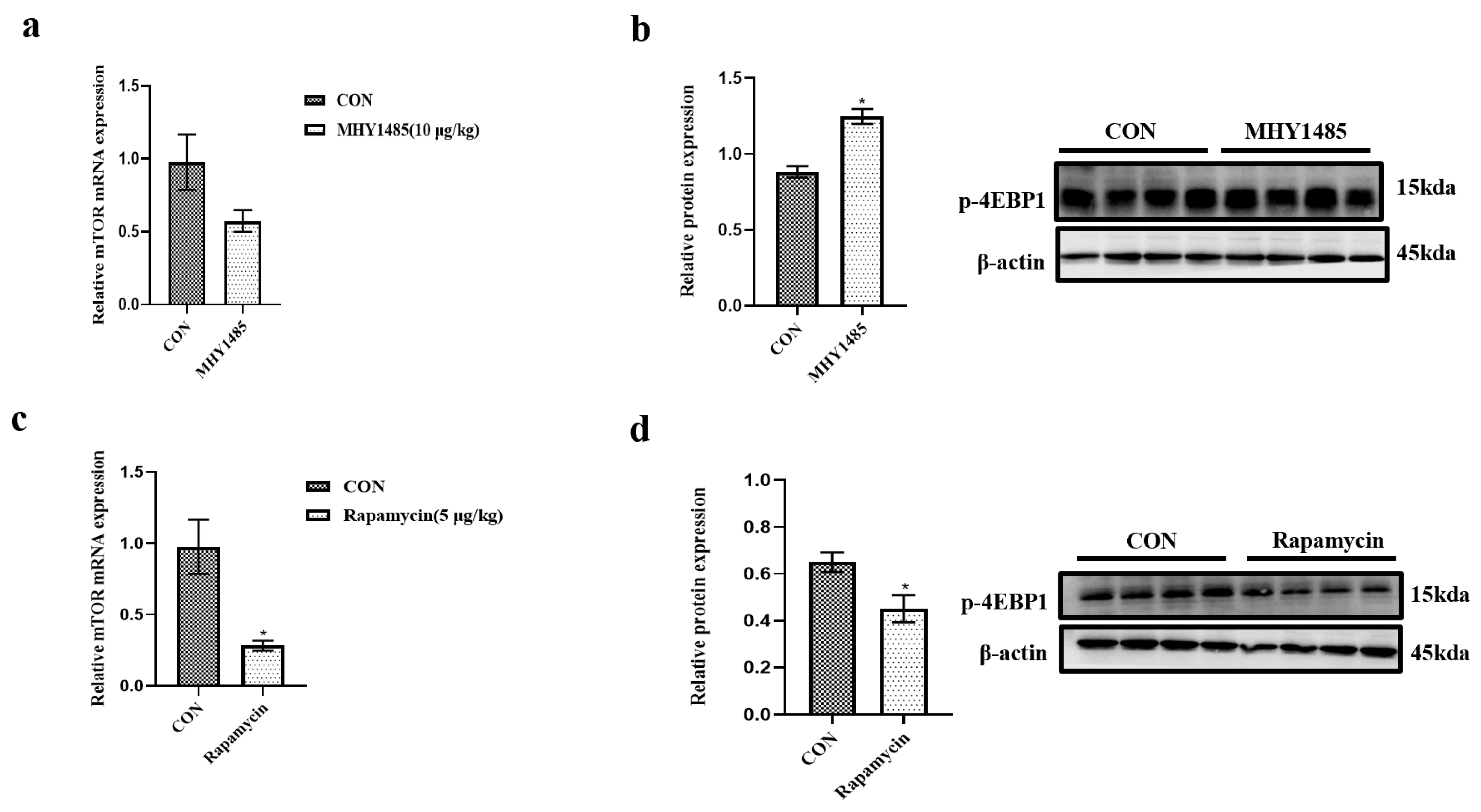
The mRNA expression of mTOR in the MHY1485 treatment group displayed an increased tendency; however, there was no significant difference (Figure 4a; p > 0.05). The phosphorylation of 4E-BP1 was significantly increased after MHY1485 treatment (Figure 4b; p < 0.05). Rapamycin significantly reduced the expression of mTOR mRNA compared to the control group (Figure 4c; p < 0.05). Meanwhile, the rapamycin-treated group had significantly lower phosphorylation of 4E-BP1 than the control group (Figure 4d; p < 0.05).

3.5. Effects of MHY1485 and Rapamycin on the Expression of AMPKα and LC3

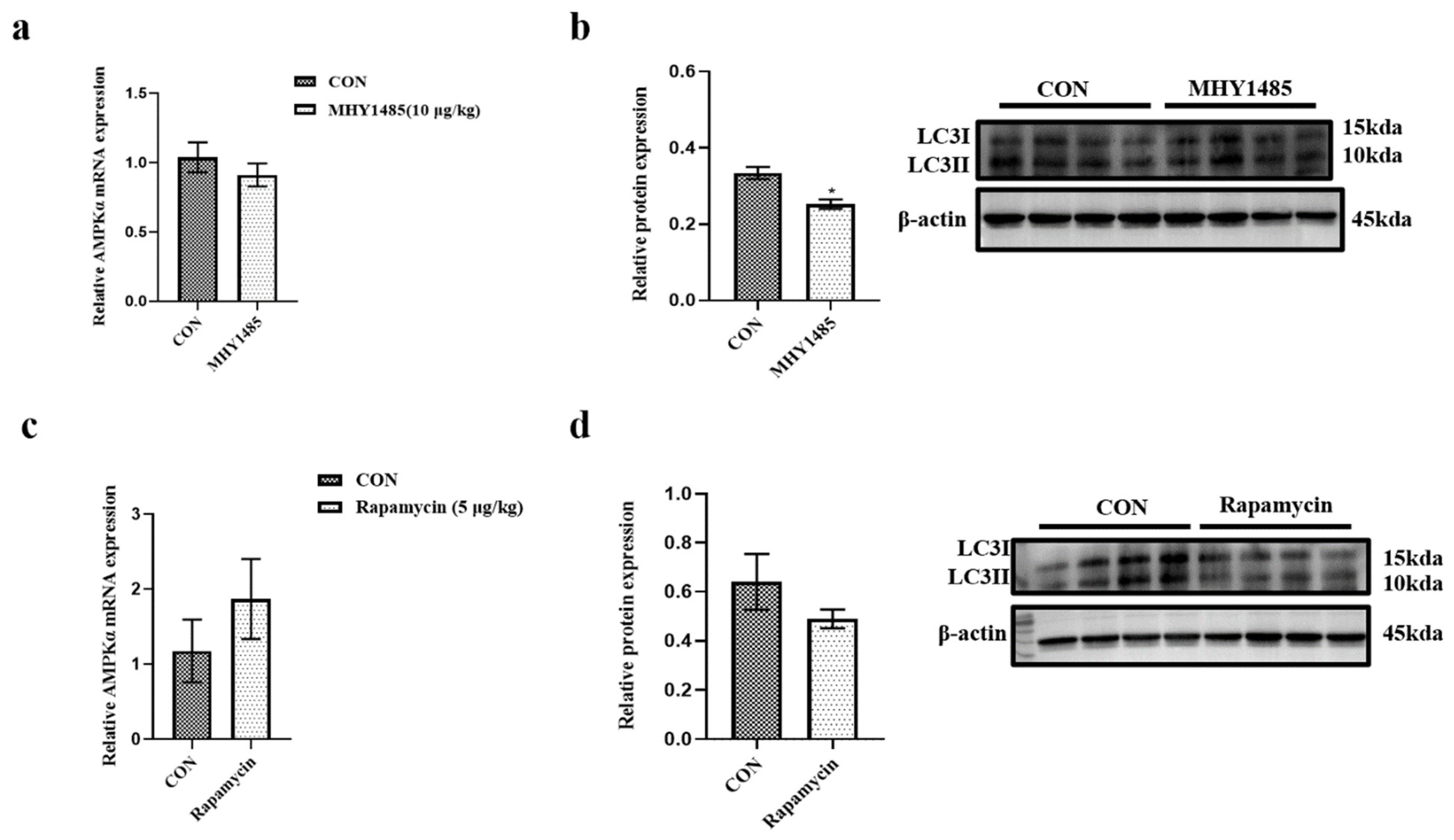
MHY1485 decreased the mRNA expression of AMPKα, but there was no significant difference (Figure 5a; p > 0.05). MHY1485 significantly down-regulated the LC3II/I ratio compared to the control group (Figure 5b; p < 0.05). The AMPKα mRNA level and the LC3II/I ratio were not significantly changed in the rapamycin group compared with the control group (Figure 5c,d; p > 0.05).

3.6. Effects of MHY1485 and Rapamycin on the Expression of Genes Related to Nutritional Metabolism

The mRNA expression of glucokinase (GK) was dramatically raised while G6P was dramatically decreased in the MHY1485 when compared to the control group (Figure 6a; p < 0.05). In addition, MHY1485 significantly increased the mRNA expression of SREBP and SCD compared with the control group (Figure 6a; p < 0.05). MHY1485 did not significantly influence ACC mRNA expression (Figure 6a; p > 0.05). The mRNA expression of tricarboxylic acid cycle (TCA)-related genes, including citrate synthase (CS) and nicotinamide adenine dinucleotide phosphate (NADP), was not significantly increased by MHY1485 treatment (Figure 6a; p > 0.05).
The mRNA expression of PK, GK, GLUT1, and G6P was significantly inhibited by rapamycin compared to the control (Figure 6b; p < 0.05), and the mRNA expression of phosphoenolpyruvate carboxykinase (PEPCK) was not significantly changed (Figure 6b; p > 0.05). In addition, rapamycin significantly reduced the mRNA expression of key enzymes involved in fatty acid synthesis, including SCD and ACC (Figure 6b; p < 0.05). The mRNA expression of SREBP was not significantly altered by rapamycin compared to the control (Figure 6b; p > 0.05). The mRNA expression of TCA cycle-related genes CS and NADP was not significantly changed in the rapamycin-treated group compared with the control group (Figure 6b; p > 0.05).

4. Discussion

In this study, mTOR sequences were obtained from the genome database of S.constricta and analyzed. First, S.constricta mTOR contains the unknown DUF3385, FAT, FRB, PI3Kc, and FATC domains, which are similar to all TOR from yeast to mammals, indicating that TOR proteins are highly conserved in all eukaryotes [4,36]. The FAT domain contains a solenoid structure formed by 28 α-helices and sandwiched between the PI3Kc domain, which acts as a protein interaction space [37]. PI3Kc belongs to the phosphatidyl inositol kinase-related kinase (PIKK) family [38]. FRB domains have conserved rapamycin binding sites, and rapamycin binds to FRB domains to inhibit mTOR activity [37,39]. FATC interacts with FAT to form a spatial structure that exposes the catalytic domain of mTOR, which is essential for mTOR activity. These domains are essential for the mTOR protein to function [40]. These results indicate that mTOR has high homology with TOR proteins from other species at the molecular level.
Phylogenetic tree analysis showed that the mTOR protein of S.constricta was closely related to the invertebrates Crassostrea gigas and Lingula anatina and was further clustered with other vertebrate species. The expression of the mTOR gene was widely expressed in the detected tissues, and the expression of the mTOR gene in muscle was significantly higher than that in other tissues, indicating that the expression of mTOR was tissue-specific. Similar results have been reported from Penaeus chinensis [41], Cyprinus carpio [42], and Litopenaeus Vannamei [36], whose mTOR expression is high in muscle. Muscle is the tissue with the greatest growth rate, so mTOR transcription levels may be related to its physiological functions in growth and nutrient regulation [41].
At present, mTOR’s role in the regulation of nutrient metabolism has not been studied in mollusks. In this study, the expressions of mTOR, LC3, and AMPKα in S. constricta were significantly changed during starvation and refeeding. We used phosphorylation-specific antibodies against mTOR polypeptides phosphorylated at S2159 to detect mTOR phosphorylation. It was found that S2159/T2164 phosphorylation regulated mTOR-raptor and raptor-PRAS40 interactions and enhanced mTOR autophosphorylation at S2481, and S2159/T2164 phosphorylation synergically promoted mTOR activation [43]. Acute starvation was found to transmit signals to the mTOR pathway through IGF, thus affecting its growth and development in Macrobrachium nipponense [44]. During starvation, the expression level of mTOR was always low, while the expression levels of the LC3 II/I ratio and AMPKα were greatly elevated. This indicated that the anabolism regulated by mTOR was transformed into catabolism in S. constricta. When cellular energy status was impaired, AMPKα was activated to reduce non-essential energy expenditure and stimulate energy production through catabolism [45]. Our research also suggested that mTOR was inhibited and energy metabolism was affected when S. constricta was deficient in nutrients; however, different from mTOR, AMPK was activated as the fasting period lengthened and energy levels dropped. Under the synergistic action of mTOR and AMPKα, the autophagy level of S. constricta increased to maintain basic life activities. After refeeding, the expression of mTOR was significantly increased, while the expression of LC3 II/I ratio and AMPKα were significantly reduced to the level at the beginning of starvation and remained at a low level during the feeding period. In mammals, activated mTOR inhibits autophagy by phosphorylation and inhibition of ULK1, ATG14, and the transcription factor EB (TFEB) [46,47]. Activated AMPKα can promote autophagy by activating TSC2 or by phosphorylating raptor to inhibit mTOR [48]. In bivalve shellfish, the regulatory mechanism of the mTOR signaling pathway is less clear and needs further study.
Numerous studies have confirmed that mTOR1 regulates the balance of cellular anabolism and catabolism in response to the external environment. Mtor1 can sense intracellular signals such as nutrients, growth factors, and stress to control protein metabolism, glucose metabolism, lipid metabolism, and autophagy [49]. In mammals, inhibiting mTOR1 can reduce glycolysis [7] and lipogenesis [50,51] while promoting fatty acid oxidation [52]. Rapamycin is a commonly used specific inhibitor of mTOR1 and promotes autophagy in many species, including yeast, worms, flies, and mammals [4,53]. Here, our data also demonstrated that rapamycin could inhibit the mTOR signaling pathway and related nutritional metabolism in S. constricta. A low nanomolar dose of rapamycin had a weak effect on mTOR and could not inhibit 4EBP1 phosphorylation. High micromolar doses of rapamycin lead to the instability or non-formation of the mTOR complex, which inhibits 4E-BP1 phosphorylation [54]. The same was true for rapamycin in S. constricta. The phosphorylated 4E-BP1 expression of S. constricta did not change after the treatment of low molar doses (0.5 and 1 µg/kg), while the phosphorylated 4EBP1 expression of S. constricta significantly decreased after the treatment of high micromolar doses (5, 10, 20, and 40 µg/kg). Different doses had different effects on the mTOR signaling pathway of S. constricta. The rapamycin/FK-binding protein 12 complex binds to mTOR1, thereby inhibiting raptor-mTOR1 interactions and suppressing mTOR function [40]. In the present study, MHY1485 was found to activate the mTOR signaling pathway. This is similar to the previous studies in mice, human cancer cells, chicken cells, and so on [23,55]. Choi et al. indicated that MHY1485 binds to the ATP-binding site of mTOR, thereby activating its functions [26].
4E-BP1 is a typical downstream regulator of mTOR. Phosphorylated 4E-BP1 activates translation initiation and promotes protein synthesis [1]. Studies showed that rapamycin effectively inhibited the phosphorylation of 4E-BP1 [56,57]. The phosphorylation level of 4E-BP1 was significantly reduced in juvenile turbot fed a rapamycin-supplemented diet for 3 h, indicating that the mTOR signaling pathway was successfully inhibited [58]. Rapamycin treatment of mouse cell lines for one hour significantly inhibited the expression of phosphorylated 4E-BP1 and thus reduced cell migration [59]. Rapamycin significantly inhibited the expression of phosphorylated 4E-BP1 in Drosophila cells [60]. Rapamycin also significantly reduced the expression of phosphorylated 4E-BP1 in human lung cancer cells [61]. In agreement with the above-mentioned studies, the level of 4E-BP1 phosphorylation was dramatically reduced by rapamycin in the present study. Meanwhile, our results showed that MHY14854 had a promotion effect on the phosphorylation of 4E-BP1 in S. constricta. Taken together, these results indicated that mTOR could regulate protein synthesis at least partially through the 4E-BP1 pathway in S. constricta.
Glycolysis converts glucose or glycogen to pyruvate. GK and PK are two restriction enzymes of the glycolytic pathway [62,63,64]. In the present study, MHY1485 significantly up-regulated the expression of GK, while rapamycin significantly down-regulated the expression of GK and PK. This is consistent with what has been observed in fish and mice. MHY1485 enhanced the activity of the glycolytic enzyme hexokinase (HK) in mouse mammary epithelial cell lines and thus enhanced the glycolytic rate [65]. Chronic rapamycin markedly decreased the relative expression of PK and GK in flatfish [58]. These results suggest that mTOR probably mediates glucose utilization in S. constricta. In this study, rapamycin significantly reduced the relative expression of GLUT1 mRNA. Sipula et al. showed that rapamycin inhibited the mTOR signaling pathway and thus prevented GLUT1 movement [66]. Taha et al. showed that rapamycin regulates glucose transport by partially inhibiting GLUT1 synthesis at the transcription and translation levels [67]. We hypothesized that rapamycin inhibited mTOR and subsequently decreased the expression of the glucose transporter, thereby reducing the glycolysis rate in S. constricta.
SREBP and its target genes (SCD and ACC) are critical for lipid synthesis [68]. Previous studies suggested that mTOR activation induces nuclear accumulation of SREBP1 and up-regulates the expression of SREBP1 target genes [8] in Drosophila cells. In rainbow trout (Oncorhynchus mykiss) hepatocytes, insulin and amino acids increased adipogenesis and up-regulated SREBP1 expression by activating mTOR signaling, while rapamycin eliminated this activation [69]. In this study, the mRNA expression of SREBP was not reduced by rapamycin, but it was indeed increased by the activator MHY1485. One possibility is that SREBP is primarily regulated by S6K, another downstream target of mTOR in S. constricta. Rapamycin has a different sensitivity to 4E-BP1 and S6K in HEK293 cells [56,57]. In human cancer cell lines, the phosphorylation of S6K was inhibited by a low concentration of short-aging rapamycin, and the phosphorylation of 4E-BP1 was inhibited by a high concentration of long-aging rapamycin, so the effect of rapamycin was limited by concentration and time [70]. It is possible that rapamycin does not affect S6K after prolonged action. Another possibility is the resistance of SREBP expression to rapamycin [7]. SCD1 catalyzes the desaturation of saturated fatty acids into monounsaturated fatty acids. In mouse breast cancer cell lines, rapamycin significantly inhibited SCD1 mRNA expression via the mTOR/4E-BP1 axis [71]. In this study, the activator MHY1485 significantly increased the expression of SCD1 mRNA, while rapamycin significantly inhibited the expression of SCD1 mRNA. The results raise the possibility that mTOR is involved in the regulation of fatty acid unsaturation in S. constricta. Brown et al. showed that rapamycin significantly reduced the expression of ACC in mouse hepatocytes [72]. In this study, rapamycin also significantly inhibited the mRNA expression of ACC in S. constricta. ACC is the first rate-limiting enzyme for de novo fatty acid synthesis. It is possible that ACC is an important target of mTOR to regulate lipid synthesis in S. constricta.
mTOR not only regulates the synthesis of macromolecules but also regulates autophagy. In the absence of nutrients, autophagy can hydrolyze its cellular contents to produce nutrients and energy to maintain necessary cellular activities. Ja Choi et al. showed that MHY1485 inhibited autophagy by inhibiting lysosomal fusion in mouse cells and inducing mTOR activity [26]. After injection of MHY1485, mTOR was activated, and the formation of autophagosomes was inhibited in sea cucumber (Apostichopus japonicus) [73]. In this study, the protein LC3II/I ratio was significantly decreased by MHY1485. Studies have shown that mTOR blocks autophagy through the recruitment and redistribution of membrane vesicles [74]. These suggested that the activation of mTOR by MHY1485 could suppress autophagy in S. constricta.
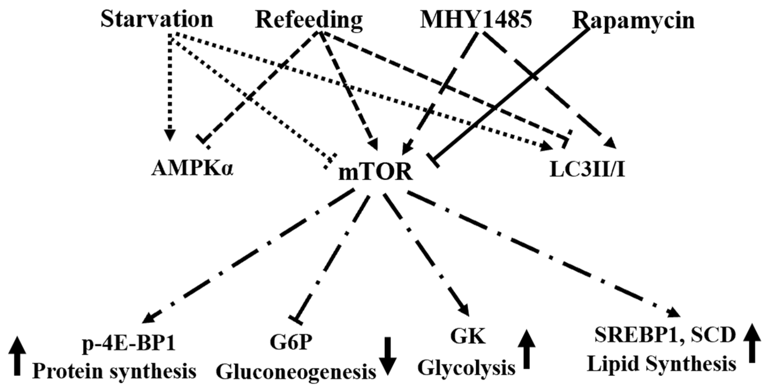
In conclusion, the domains of mTOR in S. constricta are highly conserved with those of TOR proteins in other species. The protein expression of p-mTOR, the LC3II/I ratio, and the expression of AMPKα mRNA were significantly changed during starvation and refeeding. In addition, MHY1485 can inhibit the activity of autophagy protein factor and activate the expression of glycolysis-related genes. Rapamycin can inhibit the expression of genes related to glycolysis and lipid synthesis. These results (Figure 7) indicate a conserved role of mTOR in regulating nutritional metabolisms such as glucose metabolism, lipid synthesis, and autophagy in S. constricta.

Author Contributions

Conceptualization, Q.Z.; methodology, Q.Z.; software, Q.Z.; validation, Q.Z. and Y.Q.; formal analysis, Y.Q.; investigation, Y.Q.; resources, D.C. and X.Y.; data curation, Q.Z.; writing—original draft preparation, Q.Z.; writing—review and editing, K.L.; visualization, K.L.; supervision, J.X.; project administration, J.X.; funding acquisition, Y.L. All authors have read and agreed to the published version of the manuscript.

Funding

This research was supported by the Ningbo Science and Technology Research Projects, China (2019B10006), the National Key Research and Development Program of China (2019YFD0900400), the Ningbo Public Welfare Science and Technology Plan Project (2022S157), and the Earmarked Fund for CARS-49.

Data Availability Statement

The datasets used and/or analyzed during the current study are available from the corresponding author upon reasonable request.

Acknowledgments

We thank Peng Shi, Guochao Ye, and Lina Bai for their invaluable assistance with this study.

Conflicts of Interest

The authors declare no conflict of interest.

References

  1. González, A.; Hall, M.N.; Lin, S.-C.; Hardie, D.G. AMPK and TOR: The yin and yang of cellular nutrient sensing and growth control. Cell Metab. 2020, 31, 472–492. [Google Scholar] [CrossRef] [PubMed]
  2. Loewith, R.; Jacinto, E.; Wullschleger, S.; Lorberg, A.; Crespo, J.L.; Bonenfant, D.; Oppliger, W.; Jenoe, P.; Hall, M.N. Two TOR complexes, only one of which is rapamycin sensitive, have distinct roles in cell growth control. Mol. Cell 2002, 10, 457–468. [Google Scholar] [CrossRef] [PubMed]
  3. Saxton, R.A.; Sabatini, D.M. mTOR signaling in growth, metabolism, and disease. Cell 2017, 168, 960–976. [Google Scholar] [CrossRef] [PubMed]
  4. Laplante, M.; Sabatini, D.M. mTOR signaling in growth control and disease. Cell 2012, 149, 274–293. [Google Scholar] [CrossRef] [PubMed]
  5. Nojima, H.; Tokunaga, C.; Eguchi, S.; Oshiro, N.; Hidayat, S.; Yoshino, K.I.; Hara, K.; Tanaka, N.; Avruch, J.; Yonezawa, K. The Mammalian Target of Rapamycin (mTOR) Partner, Raptor, Binds the mTOR Substrates p70 S6 Kinase and 4E-BP1 through Their TOR Signaling (TOS) Motif. J. Biol. Chem. 2003, 278, 15461–15464. [Google Scholar] [CrossRef]
  6. Gordan, J.D.; Thompson, C.B.; Simon, M.C. HIF and c-Myc: Sibling Rivals for Control of Cancer Cell Metabolism and Proliferation. Cancer Cell 2007, 12, 108–113. [Google Scholar] [CrossRef]
  7. Düvel, K.; Yecies, J.L.; Menon, S.; Raman, P.; Lipovsky, A.I.; Souza, A.L.; Triantafellow, E.; Ma, Q.; Gorski, R.; Cleaver, S. Activation of a metabolic gene regulatory network downstream of mTOR complex 1. Mol. Cell 2010, 39, 171–183. [Google Scholar] [CrossRef]
  8. Porstmann, T.; Santos, C.R.; Griffiths, B.; Cully, M.; Wu, M.; Leevers, S.; Griffiths, J.R.; Chung, Y.-L.; Schulze, A. SREBP activity is regulated by mTORC1 and contributes to Akt-dependent cell growth. Cell Metab. 2008, 8, 224–236. [Google Scholar] [CrossRef]
  9. Kim, J.; Kundu, M.; Viollet, B.; Guan, K.-L. AMPK and mTOR regulate autophagy through direct phosphorylation of Ulk1. Nat. Cell Biol. 2011, 13, 132–141. [Google Scholar] [CrossRef]
  10. Martina, J.A.; Chen, Y.; Gucek, M.; Puertollano, R. MTORC1 functions as a transcriptional regulator of autophagy by preventing nuclear transport of TFEB. Autophagy 2012, 8, 903–914. [Google Scholar] [CrossRef]
  11. Kabeya, Y.; Mizushima, N.; Ueno, T.; Yamamoto, A.; Kirisako, T.; Noda, T.; Kominami, E.; Ohsumi, Y.; Yoshimori, T. LC3, a mammalian homologue of yeast Apg8p, is localized in autophagosome membranes after processing. EMBO J. 2000, 19, 5720–5728. [Google Scholar] [CrossRef]
  12. Kadowaki, M.; Karim, M.R. Cytosolic LC3 ratio as a quantitative index of macroautophagy. Methods Enzymol. 2009, 452, 199–213. [Google Scholar]
  13. Su, Y.; Wang, T.; Wu, N.; Li, D.; Fan, X.; Xu, Z.; Mishra, S.K.; Yang, M. Alpha-ketoglutarate extends Drosophila lifespan by inhibiting mTOR and activating AMPK. Aging 2019, 11, 4183. [Google Scholar] [CrossRef]
  14. Potter, S.; Sifers, J.; Yocom, E.; Blümich, S.L.; Potter, R.; Nadolski, J.; Harrison, D.A.; Cooper, R.L. Effects of inhibiting mTOR with rapamycin on behavior, development, neuromuscular physiology and cardiac function in larval Drosophila. Biol. Open 2019, 8, bio046508. [Google Scholar] [CrossRef]
  15. Hansen, M.; Taubert, S.; Crawford, D.; Libina, N.; Lee, S.J.; Kenyon, C. Lifespan extension by conditions that inhibit translation in Caenorhabditis elegans. Aging Cell 2007, 6, 95–110. [Google Scholar] [CrossRef]
  16. Wang, X.; Lei, X.-y.; Guo, Z.-x.; Wang, S.; Wan, J.-w.; Liu, H.-j.; Chen, Y.-k.; Wang, G.-q.; Wang, Q.-j.; Zhang, D.-m. The immuneoreaction and antioxidant status of Chinese mitten crab (Eriocheir sinensis) involve protein metabolism and the response of mTOR signaling pathway to dietary methionine levels. Fish Shellfish. Immunol. 2022, 127, 703–714. [Google Scholar] [CrossRef]
  17. MacLea, K.S.; Abuhagr, A.M.; Pitts, N.L.; Covi, J.A.; Bader, B.D.; Chang, E.S.; Mykles, D.L. Rheb, an activator of target of rapamycin, in the blackback land crab, Gecarcinus lateralis: Cloning and effects of molting and unweighting on expression in skeletal muscle. J. Exp. Biol. 2012, 215, 590–604. [Google Scholar] [CrossRef]
  18. Abuhagr, A.M.; MacLea, K.S.; Chang, E.S.; Mykles, D.L. Mechanistic target of rapamycin (mTOR) signaling genes in decapod crustaceans: Cloning and tissue expression of mTOR, Akt, Rheb, and p70 S6 kinase in the green crab, Carcinus maenas, and blackback land crab, Gecarcinus lateralis. Comp. Biochem. Physiol. Part A Mol. Integr. Physiol. 2014, 168, 25–39. [Google Scholar] [CrossRef]
  19. Zhao, W.; Luo, H.; Zhu, W.; Yuan, X.; Shao, J. Effects of Time-Dependent Protein Restriction on Growth Performance, Digestibility, and mTOR Signaling Pathways in Juvenile White Shrimp Litopenaeus vannamei. Front. Physiol. 2021, 12, 661107. [Google Scholar] [CrossRef]
  20. Liu, X.; Wang, M.; Sun, G.; Wang, B.; Jiang, K.; Shao, J.; Qi, C.; Zhao, W.; Han, S.; Liu, M. Mechanistic target of rapamycin inhibition with rapamycin induces autophagy and correlative regulation in white shrimp (Litopenaeus vannamei). Aquac. Nutr. 2018, 24, 1509–1520. [Google Scholar] [CrossRef]
  21. Picot, S.; Faury, N.; Arzul, I.; Chollet, B.; Renault, T.; Morga, B. Identification of the autophagy pathway in a mollusk bivalve, Crassostrea gigas. Autophagy 2020, 16, 2017–2035. [Google Scholar] [CrossRef]
  22. Gao, Z.; Pan, L.; Xu, R.; Zhou, Y.; Li, D. Insights into disruption of lipid metabolism in digestive gland of female scallop Chlamys farreri under B [a] P exposure. Environ. Pollut. 2022, 299, 118904. [Google Scholar] [CrossRef]
  23. Zhao, S.; Chen, C.; Wang, S.; Ji, F.; Xie, Y. MHY1485 activates mTOR and protects osteoblasts from dexamethasone. Biochem. Biophys. Res. Commun. 2016, 481, 212–218. [Google Scholar] [CrossRef]
  24. Hao, E.-y.; Wang, D.-H.; Chang, L.-y.; Huang, C.-x.; Chen, H.; Yue, Q.-x.; Zhou, R.-Y.; Huang, R.-l. Melatonin regulates chicken granulosa cell proliferation and apoptosis by activating the mTOR signaling pathway via its receptors. Poult. Sci. 2020, 99, 6147–6162. [Google Scholar] [CrossRef]
  25. Liu, Q.; Miller, L.C.; Blecha, F.; Sang, Y. Reduction of infection by inhibiting mTOR pathway is associated with reversed repression of type I interferon by porcine reproductive and respiratory syndrome virus. J. Gen. Virol. 2017, 98, 1316. [Google Scholar] [CrossRef]
  26. Choi, Y.J.; Park, Y.J.; Park, J.Y.; Jeong, H.O.; Kim, D.H.; Ha, Y.M.; Kim, J.M.; Song, Y.M.; Heo, H.-S.; Yu, B.P. Inhibitory effect of mTOR activator MHY1485 on autophagy: Suppression of lysosomal fusion. PLoS ONE 2012, 8, e43418. [Google Scholar] [CrossRef]
  27. Mikdache, A.; Boueid, M.-J.; van der Spek, L.; Lesport, E.; Delespierre, B.; Loisel-Duwattez, J.; Degerny, C.; Tawk, M. Rgs4 is a regulator of mTOR activity required for motoneuron axon outgrowth and neuronal development in zebrafish. Sci. Rep. 2021, 11, 13338. [Google Scholar] [CrossRef]
  28. Thomas, G.V. mTOR and cancer: Reason for dancing at the crossroads? Curr. Opin. Genet. Dev. 2006, 16, 78–84. [Google Scholar] [CrossRef]
  29. Johnson, S.C.; Yanos, M.E.; Bitto, A.; Castanza, A.; Gagnidze, A.; Gonzalez, B.; Gupta, K.; Hui, J.; Jarvie, C.; Johnson, B.M. Dose-dependent effects of mTOR inhibition on weight and mitochondrial disease in mice. Front. Genet. 2015, 6, 247. [Google Scholar] [CrossRef]
  30. Hirose, K.; Shiomi, T.; Hozumi, S.; Kikuchi, Y. Mechanistic target of rapamycin complex 1 signaling regulates cell proliferation, cell survival, and differentiation in regenerating zebrafish fins. BMC Dev. Biol. 2014, 14, 42. [Google Scholar] [CrossRef]
  31. Wang, A.; Mouser, J.; Pitt, J.; Promislow, D.; Kaeberlein, M. Rapamycin enhances survival in a Drosophila model of mitochondrial disease. Oncotarget 2016, 7, 80131. [Google Scholar] [CrossRef]
  32. Christie, G.R.; Hajduch, E.; Hundal, H.S.; Proud, C.G.; Taylor, P.M. Intracellular sensing of amino acids in Xenopus laevis oocytes stimulates p70 S6 kinase in a target of rapamycin-dependent manner. J. Biol. Chem. 2002, 277, 9952–9957. [Google Scholar] [CrossRef]
  33. Dikicioglu, D.; Dereli Eke, E.; Eraslan, S.; Oliver, S.G.; Kirdar, B. Saccharomyces cerevisiae adapted to grow in the presence of low-dose rapamycin exhibit altered amino acid metabolism. Cell Commun. Signal. 2018, 16, 85. [Google Scholar] [CrossRef]
  34. Xu, H.; Liu, L.; Su, Y.; Liang, Y.; Yang, J.J.A.B. Effects of rapamycin on life span and on expression of TOR and S6K in Brachionus calyciflorus (Rotifera). Aquat. Biol. 2017, 26, 49–56. [Google Scholar] [CrossRef]
  35. Peng, C.; Zhao, X.; Liu, S.; Shi, W.; Han, Y.; Guo, C.; Jiang, J.; Wan, H.; Shen, T.; Liu, G. Effects of anthropogenic sound on digging behavior, metabolism, Ca2+/Mg2+ ATPase activity, and metabolism-related gene expression of the bivalve Sinonovacula constricta. Sci. Rep. 2016, 6, 24266. [Google Scholar] [CrossRef]
  36. Ha, J.; Kim, J. Novel pharmacological modulators of autophagy: An updated patent review (2012–2015). Expert Opin. Ther. Pat. 2016, 26, 1273–1289. [Google Scholar] [CrossRef]
  37. Yang, H.; Rudge, D.G.; Koos, J.D.; Vaidialingam, B.; Yang, H.J.; Pavletich, N.P. mTOR kinase structure, mechanism and regulation. Nature 2013, 497, 217–223. [Google Scholar] [CrossRef]
  38. Su, B.; Jacinto, E. Mammalian TOR signaling to the AGC kinases. Crit. Rev. Biochem. Mol. Biol. 2011, 46, 527–547. [Google Scholar] [CrossRef]
  39. Rodriguez Camargo, D.C.; Link, N.M.; Dames, S.A. The FKBP–rapamycin binding domain of human TOR undergoes strong conformational changes in the presence of membrane mimetics with and without the regulator phosphatidic acid. Biochemistry 2012, 51, 4909–4921. [Google Scholar] [CrossRef]
  40. Oshiro, N.; Yoshino, K.i.; Hidayat, S.; Tokunaga, C.; Hara, K.; Eguchi, S.; Avruch, J.; Yonezawa, K. Dissociation of raptor from mTOR is a mechanism of rapamycin-induced inhibition of mTOR function. Genes Cells 2004, 9, 359–366. [Google Scholar] [CrossRef]
  41. Sun, S.; Wang, B.; Jiang, K.; Sun, J.; Liu, M.; Wang, L. Target of rapamycin (TOR) in Fenneropenaeus chinensis: cDNA cloning, characterization, tissue expression and response to amino acids. Aquac. Nutr. 2015, 21, 1–9. [Google Scholar] [CrossRef]
  42. Jiang, J.; Feng, L.; Liu, Y.; Jiang, W.-D.; Hu, K.; Li, S.-H.; Zhou, X.-Q. Mechanistic target of rapamycin in common carp: cDNA cloning, characterization, and tissue expression. Gene 2013, 512, 566–572. [Google Scholar] [CrossRef]
  43. Ekim, B.; Magnuson, B.; Acosta-Jaquez, H.A.; Keller, J.A.; Feener, E.P.; Fingar, D.C. mTOR kinase domain phosphorylation promotes mTORC1 signaling, cell growth, and cell cycle progression. Mol. Cell. Biol. 2011, 31, 2787–2801. [Google Scholar] [CrossRef]
  44. Li, F.; Fu, C.; Xie, Y.; Wang, A.; Li, J.; Gao, J.; Cui, X. Transcriptional responses to starvation stress in the hepatopancreas of oriental river prawn Macrobrachium nipponense. Environ. Pollut. 2019, 252, 14–20. [Google Scholar] [CrossRef]
  45. Viollet, B. The energy sensor AMPK: Adaptations to exercise, nutritional and hormonal signals. In Hormones, Metabolism and the Benefits of Exercise; Springer: Cham, Switzerland, 2017; pp. 13–24. [Google Scholar]
  46. Nazio, F.; Strappazzon, F.; Antonioli, M.; Bielli, P.; Cianfanelli, V.; Bordi, M.; Gretzmeier, C.; Dengjel, J.; Piacentini, M.; Fimia, G.M. mTOR inhibits autophagy by controlling ULK1 ubiquitylation, self-association and function through AMBRA1 and TRAF6. Nature Cell Biol. 2013, 15, 406–416. [Google Scholar] [CrossRef]
  47. Settembre, C.; Fraldi, A.; Medina, D.L.; Ballabio, A. Signals from the lysosome: A control centre for cellular clearance and energy metabolism. Nat. Rev. Mol. Cell Biol. 2013, 14, 283–296. [Google Scholar] [CrossRef]
  48. Hardie, D.G.; Ross, F.A.; Hawley, S.A. AMPK: A nutrient and energy sensor that maintains energy homeostasis. Nat. Rev. Mol. Cell Biol. 2012, 13, 251–262. [Google Scholar] [CrossRef]
  49. Howell, J.J.; Manning, B.D. mTOR couples cellular nutrient sensing to organismal metabolic homeostasis. Trends Endocrinol. Metab. 2011, 22, 94–102. [Google Scholar] [CrossRef]
  50. Peng, T.; Golub, T.R.; Sabatini, D.M. The immunosuppressant rapamycin mimics a starvation-like signal distinct from amino acid and glucose deprivation. Mol. Cell. Biol. 2002, 22, 5575–5584. [Google Scholar] [CrossRef]
  51. Bazer, F.W.; Wang, X.; Johnson, G.A.; Wu, G.J.A.N. Select nutrients and their effects on conceptus development in mammals. Anim. Nutr. 2015, 1, 85–95. [Google Scholar] [CrossRef]
  52. Um, S.H.; Frigerio, F.; Watanabe, M.; Picard, F.; Joaquin, M.; Sticker, M.; Fumagalli, S.; Allegrini, P.R.; Kozma, S.C.; Auwerx, J.J.N. Absence of S6K1 protects against age-and diet-induced obesity while enhancing insulin sensitivity. Nature 2004, 431, 200–205. [Google Scholar] [CrossRef]
  53. Kim, S.G.; Buel, G.R.; Blenis, J. Nutrient regulation of the mTOR complex 1 signaling pathway. Mol. Cells 2013, 35, 463–473. [Google Scholar] [CrossRef]
  54. Mukhopadhyay, S.; Frias, M.A.; Chatterjee, A.; Yellen, P.; Foster, D.A. The enigma of rapamycin dosage. Mol. Cancer Ther. 2016, 15, 347–353. [Google Scholar] [CrossRef]
  55. Cheng, Y.; Kim, J.; Li, X.X.; Hsueh, A.J. Promotion of ovarian follicle growth following mTOR activation: Synergistic effects of AKT stimulators. PLoS ONE 2015, 10, e0117769. [Google Scholar] [CrossRef]
  56. Choo, A.Y.; Yoon, S.-O.; Kim, S.G.; Roux, P.P.; Blenis, J. Rapamycin differentially inhibits S6Ks and 4E-BP1 to mediate cell-type-specific repression of mRNA translation. Proc. Natl. Acad. Sci. USA 2008, 105, 17414–17419. [Google Scholar] [CrossRef]
  57. Zhao, J.; Benakanakere, M.R.; Hosur, K.B.; Galicia, J.C.; Martin, M.; Kinane, D.F. Mammalian target of rapamycin (mTOR) regulates TLR3 induced cytokines in human oral keratinocytes. Mol. Immunol. 2010, 48, 294–304. [Google Scholar] [CrossRef]
  58. Wang, Q.; He, G.; Mai, K.; Xu, W.; Zhou, H.; Wang, X.; Mei, L. Chronic rapamycin treatment on the nutrient utilization and metabolism of juvenile turbot (Psetta maxima). Sci. Rep. 2016, 6, 28068. [Google Scholar] [CrossRef]
  59. Wan, X.; Mendoza, A.; Khanna, C.; Helman, L.J. Rapamycin inhibits ezrin-mediated metastatic behavior in a murine model of osteosarcoma. Cancer Res. 2005, 65, 2406–2411. [Google Scholar] [CrossRef]
  60. Tran, T.A.; Kinch, L.; Peña-Llopis, S.; Kockel, L.; Grishin, N.; Jiang, H.; Brugarolas, J. Platelet-derived growth factor/vascular endothelial growth factor receptor inactivation by sunitinib results in Tsc1/Tsc2-dependent inhibition of TORC1. Mol. Cell. Biol. 2013, 33, 3762–3779. [Google Scholar] [CrossRef]
  61. Sun, S.-Y.; Rosenberg, L.M.; Wang, X.; Zhou, Z.; Yue, P.; Fu, H.; Khuri, F.R. Activation of Akt and eIF4E survival pathways by rapamycin-mediated mammalian target of rapamycin inhibition. Cancer Res. 2005, 65, 7052–7058. [Google Scholar] [CrossRef]
  62. Massa, M.L.; Gagliardino, J.J.; Francini, F. Liver glucokinase: An overview on the regulatorymechanisms of its activity. IUBMB life 2011, 63, 1–6. [Google Scholar] [CrossRef]
  63. Feksa, L.R.; Cornelio, A.R.; Vargas, C.R.; de Souza Wyse, A.T.; Dutra-Filho, C.S.; Wajner, M.; Wannmacher, C.M.D. Alanine prevents the inhibition of pyruvate kinase activity caused by tryptophan in cerebral cortex of rats. Metab. Brain Dis. 2003, 18, 129–137. [Google Scholar] [CrossRef]
  64. Ferre, T.; Riu, E.; Bosch, F.; Valera, A. Evidence from transgenic mice that glucokinase is rate limiting for glucose utilization in the liver. FASEB J. 1996, 10, 1213–1218. [Google Scholar] [CrossRef]
  65. Lan, R.; Wan, Z.; Xu, Y.; Wang, Z.; Fu, S.; Zhou, Y.; Lin, X.; Han, X.; Luo, Z.; Miao, J. Taurine reprograms mammary-gland metabolism and alleviates inflammation induced by Streptococcus uberis in mice. Front. Immunol. 2021, 12, 696101. [Google Scholar] [CrossRef]
  66. Sipula, I.J.; Brown, N.F.; Perdomo, G. Rapamycin-mediated inhibition of mammalian target of rapamycin in skeletal muscle cells reduces glucose utilization and increases fatty acid oxidation. Metabolism 2006, 55, 1637–1644. [Google Scholar] [CrossRef]
  67. Taha, C.; Liu, Z.; Jin, J.; Al-Hasani, H.; Sonenberg, N.; Klip, A. Opposite translational control of GLUT1 and GLUT4 glucose transporter mRNAs in response to insulin: Role of mammalian target of rapamycin, protein kinase b, and phosphatidylinositol 3-kinase in GLUT1 mRNA translation. J. Biol. Chem. 1999, 274, 33085–33091. [Google Scholar] [CrossRef]
  68. Sul, H.S.; Wang, D. Nutritional and hormonal regulation of enzymes in fat synthesis: Studies of fatty acid synthase and mitochondrial glycerol-3-phosphate acyltransferase gene transcription. Annu. Rev. Nutr. 1998, 18, 331–351. [Google Scholar] [CrossRef]
  69. Dai, W.; Panserat, S.; Plagnes-Juan, E.; Seiliez, I.; Skiba-Cassy, S. Amino acids attenuate insulin action on gluconeogenesis and promote fatty acid biosynthesis via mTORC1 signaling pathway in trout hepatocytes. Cell. Physiol. Biochem. 2015, 36, 1084–1100. [Google Scholar] [CrossRef]
  70. Yellen, P.; Saqcena, M.; Salloum, D.; Feng, J.; Preda, A.; Xu, L.; Rodrik-Outmezguine, V.; Foster, D.A. High-dose rapamycin induces apoptosis in human cancer cells by dissociating mTOR complex 1 and suppressing phosphorylation of 4E-BP1. Cell Cycle 2011, 10, 3948–3956. [Google Scholar] [CrossRef]
  71. Luyimbazi, D.; Akcakanat, A.; McAuliffe, P.F.; Zhang, L.; Singh, G.; Gonzalez-Angulo, A.M.; Chen, H.; Do, K.-A.; Zheng, Y.; Hung, M.-C. Rapamycin Regulates Stearoyl CoA Desaturase 1 Expression in Breast CancerRapamycin Regulates SCD1 Expression. Mol. Cancer Ther. 2010, 9, 2770–2784. [Google Scholar] [CrossRef]
  72. Brown, N.F.; Stefanovic-Racic, M.; Sipula, I.J.; Perdomo, G. The mammalian target of rapamycin regulates lipid metabolism in primary cultures of rat hepatocytes. Metabolism 2007, 56, 1500–1507. [Google Scholar] [CrossRef]
  73. Shao, Y.; Che, Z.; Chen, K.; Li, C.; Zhao, X. Target of rapamycin signaling inhibits autophagy in sea cucumber Apostichopus japonicus. Fish Shellfish. Immunol. 2020, 102, 480–488. [Google Scholar]
  74. Huang, J.; Brumell, J.H. Bacteria–autophagy interplay: A battle for survival. Nat. Rev. Microbiol. 2014, 12, 101–114. [Google Scholar] [CrossRef]
Figure 1. (a). Domains of mTOR. The basic modular architecture research tool predicts mTOR domains. (b). Prediction of protein three-dimensional structure of mTOR AA 824–2471 using SWISS-MODEL online website. (c). Phylogenetic relationship between mTOR amino acid sequence of S. constricta and various biological homologs. Amino acid sequences come from NCBI database. Carassius auratus (XP 026118234.1), Zebrafish (NP 001070679.2), Channa argus (KAF3696586.1), Homo sapiens (NP 004949.1), Sus scrofa (XP 003127632.3), Strongylocentrotus purpuratus (XP 003127632.3), Lingula anatina (XP 013395747.1), Crassostrea gigas (XP 011432165.2), Penaeus vannamei (QHT73480.1), Penaeus chinensis (AHX84170.1), Dirofilaria immitis (MCP9257960.1). (d). Expression patterns of mTOR mRNA in different tissues from S. constricta. Bars with different letters represent significant differences between different tissues (p < 0.05). DUF, domains of unknown function; FAT, focal adhesion targeting domain; PI3Kc, phosphoinositide 3-kinase catalytic domain; FATC, C-terminal adhesion targeting domain. mTOR, mammalian rapamycin target protein.
Jmse 11 01040 g001
Figure 2. Starvation-re-feeding affected the expression of p-mTOR, LC3II/I, and AMPKα at different time periods. (a). The expression of p-mTOR protein at different time periods. (b). The expression of LC3II/I protein at different time periods. (c). The expression of AMPKα mRNA at different time periods. p-mTOR, phosphorylated mammalian rapamycin target protein; LC3, microtubule-associated protein 1 light chain 3; AMPK, AMP-activated protein kinase; R, refeeding. Bar containing one of the same marked letters are not significant differences (p > 0.05), while those with different marked letters are significant differences (p < 0.05).
Jmse 11 01040 g002
Figure 3. Effects of MHY1485 and rapamycin on phosphorylation of 4E-BP1 in S. constricta at different times. (a). Treated with 40, 20, 10, 5 and 1 µg/kg MHY1485 for 12 h. (b). Treated with 40 and 10 µg/kg of the MHY1485 for 6 h. (c). Treated with 40, 20, and 10 µg/kg of the rapamycin for 12 h. (d). Treated with 10, 5, 1, 0.5 µg/kg of the rapamycin for 6 h. The data presented are means ± SEM (n = 3). * indicates that these bars are noticeably different (p < 0.05). ** indicates that these bars are noticeably different (p < 0.01) CON, control; p-4EBP1, eukaryotic initiation factor 4E-binding protein 1.
Jmse 11 01040 g003
Figure 4. MHY1485 and rapamycin affect the mTOR signaling pathway in S. constricta. (a). Effect of MHY1485 on the expression of the mTOR mRNA. (b). Effect of MHY1485 on the expression of the phosphorylated 4E-BP1. (c). Effect of rapamycin on the expression of the mTOR mRNA. (d). Effect of rapamycin on the expression of the phosphorylated 4E-BP1. The data presented are means ± SEM (n = 4). * means these bars are markedly different (p < 0.05). CON, control; p-4EBP1, eukaryotic initiation factor 4E-binding protein 1.
Jmse 11 01040 g004
Figure 5. Effects of MHY1485 and rapamycin on the AMPKα mRNA and LC3II/I protein in S. constricta. (a). Effect of MHY148 on the expression of the AMPKα mRNA. (b). Effect of MHY1485 on the expression of the AMPKα mRNA. (c). Effect of rapamycin on the expression of AMPKα mRNA. (d). Effect of rapamycin on the expression of LC3II/I protein. The data presented are means ± SEM (n = 4). * means these bars are markedly different (p < 0.05). CON, control; LC3, microtubule-associated protein 1 light chain 3; AMPK, AMP-activated protein kinase.
Jmse 11 01040 g005
Figure 6. (a). The mRNA expressions of PK, GK, GLUT1, G6P, PEPCK, SCD, ACC, SREBP, sterol-regulatory element-binding protein; CS, and NADP were analyzed by quantitative real-time PCR after MHY1485 treatment. (b). The mRNA expressions of PK, GK, GLUT1, G6P, PEPCK, SCD, ACC, SREBP, CS, and NADP were analyzed by quantitative real-time PCR after rapamycin treatment. The data presented are mean ± SEM (n = 6), and * means these bars are markedly different (p < 0.05). CON, control; PK, pyruvate kinase; GK, glucokinase; GLUT1, glucose transporter 1; G6P, glucose-6-phosphate dehydrogenase; PEPCK, phosphoenolpyruvate carboxykinase; SCD, stearoyl-CoA desaturase; ACC, acetyl-CoA carboxylase; CS, citrate synthase; NADP, nicotinamide adenine dinucleotide phosphate.
Jmse 11 01040 g006
Figure 7. A model of mTOR regulating nutritional metabolism. Starvation and inhibitors can regulate downstream signaling molecules by inhibiting mTOR; refeeding and activators can regulate downstream signaling molecules by activating mTOR. The black triangular head arrows indicate activation and the horizontal head arrows indicate inhibition. mTOR, mammalian rapamycin target protein; LC3, microtubule-associated protein 1 light; AMPK, AMP-activated protein kinase; chain 3; p-4E-BP1, eukaryotic initiation factor 4E-binding protein 1; GK, glucokinase; SREBP, sterol-regulatory element-binding protein; SCD, stearoyl-CoA desaturase.
Jmse 11 01040 g007
Table 1. Primers for real-time PCR.
Title 1TitleTitle 3
β-actinFCCATCTACGAAGGTTACGCCC
RTCGTAGTGAAGGAGTAGCCTCTTT
mTORF
R
CAGCGGTCTGGTAATGTGAAGG
TAGCAACTTAGGTGACACATACTGG
AMPKFACCACAGGCACCTCAGTAAACA
RAGGATGCGTGCGTGAAGTTA
PKF
R
TCGTGTAATGGCAATAATCG
GTAGAAGCATCGTTCAAGTC
GKF
R
GTTCGCCCGTTTATGCTTGG
CAAGTCCAGGGCGAGAAAGT
GLUT1F
R
CGTTATCCTCGTCGCTTCCA
CCACCATTGCTTCTGTTGGC
G6PF
R
CCCTCGTCTTGTCTGGCATT
CAGCATCCCTGTACACAGCA
PEPCKF
R
GGGAGGACAAGAAGGGAGT
ATTGTATCCCATGAAAGGTCTC
SREBPF
R
GCTCCTACTCTGTTATCCGATTGTG
TCCTGAGACTGGCGAGATCCTT
ACCF
R
TGGATGGCAATGTTGATGA
GGCACTGATGGTAGAGAAG
SCDF
R
CACCGCATCCCCGAAAAATC
AGGCGCAAATTATGGTTGCC
CSF
R
CAGTTCAGTGCTGCCATA
CAAGTTACGGTAGATGATAGAC
NADPF
R
ATGTTGCTAAGGATGTTACC
TTAGGAGATGGACTGTTCTT
Disclaimer/Publisher’s Note: The statements, opinions and data contained in all publications are solely those of the individual author(s) and contributor(s) and not of MDPI and/or the editor(s). MDPI and/or the editor(s) disclaim responsibility for any injury to people or property resulting from any ideas, methods, instructions or products referred to in the content.