Article

Establishment and Optimization of an Aggregate Culture System of Testicular Cells from Marine Medaka, Oryzias dancena

1
Department of Fisheries Biology, Pukyong National University, Busan 48513, Republic of Korea
2
Department of Marine Biotechnology, Institute of Marine and Environmental Technology, University of Maryland Baltimore Country, Baltimore, MD 21202, USA
3
Division of Fisheries Life Science, Major in Aquaculture and Applied Life Science, Pukyong National University, Busan 48513, Republic of Korea
*
Author to whom correspondence should be addressed.
Current address: Biotechnology Research Division, National Institute of Fisheries Science, Busan 46083, Republic of Korea.
J. Mar. Sci. Eng. 2023, 11(11), 2077; https://doi.org/10.3390/jmse11112077
Submission received: 19 September 2023 / Revised: 23 October 2023 / Accepted: 28 October 2023 / Published: 30 October 2023

Abstract

:
Although testicular organoids have remarkable potential as testicular models in vitro, there have been few studies about testicular organoids in teleost fish. As a first step to establish a stable culture system for fish testicular organoids, we investigated the efficient conditions for an aggregate culture of dispersed testicular cells from adult marine medaka (Oryzias dancena) by evaluating the effects of culture methods and media composition on an aggregate culture. As the results, we found that culturing dispersed testicular cells in an ultra-low attachment 96 well without Matrigel was most effectively able to induce the formation of testicular cell aggregates among the five different methods tested. Subsequently, through media testing, we confirmed that the modified ESM2 was more optimal for this aggregate culture than the media conventionally used in porcine, human, and rat testicular aggregate cultures. Furthermore, we demonstrated that three supplements in the modified ESM2 including fish serum (FS), basic fibroblast growth factor (bFGF), and embryo extracts (EE) did not influence the number and size of the testicular aggregates formed, but fetal bovine serum and other supplements including β-mercaptoethanol, non-essential amino acids, sodium pyruvate, and sodium selenite were affected significantly. Nevertheless, the removal of three supplements (FS, bFGF, and EE) during culture negatively affected scp3 and sox9a expression levels, indicating their necessity. Finally, we identified that the sperms derived from in vitro cultured testicular aggregates were able to produce offspring after fertilization with naturally matured oocytes. The results from this study will provide fundamental information to develop the techniques for fish testicular organoid culture, which will eventually contribute to the development of reproductive biotechnology for aquaculture and the conservation of endangered fish species.

1. Introduction

In the testis, spermatogenesis occurs by complex signals between germ cells and somatic cells [1]. During spermatogenesis, diploid spermatogonia undergo asymmetric divisions to produce haploid sperm cells, carrying genetic information to the next generation [2]. Therefore, the development of an in vitro testicular model capable of realizing in vitro spermatogenesis would be useful for research regarding male infertility treatment and animal reproductive biotechnology. So far, there have been many efforts to realize in vitro spermatogenesis by using two-dimensional (2D) or 3D culture methods [3,4,5]. However, it has been shown that a 2D culture method was not suitable for realizing in vitro spermatogenesis, possibly due to the absence of a proper spatial arrangement between germ cells and somatic cells [6,7]. Unlike this, testis organ culture, a 3D culture method, successfully produced fertilizable sperm through in vitro spermatogenesis [4], but this method has a drawback, as it is an in vitro model system that cannot accomplish a sustainable subculture, which is important in terms of preventing repeated animal sacrifices. These mean that a good in vitro testicular model for in vitro spermatogenesis needs both germ–somatic cell interactions and culture sustainability.
Testicular organoids that mimic testicular structures and functions in vitro can be good candidates that meet such requirements. They can retain germ–somatic cell interactions in vitro and may be subcultured continuously under optimized culture conditions. Previously, several studies have performed the culture of testicular organoids in mammals. The embedding of primary testicular cells, which were isolated from 18-day-old male rats, in collagen and Matrigel matrix induced cyst-like structures of testicular aggregates and increased the haploid cell population compared to a 2D culture [8]. A three-layer gradient system (3-LGS) induced the dispersed cells from rat testis to form testicular organoids that reorganized into spherical–tubular structures and maintained their germ cell population for 21 days [7]. In pigs, highly biomimetic organoids could be made through the formation of testicular spheroids in ultra-low attachment 96-well plates (ULA) and subsequent culture in an air–liquid interface [9].
In spite of its remarkable potential in biotechnology applications, there have only been a few studies about culturing testicular organoids in teleost fish. Studies on Gnathopogon caerulescens [10] and Bostrychus sinensis [11] have shown in vitro production of functional sperm by the formation of testicular spheroids, but methodological details for spheroid formation regarding culture method and media composition were not closely investigated.
Therefore, in this study, as a first step to establish a stable culture system for fish testicular organoids, we evaluated the effects of various culture methods and media compositions on the formation of testicular aggregates in adult marine medaka (Oryzias dancena). In order to accomplish this, we carried out the following:
  • We first attempted to find an efficient way to prepare the dispersed testicular cells, from which sperm cells were removed.
  • Then, we evaluated their self-aggregation under the following three methods: (1) Matrigel suspension, (2) 3-LGS, and (3) culturing into ULA.
  • Subsequently, the effects of media composition on size and number of testicular aggregates were investigated to find optimal culture conditions.
  • Finally, we tested if this culture condition is able to produce fertilizable sperm from the cultured testicular aggregates.

2. Materials and Methods

2.1. Fish

Marine medaka (O. dancena) were bred in the Laboratory of Cell Biotechnology, Pukyong National University (Busan, Republic of Korea). They were fed five times a day with brine shrimp larvae (Artemia nauplius) and/or an artificial diet for flounder larvae (EWHA, Busan, Republic of Korea). Water salinity and temperature were adjusted to 5 psu and 26 °C, respectively. Photoperiod was kept as 14 h light and 10 h dark during experiments. The males of 3.2 to 3.5 cm in length were separated from females 3 days before the experiments. All procedures dealing with animals complied with the guidelines provided by Pukyong National University, and the Institutional Animal Care and Use Committee (IACUC) of Pukyong National University approved our research proposal (approval No. PKNUIACUC-2021-31).

2.2. Cell Culture Media

To induce testicular aggregates, two types of culture media including basic medium (BM) and the modified ESM2 (mESM2) were used. BM was comprised of Leiboviz’s L15 Medium (L15; Gibco, Grand Island, NY, USA) supplemented with 10% (v/v) KnockOutTM Serum Replacement (KSR; Gibco) and 1% penicillin and streptomycin (P/S; Gibco). ESM2 was originally developed for fish embryonic stem cell culture in a previous study [12], and the formula of mESM2 was almost the same as ESM2, except for medaka embryo extract (EE)-donor species and EE concentration. Briefly, it consisted of Dulbecco’s Modified Eagle Medium (DMEM; Gibco) supplemented with 15% fetal bovine serum (FBS; Gibco), 20 mM hydroxyethyl piperazine ethane sulfonic acid (HEPES; Thermo Fisher Scientific, Waltham, MA, USA), 1 mM non-essential amino acid (NEAA; Gibco), 1 mM sodium pyruvate (Sp; Gibco), 2 nM sodium selenite (Ss; Sigma-Aldrich, St. Louis, MO, USA), 100 μM β-mercaptoethanol (BME; Gibco), 10 ng/mL human basic fibroblast growth factor (bFGF; Gibco), 1% rainbow trout serum (FS; Caisson Laboratories, Inc., North Logan, UT, USA), 50 μg/mL EE, and 1% P/S. EE was prepared from O. dancena embryos as described previously [13].

2.3. Cell Isolation and Percoll Density Gradient Centrifugation

To determine the optimal trypsin-EDTA concentration for cell isolation from testis tissues, testes were digested by different concentrations of trypsin-EDTA, and the number and viability of the cells isolated were measured as follows. Adult O. dancena males were anesthetized in 4 °C water for 5 min. Subsequently, they were immersed in 70% ethanol for 10 s for microbial decontamination, and their testes were extracted. The testes were washed 3 times in Dulbecco’s phosphate-buffered saline (DPBS; Gibco) containing 1% P/S and transferred into 35 mm Petri dishes (SPL Life Science, Pocheon, Republic of Korea), in which they were dissected by a surgical blade and digested by various concentrations (0.025%, 0.05%, 0.1%, and 0.2% (v/v)) of trypsin-EDTA (Gibco) for 1 h. After trypsin inactivation by one volume of the media containing 10% FBS and 1% P/S, cell suspensions were filtered by 40 μm cell strainers (Falcon, Corning, NY, USA), and the cells were harvested by centrifugation (400× g, 5 min). After the cells were resuspended in 300 μL of media containing 10% FBS and 1% P/S, the number of cells was counted by hemocytometer (Marienfeld-Superior, Lauda-Königshofen, Germany), and cell viability was measured by trypan blue (Gibco) assay. After trypan blue staining, stained and non-stained cells were regarded as dead and live cells, respectively, and the viability was presented as the percentage of live cells. To remove sperm and spermatids from the isolated cell population, Percoll density gradient centrifugation was performed. For this, cell suspension was loaded onto the top of a discontinuous 5-step Percoll (Sigma-Aldrich) solution consisting of 1 mL each of 20%, 30%, 40%, 50%, and 60% in 15 mL conical tubes (Falcon) and centrifuged at 800× g for 30 min. The cells from each layer were harvested separately, and cellular morphology was observed under an inverted microscope (Nikon, Eclipse TS100, Tokyo, Japan). For the experiments regarding testicular aggregate formation, layers from the top to the 50% point (1st to 4th layers in Figure 1B) were removed into 15 mL conical tube containing 11 mL DPBS, and then the cells were harvested by centrifugation (400× g for 10 min). The cells were resuspended in 1 mL DPBS and used for subsequent experiments.

2.4. Induction of Testicular Cell Aggregates

To induce the formation of testicular cell aggregates from the isolated cells, three methods, which were previously reported, were used with slight modification: (1) Matrigel (Corning, Corning, NY, USA) suspension method [8], (2) 3-LGS [7], and (3) method using ULA (Corning), each wells of which are coated with hydrophilic hydrogels [9]. All Matrigel used in these experiments was diluted by 1:1 ratio with L15. For Matrigel suspension method, isolated testicular cells were suspended in 5 μL of Matrigel (final cell concentrations: 1.25 × 10⁵ cells/μL, 2.5 × 10⁵ cells/μL, and 3.75 × 10⁵ cells/μL). Cell suspension was placed as 5 μL single drop on 35 mm cell culture dishes (SPL Life Science) or 35 mm Petri dishes (SPL Life Science) and gelled for 20 min at 28 °C. After that, the dishes were filled with 2 mL of BM. For Matrigel suspension method using agarose (SeaKem LE agarose; Lonza, Rockland, ME, USA) gel stands to prevent seeded cells from adhering to the bottom completely, agarose gel stands were prepared as follows. Distilled water containing 1.5% agarose was boiled and poured into a 100 mm Petri dish (SPL Life Sciences), and after its gelation by cooling, agarose gel stand was made by cutting it into 10 mm (width) × 10 mm (length) × 5 mm (height) in size. Agarose gel stands were used after being placed in a 35 mm Petri dish with 1.6 mL of BM overnight for the gels to hold media completely. Then, 5 μL Matrigel drops containing testicular cells were made on the agarose gel stands. Final cell concentrations of Matrigel drops and gelling condition were the same as the above.
For 3-LGS, 3 Matrigel layers were constructed on the center of the insert membrane of a 24-well hanging cell insert (Corning) placed upside down. First, 5 μL Matrigel drop was applied on the center of the insert membrane and gelled for 20 min at 28 °C. Next, 3 μL of Matrigel containing the isolated testicular cells was placed as a drop on the first Matrigel drop and gelled for 20 min at 28 °C. Finally, the previous two Matrigel drops were covered with 8 μL of Matrigel and gelled for 20 min at 28 °C. Subsequently, a hanging cell insert was inserted in a 24-well plate filled with 600 μL of BM, and the cells were cultured.
For the method using ULA, the isolated cells were suspended with 5 μL of Matrigel or 150 μL of BM and seeded into ULA (final cell concentration: 2.5 × 10⁵ cells/μL). In case of Matrigel suspension, it was gelled with same conditions described above, and then 150 μL of BM was added. Culture of testicular cells under each method was performed for 14 days at 28 °C in an incubator with air atmosphere, and culture media were changed every three days. In ULA method, half of the medium was changed.

2.5. Measurement of Number, Size, and Viability of Testicular Aggregates

Testicular aggregates were collected from ULA and washed with 2 mL of DPBS in a 35 mm Petri dish. Testicular aggregates were moved into 96-well plates (Thermo Fisher Scientific), and pictures of all aggregates were captured for measurement of their number and size. The number was counted visually on the pictures, and the size was measured by TSView7 software, Version 7 (Fuzhou Xintu Photonics Co., Ltd., Fuzhou, Fujian, China). All testicular aggregates (30 to 60 aggregates, depending on experimental group) were subjected to size measurement. The size was defined as the mean of the lengths (width and height of aggregate on the picture) and presented in μm. For measurement of cell viability, collected testicular aggregates were moved in a 1.5 mL tube (Corning) and digested by 500 μL of 0.05% (v/v) trypsin-EDTA for 30 min with pipetting every 10 min. After that, trypsin-EDTA was inactivated by adding one volume of DMEM containing 10% (v/v) FBS and 1% (v/v) P/S, and the dispersed cells were collected by centrifugation (400× g for 5 min). After resuspension of the cells with 500 μL of PBS, trypan blue assay was performed as described above. Localization of live and dead cells in testicular aggregates was analyzed using Live/Dead Viability/Cytotoxicity kit (Molecular Probes, Eugene, OR, USA) following manufacturer’s instructions. After staining of testicular aggregates with fluorescent dyes indicating live and dead cells (4 μM of ethidium homodimer-1 for live and 2 μM of calcein AM for dead), localization of live and dead cells within testicular aggregates was observed using a fluorescent microscope (Olympus, Hamburg, Germany). For negative control, testicular aggregates were immersed in 70% ethanol for one hour and stained by Live/Dead Viability/Cytotoxicity kit.

2.6. Quantitative Reverse Transcription Polymerase Chain Reaction (qRT-PCR)

Total RNA was extracted from the samples using RNeasy Micro Kit (Qiagen, Hilden, Germany), and 100 ng of total RNA was treated with DNase I (Sigma-Aldrich) according to manufacturer’s instructions. The cDNA was synthesized using GoScript TM reverse transcription system (Promega, Madison, WI, USA). Subsequently, 1 ng of cDNA was subjected to qRT-PCR using LightCycler® 480 SYBR Master (Roche Diagnostics, Mannheim, Germany) according to manufacturer’s instructions. Primer sequences used in this study were presented in Table 1. The qRT-PCR condition was as follows: initial denaturation (94 °C for 3 min), 40 cycles of amplification (denaturation: 94 °C for 30 s; annealing: 60 °C for 30 s; and elongation: 72 °C for 30 s). The relative expressions of each gene were calculated by 2−ΔΔCt method, where Ct = threshold cycle for target amplification, ΔCt = Cttarget gene − Ctinternal reference (18s rRNA), and ΔΔCt = ΔCtsample − ΔCtcalibrator [14].

2.7. Arifical Fertilization

Mature females of O. dancena were separated from mature males for a day. Mature females were collapsed in cold ice water for 10 s. Subsequently, collapsed females were placed on aluminum foil, and residual water from their bodies was absorbed by wiper papers (KIMTECH, Seoul, Republic of Korea) to prevent unintended cortical reaction of unfertilized eggs. After that, unfertilized eggs were collected by gently pushing on their abdomens. Immediately, 30–50 unfertilized eggs were gently moved into 1.5 mL tubes (Corning), and the suspension of testicular aggregates, which were harvested from three wells of ULA (total volume: 450 μL), was added to the unfertilized eggs. After 10 min at room temperature, artificially fertilized eggs were removed into 60 mm Petri dishes containing 5 mL of autoclaved breeding water and cultured at 28 °C in an incubator at air atmosphere. When the embryos developed to two-cell stage, they were transferred into 24-well plates, individually, to prevent possible negative effects from unfertilized or dead eggs. For negative control, unfertilized eggs were immersed in 450 μL of mESM2 for 10 min and then cultured in the same conditions as mentioned above. To prepare fresh sperms for artificial fertilization, an extracted testis was placed on IVF dish (SPL life science) and chopped with a surgical blade (No. 20; Feather Safety Razor, Osaka, Japan). Then, they were suspended with 450 μL of mESM2 and subjected to artificial fertilization in the same conditions as mentioned above. In case of naturally fertilized embryos, they were collected from the female gonopore after mating and fertilization and were cultured in the same conditions above. Embryonic development was observed using a stereomicroscope (SMZ745T; Nikon).

2.8. Statistical Analysis

SPSS program (SPSS Inc., Chicago, IL, USA) was used to analyze numerical data. The data were analyzed by one-way ANOVA or t-test. When the data were analyzed by one-way ANOVA, the data were followed by Duncan’s method. Significant differences were determined when p values were less than 0.05.

3. Results

3.1. Testicular Cell Isolation and Removal of Sperm Cells

To determine the optimal trypsin-EDTA concentration for isolating testicular cells from adult O. dancena testes, testes were digested under various concentrations of trypsin-EDTA, and the number of cells that were isolated and their viabilities were measured and compared to each other. The results showed that 0.05% and 0.1% trypsin-EDTA resulted in significantly higher numbers of isolated cells compared to 0.025% and 0.2% trypsin-EDTA (Figure 1A; 3.02 to 3.19 × 10⁵ cells/mg vs. 1.70 to 2.02 × 10⁵ cells/mg, p < 0.05). Cell viability was measured as being above 89.9% in all experimental groups, and no significant difference was observed among them (Figure 1A). Therefore, we confirmed that 0.05% trypsin-EDTA was optimal to isolate testicular cells. Next, to remove sperm cells from isolated testicular cells, the cells were separated by density gradient centrifugation using a discontinuous five-step Percoll solution (Figure 1B), and the morphologies of the cells from each layer were observed. As shown in Figure 1C, actively moving or moveless sperms were almost entirely observed in the fifth and sixth layers, whereas they were not observed in the other layers (first to fourth layers). For the following experiments, we used the testicular cells collected from the first to fourth layers after Percoll density gradient centrifugation of a total of the isolated testicular cells.

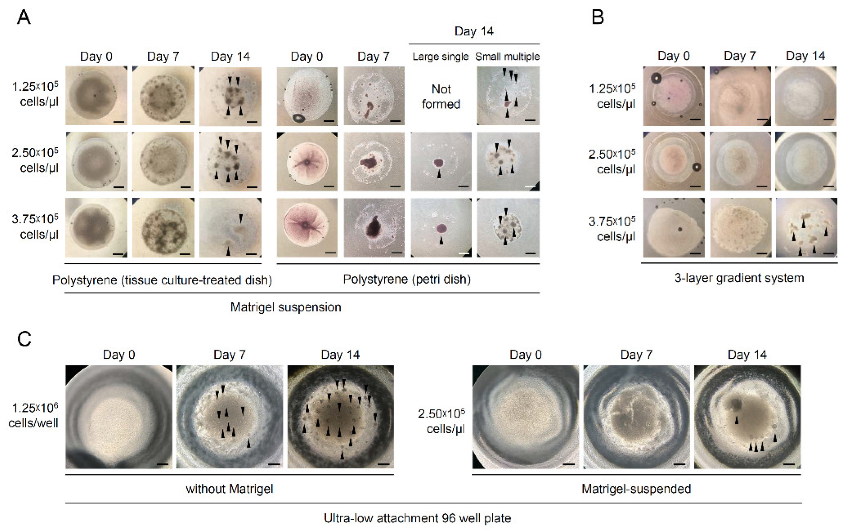
3.2. Effects of Culture Methods on the Formation of O. dancena Testicular Aggregates

To find an efficient method for self-aggregation of testicular cells, several different 3D culture methods were evaluated. For Matrigel suspension methods, Matrigel drops containing testicular cells of three different densities (1.25, 2.5, and 3.75 × 10⁵ cells/μL) were placed on three different surface materials including two types of polystyrene (tissue-culture-treated dish and Petri dish) and agarose gel stand, and after culturing for 14 days, the formation of testicular aggregates was evaluated. In the methods using a polystyrene surface, the isolated testicular cells started to congregate as small dots from the first day of seeding. On day 14, spherical testicular aggregates were observed regardless of dish type (Figure 2A). Progressive degradation of Matrigel drops was observed from day 3, but they were not completely degraded up to day 14. This degradation phenomenon was accelerated as cell concentrations were increased. Nevertheless, testicular aggregates were observed with 80 to 100% efficiencies in every cell concentration on day 14 (Table 2). In most cases, small multiple aggregates were formed, but large single aggregates were also observed in several groups, including the 2.5 × 10⁵ and 3.75 × 10⁵ cells/μL cell concentration groups, in a Petri dish (Figure 2A and Table 2). On the other hand, Matrigel suspension on agarose gel stands could not induce testicular cells to aggregate (Table 2). In 3-LGS, only one trial of the 3.75 × 10⁵ cells/μL concentration group could induce small multiple testicular aggregates (Figure 2B and Table 2). In the method using ULA without Matrigel, 1.25 × 10⁶ testicular cells started to form aggregates on day 7, and then small multiple testicular aggregates were induced on day 14 in all trials (100% formation rate). Although a similar morphological aspect for aggregate formation was also observed when Matrigel was added, the formation rate of testicular aggregates was significantly decreased to 37.5% (Figure 2C and Table 2).

3.3. Effects of Media Types on the Number and Size of Testicular Aggregates Formed

To find more optimal culture media for the formation of testicular aggregates, the effects of mESM2 on the culture of testicular aggregates were investigated. For this, the isolated testicular cells were cultured in the ULA for 14 days within BM or mESM2 and then the morphology, number, and size of the aggregates were evaluated. Two morphologically different aggregates, designated as hollow and compacted types, were observed regardless of media types (Figure 3A). Hollow-type aggregates showed a spheroid shape, but the inside was empty or had only a few testicular cells. Unlike this, compacted-type aggregates did not show such empty space inside them, as they were filled with closely packed testicular cells. Significant differences were observed in the rate of compacted-type aggregates depending on media type. For aggregates cultured in BM, 43.7 ± 14.4% and 47.3 ± 4.4% were compacted type on day 7 and day 14, respectively. In contrast, 96.1 ± 4.4% and 99.2 ± 1.5% aggregates were compacted type on day 7 and day 14, respectively, when cultured in mESM2 (Figure 3B). No significant difference was observed in the total number of aggregates formed according to media type on both day 7 and day 14 (Figure 3C). For the size of aggregates, bigger size was observed in the aggregates cultured in mESM2 than those in BM on day 7, but the difference was not shown on day 14. Regardless of media type, the sizes of aggregates were significantly increased on day 14 compared to day 7 of culture (Figure 3D).

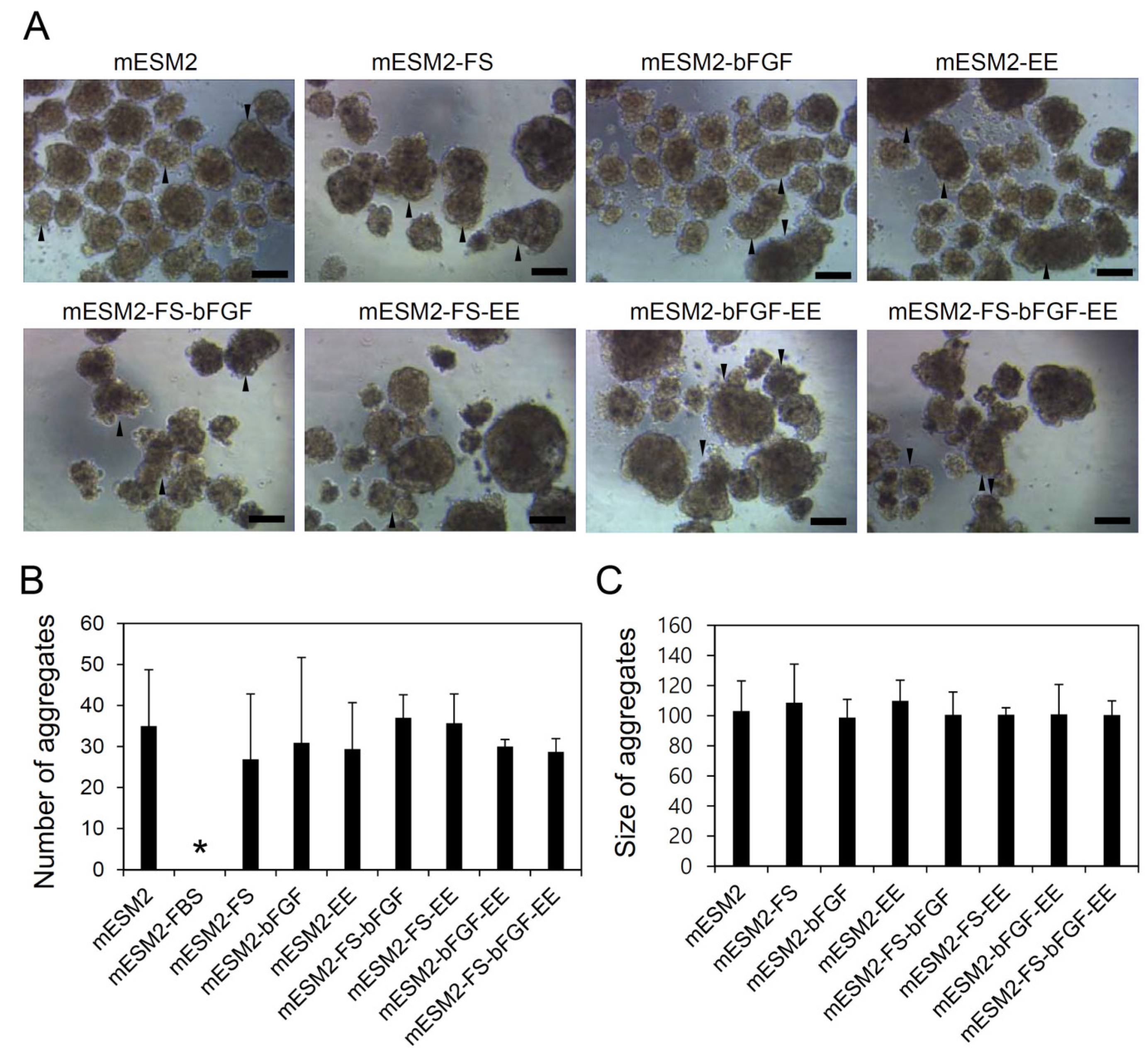
3.4. Effects of the Composition of mESM2 on the Culture of Testicular Aggregates: Part I

mESM2 was composed of various types of supplements. For the test of the effects of such supplements on testicular aggregate culture, in this study, we divided the supplements of mESM2 into two categories: protein-containing supplements (including FBS, FS, bFGF, and EE) and non-protein supplements (including BME, NEAA, Ss, and Sp). At first, to evaluate the effects of protein-containing supplements, testicular cells were cultured in ULA for 14 days under nine different media (mESM2 as a control group and eight mESM2 derivatives as the treatment groups, from which single or multiple protein-containing supplements were excluded), and the morphology, number, and size of the aggregates were compared to each other. The results showed that the media without FBS did not induce the formation of testicular aggregates, unlike the other media which did induce it. As shown in Figure 4A, the testicular aggregates showed compacted type, and the adhesion of two or more aggregates was occasionally observed regardless of media type. No significant difference was observed in the number of aggregates formed among eight media including mESM2 and mESM2 derivatives that removed FS, bFGF, and/or EE (Figure 4B). For the size of aggregates, similar size was observed among eight media groups without statistical difference (Figure 4C).
To further evaluate the role of FS, bFGF, and EE in testicular aggregate culture, the isolated testicular cells were additionally cultured in mESM2 and mESM2-FS-bFGF-EE for 14 days, and the viability and gene expression of the testicular aggregates were measured and compared to each other. As shown in Figure 5A, regardless of media type, most cells comprising testicular aggregates were live, as indicated with green fluorescence staining, and only a few cells inside of it were dead (red fluorescence). When quantitative assessment using trypan blue assay was performed, high viability was observed in both groups without significant difference (Figure 5B; 90.66 ± 0.63% in mESM2 vs. 91.08 ± 1.12% in mESM2-FS-bFGF-EE). Then, the testicular aggregates were subjected to qRT-PCR to analyze the mRNA expression levels of three genes including nanos2 (spermatogonia-specific), scp3 (mid-meiotic-germ-cells-specific), and sox9a (testicular-somatic-cells-specific). As shown in Figure 5C, no significant difference was detected in nanos2 mRNA expression between the two groups. However, the expression levels of scp3 and sox9a were significantly higher in the testicular aggregates cultured in mESM2 than the ones in mESM2-FS-bFGF-EE (2.68 and 2.14 folds in scp3 and sox9a, respectively). These results indicated that FS, bFGF, and EE promoted germ cell differentiation and influenced somatic cell function within the testicular aggregates even though they did not influence the number and size of testicular aggregates in culture.

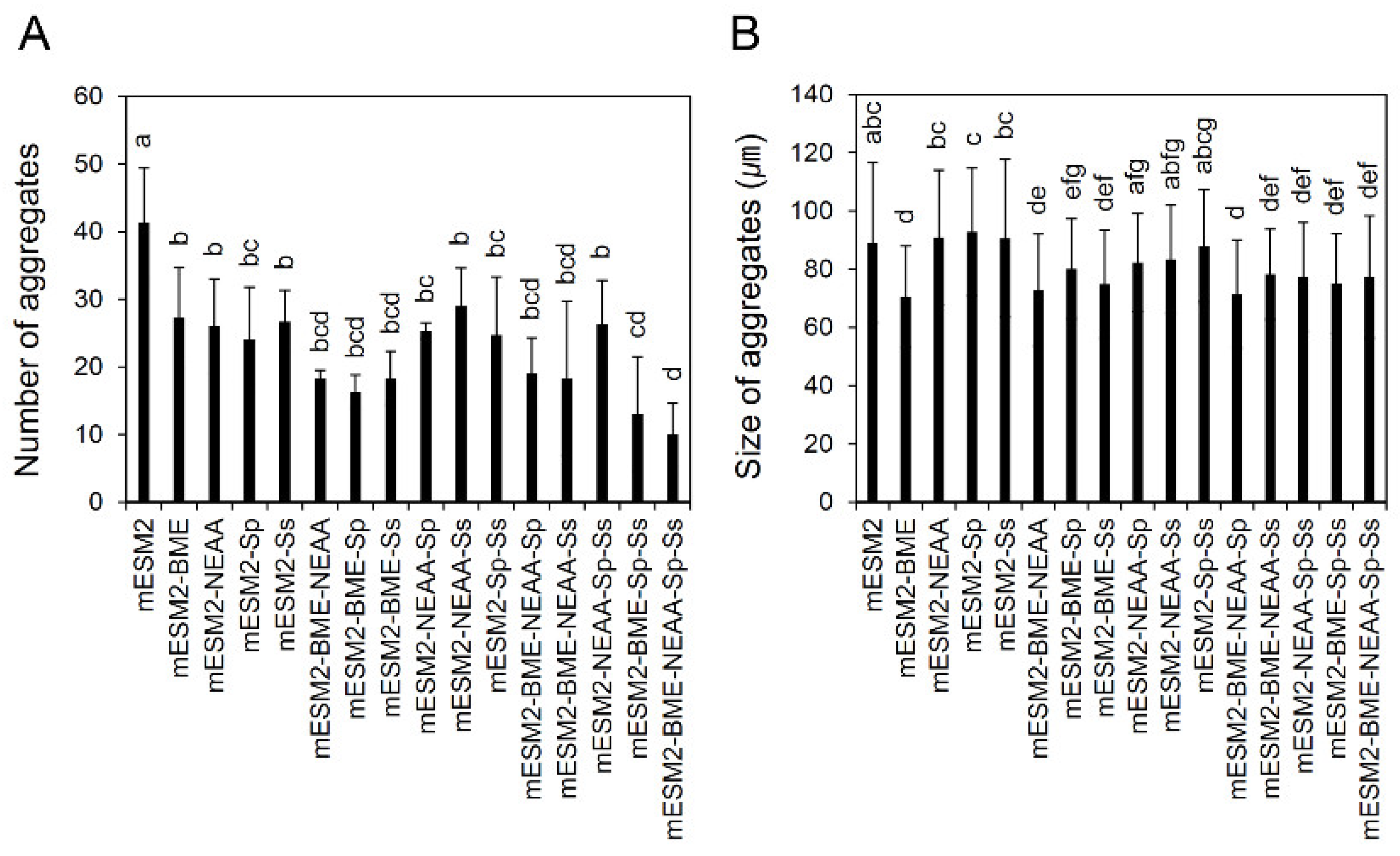
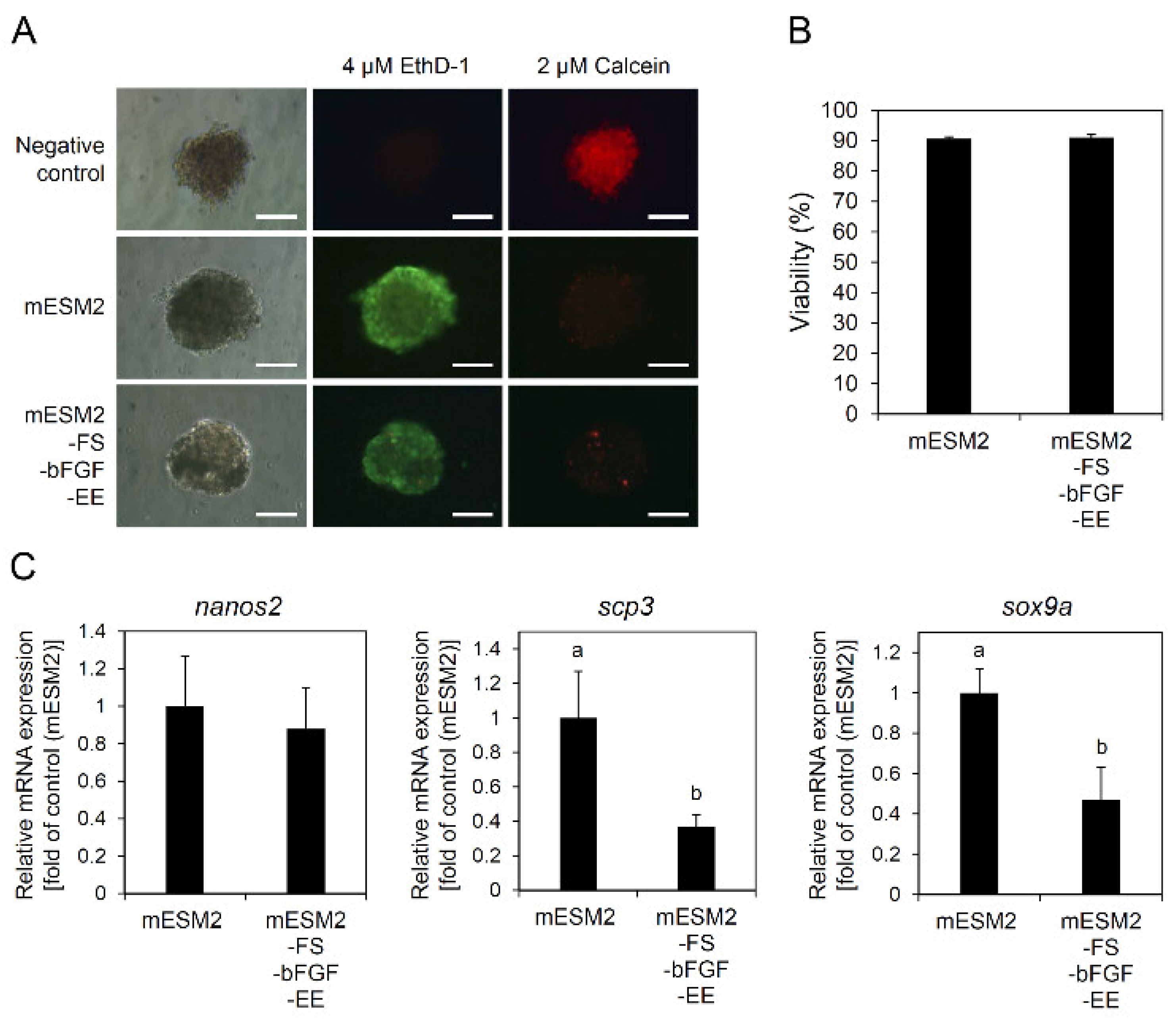
3.5. Effects of the Composition of mESM2 on the Culture of Testicular Aggregates: Part II

Subsequently, we evaluated the effects of non-protein supplements in mESM2 on the number and size of the testicular aggregates formed. For this, 15 experimental groups were set up by culturing the isolated testicular cells in 15 mESM2 derivatives from which one or more non-protein supplements were removed. In the results, spherical and compacted aggregates with occasional adhesion between them were observed in all experimental groups. However, all experimental groups showed significant decreases in the number of aggregates formed compared to the control group, where the testicular cells were cultured in mESM2 (Figure 6A). When all non-protein supplements were removed, the smallest value in aggregate number was observed. These results indicated that all non-protein supplements in mESM2 could contribute to the formation of testicular aggregates. For the size of aggregates formed, several experimental groups including -BME, -BME-NEAA, -BME-Sp, -BME-Ss, -BME-NEAA-Sp, -BME-NEAA-Ss, -NEAA-Sp-Ss, -BME-Sp-Ss, and -BME-NEAA-Sp-Ss showed significant decreases in size compared to the control group, while the other groups were not significantly different from the control group (Figure 6B). Interestingly, whether it was single or with other non-protein supplements, the removal of BME significantly affected size reduction in the aggregates. However, sole treatment of BME without other non-protein supplements did not rescue the aggregate size, while dual and triple treatments with other non-protein supplements did. These implied that BME might influence aggregate size through synergistic action with other supplements.

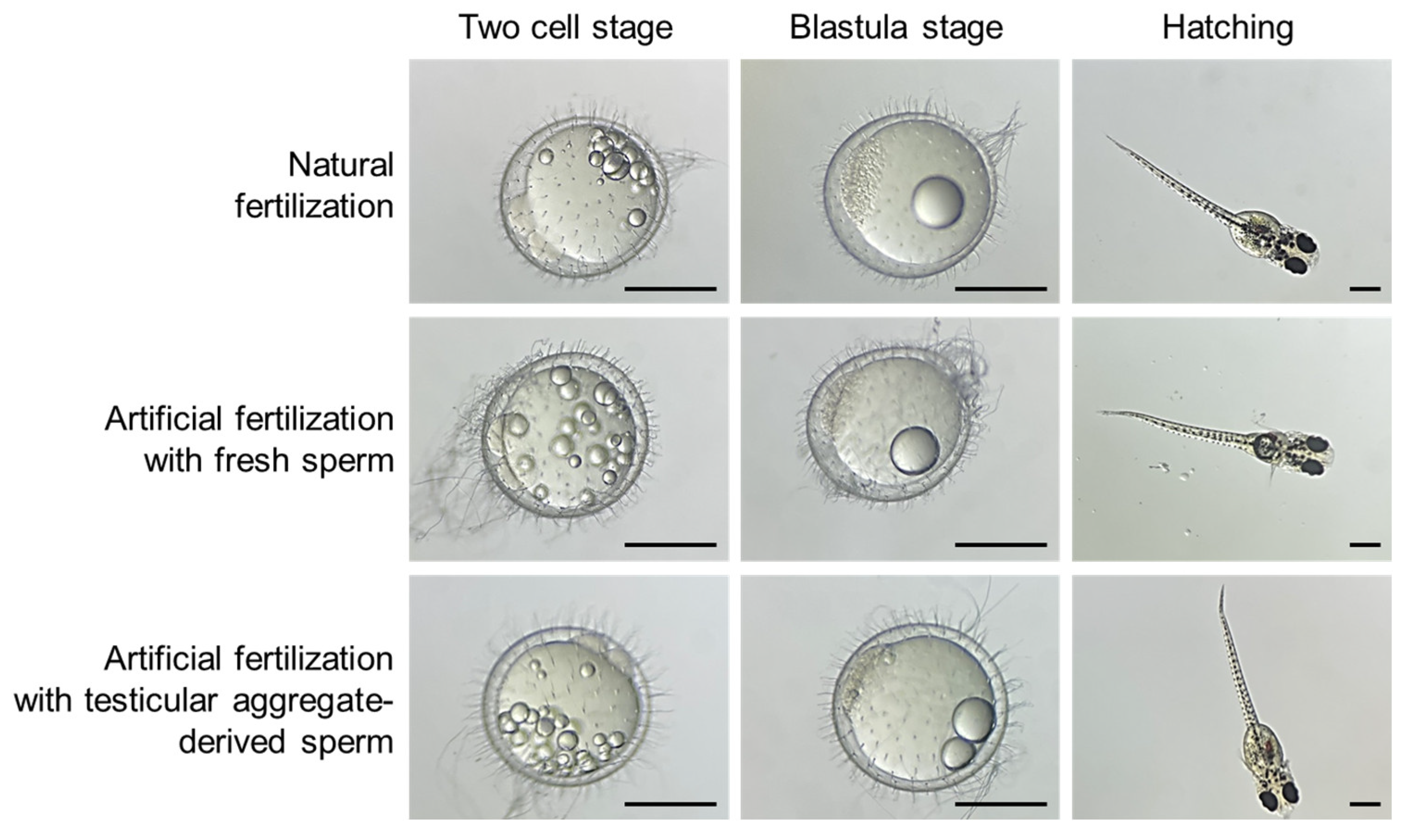
3.6. Evaluation of Fertility of the Sperms Derived from Testicular Aggregates

When the testicular aggregates were cultured for 14 days, numerous motile sperms were observed around testicular aggregates. To verify the fertility of those sperms, in vitro fertilization was performed. As shown in Table 3, when 276 unfertilized eggs were subjected to artificial fertilization with testicular-aggregate-derived sperms, 122 (44.2%) eggs developed up to a one-cell-stage embryo, but only four (1.4% of the total eggs treated) embryos reached the hatchling stage. In contrast to this, when fresh sperms were used for artificial fertilization, 107 (59.4%) out of 180 eggs developed up to a one-cell-stage embryo, and 78 (43.3% of the total eggs treated) embryos reached the hatchling stage. In case of natural fertilization, 124 (82.7%) out of 150 eggs developed up to a one-cell-stage embryo, and 111 (74.0% of the total eggs treated) embryos were hatched. As a negative control, unfertilized eggs did not develop past the one-cell stage. In all hatched larvae, regardless of treatment groups, morphological abnormalities were not observed (Figure 7).

4. Discussion

In this study, we evaluated the effects of culture methods and media compositions on the culture of marine medaka (O. dancena) testicular aggregates to find the optimal culture conditions.
In previous studies regarding the development of testicular organoid culture systems in mammals, testicular organoids were formed from the self-aggregation of testicular cells derived from immature male testes [7,9,15]. Alves-Lopes et al. explained that the high proportion of Sertoli cells within immature testes might influence the formation of testicular organoids [7]. Similarly, the testicular cells isolated from adult mice could form spheroids when they are mixed with immature somatic cells [15]. Coincident with these reports, fish testicular spheroids were made by using testicular cells derived from immature fish or fish during non-spawning season, of which the ratio of Sertoli cells/germ cells is high, in the study of teleost Gnathopogon caerulescens [10].
O. dancena has several advantages as an experimental animal model. For example, they have features like being easy to breed at lab scale, short generation time, and small genome size [16]. Moreover, O. dancena is a euryhaline fish, which can be used to investigate marine ecotoxicology [16]. Based on these advantages, this fish has been used for various cell culture studies such as the establishment of embryonic stem cells [17,18,19] and a whole testis organ culture [20,21]. Nevertheless, this fish has some defects as a fish model to study testicular aggregates, in spite of its reputation as a good experimental fish model [22]. The first thing is that the use of immature testes is restricted due to their small size. The recorded time to first spawning is 9 weeks from hatching, and at that time, their body length is known to be 22.58 ± 2.73 mm [23]. The isolation of testes in fish less than 22.58 ± 2.73 mm in length is technically very hard, which actually hampers their use. Furthermore, as daily spawning fish, O. dancena does not have a non-spawning season. To overcome these limitations, we introduced Percoll density gradient centrifugation to increase the ratio of Sertoli cells/germ cells of testicular cell populations derived from adult male O. dancena. Percoll density gradient centrifugation has been used to separate various cell types of testicular cell populations from mammals [24,25,26]. Moreover, the method has also been utilized to separate spermatogonia and/or somatic cells from fish testicular cell populations (four-eyed sleeper (Bostrychus sinensis) [11]; tench (Tinca tinca) [27]; Nile tilapia (Oreochromis niloticus) [28]). In the present study, application of Percoll density gradient centrifugation successfully separated sperm and spermatids from O. dancena testicular cell populations, which resultantly increased the ratio of Sertoli cells/germ cells. On the basis of our results that testicular cell populations containing sperm and spermatids did not form the aggregates in culture, this removal procedure of sperm and spermatids that increased the ratio of Sertoli cells/germ cells certainly contributed to the formation of testicular aggregates in this study.
Subsequently, we evaluated the effects of cultured methods on the formation of testicular aggregates. In previous studies, testicular aggregates could be formed with or without Matrigel in mammals including rats [7], humans [29], pigs [9], and bovines [30]. Thus, we evaluated three Matrigel-based methods including Matrigel suspension, 3-LGS, and culturing in ULA with Matrigel as well as a Matrigel-free method of culturing in ULA without Matrigel on the formation of the testicular aggregates. In the case of two approaches using 3-LGS and Matrigel suspension on agarose gel stands, little or no aggregate formation was observed, unlike previous studies in mammals [7,9], indicating that these methods were not proper in teleost O. dancena. In the Matrigel suspension method on agarose gel stands, we observed that the Matrigel solution containing testicular cells spread widely by making contact with hydrophilic agarose gel. It is presumed that this increased the physical distance between cells and eventually hindered the aggregate’s formation. On the other hand, testicular aggregate formation was observed with high probability (80–100%) in Matrigel suspension on polystyrene regardless of the tissue culture treated. However, this method showed several drawbacks in this application. First, it was observed that a lot of testicular cells were adhered to the plate’s bottom during culturing. It has been known that testicular somatic cells show relatively strong adherence to substrate compared with germline stem cells, which show loose adherence [31]. Therefore, most of the adhered cells would be the testicular somatic cells, indicating that this method restricted the contribution of testicular somatic cells in aggregate formation. Second, rapid Matrigel degradation was observed in this method. Matrigel began to degrade from day 3 of culture, and floating Matrigel fragments containing testicular cells were frequently observed in the culture media. This must have prevented many cells from participating in the formation of aggregates. Regarding different forms (large single and small multiple) of aggregate formation, the most likely reason is that testicular somatic cells contributed to aggregate formation differently according to the surface materials. Since a large number of testicular somatic cells adhered to tissue-culture-treated dishes, the contribution of somatic cells to aggregate formation would have been relatively low. In contrast, there was less adhesion of testicular somatic cells to the Petri dish, and more somatic cells would have contributed to aggregate formation. It is presumed that this difference may have influenced different forms of aggregates. To overcome the limitation of using Matrigel suspension methods, we tried to use ULA to induce the formation of testicular aggregates. Because ULAs have U-shaped bottoms at which the seeded cells can be gathered together, we expected that the testicular cells seeded could fully contribute to the formation of aggregates during culture. In the results, testicular aggregates were observed on day 7 with high probability (100%), and they grew significantly until day 14 without using Matrigel. These results confirmed that the method using ULA was most appropriate among the ones that we tested for culturing O. dancena testicular aggregates.
Previously, testicular organoid culture was usually performed using basal media supplemented with KSR like BM [7,32,33]. However, in fish, ESM2 and its modified forms were usually used to culture fish embryonic stem cells [34,35,36] and medaka spermatogonial stem cell lines [37]. Therefore, the effects of BM and mESM2 on the morphology, number, and size of testicular aggregates were compared in this study. Interestingly, a vast majority of aggregates were of the compacted type when cultured in mESM2, while more than half were hollow-type aggregates when cultured in BM. Although the reason why hollow-type aggregates were formed in BM was not clear, these results confirmed that mESM2 was more optimal than BM for the culture of O. dancena testicular aggregates. Furthermore, this result raised the necessity of unmasking the influences of various supplements within mESM2. In the evaluation of the effects of protein-containing supplements (FBS, FS, bFGF, and EE), the results showed that the others, except FBS, did not affect the number and size of testicular aggregates. However, the removal of FS, bFGF, and EE induced significant decreases in scp3 and sox9a expression in the testicular aggregates formed. Generally, scp3 is a marker of germ cells undergoing meiosis [21], and sox9b (amino acid sequence identity 98.32% with O. dancena sox9a) is a marker of testicular somatic cells surrounding germ cells in Japanese medaka, O. latipes [38]. Therefore, a decrease in mRNA expression of scp3 and sox9a after removal of FS, bFGF, and EE implies that these three factors might play a key role for germ cell differentiation and functional maintenance of testicular somatic cells in the testicular aggregates. Collectively, FS, bFGF, and EE in mESM2 seem to be essential in current culture conditions, even though the exact role of each factor needs to be verified. Especially, the functions of bFGF for the growth and/or differentiation of spermatogonial stem cells [39,40,41] and Sertoli cells [42] have been well known. Thus, a single effect of bFGF on the culture of O. dancena testicular aggregates needs to be preferentially addressed in further studies.
From the results of evaluating non-protein supplements (BME, NEAA, Sp, and Ss), it was demonstrated that the sizes of testicular aggregates were significantly decreased when BME was excluded from mESM2, whether it was single or with other non-protein supplements. BME is a commonly used additive which enhances cell survival and growth by reducing the concentration of reactive oxygen species and helps protein synthesis by increasing cell uptake of cytokine [43]. In murine, it has been reported that BME has positive effects on the proliferation of leukemia, lymphoma cells [44], and spleen cells [45]. In the case of teleosts, Higaki et al. demonstrated that BME is one of the factors for long-term culture of testicular and ovarian cell lines from G. caerulescens [46]. Therefore, our results indicate that oxidative stress might be one of the critical factors to decide the size of O. dancena testicular aggregates in this culture environment. However, we found that sole treatment of BME did not rescue the sizes of testicular aggregates, whereas dual and triple treatments with other non-protein supplements did. This implies that BME might work synergistically with other non-protein supplements. The exact action mechanism needs to be solved.
From this study, we identified that this culture system was able to produce fertile sperms from cultured testicular aggregates. Considering that the time that fish sperm is motile is short, from 30 s to 5 min depending on species [47], the appearance of motile sperms after culture for 14 days indicated that they were apparently derived from the cultured testicular aggregates. However, the ratio of reaching the hatching stage after artificial fertilization with aggregate-derived sperms was very low at 1.4% (4 out of 276 eggs), indicating that the addition of any specific conditions in the culture environment is required to produce more functional sperms. In previous studies about adult B. sinensis (spawning season) and adult G. caerulescens (non-spawning season), in vitro differentiated sperms from testicular aggregates showed hatching rates of 26.9 ± 4.1% and 21.7 ± 12.8% after fertilization, respectively [10,11]. In those studies, culture media contained various types of sex hormones, and among those, 11-ketotestosterone (11-KT) has been known as the essential factor for spermatogonia differentiation in several fish including eel (Anguilla japonica) [48], African catfish (Clarias gariepinus) [49], and zebrafish (Danio rerio) [50]. Additionally, the use of follicle-stimulating hormone (FSH) was also reported to promote the germline cell differentiation process [51]. In fact, 11-KT is stimulated by FSH. Therefore, additional challenges utilizing various sex hormones (especially 11-KT and FSH) should be performed for achieving stable in vitro spermatogenesis through testicular aggregate culture. Furthermore, this will contribute to the comprehensive understanding of the role of sex hormones in fish spermatogenesis, eventually leading to the development of fish reproductive biotechnology.

5. Conclusions

We successfully established and optimized a culture method and media composition for cultivating testicular aggregates from adult O. dancena by evaluating aggregate formation conditions and measuring aggregate size and number depending on media conditions. Testicular aggregates, cultured in this system for 14 days, showed high viability, maintained nanos2, scp3, and sox9a mRNA expressions, and produced fertilizable sperms. The results from this study will provide useful information for further studies regarding the testicular organoid culture of teleosts and contribute to advances in research about the in vitro spermatogenesis of teleosts. Ultimately, the establishment of stable fish testicular organoids will lead to the introduction of new directions for fish reproduction and biotechnology in aquaculture and contribute to the conservation of critically endangered fish species. Further studies should be performed to explore the practical applications of fish testicular organoids for aquaculture and species conservation after completion of basic research and development regarding in vitro culture technology.

Author Contributions

Conceptualization, S.P.G.; methodology, J.H.C. and J.H.R.; validation, J.H.C. and J.H.R.; formal analysis, J.H.C.; investigation, J.H.C.; data curation, J.H.R.; writing—original draft preparation, J.H.C.; writing—review and editing, J.H.R. and S.P.G.; supervision, S.P.G.; funding acquisition, S.P.G. All authors have read and agreed to the published version of the manuscript.

Funding

This work was supported by a research grant from Pukyong National University (2023).

Institutional Review Board Statement

The animal study protocol was approved by the Institutional Animal Care and Use Committee (IACUC) of Pukyong National University (approval No. PKNUIACUC-2021-31 and date of approval, 14 July 2021).

Informed Consent Statement

Not applicable.

Data Availability Statement

Not applicable.

Conflicts of Interest

The authors declare no conflict of interest.

References

  1. Ibtisham, F.; Honaramoo, A. Spermatogonial stem cells for the in vitro spermatogenesis and in vivo restoration of fertility. Cells 2020, 9, 745. [Google Scholar] [CrossRef] [PubMed]
  2. Shivdasani, A.A.; Ingham, P.W. Regulation of stem cell maintenance and transit amplifying cell proliferation by TGF- β signaling in Dorsophila spermatogenesis. Curr. Biol. 2003, 13, 2065–2072. [Google Scholar] [CrossRef] [PubMed]
  3. Iwanami, Y.; Kobayashi, T.; Kato, M.; Hirabayashi, M.; Hochi, S. Characteristics of rat round spermatids differentiated from spermatogonial cells during co-culture with Sertoli cells, assessed by flow cytometry, microinsemination and RT-PCR. Theriogenology 2006, 65, 288–298. [Google Scholar] [CrossRef] [PubMed]
  4. Sato, T.; Tatagiri, K.; Gohbara, A.; Inoue, K.; Ogonuki, N.; Ogura, A.; Kubota, Y.; Ogawa, T. In Vitro production of functional sperm in cultured neonatal mouse testes. Nature 2011, 471, 504–507. [Google Scholar] [CrossRef] [PubMed]
  5. Stukenborg, J.B.; Wistuba, J.; Luetjens, C.M.; Elhija, M.A.; Huleihel, M.; Lunenfeld, E.; Gromoll, J.; Nieschlag, E.; Schlatt, S. Coculture of spermatogonia with somatic cells in a novel three-dimensional soft-agar-culture-system. J. Androl. 2008, 29, 312–329. [Google Scholar] [CrossRef]
  6. Reuter, K.; Ehmcke, J.; Stukenborg, J.B.; Simoni, M.; Damm, O.S.; Redmann, K.; Schlatt, S.; Wistuba, J. Reassembly of somatic cells and testicular organogenesis in vitro. Tissue Cell 2014, 46, 86–96. [Google Scholar] [CrossRef]
  7. Alves-Lopes, J.P.; Soder, O.; Stukenborg, J.B. Testicular organoid generation by a novel in vitro three-layer gradient system. Biomaterials 2017, 130, 76–89. [Google Scholar] [CrossRef]
  8. Lee, J.H.; Kim, H.J.; Kim, H.; Lee, S.J.; Gye, M.C. In Vitro spermatogenesis by three-dimensional culture of rat testicular cells in collagen gel matrix. Biomaterials 2006, 27, 2845–2853. [Google Scholar] [CrossRef]
  9. Cham, T.C.; Ibtisham, F.; Fayaz, M.A.; Honaramooz, A. Generation of a highly biomimetic organoid, including vasculature, resembling the native immature testis tissue. Cells 2021, 10, 1696. [Google Scholar] [CrossRef]
  10. Higaki, S.; Shimada, M.; Kawamoto, K.; Todo, T.; Kawasaki, T.; Tooyama, I.; Fujioka, Y.; Sakai, N.; Takada, T. In Vitro differentiation of fertile sperm from cryopreserved spermatogonia of the endangered endemic cyprinid honmoroko (Gnathopogon caerulescens). Sci. Rep. 2017, 7, 42852. [Google Scholar] [CrossRef]
  11. Zhang, H.; Zhang, W.W.; Mo, C.Y.; Dong, M.D.; Jia, K.T.; Liu, W.; Yi, M.S. Production of functional sperm from in vitro-cultured premeiotic spermatogonia in a marine fish. Zool. Res. 2022, 43, 537–551. [Google Scholar] [CrossRef] [PubMed]
  12. Yi, M.; Hong, N.; Hong, Y. Derivation and characterization of haploid embryonic stem cell cultures in medaka fish. Nat. Protoc. 2010, 5, 1418–1430. [Google Scholar] [CrossRef] [PubMed]
  13. Ryu, J.H.; Gong, S.P. Effects of the developmental stage of extract donor embryos on the culture of marine medaka Oryzias dancena embryonic stem cell-like cells. Korean J. Fish. Aquat. Sci. 2017, 50, 160–168. [Google Scholar]
  14. Livak, K.J.; Schmittgen, T.D. Analysis of relative gene expression data using real-time quantitative PCR and the 2[-Delta Delta C(t)] method. Methods 2001, 25, 402–408. [Google Scholar] [CrossRef]
  15. Edmonds, M.E.; Woodruff, T.K. Testicular organoid formation is a property of immature somatic cells, which self-assemble and exhibit long-term hormone-responsive endocrine function. Biofabrication 2020, 12, 045002. [Google Scholar] [CrossRef]
  16. Song, H.Y.; Nam, Y.K.; Bang, I.C.; Kim, D.S. Early gonadogenesis and sex differentiation of marine medaka, Oryzias dancena (Beloniformes; Teleostei). Korean J. Ichthyol. 2009, 21, 141–148. [Google Scholar]
  17. Lee, D.; Ryu, J.H.; Lee, S.T.; Nam, Y.K.; Kim, D.S.; Gong, S.P. Identification of embryonic stem cell activities in an embryonic cell line derived from marine medaka (Oryzias dancena). Fish. Physiol. Biochem. 2015, 41, 1569–1576. [Google Scholar] [CrossRef]
  18. Choi, J.H.; Gong, S.P. Effects of temperatures and basal media on primary culture of the blastomeres derved from the embryos at blastula stage in marine medaka Oryzias dancena. J. Embryo. Transf. 2018, 33, 343–348. [Google Scholar]
  19. Son, M.J.; Gong, S.P. Feeder cell-dependent primary culture of single blastula-dervied embryonic cell lines from marine medaka (Oryzias dancena). In Vitro Cell. Dev. Biol. Anim. 2022, 9, 840–850. [Google Scholar] [CrossRef]
  20. Kang, J.H.; Ryu, J.H.; Nam, Y.K.; Gong, S.P. Effectiveness of a medaka whole testis organ culture system using an agarose gel stand for germ cell proliferation and differentiation. J. Biomater. Tissue. Eng. 2019, 9, 206–212. [Google Scholar] [CrossRef]
  21. Lee, J.H.; Choi, J.H.; Choi, J.K.; Gong, S.P. Improved conditions of a whole testis organ culture system in terms of spermatogonial proliferation levels in marine medaka (Oryzias dancena). In Vitro Cell. Dev. Biol. Anim. 2021, 57, 808–816. [Google Scholar] [CrossRef] [PubMed]
  22. Dong, S.; Kang, M.; Wu, X.; Ye, T. Development of a promising fish model (Oryzias melastigma) for assessing multiple responses to stresses in the marine environment. Biomed. Res. Int. 2014, 2014, 563131. [Google Scholar] [CrossRef] [PubMed]
  23. Song, H.Y.; Nam, Y.K.; Bang, I.C.; Kim, D.S. Embryogenesis and early ontogenesis of a marine medaka, Oryzias dancena. Korean J. Ichthyol. 2009, 21, 227–238. [Google Scholar]
  24. de Barros, F.R.; Worst, R.A.; Saurin, G.C.; Mendes, C.M.; Assumpção, M.E.; Visintin, J.A. α-6 integrin expression in bovine spermatogonial cells purified by discontinuous Percoll density gradient. Reprod. Domest. Anim. 2012, 47, 887–890. [Google Scholar] [CrossRef]
  25. Goel, S.; Sugimoto, M.; Minami, N.; Yamada, M.; Kume, S.; Imai, H. Identification, isolation, and in vitro culture of porcine gonocytes. Biol. Reprod. 2007, 77, 127–137. [Google Scholar] [CrossRef]
  26. Heidari, B.; Gifani, M.; Shirazi, A.; Zarnani, A.H.; Baradaran, B.; Naderi, M.M.; Behzadi, B.; Borjian-Boroujeni, S.; Sarvari, A.; Lakpour, N.; et al. Enrichment of undifferentiated type A spermatogonia from goat testis using discontinuous Percoll density gradient and differential plating. J. Med. Biotechnol. 2014, 6, 94–103. [Google Scholar]
  27. Linhartová, Z.; Rodina, M.; Guralp, H.; Gazo, I.; Saito, T.; Pšenička, M. Isolation and cryopreservation of early stages of germ cells of tench (Tinca tinca). Czech J. Anim. Sci. 2014, 59, 381–390. [Google Scholar] [CrossRef]
  28. Lacerda, S.; Batlouni, S.; Assis, L.; Resende, F.; Campos-Silva, S.; Campos-Silva, R.; Campos-silva, T.; Segatelli, T.; Franca, L. Germ cell transplantation in tilapia (Oreochromis niloticus). Cybium 2008, 32, 115–118. [Google Scholar]
  29. Strange, D.P.; Zarandi, Z.P.; Trivedi, G.; Atala, A.; Bishop, C.E.; Sadri-Ardekani, H.; Verma, S. Human testicular organoid system as a novel tool to study Zika virus pathogenesis. Emerg. Microbes. Infect. 2018, 7, 82. [Google Scholar] [CrossRef]
  30. Cortez, J.; Levia, B.; Torres, C.G.; Parraguez, V.H.; de los Reyes, M.; Carrasco, A.; Peralta, O.A. Generation and characterization of bovine testicular organoids derived from primary somatic cell populations. Animals 2022, 12, 2283. [Google Scholar] [CrossRef]
  31. Ryu, J.H.; Gong, S.P. Enhanced enrichment of medaka ovarian germline stem cells by a combination of density gradient centrifugation and differential plating. Biomolecules 2020, 10, 1477. [Google Scholar] [CrossRef] [PubMed]
  32. Richer, G.; Baert, Y.; Goossens, E. In Vitro spermatogenesis through testis modelling: Toward the generation of testicular organoids. Andrology 2019, 8, 879–891. [Google Scholar] [CrossRef] [PubMed]
  33. Topraggaleh, T.R.; Valojerdi, M.R.; Montazeri, L.; Baharvand, H. A testis-derived macroporous 3D scaffold as a platform for the generation of mouse testicular organoids. Biomater. Sci. 2019, 7, 1422–1436. [Google Scholar] [CrossRef] [PubMed]
  34. Hong, Y.; Schartl, M. Establishment and growth responses of early medaka fish (Oryzias latipes) embryonic cells in feeder layer-free culture. Mol. Mar. Biol. Biotechnol. 1996, 5, 93–104. [Google Scholar]
  35. Fan, Z.; Liu, L.; Huang, X.; Zhao, Y.; Zhou, L.; Wang, D.; Wei, J. Establishment and growth responses of Nile tilapia embryonic stem-like cell lines under feeder-free condition. Develop. Growth Differ. 2017, 59, 83–93. [Google Scholar] [CrossRef]
  36. Ho, S.Y.; Goh, C.W.; Gan, J.Y.; Lee, Y.S.; Lam, M.K.; Hong, N.; Hong, Y.; Chan, W.K.; Shu-Chien, A.C. Derivation and long-term culture of an embryonic stem cell-like line from zebrafish blastomeres under feeder-free condition. Zebrafish 2014, 11, 407–420. [Google Scholar] [CrossRef]
  37. Hong, Y.; Liu, T.; Zhao, H.; Xu, H.; Wang, W.; Liu, R.; Chen, T.; Deng, J.; Gui, J.F. Establishment of a normal medakafish spermatogonial stem cell line capable of sperm production in vitro. Proc. Natl. Acad. Sci. USA 2004, 101, 8011–8016. [Google Scholar] [CrossRef]
  38. Nakamura, S.; Watakabe, I.; Nishimura, T.; Toyoda, A.; Taniguchi, Y.; Tanaka, M. Analysis of medaka sox9 orthologue reveals a conserved role in germ cell maintenance. PLoS ONE 2012, 7, e29982. [Google Scholar] [CrossRef]
  39. Kubota, H.; Avarbock, M.R.; Brinste, R.L. Growth factors essential for self-renewal and expansion of mouse spermatogonial stem cells. Proc. Natl. Acad. Sci. USA 2004, 101, 16489–16494. [Google Scholar] [CrossRef]
  40. Mizapour, T.; Movahedin, M.; Tengku Ibrahim, T.A.; Koruji, M.; Haron, A.W.; Nowroozi, M.R.; Rafieian, S.H. Effects of basic fibroblast growth factor and leukaemia inhibitory factor on proliferation and short-term culture of human spermatogonial stem cells. Andrologia 2012, 44, 41–55. [Google Scholar] [CrossRef]
  41. Masaki, K.; Sakai, M.; Kuroki, S.; Jo, J.I.; Hoshina, K.; Fujimori, Y.; Oka, K.; Amano, T.; Yamanaka, T.; Tachibana, M.; et al. FGF2 has distinct molecular functions from GDNF in mouse germline niche. Stem Cell Rep. 2018, 10, 1782–1792. [Google Scholar] [CrossRef] [PubMed]
  42. Guo, Y.; Hai, Y.; Yao, C.; Chen, Z.; Hou, J.; Li, Z.; He, Z. Long-term culture and significant expansion of human Sertoli cells whilst maintaining stable global phenotype and AKT and SMAD1/5 activation. Cell Commun. Signal. 2015, 13, 20. [Google Scholar] [CrossRef] [PubMed]
  43. Bui-Marinos, M.P.; Todd, L.A.; Douglas, A.J.; Katzenback, B.A. So, you want to create a frog cell line? A guide to establishing frog skin cell lines from tissue explants. MethodsX 2022, 9, 101693. [Google Scholar] [CrossRef]
  44. Hewlett, G.; Opitz, G.H.; Schlumberger, H.G.; Lemke, H. Growth regulation of a murine lymphoma cell line by a 2-mercaptoethanol or macrophage-activated serum factor. Eur. J. Immum. 1977, 7, 781–785. [Google Scholar] [CrossRef] [PubMed]
  45. Burger, M.; Hess, M.W.; Cottier, H. Role of 2-mercaptoethanol in the stimulation of spleen cell culture: Increased uptake of cysteine into the TCA-soluble pool. Immun. Lett. 1980, 4, 193–197. [Google Scholar] [CrossRef] [PubMed]
  46. Higaki, S.; Koyama, Y.; Shirai, E.; Yokota, T.; Fujioka, Y.; Sakai, N.; Takada, T. Establishment of testicular and ovarian cell lines from Honmoroko (Gnathopogon caerulescens). Fish. Physiol. Biochem. 2013, 39, 701–711. [Google Scholar] [CrossRef]
  47. Yang, H.; Tiersch, T.R. Sperm motility initiation and duration in a euryhaline fish, medaka (Oryzias latipes). Theriogenology 2009, 72, 386–392. [Google Scholar] [CrossRef]
  48. Miura, C.; Miura, T. Analysis of spermatogenesis using an eel model. Aqua-BioSci. Monogr. 2011, 4, 105–129. [Google Scholar] [CrossRef]
  49. Cavaco, J.E.; Bogerd, J.; Goos, H.; Schulz, R.W. Testosterone inhibits 11-ketotestosterone-induced spermatogenesis in African catfish (Clarias gariepinus). Biol. Reprod. 2001, 65, 1807–1812. [Google Scholar] [CrossRef]
  50. Leal, M.C.; de Waal, P.P.; García-López, A.; Chen, S.X.; Bogerd, J.; Schulz, R.W. Zebrafish primary testis tissue culture: An approach to study testis function ex vivo. Gen. Comp. Endocrinol. 2009, 162, 134–138. [Google Scholar] [CrossRef]
  51. Nóbrega, R.H.; Morais, R.D.V.d.S.; Crespo, D.; de Waal, P.P.; de França, L.R.; Schulz, R.W.; Bogerd, J. Fsh stimulates spermatogonial proliferation and differentiation in zebrafish via Igf3. Endocrinology 2015, 156, 38043817. [Google Scholar] [CrossRef] [PubMed]
Figure 1. Testicular cell isolation and sperm removal. (A) Total number of cells isolated from testes and their viability according to different concentrations of trypsin-EDTA. Testicular cells were isolated more effectively in 0.05% and 0.1% trypsin-EDTA solutions than in the others. No significant difference was detected in cell viability among groups. All values are expressed as mean ± standard deviation of three independent experiments. ab Different letters indicate significant differences (p < 0.05). (B) A picture taken after Percoll density gradient centrifugation. (C) Pictures of isolated testicular cells. Sperm and spermatids were almost observed in the 5th and 6th layers. Scale bar = 20 μm.
Jmse 11 02077 g001
Figure 2. Evaluation of the formation of testicular aggregates depending on various methods. (A) Morphology of testicular cells cultured for 14 days by Matrigel suspension. Matrigel drops were made in tissue-culture-treated dishes and Petri dishes, respectively. (B) Morphology of testicular cells cultured for 14 days by 3-layer gradient system (3-LGS). (C) Morphology of testicular cells cultured for 14 days in ultra-low attachment 96-well plate (ULA). Isolated testicular cells were seeded in ULA with or without Matrigel. Arrow heads indicate testicular aggregates. Scale bar = 500 μm for all.
Jmse 11 02077 g002
Figure 3. Evaluation of media types for the formation of testicular aggregates. (A) Representative images of hollow type or compacted type of testicular aggregates. Scale bar = 100 μm. (B) Percentages of compacted testicular aggregates cultured in BM or mESM2 at 7 days and 14 days. (C) Numbers of induced testicular aggregates cultured in BM or mESM2 at 7 days and 14 days. (D) Sizes of induced testicular aggregates cultured in BM or mESM2 at 7 days and 14 days. All values are expressed as mean ± standard deviation of three or four independent experiments. abc Different letters indicate significant differences (p < 0.05).
Jmse 11 02077 g003
Figure 4. Effects of protein supplements in mESM2 on number and size of testicular aggregates. The size and numbers of testicular aggregates were measured on day 14 after the isolated testicular cells were cultured in ULA with mESM2 in which single or combinations of protein supplements were excluded. (A) Morphology of testicular aggregates cultured in each type of mESM2 media. Arrow heads indicate the testicular aggregates where two or more aggregates stick together. Scale bar = 100 μm. (B) The number of testicular aggregates cultured in each media. (C) The size of testicular aggregates cultured in each media. FBS: fetal bovine serum; FS: fish serum; bFGF: basic fibroblast growth factor; EE: embryo extract. All values are expressed as mean ± standard deviation of three independent experiments at least. * Asterisk indicates significant differences (p < 0.05).
Jmse 11 02077 g004
Figure 5. Effects of FS, bFGF, and EE on the viability and gene expression of testicular aggregates formed. Testicular aggregates were induced in ULA with mESM2 or mESM2-FS-bFGF-EE culture media for 14 days, and then viability and relative mRNA expression levels were compared between testicular aggregates cultured in each media. (A,B) Viability of testicular aggregates. Live/dead staining (A) and trypan blue assay (B) were performed to visualize and compare the viability of the cells comprising testicular aggregates cultured in each media. No significant difference was observed between the two groups. Scale bar = 100 μm. (C) Relative mRNA expression of nanos2, scp3, and sox9a. All values are expressed as mean ± standard deviation of three independent experiments. ab Different letters indicate significant differences (p < 0.05).
Jmse 11 02077 g005
Figure 6. Evaluation of the effects of non-protein supplements in mESM2 on the number and the size of testicular aggregates. (A) The numbers of testicular aggregates formed in mESM2 and its derivatives from which single or multiple non-protein supplements were excluded. (B) The sizes of testicular aggregates cultured in mESM2 and its derivatives. BME: beta-mercaptoethanol; NEAA: non-essential amino acid; Sp: sodium pyruvate; Ss: sodium selenite. All values are expressed as mean ± standard deviation of three independent experiments. abcdefg Different letters indicate significant differences (p < 0.05).
Jmse 11 02077 g006
Figure 7. Representative images of O. dancena embryos derived from natural fertilization and artificial fertilization with fresh sperm or with testicular-aggregate-derived sperm. Scale bar = 500 μm.
Jmse 11 02077 g007
Table 1. Primer sequences used in this study for qRT-PCR.
GenesPrimer Sequences (5′ > 3′)Product Size (bp)Accession
Number
nanos2Forward: AAACTACACCTGTCCCATCTG111XM_036217407.1
Reverse: AACTTGTAGGAGGGCAGCATC
scp3Forward: CAGCTGCTAGCTTTGAGGAA224XM_024295185.2
Reverse: CTGAGAGAACTGCTGCATTG
sox9aForward: GGAGAAGTGTCACTCCGACG138XM_024272555.2
Reverse: TTGTAGTCCGGACCCCTGAT
18s rRNAForward: TCCAGCTCCAATAGCGATTCACC253HM347347.1
Reverse: AGAACCGGAGTCCTATTCCA
Table 2. Aggregate formation from Oryzias dancena dispersed testicular cells by different culture methods and surface materials.
Culture MethodsSurface MaterialsMatrigel + Cell Drop Volume (μL)Suspended Cell Concentration (Cells/μL)Final No. of Cells Seeded (Cells)No. of
Trials
No. (%) a of Aggregate FormationNo. (%) b of Aggregate Type
Large SingleSmall Multiple
Matrigel suspensionPolystyrene
(tissue-culture-treated dish)
51.25 × 1056.25 × 10554 (80)0 (0)4 (100)
2.50 × 1051.25 × 10633 (100)0 (0)3 (100)
3.75 × 1051.875 × 10633 (100)0 (0)3 (100)
Matrigel suspensionPolystyrene
(Petri dish)
51.25 × 1056.25 × 10555 (100)0 (0)5 (100)
2.50 × 1051.25 × 1061010 (100)2 (20)8 (80)
3.75 × 1051.875 × 10655 (100)2 (40)3 (60)
Matrigel suspensionAgarose gel51.25 × 1056.25 × 10540 (0)0 (0)0 (0)
2.50 × 1051.25 × 10640 (0)0 (0)0 (0)
3.75 × 1051.875 × 10630 (0)0 (0)0 (0)
Three-layer gradient systemMatrigel31.25 × 1053.75 × 10540 (0)0 (0)0 (0)
2.50 × 1057.5 × 10540 (0)0 (0)0 (0)
3.75 × 1051.125 × 10631 (33.3)0 (0)1 (100)
Ultra-low attachment 96-well plateHydrophilic gel--1.25 × 10655 (100)0 (0)5 (100)
52.50 × 1051.25 × 10683 (37.5)0 (0)3 (100)
a Percentage of number of trials. b Percentage of number of aggregate formations.
Table 3. The results of the fertility test, depending on fertilization method.
Fertilization MethodNo. of EggsNo. (%) a of One-Cell StageNo. (%) a of Two-Cell StageNo. (%) a of Blastula StageNo. (%) a of Hatched Larvae
Natural fertilization by mating150124 (82.7)124 (82.7)114 (76.0)111 (74.0)
Artificial fertilization with fresh sperm180107 (59.4)81 (45.0)80 (44.4)78 (43.3)
Artificial fertilization with aggregate-derived sperm276122 (44.2)6 (2.2)4 (1.4)4 (1.4)
Unfertilization10351 (49.5)0 (0)0 (0)0 (0)
a Percentage of number of eggs.
Disclaimer/Publisher’s Note: The statements, opinions and data contained in all publications are solely those of the individual author(s) and contributor(s) and not of MDPI and/or the editor(s). MDPI and/or the editor(s) disclaim responsibility for any injury to people or property resulting from any ideas, methods, instructions or products referred to in the content.

