Article

Induction of Reproductive Sterility in Coho Salmon (Oncorhynchus kisutch) by an Immersion-Based Gene Silencing Technology

1
Department of Marine Biotechnology, Institute of Marine and Environmental Technology, University of Maryland Baltimore County, Baltimore, MD 21202, USA
2
School of Biological and Behavioural Sciences, Queen Mary University of London, London E1 4NS, UK
3
Institute of Marine and Environmental Technology, University of Maryland Baltimore County, Baltimore, MD 21202, USA
4
Department of Pathobiology and Population Sciences, Royal Veterinary College, London AL9 7TA, UK
*
Author to whom correspondence should be addressed.
J. Mar. Sci. Eng. 2023, 11(12), 2208; https://doi.org/10.3390/jmse11122208
Submission received: 21 September 2023 / Revised: 2 November 2023 / Accepted: 15 November 2023 / Published: 21 November 2023

Abstract

:
Undesired maturation and reproduction are major challenges in fish aquaculture regarding genetic introgression, precocious maturation, and reproduction-related mortality, which can have profound ecological or economic impacts. Farming reproductively sterile fish can effectively mitigate these challenges. In this paper, we transferred and applied a novel immersion-based, non-transgenic gene silencing technology to sterilize coho salmon for the first time. Unfertilized eggs were bath immersion-treated with csdnd-MO-Vivo in different immersion media. Eyed rates of treated groups ranged from 0.9 to 63.5%. Sterile fish lacking germ cells, and those with arrested germ cells/atretic oocytes, were obtained at 14 and 20 months of age, albeit at a low percentage (2.3 to 10.0% based on females). Gonadal histology and vasa/nanos3 gene expression profile were provided for comparing fertile and sterile gonads, as well as retarded ovaries. Future directions and strategies for optimizing the technology and improving sterility induction were also proposed. The successful production of sterile coho salmon achieved in this study demonstrates the proof of principle for this new sterilization technology. As we continue to expand upon these findings and refine the technology, achieving coho salmon sterile population farming would facilitate the future transfer and application to other commercially important aquaculture fish.

1. Introduction

As seafood consumption shifts from harvested fishery production to artificially propagated species, aquaculture is becoming increasingly important to resolve the current and projected shortfalls in aquatic food production. Optimization of aquaculture is necessary to maximize production to meet the increasing demands of the world’s growing population while minimizing ecological impact under sustainability constraints. One approach to increase aquaculture production is through selectively bred (non-native species in some cases) and genetically engineered species that exhibit enhanced growth characteristics and/or disease resistance [1,2,3,4].
However, these fish, often genetically distinct from their natural conspecifics, when they escape from captivity, may propagate and/or interbreed with wild stocks, subsequently changing the genetic composition of populations [5,6]. Since intensifying aquaculture activity in a sustainable manner is essential for ensuring our future seafood supply and security, it is imperative to have highly effective containment methods in place to prevent escaped aquaculture species from propagating in the natural environment. The most effective and cost-efficient genetic-containment strategy for large-scale commercial aquaculture operations is using sterile populations of farmed fish.
On the other hand, many farmed species attain sexual maturity before reaching market size, which is associated with a substantial decrease in somatic growth due to the diversion of energy into the development of the gonads [7,8] (Supplementary Figure S1 illustrates an example of precocious maturation in a male coho salmon, Oncorhynchus kisutch). The period of intensive gonadal growth also results in the deterioration of flesh quality and an increase in susceptibility to stress and disease [9,10,11,12]. Sterilization minimizes energy input towards gonadal growth while enhancing muscle (flesh) development and promoting health [13]. Thus, farming sterile fish populations can alleviate undesired maturation and reproduction challenges associated with genetic introgression and precocious maturation.
One of the most common and practical methods for producing large-scale sterile fish for aquaculture is chromosome set manipulation, more specifically, triploidization. Nevertheless, triploid male fish are seldom completely sterile [14]. Moreover, triploid fish are typically more sensitive to suboptimal environmental conditions and often do not perform as well as the normal diploids under commercial culture conditions [14,15,16,17]. Alternatively, many emerging strategies take advantage of genetic engineering methods to induce sterility (e.g., [18]). However, regulatory complexity and consumer resistance associated with genetically engineered fish remain hurdles that limit their application.
Using zebrafish as the model, our lab has developed an immersion-based technology to produce sterile fish by disrupting primordial germ cell (PGC) development without any genetic modification of the fish [19]. Specifically, we delivered morpholino oligonucleotides (MO) into the embryos, targeting the translation of dead end (dnd) gene to disrupt PGC development in zebrafish, leading to the elimination of germ cells and the development of sterile fish. As a transient gene-silencing method, it does not involve any genetic engineering, thus alleviating public concerns associated with food safety and environmental responsibility in aquaculture. In this study, we applied our novel immersion-based gene silencing sterilization technology to coho salmon for the first time with some modifications. We evaluated the gonadal development of two batches of treated fish at 14 and 20 months old, respectively. Gonadal histology and RT-qPCR for germ cell marker gene expression revealed sterile coho salmon were obtained without germ cell development or with arrested prophase I germ cells, as well as with atretic oocytes. We have also proposed future directions and strategies to improve the sterility rates and refine the technology.

2. Materials and Methods

2.1. dnd Gene Target Region Verification and MO Design

Coho salmon dnd gene mRNA sequences with two transcript variants were accessed from the National Center for Biotechnology Information (NCBI) website (https://www.ncbi.nlm.nih.gov/, accessed on 10 October 2019). The accession numbers for two transcript variants were XM_020452699.1 (variant X1) and XM_020452700.1 (variant X2). In order to design a translation blocker MO, the sequence near the start codon needs to be verified. Two primers WBCS1 and WBCS2 were designed (Table 1) to amplify the sequence spanning the predicted start codons in both variants X1 and X2.
Coho salmon ovary samples were provided by Riverence Holdings LLC. Total RNA was extracted by Trizol (38% Phenol, 0.8 M guanidine thiocyanate, 0.4 M ammonium thiocyanate, 0.1 M sodium acetate, 5% glycerol, 0.7% 2-mercaptoethanol) according to the standard protocol. The SuperScript™ III Reverse Transcriptase (Invitrogen, Waltham, MA, USA) was used to reverse-transcribe extracted ovary RNA following the manufacturer’s instructions. PCR amplifications of the cDNA by Advantage® 2 Polymerase (Takara Bio, Mountain View, CA, USA) were set up, and amplicons were subsequently cloned into pGEMT vector (Promega, Madison, WI, USA) and sequenced. The sequence of the region around the predicted start codon is identical to XM_020452700.1 and was submitted to design a translation blocker MO by Gene Tools LLC. (Philomath, OR, USA), which was subsequently conjugated to the molecular transporter Vivo and delivered as csdnd-MO-Vivo. The csdnd-MO sequence is 5′-CTGACTTGAACGCTCCTCCATTATC-3′ (the underlined “CAT” denotes the binding site for the targeting AUG start codon). A control-MO-Vivo (sequence: 5′-CTTTCCATCGTCTCCTCCGGTGTAG-3′) was also introduced as a control.

2.2. Immersion Treatment

Gametes were provided by Nez Perce Fisheries, Washington Department of Fish and Wildlife Voights Creek Hatchery, Minter Creek Hatchery, and Bingham Creek Hatchery. Briefly, the green eggs and milt were collected from three females and three males for each batch by stripping broodstock separately to avoid fertilization. Approximately 500 mL of eggs alongside ovarian fluid from each female, or 5 to 10 mL milt from each male, was collected into a ziplock bag. All of the bags were inflated with oxygen and sealed. Gametes were then kept cold and overnight shipped to the Institute of Marine and Environmental Technology in Baltimore, MA, USA.
Upon arrival, eggs were kept in the original ovarian fluid at 4 °C until use. Before treatment, a wash medium (123 mM NaCl, 25 mM glycine, 2 mM glucose, 6 mM Tris base, 4 mM Tris-HCl, 5 mM KCl, 1 mM MgSO4·7H2O, 2 mM CaCl2·2H2O; 295 mOsm/kg; pH 8.3) was used to replace ovarian fluid. Ovarian fluid was collected after brief centrifugation to remove the debris. After washing with the wash medium a few times, eggs were immersion-treated in different immersion medium containing 12.5 to 20 µM csdnd-MO-Vivo for 12 to 36 h at 4 °C or 8 °C with gentle shaking (at around 70 rpm depending on immersion volume) over an orbital shaker. About 200 to 400 eggs were treated for each group in sealed 2-cup glass bowls with 25–50 mL immersion medium. Control groups without csdnd-MO-Vivo treatment were also included for different immersion media.
The groups in Batch B were treated in November 2019 (Table 2) in medium B containing 35% ovarian fluid and 65% fertilization diluent (85 mM NaCl, 50 mM glycine and 20 mM Tris base, with an osmolality of 240 mOsm/kg and pH 8.9) [23,24]. The groups in Batch C were treated in November 2020 and January 2021 (Table 2) in media with cell culture-based component Dulbecco′s Modified Eagle′s Medium (DMEM, Sigma-Aldrich D5523, St. Louis, MO, USA). We developed two new immersion media, C1 (5g/L DMEM, 22 mM NaHCO3, 3 mM glucose, 43 mM NaCl, 25 mM glycine, 10 mM Tris; 280 mOsm/kg; pH 8.5) and C2 (9g/L DMEM, 6 mM glucose, 25 mM glycine, 10 mM Tris; 280 mOsm/kg; pH 8.5), to replace ovarian fluid containing immersion medium.
Oxygen was filled into glass bowls at the beginning of treatment, as well as every 12 h if the treatments were longer than 12 h. After immersion treatment, the original immersion medium containing csdnd-MO-Vivo was gradually replaced by a fresh immersion medium over about 2 h. The milt from each male was kept separately at 4 °C in oxygen-filled bags before use. Sperm activity was checked for each male before using it for fertilization. One hundred microliters of milt from each of the three males was mixed and diluted in the wash medium, and immediately added to the glass bowl to fertilize treated eggs. After incubation with gentle mixing for 5 min, eggs were transferred into a Heath tray system with 8 °C freshwater for water activation and incubation. The number of eyed embryos was documented, and eyed rates were calculated as the percentage of embryos that survived and developed to the eyed larvae stage. A schematic diagram illustrating the immersion procedures is given in Figure 1A.

2.3. Animal Husbandry

After hatching and yolk absorption, larvae were transferred into 90-gallon tanks hosting four small compartments in a recirculating aquaculture system. The number of larvae in each group was thinned down to up to 100 animals in each small compartment. Water parameters (salinity, temperature, pH, ammonia levels, nitrite levels, and alkalinity) were closely monitored and adjusted accordingly. Water temperature was gradually increased from 10 °C to 14 °C over the course of a few weeks. Fish were reared under standard feeding and natural photoperiod regimes until sampling. For groups from batch B, fish were raised up to 14 months old, whereas fish were sampled at 20 months old in batch C groups.

2.4. Animal Sampling

All of the fish were first tagged under anesthesia using tricaine (MS-222, Sigma-Aldrich) (pH balanced by sodium bicarbonate), and a piece of caudal fin was clipped for gDNA extraction and sex genotyping (Figure 1B). Animals were afterwards euthanatized by an overdose of MS-222 before dissection and gonadal tissue sampling. Since all fertile females were characterized by two prominent ovarian bulbs, sterile female gonadal tissues could be easily distinguished due to their much smaller size. All potential sterile ovaries were sampled for histology and RT-qPCR (Figure 1C,D). However, for males, all testes were still small at these stages, and sterile testes were not easily separated from fertile testes upon dissection; therefore, testes of treated fish were only randomly sampled and examined. Five testes and five ovaries from untreated fertile males and females were also sampled. All experimental protocols were approved by the Institutional Animal Care and Use Committee at the University of Maryland School of Medicine.

2.5. Sex Genotyping

Caudal fin samples were first incubated at 95 °C for 10 min to inactivate DNase, and then incubated in gDNA lysis buffer (50 mM KCl, 10 mM Tris-HCl with pH 8.0, 0.3% Tween-20, and 0.3% NP40) with 20 units/mL proteinase K (New England BioLabs, Ipswich, MA, USA) at 55 °C for at least 3–4 h. After digestion, fin tissue lysates were boiled at 95–100 °C for 15 min to deactivate proteinase K and centrifuged for 5 min at 10,000× g. The gDNA in the supernatant was genotyped by PCR to determine the sex. Primers targeting growth hormone genes (GH-I and -II and -Ψ) from Du et al., (1993) [20], and sdY genes from Yano et al., (2013) [21] were employed (Table 1). PCR products were visualized by agarose gel electrophoresis, and the sex of each fish was documented (gel photos in Figure 1B).

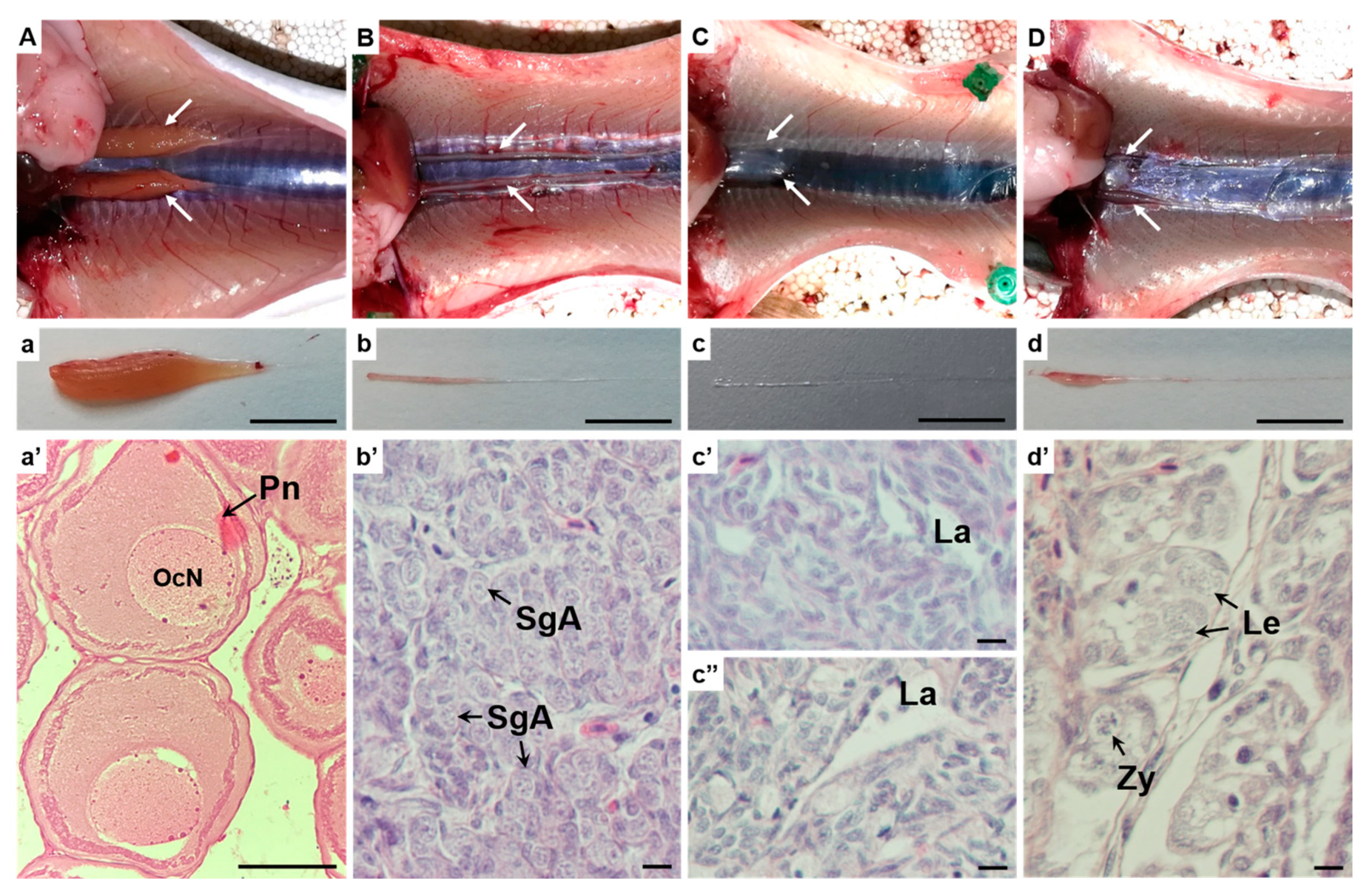
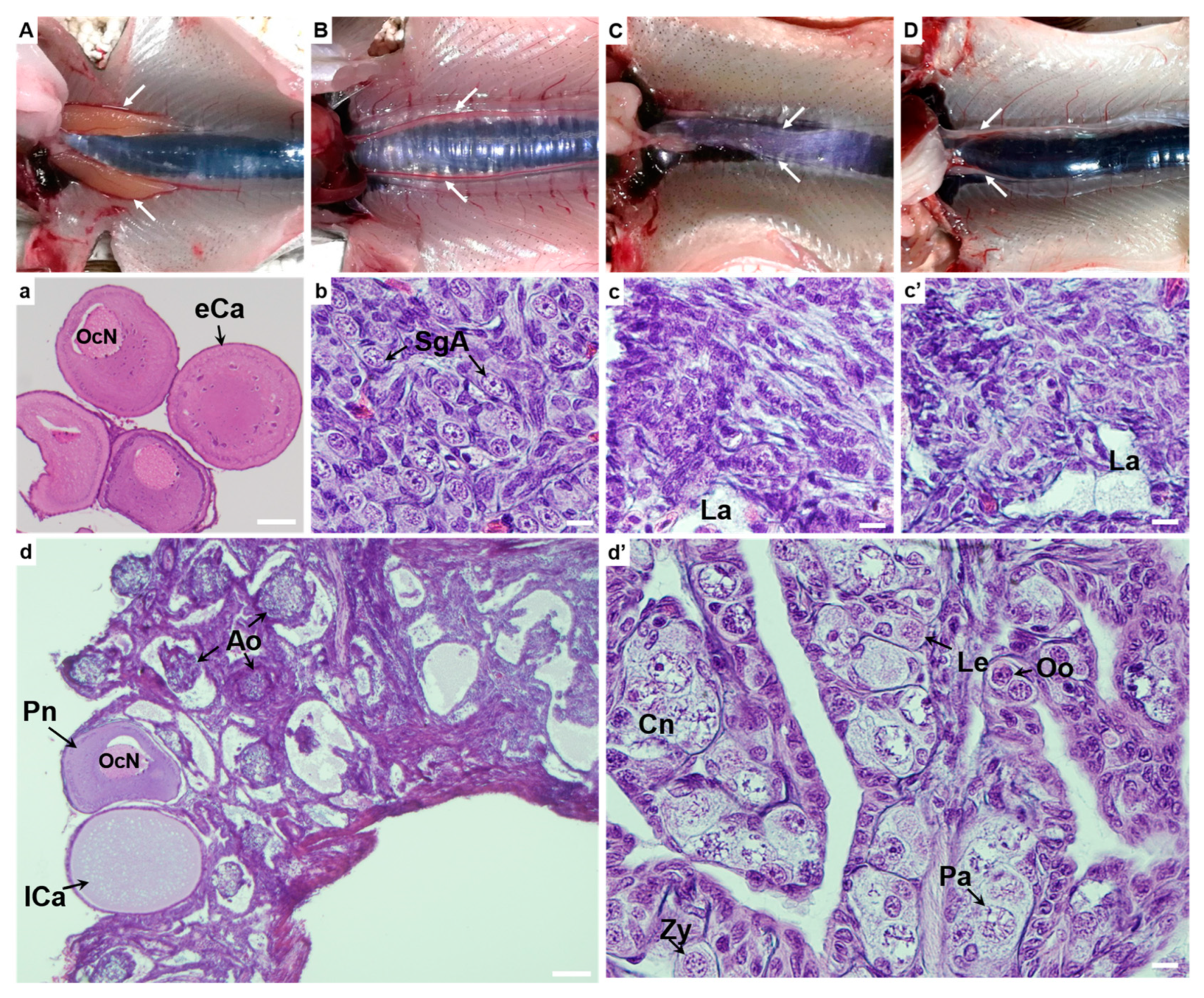
2.6. Gonad Histology

Gonads were excised out and fixed in Bouin’s fixative solution (Sigma-Aldrich) for 24 h at room temperature, followed by paraffin embedding. Five-micrometer sections were cut and mounted on plus-charged glass slides and dried overnight over a heat plate at 40 °C. The sections were then rehydrated and stained with standard hematoxylin-eosin staining and mounted for detecting any germ cells in the gonads. The images were acquired using a Zeiss Axioplan2 microscope (Carl Zeiss AG, Oberkochen, Germany) and CCD Olympus DP70 camera (Olympus Corporation, Tokyo, Japan).

2.7. RT-qPCR Assay

Gonads were also flash-frozen in a dry ice–ethanol bath and stored at −80 °C until further analysis. Total RNA was extracted by Trizol, and 2 μg RNA was reverse-transcribed to cDNA using QuantiNova Reverse Transcription Kit (Qiagen, Hilden, Germany) containing a gDNA removal step. Primer sequences for the target genes, including vasa, nanos3 and a housekeeping gene α subunit of elongation factor 1 (ef1a), are given in Table 1. Amplification of cDNA was performed on a 7500 Fast Real-Time PCR System (Applied Bioscience) with a Fast SYBR™ Green Master Mix kit (Thermo Fisher Scientific, Waltham, MA, USA) using the following conditions: 20 s 95 °C activation, 3 s 95 °C denaturation, and 30 s 60 °C annealing/amplification, with the last two steps repeated 45 times. A melting curve running program was also added to the end of amplification to confirm that only a single PCR product was amplified. In each reaction round, negative controls, including non-RT controls (without reverse transcriptase) and non-template controls, were added to determine gDNA and other contaminations. CT values for target genes in each sample were normalized against the internal control ef1a, and the relative expression level was calculated by 2−△△Ct method. Statistically significant differences between female, male, and sterile gonads were determined by one-way ANOVA, followed by Tukey’s HSD post hoc test, except for the nanos3 gene in batch B, where Kruskal–Wallis rank sum test and pairwise Wilcoxon Rank Sum tests were applied due to the violation of ANOVA test assumptions. The significance level was set as * indicating p value ≤ 0.05, ** indicating p value ≤ 0.005, *** indicating p value ≤ 0.0005.

3. Results

3.1. Immersion Treatment

The eyed rates in different control and treated groups are shown in Table 2. All groups treated for 48 h exhibited low eyed rates (<5%). As a result, subsequent treatments in both batches B and C were reduced to 12 to 36 h, which improved survival until the eye stage. In batch C, the ovarian fluid component in medium B was later replaced by DMEM due to inconsistent availability, biochemical parameters, and composition of ovarian fluid among different females. Immersion media C1 and C2 with DMEM were found to be competent to maintain the viability of green eggs during the incubation procedure.

3.2. Sex Genotyping

Fish were sex genotyped before dissection. Representative gel photos for sex genotyping are demonstrated in Figure 1B. Female gDNA samples yielded amplification of GH-I and GH-II genes, whereas males exhibited an additional product of the male-specific pseudo growth hormone gene, GH-Ψ (the left gel photo in Figure 1B). The sexually dimorphic on the Y-chromosome (sdY) gene was only amplified, as a product around 700 bp in size, in male but not female fin samples (the right gel photo in Figure 1B). The sex determined by these two marker genes was consistent for all of the fish examined.

3.3. Dissection and Gonadal Histology

In batch B, fish were sampled at 14 months old. Upon dissection, all fertile female fish were identified by two apparent ovarian bulbs, while many, although not all, fertile male fish displayed two small lobes of the testes (Figure 2A,B). In contrast, gonad development in sterile fish was absent except for two barely visible pieces of thin filament-like tissue (Figure 2C). There was a remarked size difference between fertile (Figure 2a,b) and sterile gonads (Figure 2c) when excised from the fish. Gonad histology further confirmed the presence of previtellogenic oocytes at the perinucleolar stage (Figure 2a′) in control females and spermatogonium A (Figure 2b′) in control males. In contrast, both sterile male gonads (Figure 2c′) and sterile female gonads (Figure 2c″) were devoid of germ cells. Sterile gonads were less organized, with the presence of lacuna, connective tissue elements, as well as some cells that may have represented gonadal somatic cells. Interestingly, another type of sterile female was observed as oogenesis retardation, where germ cells were arrested at different prophases in meiosis I (Figure 2d′). The retarded ovaries (Figure 2d) were larger than sterile gonads but still significantly smaller than fertile ovarian bulbs at the perinucleolar oocyte stage.
In batch C, fish were sampled at 20 months old. Upon dissection, ovaries affected by the treatments (Figure 3C,D) were distinguishable from fertile ovaries (Figure 3A). Control females exhibited ovaries at the early cortical alveoli stage, marked by the accumulation of cortical alveoli (Figure 3a). Fertile testes remained at similar stages compared to the earlier sampled batch B without further development (Figure 3B,b). Most retarded ovaries were found arrested at different stages of prophase I oocytes (from leptotene to pachytene, Figure 3d′). However, one retarded female had two primary oocytes (perinucleolar and late cortical alveoli), with the rest of the oocytes all being atretic (Figure 3d), indicating their incompetence to develop into functional ovaries, thus representing sterile gonads. Table 2 provides the number of sterile fish found in each group and sterility rates (based on females only).

3.4. Gene Expression in Gonadal Tissues

Relative expression levels of nanos3 and vasa normalized by ef1a are presented separately for batch B and batch C in Figure 4. Significant differences were observed in the mRNA levels of both vasa (Figure 4A,C) and nanos3 (Figure 4B,D) among different groups. For vasa expression, fertile ovaries and testes had similar levels at 14 months old (p = 0.17), while fertile ovaries displayed lower vasa expression compared to fertile testes at 20 months old (p = 0.01), due to the advancement of oogenesis from perinucleolar to cortical alveoli stage. Sterile gonads lacking germ cells showed no expression of vasa mRNA, while retarded ovaries with arrested germ cells exhibited similar vasa expression as fertile females due to early activity of their germ cells (Figure 4A,C, p > 0.05). In terms of nanos3 expression, sterile ovaries exhibited no nanos3 expression at 14 months old (Figure 4B), and extremely low levels at 20 months old, significantly lower than fertile ovaries (Figure 4D, p < 0.005). Ovaries displaying retardation had extremely low levels of nanos3 expression that were significantly lower than in fertile ovaries at 20 months old (p < 0.0005).

4. Discussion

As the first example reported of applying this novel immersion sterilization technology to an aquaculture species with elaborated protocols, we demonstrated the proof of concept of the transferability of this technology. We obtained sterile coho salmon individuals without germ cells, those with oocytes arrested at prophase I, and with atretic oocytes as well, though in small numbers. As dnd is an evolutionarily conserved element essential for PGC development in multiple fish species [18,25,26,27,28,29], this sterilization technology could be applied to other commercially important fish species. As we pursue strategies for optimization, including those mentioned here and elsewhere [30], we will continue to build on these results to achieve high sterility induction in salmonids and extend the methodology to other fish species. Achieving reproductively sterile fish farming by immersion sterilization is expected to mitigate the issues associated with undesired maturation and reproduction, and provide premium sterile fish farming paradigms for ecologically and economically sustainable aquaculture globally.
The emerging immersion-based approach targeting dnd to sterilize fish without introducing any genetic modifications presents a promising solution for mitigating the limitations and disadvantages of triploidization and genetic engineering approaches. This novel sterilization technology has many advantages, including (1) targeting dnd, which is highly conserved across species, to disrupt PGC development, offering a solid foundation for technology transfer to other fish, (2) achieving sterility without introducing any genetic modifications, thus mitigating public concerns over food safety and environmental sustainability, (3) producing sterile populations in an efficient manner that is practical for large-scale aquaculture operations, and (4) acquiring sterility in an inducible way, while fertile broodstock can be maintained by simply omitting the treatment.
In the case of zebrafish, where the technology was first demonstrated [19], a post-fertilization treatment was conducted on early embryos. However, for coho salmon, only the pre-fertilization treatment (treating eggs before fertilization) was feasible since the embryos after fertilization were sensitive to the movement applied during immersion treatment. Therefore, eggs must be incubated in an appropriate medium during treatment to maintain their viability before fertilization. A common practice to achieve this is by keeping them in the ovarian fluid, imitating the environment they originated from [31,32]. Therefore, we included ovarian fluid as a major component of the immersion medium during the 2019 winter treatments. The presence of ovarian fluid in the immersion medium was indeed shown to maintain some viability of the green eggs compared to other media we used.
However, obtaining enough ovarian fluid can be challenging. In addition, the characteristics of the ovarian fluid, such as clarity, ionic composition, pH, and osmolarity, vary among different females and at different maturation stages [3], leading to potential inconsistency affecting egg viability and MO uptake during immersion treatments. Moreover, the use of ovarian fluid increases the risk of introducing diseases into the fish culture system [33]. Therefore, adopting a well-defined medium, such as a fish cell culture medium like Leibovitz’s medium (L-15) and Eagle’s minimum essential medium (MEM), modified to resemble components and physicochemical properties of ovarian fluid [34], may provide a more reliable and consistent treatment condition. Indeed, the immersion medium C1 containing DMEM, for example, demonstrated competence in maintaining the viability of green eggs during incubation (Table 2).
The immersion conditions, including temperature, the concentration of csdnd-MOVivo, and duration of treatment, collectively determine survival and sterility rates, between which there is a trade-off. In zebrafish, a higher concentration of zfdnd-MO-Vivo or longer immersion treatment produced more sterile fish but led to lower embryo survival (citation). This drop in survival may be caused by the endocytosis activity imposed by Vivo during early embryogenesis, which may retard and irreversibly arrest development. Therefore, the optimal dosage and immersion conditions need to be determined according to not only their effectiveness for sterility induction but also the embryo viability after treatments. When we shortened the treatment to 12 and 24 h in subsequent treatments in batch C, survival rates in the eye stage increased to 19.3–63.5% compared to extremely low eyed rates (<5%) for groups treated for 48 h in batch B. The survival rates may be further improved by shortening treatment time, as we have found the MO uptake by chorion is a relatively fast process.
In addition to egg viability, the immersion medium must also prevent water hardening of the eggs. Teleost eggs, when spawned from the isotonic environment of the coelom into freshwater, regardless of whether or not fertilization occurs, undergo rapid changes generally referred to as activation and water hardening. This change renders the chorion to act as a semipermeable barrier [35,36] and become noticeably less permeable in many teleost fish [37,38,39,40,41,42,43]. In zebrafish, decreased permeability of chorion after water hardening caused insufficient MO uptake and reduced sterilization rates [19]. Therefore, the immersion medium needs to be isotonic to coelomic/ovarian fluid to prevent water hardening in this regard. On the other hand, enzymatic removal of chorion has also been attempted using natural hatching enzymes, trypsin, pancreatin, or a combination of them [44,45,46]. A milder treatment with these enzymes during or before treatment may soften the chorion, promoting its permeability and enhancing MO uptake for a higher sterility rate.
Coho salmon, a native Pacific salmonid species with a long cultural history, has gained increasing interest in aquaculture in recirculating aquaculture systems (RAS) in recent years [47,48]. Although fish reared in RAS are less likely to escape into the wild compared to those reared using the net pen culture method, many salmonids experience a high level of precocious maturation in both sexes when raised in RAS [49,50], which can severely compromise their growth and flesh quality, thus economic profitability. Farming sterile coho salmon offers one of the most effective methods to prevent unwanted maturation and reproduction while promoting cost-effective and ecologically responsible aquaculture. Here, we applied our novel immersion-based gene silencing sterilization technology to coho salmon for the first time to produce sterile coho salmon and evaluated their gonadal development.
The excessively long reproduction cycle of many aquaculture fish may hinder the application and refinement when transferring this technology. In the case of coho salmon, testes were still tiny and underdeveloped even at 20 months old, making it difficult to distinguish sterile males upon dissection. In contrast, sterile females without germ cells and those with oogenesis retardation showed remarkably smaller ovaries compared to fertile females. As the two prominent ovarian bulbs can develop in as early as 6 months in females, using an all-female population for immersion treatment would shorten the time needed for gonadal development assessment and accelerate the application and optimization process.

Supplementary Materials

The following supporting information can be downloaded at: https://www.mdpi.com/article/10.3390/jmse11122208/s1, Figure S1: A striking example of a precocious male coho salmon reared in our recirculating aquaculture system.

Author Contributions

Conceptualization, T.-T.W., Y.Z. and L.X.; methodology, T.-T.W., L.X. and Y.Z.; software, L.X.; validation, T.-T.W.; formal analysis, L.X. and T.-T.W.; investigation, L.X., T.-T.W. and M.Z.; resources, T.-T.W., L.X. and Y.Z.; data curation, T.-T.W.; writing—original draft preparation, L.X. and M.Z.; writing—review and editing, T.-T.W., L.X., M.Z. and Y.Z.; visualization, M.Z. and L.X.; supervision, T.-T.W. and Y.Z.; project administration, T.-T.W. and Y.Z.; funding acquisition, T.-T.W. and Y.Z. All authors have read and agreed to the published version of the manuscript.

Funding

This research was funded by University of Maryland Baltimore County STRT7TEN-1113 to T.-T.W. In addition, L.X. and M.Z. were supported by scholarships from the China Scholarship Council.

Institutional Review Board Statement

All experimental protocols involved in this study were approved by the Institutional Animal Care and Use Committee at the University of Maryland School of Medicine IACUC# 0521013.

Data Availability Statement

Data are contained within the article.

Acknowledgments

The authors acknowledge Joseph Kalisch, Joel Jaquez, Sherman Davis and Angela Stefani from Washington Department of Fish and Wildlife, Michael Bisbee and Tui Moliga from Nez Perce Tribe Fisheries, Daniel Green from Oregon Department of Fish and Wildlife and Riverence Holdings LLC. for collecting and shipping coho salmon gametes. We also thank the staff of the IMET Aquaculture Research Center for maintaining the fish. We acknowledge financial support from IMET MEES Student Enhancement Awards (Baltimore, MD, USA).

Conflicts of Interest

The authors declare no conflict of interest.

References

  1. Du, S.J.; Gong, Z.; Fletcher, G.L.; Shears, M.A.; King, M.J.; Idler, D.R.; Hew, C.L. Growth Enhancement in Transgenic Atlantic Salmon by the Use of an “All Fish” Chimeric Growth Hormone Gene Construct. Bio/Technol. 1992, 10, 176–181. [Google Scholar] [CrossRef]
  2. Lind, C.E.; Ponzoni, R.W.; Nguyen, N.H.; Khaw, H.L. Selective Breeding in Fish and Conservation of Genetic Resources for Aquaculture. Reprod. Domest. Anim. 2012, 47, 255–263. [Google Scholar] [CrossRef]
  3. Dunham, R.A. Transgenic Fish Resistant to Infectious Diseases, Their Risk and Prevention of Escape into the Environment and Future Candidate Genes for Disease Transgene Manipulation. Comp. Immunol. Microbiol. Infect. Dis. 2009, 32, 139–161. [Google Scholar] [CrossRef]
  4. Regan, T.; Bean, T.P.; Ellis, T.; Davie, A.; Carboni, S.; Migaud, H.; Houston, R.D. Genetic Improvement Technologies to Support the Sustainable Growth of UK Aquaculture. Rev. Aquac. 2021, 13, 1958–1985. [Google Scholar] [CrossRef]
  5. Muir, W.M.; Howard, R.D. Possible Ecological Risks of Transgenic Organism Release When Transgenes Affect Mating Success: Sexual Selection and the Trojan Gene Hypothesis. Proc. Natl. Acad. Sci. USA 1999, 96, 13853–13856. [Google Scholar] [CrossRef]
  6. Karlsson, S.; Diserud, O.H.; Fiske, P.; Hindar, K. Widespread Genetic Introgression of Escaped Farmed Atlantic Salmon in Wild Salmon Populations. ICES J. Mar. Sci. 2016, 73, 2488–2498. [Google Scholar] [CrossRef]
  7. Tveiten, H.; Johnsen, H.K.; Jobling, M. Influence of Maturity Status on the Annual Cycles of Feeding and Growth in Arctic Charr Reared at Constant Temperature. J. Fish. Biol. 1996, 48, 910–924. [Google Scholar] [CrossRef]
  8. Kadri, S.; Mitchell, D.F.; Metcalfe, N.B.; Huntingford, F.A.; Thorpe, J.E. Differential Patterns of Feeding and Resource Accumulation in Maturing and Immature Atlantic Salmon, Salmo salar. Aquaculture 1996, 142, 245–257. [Google Scholar] [CrossRef]
  9. Aksnes, A.; Gjerde, B.; Roald, S.O. Biological, Chemical and Organoleptic Changes during Maturation of Farmed Atlantic Salmon, Salmo salar. Aquaculture 1986, 53, 7–20. [Google Scholar] [CrossRef]
  10. Zohar, Y. Endocrinology and Fish Farming: Aspects in Reproduction, Growth, and Smoltification. Fish. Physiol. Biochem. 1989, 7, 395–405. [Google Scholar] [CrossRef]
  11. Harris, J.; Bird, D.J. Modulation of the Fish Immune System by Hormones. Vet. Immunol. Immunopathol. 2000, 77, 163–176. [Google Scholar] [CrossRef]
  12. Skarstein, F.; Folstad, I.; Liljedal, S. Whether to Reproduce or Not: Immune Suppression and Costs of Parasites during Reproduction in the Arctic Charr. Can. J. Zool. 2001, 79, 271–278. [Google Scholar] [CrossRef]
  13. Taranger, G.L.; Carrillo, M.; Schulz, R.W.; Fontaine, P.; Zanuy, S.; Felip, A.; Weltzien, F.A.; Dufour, S.; Karlsen, Ø.; Norberg, B.; et al. Control of Puberty in Farmed Fish. Gen. Comp. Endocrinol. 2010, 165, 483–515. [Google Scholar] [CrossRef]
  14. Piferrer, F.; Beaumont, A.; Falguière, J.C.; Flajšhans, M.; Haffray, P.; Colombo, L. Polyploid Fish and Shellfish: Production, Biology and Applications to Aquaculture for Performance Improvement and Genetic Containment. Aquaculture 2009, 293, 125–156. [Google Scholar] [CrossRef]
  15. Johnson, O.W.; Dickhoff, W.W.; Utter, F.M. Comparative Growth and Development of Diploid and Triploid Coho Salmon, Oncorhynchus kisutch. Aquaculture 1986, 57, 329–336. [Google Scholar] [CrossRef]
  16. Ojolick, E.J.; Cusack, R.; Benfey, T.J.; Kerr, S.R. Survival and Growth of All-Female Diploid and Triploid Rainbow Trout (Oncorhynchus mykiss) Reared at Chronic High Temperature. Aquaculture 1995, 131, 177–187. [Google Scholar] [CrossRef]
  17. Fjelldal, P.G.; Hansen, T. Vertebral Deformities in Triploid Atlantic Salmon (Salmo salar L.) Underyearling Smolts. Aquaculture 2010, 309, 131–136. [Google Scholar] [CrossRef]
  18. Wargelius, A.; Leininger, S.; Skaftnesmo, K.O.; Kleppe, L.; Andersson, E.; Taranger, G.L.; Schulz, R.W.; Edvardsen, R.B. Dnd Knockout Ablates Germ Cells and Demonstrates Germ Cell Independent Sex Differentiation in Atlantic Salmon. Sci. Rep. 2016, 6, 21284. [Google Scholar] [CrossRef]
  19. Wong, T.-T.; Zohar, Y. Production of Reproductively Sterile Fish by a Non-Transgenic Gene Silencing Technology. Sci. Rep. 2015, 5, 15822. [Google Scholar] [CrossRef]
  20. Du, S.J.; Devlin, R.H.; Hew, C.L. Genomic Structure of Growth Hormone Genes in Chinook Salmon (Oncorhynchus tshawytscha): Presence of Two Functional Genes, GH-I and GH-II, and a Male-Specific Pseudogene, GH-ψ. DNA Cell Biol. 1993, 12, 739–751. [Google Scholar] [CrossRef]
  21. Yano, A.; Nicol, B.; Jouanno, E.; Quillet, E.; Fostier, A.; Guyomard, R.; Guiguen, Y. The Sexually Dimorphic on the Y-chromosome Gene (SdY) Is a Conserved Male-specific Y-chromosome Sequence in Many Salmonids. Evol. Appl. 2013, 6, 486–496. [Google Scholar] [CrossRef]
  22. Moore, L.J.; Somamoto, T.; Lie, K.K.; Dijkstra, J.M.; Hordvik, I. Characterisation of Salmon and Trout CD8α and CD8β. Mol. Immunol. 2005, 42, 1225–1234. [Google Scholar] [CrossRef]
  23. Trummel, D.E.; Fulcher, K.D.; Beck, J.C.; Cloud, J.G. Fertility of Rainbow Trout Males Relative to Differences in the Proportion of Sperm That Binds to a Specific Antibody. Aquaculture 1992, 104, 175–182. [Google Scholar] [CrossRef]
  24. Krise, W.F.; Hendrix, M.A.; Bonney, W.A.; Baker-Gordon, S.E. Evaluation of Sperm-Activating Solutions in Atlantic Salmon Salmo salar Fertilization Tests. J. World Aquac. Soc. 1995, 26, 384–389. [Google Scholar] [CrossRef]
  25. Fujimoto, T.; Nishimura, T.; Goto-Kazeto, R.; Kawakami, Y.; Yamaha, E.; Arai, K. Sexual Dimorphism of Gonadal Structure and Gene Expression in Germ Cell-Deficient Loach, a Teleost Fish. Proc. Natl. Acad. Sci. USA 2010, 107, 17211–17216. [Google Scholar] [CrossRef]
  26. Goto, R.; Saito, T.; Takeda, T.; Fujimoto, T.; Takagi, M.; Arai, K.; Yamaha, E. Germ Cells Are Not the Primary Factor for Sexual Fate Determination in Goldfish. Dev. Biol. 2012, 370, 98–109. [Google Scholar] [CrossRef]
  27. Linhartová, Z.; Saito, T.; Kašpar, V.; Rodina, M.; Prášková, E.; Hagihara, S.; Pšenička, M. Sterilization of Sterlet Acipenser ruthenus by Using Knockdown Agent, Antisense Morpholino Oligonucleotide, against Dead End Gene. Theriogenology 2015, 84, 1246–1255.e1. [Google Scholar] [CrossRef]
  28. Weidinger, G.; Stebler, J.; Slanchev, K.; Dumstrei, K.; Wise, C.; Lovell-Badge, R.; Thisse, C.; Thisse, B.; Raz, E. Dead End, a Novel Vertebrate Germ Plasm Component, Is Required for Zebrafish Primordial Germ Cell Migration and Survival. Curr. Biol. 2003, 13, 1429–1434. [Google Scholar] [CrossRef]
  29. Yoshizaki, G.; Takashiba, K.; Shimamori, S.; Fujinuma, K.; Shikina, S.; Okutsu, T.; Kume, S.; Hayashi, M. Production of Germ Cell-Deficient Salmonids by Dead End Gene Knockdown, and Their Use as Recipients for Germ Cell Transplantation. Mol. Reprod. Dev. 2016, 83, 298–311. [Google Scholar] [CrossRef]
  30. Xu, L.; Zhao, M.; Ryu, J.H.; Hayman, E.S.; Fairgrieve, W.T.; Zohar, Y.; Luckenbach, J.A.; Wong, T.-T. Reproductive Sterility in Aquaculture: A Review of Induction Methods and an Emerging Approach with Application to Pacific Northwest Finfish Species. Rev. Aquac. 2023, 15, 220–241. [Google Scholar] [CrossRef]
  31. Bongers, A.B.J.; in’t Veld, E.P.C.; Abo-Hashema, K.; Bremmer, I.M.; Eding, E.H.; Komen, J.; Richter, C.J.J. Androgenesis in Common Carp (Cyprinus carpio L.) Using UV Irradiation in a Synthetic Ovarian Fluid and Heat Shocks. Aquaculture 1994, 122, 119–132. [Google Scholar] [CrossRef]
  32. Corley-Smith, G.E.; Lim, C.J.; Brandhorst, B.P. Production of Androgenetic Zebrafish (Danio rerio). Genetics 1996, 142, 1265–1276. [Google Scholar] [CrossRef]
  33. Sakai, N.; Burgess, S.; Hopkins, N. Delayed in Vitro Fertilization of Zebrafish Eggs in Hank’s Saline Containing Bovine Serum Albumin. Mol. Mar. Biol. Biotechnol. 1997, 6, 84–87. [Google Scholar]
  34. Rosengrave, P.; Taylor, H.; Montgomerie, R.; Metcalf, V.; McBride, K.; Gemmell, N.J. Chemical Composition of Seminal and Ovarian Fluids of Chinook Salmon (Oncorhynchus tshawytscha) and Their Effects on Sperm Motility Traits. Comp. Biochem. Physiol. A Mol. Integr. Physiol. 2009, 152, 123–129. [Google Scholar] [CrossRef]
  35. Ernst, V.V.; Neff, J.M.; Anderson, J.W. The Effects of the Water-Soluble Fractions of No. 2 Fuel Oil on the Early Development of the Estuarine Fish, Fundulus Grandis Baird and Girard. Environ. Pollut. (1970) 1977, 14, 25–35. [Google Scholar] [CrossRef]
  36. Manner, H.W.; Vancura, M.; Muehleman, C. The Ultrastructure of the Chorion of the Fathead Minnow, Pimephales promelas. Trans. Am. Fish. Soc. 1977, 106, 110–114. [Google Scholar] [CrossRef]
  37. Gellert, G.; Heinrichsdorff, J. Effect of Age on the Susceptibility of Zebrafish Eggs to Industrial Wastewater. Water Res. 2001, 35, 3754–3757. [Google Scholar] [CrossRef]
  38. González-Doncel, M.; Larrea, M.; Sánchez-Fortún, S.; Hinton, D.E. Influence of Water Hardening of the Chorion on Cadmium Accumulation in Medaka (Oryzias latipes) Eggs. Chemosphere 2003, 52, 75–83. [Google Scholar] [CrossRef]
  39. Harvey, B.; Chamberlain, J.B. Water Permeability in the Developing Embryo of the Zebrafish, Brachydanio rerio. Can. J. Zool. 1982, 60, 268–270. [Google Scholar] [CrossRef]
  40. Mangor-Jensen, A. Water Balance in Developing Eggs of the Cod Gadus morhua L. Fish. Physiol. Biochem. 1987, 3, 17–24. [Google Scholar] [CrossRef]
  41. Prescott, D.M. Effect of Activation on the Water Permeability of Salmon Eggs. J. Cell. Comp. Physiol. 1955, 45, 1–12. [Google Scholar] [CrossRef]
  42. Rudy, P.P.; Potts, W.T. Sodium Balance in the Eggs of the Atlantic Salmon, Salmo salar. J. Exp. Biol. 1969, 50, 239–246. [Google Scholar] [CrossRef]
  43. Terner, C. Studies of Metabolism in Embryonic Development-I. The Oxidative Metabolic of Unfertilized and Embryonated Eggs of the Rainbow Trout. Comp. Biochem. Physiol. 1968, 24, 933–940. [Google Scholar] [CrossRef]
  44. Onozato, H.; Usui, K.; Hamada, K. A Method for Dechorionation in Goldfish Carassius auratus. Nippon Suisan Gakkaishi 1986, 52, 1929–1934. [Google Scholar] [CrossRef]
  45. Sakai, Y.T. Method for Removal of Chorion and Fertilization of the Naked Egg in Oryzias latipes. Embryologia 1961, 5, 357–368. [Google Scholar] [CrossRef]
  46. Yoshizaki, G.; Oshiro, T.; Takashima, F. Prevention of Hardening of Chorion and Dechorionation for Microinjection into Fish Eggs. Nippon Suisan Gakkaishi 1989, 55, 369. [Google Scholar] [CrossRef]
  47. Bendriem, N.; Roman, R.; Sumaila, U.R. Enhancing Land-Based Culture of Coho Salmon through Genomic Technologies: An Economic Analysis. Aquac. Econ. Manag. 2021, 25, 27–52. [Google Scholar] [CrossRef]
  48. Gaffney, L.P.; Franks, B.; Weary, D.M.; Von Keyserlingk, M.A.G. Coho Salmon (Oncorhynchus kisutch) Prefer and Are Less Aggressive in Darker Environments. PLoS ONE 2016, 11, e0151325. [Google Scholar] [CrossRef]
  49. Crouse, C.; Davidson, J.; May, T.; Summerfelt, S.; Good, C. Production of Market-Size European Strain Atlantic Salmon (Salmo salar) in Land-Based Freshwater Closed Containment Aquaculture Systems. Aquac. Eng. 2021, 92, 102138. [Google Scholar] [CrossRef]
  50. Good, C.; Davidson, J. A Review of Factors Influencing Maturation of Atlantic Salmon, Salmo salar, with Focus on Water Recirculation Aquaculture System Environments. J. World Aquac. Soc. 2016, 47, 605–632. [Google Scholar] [CrossRef]
Figure 1. Flowchart of the immersion and sampling procedures involved in this study. (A) Schematic depiction of the application of immersion-based sterilization technology in coho salmon. Unfertilized green eggs were treated in an immersion medium containing csdnd-MO-Vivo. After washing, treated eggs were fertilized and reared up to 14 or 20 months old for gonadal development assessment. (B) All fish were individually tagged and sex genotyped before the assessment of gonadal development. The left gel photo demonstrates sex genotyping using a primer pair that amplifies the growth hormone genes. Males have an extra band compared to females, corresponding to the pseudo growth hormone gene (GH-Ψ) amplicon, which falls between 200 and 300 bp. The right gel photo shows sex genotyping with a primer pair for amplifying sexually dimorphic on the Y-chromosome (sdY) gene. Males have a specific sequence in the Y-chromosome that can be amplified by this primer pair, which is absent in females. L, DNA ladder; F, female; M, male. Numbers on the side denote the 100 bp, 500 bp and 1000 bp bands of the DNA ladder. After dissection, gonadal tissues were sampled and subjected to gonadal histology (C) and RT-qPCR assay (D) for vasa and nanos3 expression.
Jmse 11 02208 g001
Figure 2. Gross morphology of gonads and gonadal histology in 14-month-old fertile and treated sterile coho salmon. (A) A control female; (B) a control male; (C) the gonads of sterile female and male fish had similar appearance; (D) a retarded female. Arrows in (AD) point to the gonads; (ad) show the excised gonads from fertile female, fertile male, sterile, and retarded female, respectively. Two ovarian bulbs were present in control females (A,a). Gonads of sterile fish (C,c) are filament-like and much smaller than testes (B,b), which can be easily distinguished. Retarded ovaries (D,d), although exhibiting some development at the anterior end, remained considerably smaller than the fertile ovaries. Gonad histology sections of a control female (a′), a control male (b′), a sterile male (c′), a sterile female (c″), and a retarded female (d′) are shown. Germ cells (perinucleolar oocytes and spermatogonium) were present in control fish, while sterile gonads were devoid of germ cells and less organized, with the presence of lacunas. The ovary with oogenesis retardation exhibited oocytes arrested at different prophases at meiosis I stage (d′). OcN, oocyte nucleus; Pn, perinucleolar oocyte; SpA, spermatogonium A; La, lacuna; Le, leptotene; Zy, zygotene. Scale bar = 1 cm in (ad). Scale bar = 100 µm in (a′). Scale bar = 10 µm in (b′,c′,c″,d′).
Jmse 11 02208 g002
Figure 3. Gross morphology of gonads and gonadal histology in 20-month-old fertile and treated sterile coho salmon. The gross morphology of gonads was similar to those at 14 months old. Arrows in (AD) point to the gonads. Control females (A) at this age had progressed to the early cortical alveoli stage (a), while control testes (B) were still small and had only germ cells at the spermatogonium A stage (b). Sterile male (C,c) and female gonads (c′) were both devoid of germ cells and exhibited less organization, with the presence of lacunas. Most retarded ovaries (D) had oocytes at different prophase I stages (d′), while one displayed two primary oocytes at perinucleolar and late cortical alveoli stages, with the remaining oocytes all atretic oocytes (d). OcN, oocyte nucleus; eCa, early cortical alveoli oocyte; SpA, spermatogonium A; La, lacuna; Pn, perinucleolar oocyte; Ao, atretic oocyte; lCa, late cortical alveoli oocyte; Cn: cell nest in different meiosis stages; Oo, oogonium; Le, leptotene; Zy, zygotene; Pa, patchytene. Scale bar = 100 µm in (a,d). Scale bar = 10 µm in (b,c,c′,d′).
Jmse 11 02208 g003
Figure 4. Relative expression levels of vasa and nanos3 genes were measured and compared among different groups. Data are presented as relative expression normalized against ef1a expression and separated by vasa (A,C) and nanos3 (B,D), as well as by batch B at 14 months old (A,B) and batch C at 20 months old (C,D). Sterile gonads had no expression of vasa, while retarded ovaries with arrested germ cells showed vasa expression levels similar to those of fertile females due to the early activity of their germ cells at both stages. In terms of nanos3, both sterile and retarded ovaries exhibited no nanos3 expression at 14 months old, and extremely low levels at 20 months old, significantly lower than that observed in fertile ovaries. Statistical significance was determined by one-way ANOVA, followed by Tukey’s HSD post hoc test in A, B, and C, and by Kruskal–Wallis rank sum test and pairwise Wilcoxon Rank Sum tests in D. The significance levels are denoted as * indicating p value ≤ 0.05, ** indicating p value ≤ 0.005, *** indicating p value ≤ 0.0005.
Jmse 11 02208 g004
Table 1. Primers used to amplify coho salmon genes.
Target GeneAccession NumberPrimer NameDirectionSequences (5′ to 3′)Tm (°C)Anticipated
Amplicon Size (bp)
Start codon verification
dndXM_020452699.1WBCS1ForwardCACCTAGAACTACCTGTCGAAAC55566
WBCS2ReverseGCTGTCGTACTTGGCGTAG
Sex genotyping
GH1; GH2; GHΨ 1Gene IDs: 109893213; 109898300WBCS11ForwardCCTGGATGACAATGACTCTCA60779; 404; 273
WBCS12ReverseCTACAGAGTGCAGTTGGCCTC
sdY 2-WBCS17ForwardATGGCTGACAGAGAGGCCAGAATCCAA55~700
WBCS18ReverseTGCTCTCTGTTGAAGAGCATCAC
Gonadal tissue gene expression
vasaXM_020457444.2qrCS5ForwardTTTGGGAGACCGACTGATAAAG60124
qrCS6ReverseCACCAGCACCTGAAGAGAAA
nanos3XM_031810988.1WBTP51ForwardTCATGACTCCGGGATATGCT60115
WBTP52ReverseGGGTTCCATTTCGTGCCATA
ef1a 3XM_031793750.1qrCS13ForwardCCCCTCCAGGATGTTTACAAA6057
qrCS14ReverseCACACGGCCCACAGGTACA
1 Primers WBCS11 and WBCS12 are from Du et al., (1993) [20]; 2 primers WBCS17 and WBCS18 are from Yano et al., 2013 [21]; 3 primers qrCS13 and qrCS14 are from Moore et al., (2005) [22]. Tm, annealing temperature used.
Table 2. List of immersion media, treatment conditions, eyed rates, the number of fish dissected, and sterile fish found in different groups.
GroupImmersion Mediumcsdnd-MO-Vivo (μM)Tm (°C)Time (Hours)Eyed Rates 1Fish DissectedSterile FishSterility Rates 2
FemaleMaleFemaleMaleRetarded Female
Batch B
CSB1----146/261 = 55.9%1113 0
CSB16B17.54485/557 = 0.9%23 1 -
CSB27B2042469/514 = 13.4%24261 12/24 = 8.3%
CSB39B12.583653/258 = 20.5%161912 1/16 = 6.3%
Batch C
CSC13C10812198/237 = 83.5%-- -
CSC14-2C120812120/375 = 32.0%2533 1 -
CSC17C10824198/230 = 86.1%--
CSC18-1C12082471/368 = 19.3%2025 22/20 = 10.0%
CSC19C20824109/227 = 48.0%-- -
CSC20-1C220824145/389 = 37.3%28221 1/28 = 3.6%
CSC22-2C10812124/272 = 45.6%-- -
CSC27-1C12081269/337 = 20.5%3417 11/34 = 2.9%
CSC28C20812121/240 = 50.4%-- -
CSC29-2C22081281/326 = 24.58%1922 11/19 = 5.3%
CSC30-1C115 μM
control-MO-Vivo 3
812113/330 = 34.2%5843 0
CSC36-1C115824202/318 = 63.5%4337 11/43 = 2.3%
The groups in Batch B were treated in medium B containing 35% ovarian fluid and 65% fertilization diluent (medium components listed in Section 2.2). The groups in Batch C were treated in medium C1 or C2 with cell culture-based component DMEM replacing ovarian fluid (medium components listed in Section 2.2). 1 Eyed rates were calculated as the number of embryos developed to eye stage (numerator) divided by total treated eggs (denominator). 2 Sterility rates were based on females only and calculated as the number of females without germ cells and retarded females (numerator) divided by the total females dissected (denominator). 3 The CSC30-1 group was treated in 15 μM control-MO-Vivo as an MO control.
Disclaimer/Publisher’s Note: The statements, opinions and data contained in all publications are solely those of the individual author(s) and contributor(s) and not of MDPI and/or the editor(s). MDPI and/or the editor(s) disclaim responsibility for any injury to people or property resulting from any ideas, methods, instructions or products referred to in the content.

