Article

Emericellopsis maritima and Purpureocillium lilacinum Marine Fungi as a Source of Functional Fractions with Antioxidant and Antitumor Potential in Colorectal Cancer: A Preliminary Study

by
Gloria Perazzoli
1,2,3,†,
Carolina de los Reyes
4,†,
Cristina Pinedo-Rivilla
4,*,
Rosa Durán-Patrón
4,
Josefina Aleu
4,
Laura Cabeza
1,2,3,
Consolación Melguizo
1,2,3,* and
Jose Prados
1,2,3
1
Institute of Biopathology and Regenerative Medicine (IBIMER), Biomedical Research Center (CIBM), 18100 Granada, Spain
2
Department of Anatomy and Embryology, University of Granada, 18071 Granada, Spain
3
Instituto de Investigación Biosanitaria de Granada, (ibs.GRANADA), 18014 Granada, Spain
4
Organic Chemistry Department, Faculty of Sciences, University of Cádiz, 11510 Puerto Real, Spain
*
Authors to whom correspondence should be addressed.
These authors contributed equally to this work.
J. Mar. Sci. Eng. 2023, 11(10), 2024; https://doi.org/10.3390/jmse11102024
Submission received: 2 October 2023 / Revised: 16 October 2023 / Accepted: 19 October 2023 / Published: 20 October 2023
(This article belongs to the Section Marine Biology)

Abstract

:
The marine environment is a promising source of natural products with possible pharmacological applications. In this sense, marine microorganisms, especially marine fungi, can produce bioactive compounds with various therapeutic properties. Colorectal cancer (CRC) represents a major health problem worldwide, since the treatments used to date are not capable of improving patient survival; that is why natural compounds from marine fungi offer a promising alternative. This study focused on evaluating the antitumor and antioxidant activity of fractions derived from the marine fungi E. maritima and P. lilacinum in two CRC cell lines T84 and SW480. Fractions Fr-EM6, Fr-EM7, Fr-EM8 and Fr-PLMOH-3 demonstrated potent cytotoxic activity in tested CRC cell lines with no activity in the non-tumor line. In particular, the Fr-PLMOH-3 fraction from P. lilacinum showed significant antiproliferative effects on T84 and SW480 cell lines and exhibited a greater cytotoxic effect on cancer stem cells compared to tumor cells. Furthermore, the Fr-EM8 fraction from E. maritima demonstrated a strong antioxidant capacity. These findings highlight the potential of compounds of marine origin as effective and selective antitumor agents for the treatment of CRC. Further studies are needed to explore the underlying mechanisms and potential clinical applications of these bioactive fractions and compounds.

Graphical Abstract

1. Introduction

The marine environment represents a poorly investigated and highly promising source of natural products with potential pharmacological use [1]. Among these marine microorganisms there are cyanobacteria, diatoms, bacteria, and fungi that are powerful producers of bioactive compounds and exhibit extensive chemical variability and complexity, including alkaloids, polyketides, terpenes, peptides and carbohydrates [2]. In this field of research, marine fungi have attracted increasing attention in recent years due to their ability to produce therapeutic compounds with antifungal, antiviral, antibacterial, antioxidant, and antitumor properties [3]. More specifically, some marine fungi such as Acremonium sp., Acrocalymma sp., Acrocalymma Africana and Acrocalymma medicaginis significantly reduced tumor growth. Specifically, Acremonium sp. decreased the proliferation capacity of lung, skin and breast cancer cell lines by 85% to 97% [4]. In addition, secondary metabolites from Aspergillus sp., Penicillium sp. and Paradendryphiella sp. showed promising results in colorectal cancer (CRC). Specifically, Paradendryphiella sp. derivatives reached an IC50 of 25.7 nM in the HCT116 CRC cell line [5]. Likewise, natural compounds derived from Penicillium sclerotiorum decreased cell migration and cell proliferation in both HCT116 and LOVO CRC cell lines [6]. Marine fungi of the genus Purpureocillium and Emericellopsis stand out among the potential sources of bioactive compounds. The fungus formerly known as Paecilomyces lilacinus, now named P. lilacinum, found in marine sediments with low oxygen and few nutrients, is an entomopathogenic endophytic filamentous fungus [7]. This is also a well-known biocontrol fungus used against several plant pathogens in agriculture [8]. Fungi of the genus Emericellopsis, including the marine species E. maritima, are a group of 20 species. Interestingly, these fungi produce peptides such emericellipsin A, isolated from E. alkaline, which shows cytotoxic activity against cancer cells including melanoma (B16 cells), breast cancer (MCF-7 and MDA.MB-231 cells) and CRC (HCT-116 cells) [9].
In this context, marine microorganisms have been explored as a source of new therapeutic biomolecules that may be effective against CRC [10,11], the third most frequent tumor in the world, with an estimated increase in malignancy of 60% by 2030 [12]. Despite advances in the physiopathology of this tumor, which have determined the three precursor pathways for CRC lesions, chromosomal instability (70–90% of cases), serrated neoplasm (10–20% of cases) and microsatellite instability (2–7% of cases), there have been no significant advances in therapy in recent years. Currently, the curative treatment for patients with non-metastatic CRC is usually surgical resection. However, in more advanced stages of the disease, such as stage III or stage IV (metastatic), adjuvant chemotherapies are used to reduce recurrences and improve quality of life [13,14]. The most commonly used drugs are 5-fluorouracil and capecitabine, often combined with leucovorin or oxaliplatin [15]. However, chemotherapy is not entirely effective and shows serious limitations, such as drug resistance, non-specificity, and side effects that frequently lead to abandonment of treatment [16]. Therefore, it is essential to discover and develop new therapeutic and antitumor agents that are effective and safe, with greater selectivity in their cytotoxicity. Concretely, the discovery of complex molecules with interesting biological properties, including antitumor activity, highlights the enormous potential of marine microorganisms to obtain highly valuable bioactive compounds to prevent or treat cancer with less toxicity than other types of drugs [17,18].
The main objective of our study was to carry out an in vitro evaluation of the antitumor activity of several fractions and compounds derived from two marine fungi, E. maritima and P. lilacinum. Our results show that some of these samples exhibited cytotoxic potential against CRC cells, which may be of great interest for developing new therapeutic strategies for this type of tumor.

2. Materials and Methods

2.1. Fungal Material

E. maritima and P. lilacinum were isolated from sediment samples collected along an intertidal gradient of the Bay of Cádiz (Cádiz, Spain). Conidial stock suspensions of these strains are maintained viable in 80% glycerol at −40 °C.

2.2. General Experimental Procedures

One- and two-dimensional NMR spectra were recorded on Agilent 500 and 600 MHz spectrometers with SiMe4 as the internal reference. NMR assignments were made using a combination of 1D and 2D techniques and by comparison with those made for previously described compounds, where appropriate. TLC was performed on Merck Kiesegel 60 F254, 0.25 mm thick. Silica gel 60 (70–230 mesh, Merck) was used for column chromatography. HPLC was performed with a Merck-Hitachi LaChrom apparatus equipped with a UV–vis detector (L 4250) and a differential refractometer detector (RI- 7490) and an Elite LaChrom-Hitachi apparatus equipped with a differential refractometer detector (RI-2490). LiChroCART LiChrospher Si 60 (5 μm, 250 mm × 4 mm) and LiChroCART LiChrospher Si 60 (10 μm, 250 mm × 10 mm) columns were used for isolation experiments.

2.3. Media and Culture Conditions

The fungal strains were grown in 500 mL Erlenmeyer flasks containing rice solid medium (80 g white rice per 100 mL of water; ingredients were soaked overnight before autoclaving) [19].
Flasks were inoculated with 750 µL of a conidial suspension of the fungi and maintained at static conditions at 24–26 °C under artificial light (daylight lamp) for 40 days.

2.4. Extraction and Isolation of Fractions and Compounds

The rice broths (16 flasks) of the fungal strains were extracted with EtOAc. Organic extracts were washed with H2O and then treated with dry Na2SO4 to remove water. Subsequently, the solvent was evaporated at reduced pressure to yield crude extracts as yellow oils, 4.197 g from E. maritima and 0.582 g from P. lilacinum. Attending to the low weight of the extract from P. lilacinum, the rice broth was also extracted with methanol, affording 0.418 g. The crude extracts obtained were subjected to column chromatography (CC) over silica gel using an increasing gradient of EtOAc in n-hexane and then MeOH:EtAcO 1:9 (v/v) to give eight fractions (Fr-EM1 to Fr-EM8 for E. maritima and Fr-PL1 to Fr-PL8 for P. lilacinum). All the fractions were studied by 1H-NMR analysis to determine which ones were the most interesting for further purification. The MeOH extract obtained from P. lilacinum was resuspended in MeOH–H2O (7:3, v/v, 50 mg/mL), transferred onto RP-18 cartridges preconditioned with MeOH-H2O (7:3, v/v, 1 mL) and eluted with 10 mL of the same solvent, 10 mL of MeOH, and 10 mL of a solution of CHCl3:MeOH 1:1 (v/v). This procedure yielded three fractions of 254.01 mg (Fr-PLMOH-1), 83.17 mg (Fr-PLMOH-2) and 50.46 mg (Fr-PLMOH-3). On the other hand, the fraction Fr-EM3 (425.16 mg) was subdivided into two identical fractions for further purification. One subfraction was subjected to CC using Hex:EtAcO 7:3 (v/v) as eluent to yield seven fractions (Fr-EMCC-3.1 to Fr-EMCC-3.7). The remaining subfraction was purified by semipreparative HPLC, using Hex:EtAcO 8:2 (v/v) as eluent and a flow of 2.5 mL min − 1, to afford the compounds 1 (0.69 mg), 2 (0.83 mg) and 3 (0.74 mg).

2.5. Cell Culture

Cell lines were purchased from the American Type Culture Collection (ATCC, USA). The non-tumoral cell line CCD18 and two human colon adenocarcinoma cell lines (T84 and SW480) were cultured using Dulbecco’s Modified Eagle’s Medium (DMEM) (Sigma-Aldrich, Madrid, Spain) supplemented with fetal bovine serum (FBS) (10%) and antibiotic mixture (1% of penicillin–streptomycin (Sigma–Aldrich, Madrid, Spain)). Cells were maintained in an incubator at 37 °C and a 5%-CO2 humidified atmosphere.

2.6. Cytotoxicity Assay

Cells were seeded in 48-well plates at a density of 5 × 10³ cells per well for the T84 and SW480 lines with 300 μL of complete DMEM medium. Of all obtained fractions from E. maritima (Fr-EM) and P. lilacinum (Fr-PL) (see previously), only the following could be adequately resuspended and tested: Fr-EM5, Fr-EM-3.3, Fr-EMCC-3.2, Fr-EM6, Fr-EMCC-3.4, Fr-EMCC-3.6, Fr-EMCC-3.7, Fr-EM7, Fr-EM8, Fr-Pl5 y, and Fr-PLMOH-3. In addition, the three isolated compounds (1, 2, and 3) could also be assayed. Both fractions and isolated compounds were dissolved in DMSO. For the cell viability study an MTT (3-(4,5-dimethylthiazol-2-yl)-2,5-diphenyltetrazolium bromide) assay was performed 12 h after the end of treatment. Each well was incubated for 2.5 h with 20 µL of MTT. Finally, the medium was discarded, and formazan crystals were dissolved with 25 μL of Sorensen’s glycine buffer (glycine 0.1M, NaCl 0.1M, pH:10.5 with 0.1 NaOH) plus 200 μL of dimethyl sulfoxide (DMSO). The same assay was carried out for the solvent DMSO (0.1–250 μg/mL). Optical density was measured at 570 nm with a reference wavelength of 690 nm in an EX-Thermo Multiskan spectrophotometer (Waltham, MA, USA).

2.7. Cell Cycle Assay

Cells were seeded in 6-well plates at a density of 1 × 10⁵ for the T84 and SW480 cell lines. To synchronize the cell cycle twenty-four hours later, when cells were attached, the culture medium was removed and replaced with serum-free DMEM. Then, culture cells were exposed to fresh supplemented DMEM medium containing the respective treatment. After 48 h, cells were fixed with cold 70% ethanol (4 °C, 30 min). After centrifugation, the pellets were processed with PI/RNAse Solution (Immunostep, Salamanca, Spain). All samples were analyzed with FACScan (Becton Dickinson, Franklin Lakes, NJ, USA).

2.8. Cancer Stem-Like Cell (CSC) Isolation and Characterization

The SW480 cell line was selected to obtain CSCs due to its higher yield based on our experience of CRC cell lines [20]. Accordingly, supplemented (2 mM glutamine, 4 ng/mL bFGF, 10 ng/mL EGF, insulin (10 μg/mL), transferrin (5.5 μg/mL), and sodium selenite (5 ng/mL)) serum-free RPMI-1640 medium was used to culture SW480 in an incubator at 37 °C in a 5%-CO2 humidified atmosphere for eight days. On the fourth day, attached cells were removed and fresh culture medium was added. RNA extraction was carried with an RNeasy Mini Kit following the manufacturer’s instructions (Qiagen, MD, USA) to analyze the expression of CD44, CD24, NANOG, SOX2, CD133, OCT4, and the GADPH housekeeping gene (Table S1). This expression was analyzed by real-time PCR using the Taq Universal SYBR Green Supermix (Bio-Rad Laboratories, Hercules, CA, USA) on a StepOnePlus Real-Time PCR system (Thermo Fisher Scientific, Waltham, MA, USA). Using the 2(−ΔΔCt) method, relative expression levels were calculated.

2.9. Antioxidant Capacity Using H2O2

To test the antioxidant activity, the T84 human CRC cell line was selected. Cells were seeded in 96-well plates at a density of 2.5 × 10⁴ cells/well with supplemented DMEM. Serum-free DMEM was added after 24 h replacing supplemented DMEM. One day later, 0.1 and 0.05 µg/mL of Fr-EM8 and Fr-PLMOH-3 were added for one more day. New fresh serum-free medium was added, and the appropriate wells were treated for 6 h with 1.2 and 1.3 mM of H2O2. For the cell viability study, an MTT assay was performed in the same way described previously for the cytotoxicity assay.

2.10. Antioxidant Capacity Using ABTS

To measure the total antioxidant capacity, an ABTS assay using 2,2′-azino-bis (3-ethylbenzothiazoline-6-sulfonic acid) was conducted following the methodology described by Miller and Miller [21].
A standard solution of gallic acid (0–60 mg/L) was mixed with 6 µL of each compound sample (Fr-EM8: 1180 ug/mL; Fr-PLMOH-3: 336 ug/mL) and 294 µL of ABTS (500 µM/L) and incubated for 5 min. The optical density was measured at 620 nm (Mul-tiskan™ FC, Thermo Fisher Scientific, Waltham, MA, USA). For the blank measurement water was used. The results are expressed as μg of GAE (gallic acid equivalent)/mg of sample.

2.11. Statistical Analysis

All the results are expressed as mean ± standard deviation (SD) of three replicates. Statistical analysis was performed using Student’s t-test (SPSS v.15, SPSS, Chicago, IL, USA). Statistical significance was considered at p < 0.05.

3. Results

3.1. Isolated Fractions and Compounds from Marine Fungi

Of the obtained fractions from marine fungi, only eleven (nine fractions from E. maritima and two from P. lilacinum) could be adequately resuspended and tested in CRC cell lines. In addition, three known compounds (metabolites) with eremophilene skeleton were isolated and identified from the rice extract of E. maritima, isopetasone (1) [22], 3-epiisopetasol (2) [23] and 1α-hydroxydehydrofukinone (3) [24] (Figure 1). The structures of these compounds were determined by detailed analyses of their NMR spectra and comparisons with previously published data (Figures S1–S7). It is worth noting that compounds 1, 2 and 3 were isolated for the first time from an E. maritima strain. Due to the low amounts of compounds obtained, not all in vitro assays could be carried out.

3.2. In Vitro Cytotoxicity of Fractions and Compounds from Marine Fungi

As shown in Table 1, a variable degree of cytotoxicity in the fractions from marine-derived fungi was detected in the CRC cell lines. In fact, the SW480 tumor cell line exhibited higher sensitivity to most treatments, showing lower IC50 values than the T84 cell line. Only compound 2 showed greater activity against T84 cells (IC50 44.47 µg/mL) than SW480 cells (IC50 66.35 µg/mL). It is necessary to underline the potent antitumor effect observed in T84 and SW480 cell lines after exposure to Fr-PLMOH-3 from P. lilacinum (IC50: 11.31 μg/mL in T84 and 6.4 μg/mL in SW480). The remaining fractions tested showed less antiproliferative effects in T84 and SW480 cell lines, such as fraction Fr-EMCC-3.7 (IC50: 56.3 and 23.2 μg/mL in T84 and SW480 cells, respectively) and fraction Fr-EM8 (IC50: 71, 7 and 38.3 μg/mL in T84 and SW480 cells, respectively), both obtained from E. maritima. Finally, other fractions, such as Fr-EMCC-3.2 and Fr-EMCC-3.6, also obtained from E. maritima, showed poor results. In view of the excellent antiproliferative results for Fr-PLMOH-3 from P. lilacinum against CRC, this fraction was tested in the non-tumorigenic colon cell line CCD18. Interestingly, as shown in Figure S8, the Fr-PLMOH-3 IC50 value obtained for CCD18 cells reached very high concentrations (close to 100 μg/mL), which would imply a selective action against tumor cells. In addition, compounds 1 and 3 from E. maritima exhibited potent antiproliferative activity in SW480 cells, showing IC50 values of 24.15 and 14.46 µg/mL, respectively. Due to the low quantities of the compounds obtained, these samples could not be assayed in CCD18 cells. All cytotoxic graphics are shown in Figures S9 and S10.

3.3. Cell Cycle Modulation by Marine-Derived Fungus Fractions

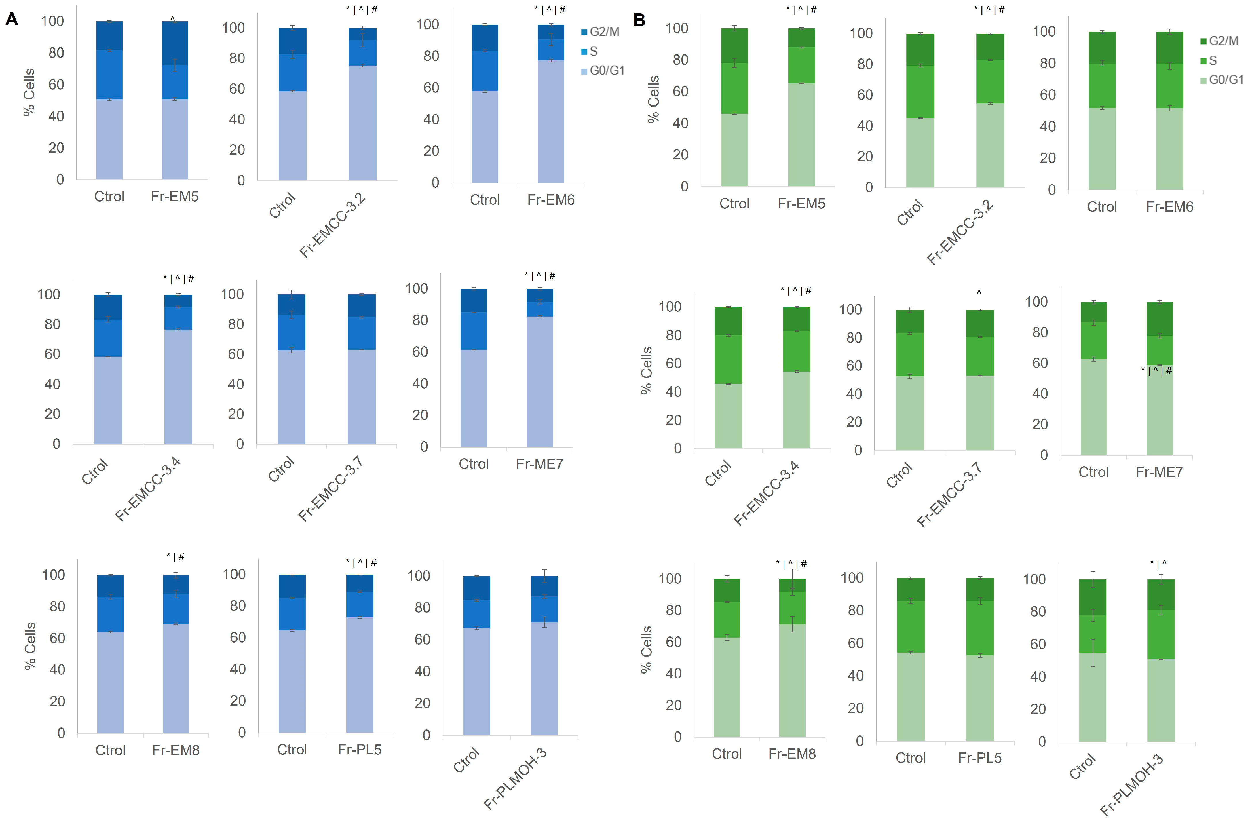
As shown in Figure 2, cell cycle analysis of T84 and SW480 CRC cell lines after fraction exposure (IC50) showed significant changes. Concretely, T84 cells showed modulation in all phases of the cell cycle after treatment with the fractions Fr-EMCC3.2, Fr-EM6, Fr-EMCC-3.4, Fr-EM7, Fr-EM8 and Fr-PL5. By contrast, Fr-EM5 only induced significant changes in the T-84 G2/M phase. In addition, Fr-EM5, Fr-EMCC-3.2, Fr-EMCC-3.4, Fr-EM7 and Fr-EM8 modulated the cell cycle in SW480 cells, highlighting the induction of a significant decrease in the S phase and G2/M phase due to Fr-EM8 and a significant increase in the S phase due to Fr-PLMOH-3. However, no data indicating clear cell cycle arrest were detected in either case compared to control.

3.4. Antiproliferative Activity of Marine-Derived Fungus Fractions against Cancer Stem Cells

To characterize CSC obtained from SW480 cells, a qPCR study was carried out. As shown in Figure 3A, a high expression of specific CSC markers, such as CD44, CD24, NANOG, SOX2, CD133 and OCT4 was detected in CSCs. SW480 CSCs were exposed to those fractions that showed higher antiproliferative activity against SW480 cells and significant variation in cell cycle phases (Fr-EM6, Fr-EM7, Fr-EM8 and Fr-PLMOH-3) (Figure 3B). Interestingly, Fr-PLMOH-3 reduced the IC50 by half in CSCs compared to SW480 tumor cells (from 12.26 to 6.53 µg/mL). In addition, fractions Fr-EM6 and Fr-EM7 were also able to induce a decrease in CSCs’ IC50 relative to SW480 cells (from 62.89 to 43.36 µg/mL and from 55.96 to 43.68 µg/mL respectively), but the difference was not statistically significant. Fr-EM8 not only does not improve the effect in this type of cell, but it was less effective (increasing from 58.54 to 66.25 µg/mL).

3.5. Antioxidant Activity of Marine-Derived Fungus Fractions

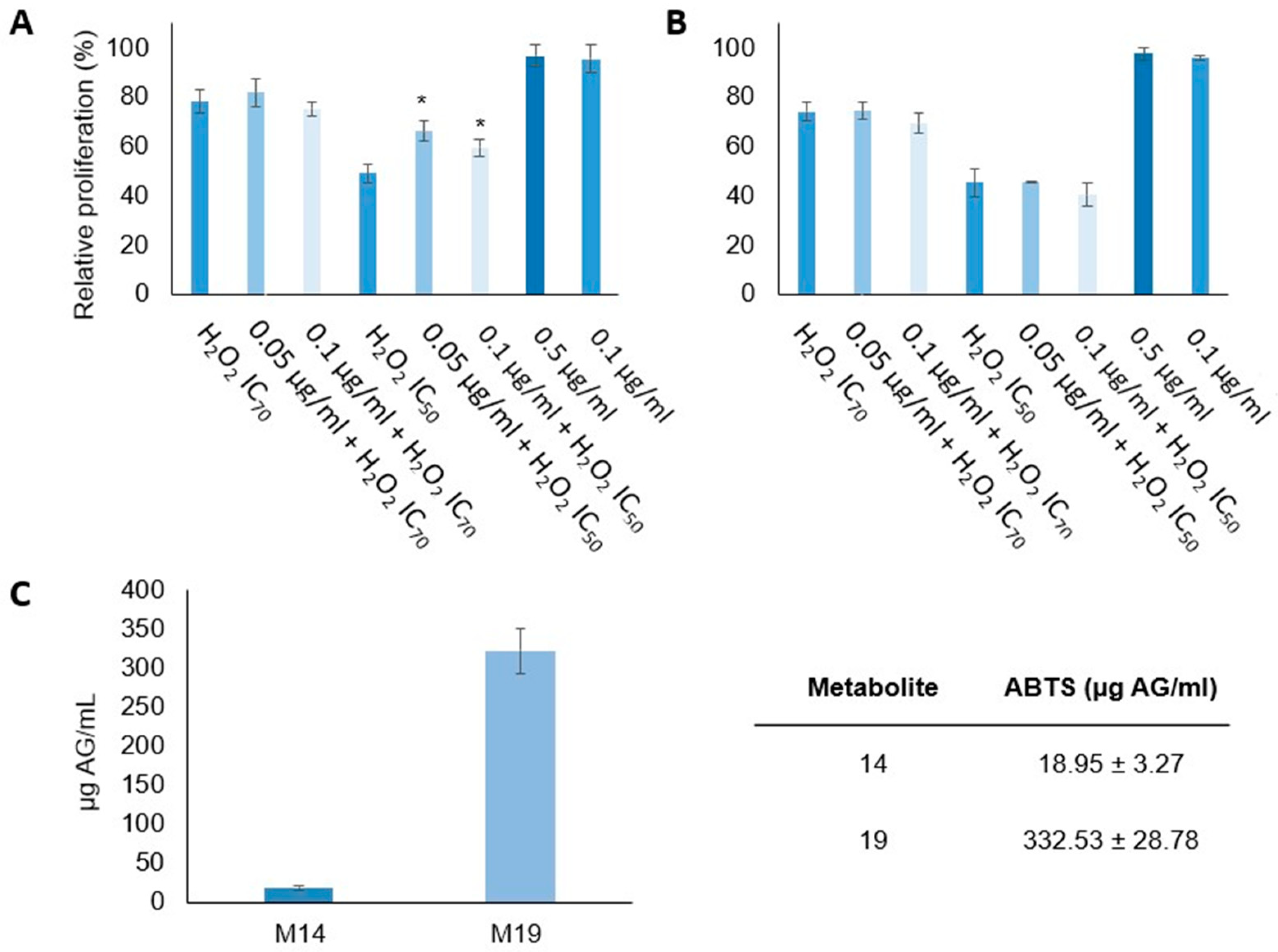
Figure 4 displays the results of testing the antioxidant capacity of selected fractions (Fr-EM8 and Fr-PLMOH-3). To carry out this assay, we selected the T84 cell line. Cells were exposed to oxidative stress using H2O2 as a free radical generator. Notably, fraction Fr-EM8 exhibited the highest antioxidant capacity. As shown in Figure 4A, this fraction significantly modulated IC50 values for H2O2 at both 0.05 and 0.1 µg/mL doses. By contrast, fraction Fr-PLMOH-3 did not exhibit significant antioxidant capacity (Figure 4B). To complement these studies, an ABTS analysis was performed for Fr-EM8 and Fr-PLMOH-3, revealing that Fr-EM8 exhibited a value of 18.95 µg AG/mL, whereas Fr-PLMOH-3 showed a value of 332.53 µg AG/mL (Figure 4C).

4. Discussion

The high mortality rates associated with CRC despite recent advances in chemotherapy and prevention show that current treatments remain ineffective. This may be due to the low selectivity of the drugs against tumor cells, their side effects, and the development of multiresistant phenotypes. This is why it is increasingly necessary to develop therapeutic alternatives that improve both the treatment and prevention of this type of tumor as well as to improve existing chemotherapeutic agents. In this context, fungi of marine origin are being studied, since therapeutic agents and bioactive compounds can be isolated from them for use against CRC [2], including the genera Emericellopsis and Purpureocillium [25,26].
In this context, a cell viability analysis was conducted on different mixtures of pure compounds (13) and mixtures of compounds (fractions) derived from E. maritima and P. lilacinum species against CRC cell lines. Previous studies reported low IC50 values (<20 μg/mL) for compounds of these two genera [25,27]. Kil et al. performed cytotoxicity analysis using lung, breast, liver and skin cancer cell lines with two compounds derived from Emericellopsis spp., JF72 and AF71. Interestingly, both metabolites induced high percentages of inhibition in all cell lines tested, although these authors did not indicate IC50 values. Relevant results in terms of cytotoxicity have also been reported for Purpureocillium spp., whose effect has been tested on triple-negative breast cancer cells [28]. These findings were consistent with results obtained for compounds 1 (24.75 and 24.15 μg/mL in T84 and SW480 cell line respectively) and 3 (32.06 and 14.46 μg/mL in T84 and SW480 cell line respectively) and the Fr-PLMOH-3 fraction (11.3 and 6.4 μg/mL in T84 and SW480 cell lines, respectively). Concretely, this fraction was tested on the non-tumor cell line CCD18, displaying a low antiproliferative activity compared to tumor cell lines (IC50 > 100 μg/mL). This fact could indicate a possible selective effect against tumor cells, since, following [29], an antitumor agent should be more cytotoxic for cancer cell lines than non-tumorigenic ones. Recently, bengamide, a natural product isolated from marine sponges of the Jaspidae family, also showed a significant antiproliferative effect in several CRC cell lines, without inducing any cytotoxicity in normal cells (blood cells) [30]. Furthermore Phi et al. [31] tested bengamide analogs in six tumor cell lines, including breast and hepatocellular carcinoma, obtaining low IC50 values. This promising result supports the need for future studies to elucidate the antitumor action of fungal compounds.
Although cell cycle studies have not detected data indicating clear cell cycle arrest in either case compared to control, uncontrolled cell cycle progression and evasion of apoptosis are common events in cell development in CRC. G0/G1 arrest occurs with other compounds derived from marine fungi that exhibit cytotoxic activity, like norsolorinic acid isolated from Aspergillus nidulans, with a reported 10% increase in the G0/G1 phase in T24 human bladder cancer cells after exposure to norsolorinic acid (IC50) [32]. Steroid derivatives of Penicillium granulatum are also capable of inhibiting cell proliferation through an arrest in this phase of the cycle, with an increase in the G0/G1 percentage of between 10–11% in the A549 lung cancer cell line [33]. In addition, sesquiterpenoid compounds derived from Aspergillus ochraceus can arrest the cell cycle at the G0/G1 phase at a concentration of 1 μM and induce delayed apoptosis after a 72 h treatment of the renal carcinoma cell line 786-O [34]. These data support those obtained for the fractions Fr-EMCC-3.2, Fr-EMCC-3.4, Fr-EM7 and Fr-EM8, which presented changes in their cell cycle phases with respect to the control in both the T84 and SW480 cell lines. There is a well-known connection between apoptosis induction or facilitation and the capacity for cell cycle modulation by some active biomolecules [35]. Apoptosis can be generated by an arrest in the G2/M phase, as Hnit et al. [36] demonstrated in prostate cancer cells, where prevention of phase change in the cell cycle triggers apoptosis. Moreover, Astuti et al. [37] showed a significant relationship between apoptosis-mediated cell death and cell cycle arrest in the S phase in breast tumor cells exposed to an extract of the fungus Aspergillus sp. However, despite the clear cell cycle effect of our fractions, their mechanisms of action are not completely diluted, so future assays will be necessary.
On the other hand, the search for antioxidant compounds in marine fungi is increasingly significant, and they can inactivate reactive oxygen and nitrogen species, preventing various degenerative diseases and cancer [38]. The antioxidant activity that has been obtained for the Fr-EM8 fraction is correlated with other isolated compounds derived from Emericellopsis spp., JF72 and AF71 that present DPPH scavenging activity values between 70 and 80% [4].
Finally, cancer stem cells (CSCs) play a fundamental role in the development and metastasis of CRC, as well as being responsible for resistance to treatment [39]. There are very few articles that have studied compounds derived from marine fungi in tumor stem cells. [40] used terrein, a metabolite obtained from the fermentation broth of Aspergillus terreus, against CSCs from the SKOV3 ovarian cancer cell line, significantly reducing the viability of these cells compared to cisplatin. Secondary metabolites derived from the marine fungus Paradendryphiella salina have been analyzed in the breast cancer MCF7 cell line and its stem cell-like MCF7-Sh-WISP2, showing a significant reduction in its IC50 from 0.4 μg/mL in the tumor cell line to 0.2 μg/mL in the cancer stem-like line [41]. In this context, the fraction Fr-PLMOH3 was able to reduce the IC50 value by half in CSCs compared with SW480 tumor cells (from 12.26 to 6.53 µg/mL), being a promising fraction for future studies.

5. Conclusions

In conclusion, this article highlights the potential of marine-derived fungi in the search for new therapeutic agents against CRC, one of the main causes of cancer death in the world. Our study demonstrated that compounds and fractions of the marine fungi E. maritima and P. lilacinum possess strong cytotoxic activity against both T84 and SW480 CRC cell lines. Interestingly, this activity was selective against CRC tumor cells. Among these fractions, Fr-PLMOH-3 stands out, which showed the lowest IC50. Furthermore, our article explored the impact of these marine-derived compounds on cancer stem cells (CSCs) responsible for tumor growth, metastasis, and resistance to conventional treatments. Our findings reveal that the Fr-PLMOH-3 fraction exhibits promising antiproliferative effects against CSCs, which could open up new avenues for CRC-targeted therapy. Furthermore, the antioxidant activity observed in Fr-EM8 suggests a potential role of marine fungi in combating oxidative stress, a factor that contributes to the development and progression of CRC. In a broader context, this research highlights the vast and underexplored potential of the marine environment as a source of new therapeutic molecules for CRC and other cancers. As the need for more effective and selective anticancer agents continues to grow, marine-derived compounds represent a promising area of exploration that could lead to the development of safer and more efficient treatments for CRC.

Supplementary Materials

The following supporting information can be downloaded at https://www.mdpi.com/article/10.3390/jmse11102024/s1. Table S1 Primer sequence used in the real-time quantitative; Figure S1. HPLC analysis from fraction Fr-EM3. Compounds 1, 2, and 3 are picks 5, 6, and 7 respectively; Figure S2. 1H-NMR from compound 1 in CDCl3; Figure S3. 13C-NMR from compound 1 in CDCl3; Figure S4. 1H-NMR from compound 2 in CDCl3; Figure S5. 13C-NMR from compound 2 in CDCl3; Figure S6. 1H-NMR from compound 3 in CDCl3; Figure S7. 13C-NMR from compound 3 in CDCl3; Figure S8. Cytotoxicity activity expressed as relative proliferation in CCD18 non- tumor cell line. Data are represented as mean ± standard deviation of triplicate cultures; Figure S9. Proliferation assay in T84 and SW480 CRC cell lines treated with functional fractions obtained from the fermentation of marine-derived fungi Emericellopsis maritima and Purpureocillium lilaci-num. DMSO was used as a solvent for all samples. Data are represented as mean ± standard deviation of triplicate cultures; Figure S10. Proliferation assay in T84 and SW480 CRC cell lines treated with compounds from the fermentation broth of marine-derived fungus Emericellopsis maritima. DMSO was used as solvent for all samples. Data are represented as mean ± standard deviation of triplicate cultures.

Author Contributions

Conceptualization, C.P.-R., C.M. and J.P.; methodology, G.P. and C.d.l.R.; validation, R.D.-P. and J.A.; formal analysis, R.D.-P., J.A. and L.C.; investigation, G.P. and C.d.l.R.; resources, R.D.-P. and J.A.; data curation, R.D.-P. and L.C.; writing—original draft preparation, G.P. and C.d.l.R.; writing—review and editing, C.P.-R., C.M. and J.P.; visualization, C.P.-R. and C.M.; supervision, R.D.-P. and J.A.; funding acquisition, G.P. and C.P.-R. All authors have read and agreed to the published version of the manuscript.

Funding

This work was co-financed by the 2014–2020 ERDF Operational Programme and by the Department of Economy, Knowledge, and Business of the University of the Regional Government of Andalusia (project reference: FEDER-UCA18-105749).

Institutional Review Board Statement

Not applicable.

Informed Consent Statement

Not applicable.

Data Availability Statement

Not applicable.

Acknowledgments

We thank the Instrumentation Scientific Center (CIC) of the University of Granada for technical assistance.

Conflicts of Interest

The authors declare no conflict of interest.

References

  1. Ruiz-Torres, V.; Encinar, J.A.; Herranz-López, M.; Pérez-Sánchez, A.; Galiano, V.; Barrajón-Catalán, E.; Micol, V. An Updated Review on Marine Anticancer Compounds: The Use of Virtual Screening for the Discovery of Small-Molecule Cancer Drugs. Molecules 2017, 22, 1037. [Google Scholar] [CrossRef]
  2. Hasan, S.; Ansari, M.I.; Ahmad, A.; Mishra, M. Major Bioactive Metabolites from Marine Fungi: A Review. Bioinformation 2015, 11, 176–181. [Google Scholar] [CrossRef]
  3. Rai, M.; Gade, A.; Zimowska, B.; Ingle, A.P.; Ingle, P. Marine-Derived Phoma-the Gold Mine of Bioactive Compounds. Appl. Microbiol. Biotechnol. 2018, 102, 9053–9066. [Google Scholar] [CrossRef]
  4. Ameen, F.; AlNAdhari, S.; Al-Homaidan, A.A. Marine Fungi Showing Multifunctional Activity against Human Pathogenic Microbes and Cancer. PLoS ONE 2022, 17, e0276926. [Google Scholar] [CrossRef]
  5. Julianti, E.; Abrian, I.A.; Wibowo, M.S.; Azhari, M.; Tsurayya, N.; Izzati, F.; Juanssilfero, A.B.; Bayu, A.; Rahmawati, S.I.; Putra, M.Y. Secondary Metabolites from Marine-Derived Fungi and Actinobacteria as Potential Sources of Novel Colorectal Cancer Drugs. Mar. Drugs 2022, 20, 67. [Google Scholar] [CrossRef]
  6. Tang, P.; Liu, D.; Wu, Z.; Cui, H.; Zhang, R.; Kuang, Z. Inhibitory Effects and Mechanism of the Natural Compound Diaporthein B Extracted from Marine-Derived Fungi on Colon Cancer Cells. Molecules 2022, 27, 2944. [Google Scholar] [CrossRef]
  7. Luangsa-Ard, J.; Houbraken, J.; van Doorn, T.; Hong, S.-B.; Borman, A.M.; Hywel-Jones, N.L.; Samson, R.A. Purpureocillium, a New Genus for the Medically Important Paecilomyces Lilacinus. FEMS Microbiol. Lett. 2011, 321, 141–149. [Google Scholar] [CrossRef]
  8. Liu, R.; Khan, R.A.A.; Yue, Q.; Jiao, Y.; Yang, Y.; Li, Y.; Xie, B. Discovery of a New Antifungal Lipopeptaibol from Purpureocillium Lilacinum Using MALDI-TOF-IMS. Biochem. Biophys. Res. Commun. 2020, 527, 689–695. [Google Scholar] [CrossRef]
  9. Kuvarina, A.E.; Gavryushina, I.A.; Kulko, A.B.; Ivanov, I.A.; Rogozhin, E.A.; Georgieva, M.L.; Sadykova, V.S. The Emericellipsins A-E from an Alkalophilic Fungus Emericellopsis Alkalina Show Potent Activity against Multidrug-Resistant Pathogenic Fungi. J. Fungi 2021, 7, 153. [Google Scholar] [CrossRef]
  10. Luque, C.; Cepero, A.; Perazzoli, G.; Mesas, C.; Quiñonero, F.; Cabeza, L.; Prados, J.; Melguizo, C. In Vitro Efficacy of Extracts and Isolated Bioactive Compounds from Ascomycota Fungi in the Treatment of Colorectal Cancer: A Systematic Review. Pharmaceuticals 2023, 16, 22. [Google Scholar] [CrossRef]
  11. Porras-Alcalá, C.; Moya-Utrera, F.; García-Castro, M.; Sánchez-Ruiz, A.; López-Romero, J.M.; Pino-González, M.S.; Díaz-Morilla, A.; Kitamura, S.; Wolan, D.W.; Prados, J.; et al. The Development of the Bengamides as New Antibiotics against Drug-Resistant Bacteria. Mar. Drugs 2022, 20, 373. [Google Scholar] [CrossRef]
  12. Sung, H.; Ferlay, J.; Siegel, R.L.; Laversanne, M.; Soerjomataram, I.; Jemal, A.; Bray, F. Global Cancer Statistics 2020: GLOBOCAN Estimates of Incidence and Mortality Worldwide for 36 Cancers in 185 Countries. CA Cancer J. Clin. 2021, 71, 209–249. [Google Scholar] [CrossRef]
  13. Nozawa, H.; Takiyama, H.; Hasegawa, K.; Kawai, K.; Hata, K.; Tanaka, T.; Nishikawa, T.; Sasaki, K.; Kaneko, M.; Murono, K.; et al. Adjuvant Chemotherapy Improves Prognosis of Resectable Stage IV Colorectal Cancer: A Comparative Study Using Inverse Probability of Treatment Weighting. Ther. Adv. Med. Oncol. 2019, 11, 1758835919838960. [Google Scholar] [CrossRef]
  14. Taieb, J.; Gallois, C. Adjuvant Chemotherapy for Stage III Colon Cancer. Cancers 2020, 12, 2679. [Google Scholar] [CrossRef]
  15. Dariya, B.; Aliya, S.; Merchant, N.; Alam, A.; Nagaraju, G.P. Colorectal Cancer Biology, Diagnosis, and Therapeutic Approaches. Crit. Rev. Oncog. 2020, 25, 71–94. [Google Scholar] [CrossRef]
  16. Van der Jeught, K.; Xu, H.-C.; Li, Y.-J.; Lu, X.-B.; Ji, G. Drug Resistance and New Therapies in Colorectal Cancer. World J. Gastroenterol. 2018, 24, 3834–3848. [Google Scholar] [CrossRef]
  17. Kokhdan, E.P.; Sadeghi, H.; Ghafoori, H.; Sadeghi, H.; Danaei, N.; Javadian, H.; Aghamaali, M.R. Cytotoxic Effect of Methanolic Extract, Alkaloid and Terpenoid Fractions of Stachys Pilifera against HT-29 Cell Line. Res. Pharm. Sci. 2018, 13, 404–412. [Google Scholar] [CrossRef]
  18. Lin, Y.; Li, B.; Zhao, J.; Wei, L.; Wang, Y.; Wang, M.; Dia, V.P.; Meng, X. Combinatorial Effect of Blueberry Extracts and Oxaliplatin in Human Colon Cancer Cells. J. Cell Physiol. 2019, 234, 17242–17253. [Google Scholar] [CrossRef]
  19. GAO, S.-S.; SHANG, Z.; LI, X.-M.; LI, C.-S.; CUI, C.-M.; WANG, B.-G. Secondary Metabolites Produced by Solid Fermentation of the Marine-Derived Fungus Penicillium Commune QSD-17. Biosci. Biotechnol. Biochem. 2012, 76, 358–360. [Google Scholar] [CrossRef]
  20. Oliver, J.A.; Ortiz, R.; Melguizo, C.; Álvarez, P.J.; Gómez-Millán, J.; Prados, J. Prognostic Impact of MGMT Promoter Methylation and MGMT and CD133 Expression in Colorectal Adenocarcinoma. BMC Cancer 2014, 14, 511. [Google Scholar] [CrossRef]
  21. Miller, N.J.; Rice-Evans, C.; Davies, M.J.; Gopinathan, V.; Milner, A. A Novel Method for Measuring Antioxidant Capacity and Its Application to Monitoring the Antioxidant Status in Premature Neonates. Clin. Sci. 1993, 84, 407–412. [Google Scholar] [CrossRef] [PubMed]
  22. Yamakawa, K.; Izuta, I.; Oka, H.; Sakaguchi, R.; Kobayashi, M.; Satoh, T. Studies on Terpenoids and Related Alicyclic Compounds. XVII. Total Syntheses of Sesquiterpenoids; (±)-Isopetasol, (±)-3-Epiisopetasol, (±)-Warburgiadione, and (±)-Petasitol. Chem. Pharm. Bull. 1979, 27, 331–340. [Google Scholar] [CrossRef]
  23. Yamakawa, K.; Izuta, I.; Oka, H.; Sakaguchi, R. Total Synthesis of (±)-Isopetasol, (±)-3-Epiisopetasol, and (±)-Warburgiadion. Tetrahedron Lett. 1974, 15, 2187–2190. [Google Scholar] [CrossRef]
  24. Bohlmann, F.; Knoll, K.-H. Natürlich vorkommende Terpen-Derivate, 200 Zwei neue Eremophilan-Derivate aus Senecio suaveolens. Liebigs Ann. Der. Chem. 1979, 1979, 470–472. [Google Scholar] [CrossRef]
  25. Bladt, T.T.; Frisvad, J.C.; Knudsen, P.B.; Larsen, T.O. Anticancer and Antifungal Compounds from Aspergillus, Penicillium and Other Filamentous Fungi. Molecules 2013, 18, 11338–11376. [Google Scholar] [CrossRef] [PubMed]
  26. Noman, E.; Al-Shaibani, M.M.; Bakhrebah, M.A.; Almoheer, R.; Al-Sahari, M.; Al-Gheethi, A.; Radin Mohamed, R.M.S.; Almulaiky, Y.Q.; Abdulaal, W.H. Potential of Anti-Cancer Activity of Secondary Metabolic Products from Marine Fungi. J. Fungi 2021, 7, 436. [Google Scholar] [CrossRef]
  27. Kawada, M.; Inoue, H.; Ohba, S.-I.; Masuda, T.; Momose, I.; Ikeda, D. Leucinostatin A Inhibits Prostate Cancer Growth through Reduction of Insulin-like Growth Factor-I Expression in Prostate Stromal Cells. Int. J. Cancer 2010, 126, 810–818. [Google Scholar] [CrossRef]
  28. Kil, Y.-S.; Risinger, A.L.; Petersen, C.L.; Mooberry, S.L.; Cichewicz, R.H. Leucinostatins from Ophiocordyceps Spp. and Purpureocillium Spp. Demonstrate Selective Antiproliferative Effects in Cells Representing the Luminal Androgen Receptor Subtype of Triple Negative Breast Cancer. J. Nat. Prod. 2020, 83, 2010–2024. [Google Scholar] [CrossRef]
  29. Mfengwana, P.H.; Mashele, S.S.; Manduna, I.T. Cytotoxicity and Cell Cycle Analysis of Asparagus Laricinus Burch. and Senecio Asperulus DC. on Breast and Prostate Cancer Cell Lines. Heliyon 2019, 5, e01666. [Google Scholar] [CrossRef]
  30. García-Pinel, B.; Porras-Alcalá, C.; Cabeza, L.; Ortiz, R.; Prados, J.; Melguizo, C.; Cheng-Sánchez, I.; López-Romero, J.M.; Sarabia, F. Bengamide Analogues Show A Potent Antitumor Activity against Colon Cancer Cells: A Preliminary Study. Mar. Drugs 2020, 18, 240. [Google Scholar] [CrossRef]
  31. Phi, T.D.; Doan Thi Mai, H.; Tran, V.H.; Truong, B.N.; Tran, T.A.; Vu, V.L.; Chau, V.M.; Pham, V.C. Design, Synthesis and Cytotoxicity of Bengamide Analogues and Their Epimers. Medchemcomm 2017, 8, 445–451. [Google Scholar] [CrossRef]
  32. Wang, C.C.C.; Chiang, Y.M.; Kuo, P.L.; Chang, J.K.; Hsu, Y.L. Norsolorinic Acid Inhibits Proliferation of T24 Human Bladder Cancer Cells by Arresting the Cell Cycle at the G0/G1 Phase and Inducing a FAS/Membrane-Bound FAS Ligand-Mediated Apoptotic Pathway. Clin. Exp. Pharmacol. Physiol. 2008, 35, 1301–1308. [Google Scholar] [CrossRef]
  33. Xie, C.-L.; Zhang, D.; Xia, J.-M.; Hu, C.-C.; Lin, T.; Lin, Y.-K.; Wang, G.-H.; Tian, W.-J.; Li, Z.-P.; Zhang, X.-K.; et al. Steroids from the Deep-Sea-Derived Fungus Penicillium Granulatum MCCC 3A00475 Induced Apoptosis via Retinoid X Receptor (RXR)-α Pathway. Mar. Drugs 2019, 17, 178. [Google Scholar] [CrossRef]
  34. Tan, Y.; Yang, B.; Lin, X.; Luo, X.; Pang, X.; Tang, L.; Liu, Y.; Li, X.; Zhou, X. Nitrobenzoyl Sesquiterpenoids with Cytotoxic Activities from a Marine-Derived Aspergillus Ochraceus Fungus. J. Nat. Prod. 2018, 81, 92–97. [Google Scholar] [CrossRef]
  35. Pucci, B.; Kasten, M.; Giordano, A. Cell Cycle and Apoptosis. Neoplasia 2000, 2, 291–299. [Google Scholar] [CrossRef]
  36. Hnit, S.S.T.; Yao, M.; Xie, C.; Ge, G.; Bi, L.; Jin, S.; Jiao, L.; Xu, L.; Long, L.; Nie, H.; et al. Transcriptional Regulation of G2/M Regulatory Proteins and Perturbation of G2/M Cell Cycle Transition by a Traditional Chinese Medicine Recipe. J. Ethnopharmacol. 2020, 251, 112526. [Google Scholar] [CrossRef]
  37. Astuti, P.; Erden, W.; Wahyono, N.; Wahyuono, S.; Hertiani, T. Pyrophen Produced by Endophytic Fungi Aspergillus Sp Isolated from Piper Crocatum Ruiz and Pav Exhibits Cytotoxic Activity and Induces S Phase Arrest in T47D Breast Cancer Cells. Asian Pac. J. Cancer Prev. 2016, 17, 615–618. [Google Scholar] [CrossRef]
  38. Vitale, G.A.; Coppola, D.; Palma Esposito, F.; Buonocore, C.; Ausuri, J.; Tortorella, E.; de Pascale, D. Antioxidant Molecules from Marine Fungi: Methodologies and Perspectives. Antioxidants 2020, 9, 1183. [Google Scholar] [CrossRef]
  39. Zhou, Y.; Xia, L.; Wang, H.; Oyang, L.; Su, M.; Liu, Q.; Lin, J.; Tan, S.; Tian, Y.; Liao, Q.; et al. Cancer Stem Cells in Progression of Colorectal Cancer. Oncotarget 2018, 9, 33403–33415. [Google Scholar] [CrossRef] [PubMed]
  40. Chen, Y.-F.; Wang, S.-Y.; Shen, H.; Yao, X.-F.; Zhang, F.-L.; Lai, D. The Marine-Derived Fungal Metabolite, Terrein, Inhibits Cell Proliferation and Induces Cell Cycle Arrest in Human Ovarian Cancer Cells. Int. J. Mol. Med. 2014, 34, 1591–1598. [Google Scholar] [CrossRef] [PubMed]
  41. Dezaire, A.; Marchand, C.H.; Vallet, M.; Ferrand, N.; Chaouch, S.; Mouray, E.; Larsen, A.K.; Sabbah, M.; Lemaire, S.D.; Prado, S.; et al. Secondary Metabolites from the Culture of the Marine-Derived Fungus Paradendryphiella Salina PC 362H and Evaluation of the Anticancer Activity of Its Metabolite Hyalodendrin. Mar. Drugs 2020, 18, 191. [Google Scholar] [CrossRef] [PubMed]
Figure 1. Structures for isopetasone (1), 3-epiisopetasol (2) and 1α-hydroxydehydrofukinone (3) isolated from Emericellopsis maritima.
Jmse 11 02024 g001
Figure 2. Cell cycle modulation by fractions from the fermentation broth of marine-derived fungi Emericellopsis maritima and Purpureocillium lilacinum. (A) T84 cell cycle after exposure to IC50 fractions. (B) SW480 cell cycle after exposure to IC50 compounds. Data are represented as mean ± standard deviation of triplicate cultures. Significant differences between treated and non-treated cells are represented by: G0/G1 cell cycle phase * p < 0.05; S cell cycle phase ^ p < 0.05; G2/M cell cycle phase # p < 0.05.
Jmse 11 02024 g002
Figure 3. Analysis of CSCs from SW480. (A) Characterization of CSCs by RT-PCR. (B) Cytotoxicity assay of CSCs exposed to fractions from the fermentation broth of the marine-derived fungi Emericellopsis maritima and Purpureocillium lilacinum. Cytotoxicity is expressed as relative proliferation of CSCs vs. SW480 cells. Data are represented as mean ± standard deviation of triplicate cultures. * p < 0.05.
Jmse 11 02024 g003
Figure 4. Analysis of the antioxidant activity of fractions from the fermentation broth of the marine-derived fungi Emericellopsis maritima and Purpureocillium lilacinum. Antioxidant activity of fractions Fr-EM8 (A) and Fr-PLMOH-3 (B) using H2O2 as a free radical generator. (C) Uptake/retention of free radicals using ABTS. Data are expressed as mean ± SD of at least three replicates. Significant differences are represented by * p < 0.05.
Jmse 11 02024 g004
Table 1. IC50 values of fractions and compounds from marine-derived fungi in T84 and SW480 CCR cell lines.
SampleT84 IC50 (µg/mL)SW480 IC50 (µg/mL)
Fr-EM5147.1 ± 0.03365.0 ± 0.039
Fr-EM3.358.90 ± 0.02327.94 ± 0.073
Fr-EMCC-3.2206.7 ± 0.022125.3 ± 0.019
Fr-EM691.6 ± 0.04848.2 ± 0.029
Fr-EMCC-3.484.0 ± 0.07575.2 ± 0.047
Fr-EMCC-3.6114.7 ± 0.086106.5 ± 0.051
Fr-EMCC-3.756.3 ± 0.05323.2 ± 0.039
Fr-EM793.8 ± 0.02251.1 ± 0.054
Fr-EM871.7 ± 0.03638.3 ± 0.027
Fr-PL588.4 ± 0.05241.4 ± 0.008
Fr-PLMOH-311.3 ± 0.0416.4 ± 0.016
124.75 ± 0.15024.15 ± 0.065
244.47 ± 0.11266.35 ± 0.030
332.06 ± 0.08214.46 ± 0.058
Disclaimer/Publisher’s Note: The statements, opinions and data contained in all publications are solely those of the individual author(s) and contributor(s) and not of MDPI and/or the editor(s). MDPI and/or the editor(s) disclaim responsibility for any injury to people or property resulting from any ideas, methods, instructions or products referred to in the content.