Article

Antimicrobial Effect of Carbon Nanodots–ZnO Nanocomposite Synthesized Using Sargassum horneri

1
Department of Natural Products, National Marine Biodiversity Institute of Korea, 75 Jangsan-ro, 101 Beon-gil, Janghang-eup, Seocheon-gun 33662, Chungcheongnam-do, Korea
2
Department of Microbial Resources, National Marine Biodiversity Institute of Korea, 75 Jangsan-ro, 101 Beon-gil, Janghang-eup, Seocheon-gun 33662, Chungcheongnam-do, Korea
3
Department of Genetic Resources, National Marine Biodiversity Institute of Korea, 75 Jangsan-ro, 101 Beon-gil, Janghang-eup, Seocheon-gun 33662, Chungcheongnam-do, Korea
*
Authors to whom correspondence should be addressed.
J. Mar. Sci. Eng. 2022, 10(10), 1546; https://doi.org/10.3390/jmse10101546
Submission received: 13 September 2022 / Revised: 14 October 2022 / Accepted: 18 October 2022 / Published: 20 October 2022
(This article belongs to the Special Issue New Insights in the Study of Harmful Algal Bloom)

Abstract

:
For several years, industrial damages caused by massive blooming and drifting of Sargassum horneri (S. horneri) called “golden tides” seaweeds have been continuously reported in Korea. National efforts have been made to produce useful cases of application by using the troublesome S. horneri. As a part of that, a CNDs–ZnO nanocomposite with antibacterial and antifungal properties was synthesized through a simple hydrothermal reaction using S. horneri, and the results were verified in this paper. The antibacterial and antifungal activities were mainly determined by the disk diffusion test against five bacterial and fungal strains, respectively. Of note, the inhibitory effect of the CNDs–ZnO on the growth of both Gram-positive (Bacillus cereus and Staphylococcus aureus) and Gram-negative (Escherichia coli, Salmonella typhimurium, and Vibrio alginolyticus) bacteria was highly effective. Moreover, the nanocomposite showed low toxicity compared to chlorine bleach. In addition, the CNDs–ZnO showed antifungal activities against both yeast (Saccharomyces cerevisiae and Rhodotorula mucilaginosa) and mold (Aspergillus flavus, Aspergillus niger, and Aspergillus terreus). This work showed the potential usability in antimicrobial application based on poor marine brown alga considered as useless in Korea. Through this paper, it seems that sufficient utility and possibility can be expected upon various unappreciated and uninterested marine species.

1. Introduction

Drug-resistant microorganisms give rise to infectious diseases. Accordingly, many research groups have developed novel antimicrobial nano-agents, such as antibiotic-loaded nanocarriers [1], metal-based nanoparticles (NPs) [2], carbon-based nanomaterials [3,4], and dendrimers [5]. Among them, carbon nanodots (CNDs) are recently known to be the most biopromising materials owing to their biomedical applications [6,7,8,9]. CNDs hold biocompatibility, low cytotoxicity, cellular uptake affinity, theranostic properties, and antimicrobial activity [8,9,10,11].
Interestingly, CNDs have been revealed to possess photocatalytic characteristics resulting in light-activated antimicrobial activities under visible light through several research studies. The fluorescence emission parameters of CNDs correlated with the photoexcited states and redox processes are major important factors to induce their antimicrobial activities [4,11,12,13,14]. These carbon-based fluorescent materials of typically less than 10 nm in diameter are usually synthesized by top–down or bottom–up methods. In particular, green synthetic CNDs using undervalued or unprofitable natural resources are recently attracting attention [15,16,17].
Among metal-based functional NPs, FDA-approved zinc oxide (ZnO) is a useful and efficient nanomaterial for a wide range of industrial applications due to its antibacterial and photo-catalytic abilities. To avoid physical and chemical synthetic methods with inevitable drawbacks requiring toxic chemicals, high energy, long time, and high expense, the biosynthesis of ZnO NPs by using plants, algae, diatoms, bacteria, and other natural resources has recently been explored for eco-friendly, clean, and low-cost synthesis [18,19,20]. Moreover, studies on ZnO nanocomposites to further improve the characteristics of ZnO NPs are currently in progress [21,22,23,24]. The usage of natural resources is not only attractive in terms of resource utilization but also highly valuable for securing the diversity of resources from an industrial perspective. In the development of various outstanding nanomaterials, the hybrid form of the two nanoparticles, that is, CNDs and ZnO NPs, is also being investigated for various research purposes [25,26,27,28,29,30,31].
From a global perspective, the ocean is extremely important to human beings in numerous ways because it plays important roles in food supply, livelihoods, recreation, and global climate control [32]. Many natural resources from the ocean have become the focus of numerous research studies owing to their potential for sustainable use. In particular, seaweeds are considered to be a valuable and attractive marine resource. Edible seaweeds contain many vitamins and minerals that are beneficial to human health, as well as rich natural bioactive compounds such as antioxidants, alkaloids, and flavonoids. In some East Asian countries including Korea, China, and Japan, seaweeds are historically popular food sources and medical ingredients [33,34,35]. Furthermore, the interest in consuming seaweeds is gradually increasing in many European countries [35,36].
In particular, Korea is one of the top seaweed-producing and -consuming countries [35]. However, since 2015 in Korea, one seaweed has been posing serious economical threats and causing damage to aquaculture, fisheries, local costal biodiversity, and the aesthetic landscapes for tourism. Large-scale masses of drifting S. horneri are accumulated along the southern and western coastlines of the Korean peninsula, including Jeju Island, and the accumulated S. horneri generates harmful effects on the local economy and coastal ecosystem [37,38]. According to an official report from the Ministry of Oceans and Fisheries in Korea, fishery damage related to S. horneri continuously increased from 356 million won (≈USD 0.28 million) in 2015 to 1.969 billion won (≈USD 1.57 million) in 2021. Therefore, national interest in S. horneri is growing, and ways to utilize the unwelcome aggregated seaweed are gradually being studied.
Consequently, this article reports the preparation of CNDs–ZnO nanocomposites (CNDs–ZnO) in a one-pot reaction with marine brown alga S. horneri functioning as a natural source for the biosynthesis of the nanocomposite. Marine algae have actually been used as effective mediators for synthesizing various nanomaterials including carbon dots [39,40,41,42,43,44]. S. horneri has been recently considered as a useless and often harmful marine alga to the coastal ecosystem in Korea. Therefore, this is the stage where we are starting to find ways to utilize S. horneri. There have been a few studies using this alga to synthesize nanoparticles [45,46]. However, S. horneri-derived CNDs–ZnO nanocomposites with both antibacterial and antifungal activities have not been reported. Based on our research, we would like to propose the potential of S. horneri for industrial applications by mediating the fabrication of functional nanomaterials, rather than classifying it as troublesome waste or using it as compost with no added value. CNDs–ZnO nanocomposites were successfully fabricated by the unwelcome S. horneri, and their antimicrobial effects against bacteria and fungi were evaluated here. In addition, the toxicity for CNDs–ZnO in a zebrafish (Danio rerio) model was evaluated. The D. rerio model is a useful alternative model organism that can provide more information about toxicity than do developing embryos. The results of these studies may suggest the potential of CNDs–ZnO for use in many fields.

2. Materials and Methods

2.1. Materials

Piled-up stacks of the brown alga S. horneri were manually collected from Byeonsan Beach, Buan-gun, Jeollabuk-do (35°40′50.6″ N 126°31′49.5″ E, GPS coordinates: 35.680707, 126.530407) and Ujeon Beach, Sinan-gun, Jeollanam-do (34°58′16.1″ N 126°08′10.6″ E, GPS coordinates: 34.971144, 126.136289). Zinc acetate dihydrate, sodium hydroxide, ethanol absolute (EtOH), zinc oxide (<100 nm of particle size), and sea salt were purchased from Sigma-Aldrich (St. Louis, MO, USA). Paper discs (D = 8 mm/t = 0.7 mm and D = 10 mm/t = 1.1 mm) were ordered from Advantec® (Tokyo, Japan). Chlorine bleach was obtained from Yuhanclorox Co., Ltd. (Seoul, Korea). UltraPureTM DNase/RNase-Free Distilled Water (ultrapure water), BD Difco™ LB (Luria-Bertani) broth, Minisart® NML syringe filters, and BD Difco™ Agar were obtained from Thermo Fisher Scientific (Waltham, MA, USA). The following bacterial strains were ordered from the Korean Collection for Type Cultures (KTCT) and American Type Culture Collection (ATCC): Escherichia coli (E. coli, KCTC1682), Bacillus cereus (B. cereus, ATCC14579), Salmonella typhimurium (S. typhimurium, KCTC1925), Staphylococcus aureus (S. aureus, KCTC1927), and Vibrio alginolyticus (V. alginolyticus, KCTC2472). The following fungal strains were purchased from Korea Culture Center of Microorganisms (KCCM) and KCTC: Aspergillus flavus (A. flavus, KCCM60330), Aspergillus niger (A. niger, KCCM60332), Aspergillus terreus (A. terreus, KCCM60335), Rhodotorula mucilaginosa (R. mucilaginosa, KCCM50677), and Saccharomyces cerevisiae (S. cerevisiae, KCTC7296). Adult zebrafish were obtained from an aquarium pet store and maintained under standard conditions at 28.5 °C with a 14 h/10 h light–dark cycle [47]. Zebrafish embryos were prepared via natural mating and developed in egg water (EM), which was made with 60 g/mL of sea salt in ultrapure water.

2.2. Synthesis of CNDs–ZnO Nanocomposite

The CNDs–ZnO using S. horneri was easily synthesized by a one-pot hydrothermal reaction. First, 2 g of S. horneri was washed thoroughly under running tap water to remove some remained salt and sand and dried at 25 °C. The washed and dried brown algae were frozen using liquid nitrogen and finely crushed in a mortar, and the fine powders were mixed with 10 wt% of zinc acetate solution in 15 mL of ethanol absolute and 15 mL of ultrapure water (volume ratio of EtOH:ultrapure water = 1:1). The mixed solution was sonicated for 30 min using an ultrasonic bath sonicator (S60H; Elma Schmidbauer GmbH, Singen, Germany) and it was transferred into a 50 mL polypropylene (PPL)-lined hydrothermal autoclave reactor (TEFIC Biotech Co., Xi′an, China) for hydrothermal reaction at 180 °C for 6 h. The heated reactor was cooled down naturally at room temperature to 25 °C. The obtained aqueous CNDs–ZnO solution was centrifuged using a FrontierTM 5718R centrifuge (Ohaus, Parsippany, NJ, USA) at 5500 rpm for 30 min. The supernatant after centrifugation was gathered and then filtered using a syringe filter with a 0.22 μm pore sized membrane. The final CNDs–ZnO solution (pH 6–6.5) was stored in the refrigerator at 4 °C until use. Using freeze-drying, the concentration of the CNDs–ZnO nanocomposite was determined to be 58–60 mg/mL.

2.3. Preparation of “CNDs Alone” and Pure ZnO Nanoparticles

CNDs based on S. horneri, which were denoted by ”CNDs alone”, were fabricated as a control to prove the antibacterial activity of CNDs–ZnO. The fabrication procedure was highly similar to that of CNDs–ZnO. First, 2 g of ground S. horneri powders was mixed with 15 mL of ethanol absolute and 15 mL of ultrapure water (volume ratio of EtOH:ultrapure water = 1:1), and the mixed solution was ultrasonicated for 30 min in an ultrasonic bath. The solution was subjected to a hydrothermal reaction at 180 °C for 6 h and subsequently cooled down to 25 °C. The final solution was obtained through centrifugation at 5500 rpm for 30 min and filtration using a 0.22 μm pore filter. Two kinds of pure ZnO solutions were prepared based on zinc acetate dehydrate and commercial ZnO NPs. The first pure ZnO NPs were fabricated under a virtually identical synthetic condition to that of CNDs–ZnO based on several references [47,48,49,50,51,52]. In brief, 2 g of zinc acetate dehydrate with 10 g of sodium hydroxide were dissolved into 10 mL of ultrapure water and 10 mL of ethanol absolute. Then, the mixed solution was heated at 180 °C for 6 h. After cooling to room temperature, the as-obtained white powders were washed with ultrapure water and ethanol absolute three times through centrifugation. Finally, the powders were reconstituted in 20 mL of mixed ultrapure water and ethanol absolute solution (volume ratio = 1:1). Another type of pure ZnO NPs (60 mg/mL of concentration) was prepared using commercialized ZnO NPs in ethanol–ultrapure water at pH 9. Both pure ZnO NPs were also used as experimental controls.

2.4. Characterization of CNDs–ZnO

Transmission electron microscopy (TEM) images of the CNDs–ZnO were taken by using a H-7650 microscope operated at 100 kV (Hitachi, Ltd., Tokyo, Japan) and EDS analysis was performed simultaneously. For TEM image analysis, the diluted CNDs–ZnO solution added with ultrapure water was dropped on the TEM grid and dried at room temperature. The particle size distribution analysis of the CNDs–ZnO was performed by the Gatan DigitalMicrograph program (v 3.30), and its histogram was represented through the Gaussian distribution function in Origin software (8.6 version, OriginLab Corp., Northampton, MA, USA). Fourier-transform infrared (FT-IR) spectroscopy and X-ray photoelectron spectroscopy (XPS) were analyzed using a Frontier MIR/FIR spectrometer from 400 cm−1 to 4000 cm−1 (PerkinElmer, Inc., Waltham, MA, USA) by the ATR method and a PHI5000 VersaProbe III photoelectron spectrometer (Ulvac-Phi, Inc., Kanagawa, Japan) including an Al Kα radiation source (hv = 1486.6 eV), respectively. For FT-IR and XPS analyses, the CNDs–ZnO solution was lyophilized through the temperature change from −40 °C to 20 °C using the FreeZone benchtop freeze dryer (Labconco, Kansas city, MO, USA). Photoluminescence (PL) spectra were acquired through a FluoroMax-4 spectrometer (HORIBA, Ltd., Kyoto, Japan) within the range from 270 nm to 700 nm in 10 nm-step increments under the excitation range between 250 nm and 550 nm. The UV–Vis absorption spectra were investigated from 220 nm to 800 nm using a BioSpectrometer® basic (Eppendorf, Hamburg, Germany). Photo-images of the fluorescent CNDs–ZnO solution were recorded using a commercial digital camera (EOS M6, Canon, Tokyo, Japan) while irradiating an LED light with 430–440 nm or UV-AC hand lamp with 365 nm (VL-6.LC; Vilber, Collégien, France).

2.5. Antibacterial Test

To examine the antibacterial activity of the as-fabricated CNDs–ZnO, three strains of Gram-negative bacteria (E. coli, S. typhimurium, and V. alginolyticus) and two strains of Gram-positive bacteria (B. cereus and S. aureus) were selected. The bacterial strains were first cultured in 20 mL of Luria–Bertani (LB) liquid media broth at 30 °C overnight. Colony-forming units (CFUs) were determined using a QUANTOM TxTM microbial cell counter (Logos Biosystems, Anyang, South Korea), and all bacterial strains were adjusted to 1 × 10⁷ cfu/mL. Then, 200 μL of each bacterial suspension was spread evenly over LB-agar plates. Paper discs (φ = 8 mm) were soaked in 30 μL of CNDs–ZnO or control solutions and then placed on LB-agar plates. The plates were incubated at 30 °C overnight. The antibacterial activity of the CNDs–ZnO solution was observed by examining the disks for an inhibition zone. Disk diffusion tests were performed with or without UV irradiation at 365 nm to investigate the photo-induced antibacterial effect attributed to the CNDs of the nanocomposite. Moreover, the CNDs–ZnO solution was lyophilized by freeze-drying, and the powder was reconstituted in ultrapure water without subsequent EtOH addition to examine its antibacterial activity through the disk diffusion test under the same method mentioned above. The experiments to measure the inhibition zones were conducted in triplicate, and the sizes of the inhibition zones were measured using a digital caliper (CD-8” ASX; Mitutoyo Corp., Kanagawa, Japan). A minimum inhibitory concentration test (MIC) for the bacterial strains was also performed using the CNDs–ZnO solution. Every strain was incubated to 1 × 10⁵ cfu/mL in LB liquid media broth at 30 °C and then transferred to a 96-well plate. A total of 11 different concentrations of CNDs–ZnO solutions were prepared by two-fold serial dilution and combined with as-prepared bacterial strains. The plates were incubated at 30 °C for 20 h, and then turbidity was observed at OD600 through a Multiskan™ GO Microplate Spectrophotometer (Thermo Scientific, Waltham, MA, USA). In addition, bacterial growth curve analysis according to the adjusted CNDs–ZnO solution was conducted using a Multiskan™ GO Microplate Spectrophotometer for 20 h.

2.6. Antifungal Test

The antifungal activities of CNDs–ZnO were determined using a disk diffusion test against two species of yeasts (R. mucilaginosa and S. cerevisiae) and three species of molds (A. flavus, A. niger, and A. terreus). The selected fungal strains were cultivated on potato dextrose agar (PDA; BD, Franklin Lakes, NJ, USA) at 28 °C for 5 days. Their spores were collected using sterile ultrapure water and counted using a hemocytometer (INCYTO, Cheonan, South Korea). Then, 1 × 10⁶ of fungal spores were prepared in 5 mL of melted PDA with 0.7% agar and overlaid onto 25 mL of PDA. Before the paper discs were placed on the PDA plates, 50 μL of CNDs–ZnO or pure ZnO as a control were dispensed to 10 mm discs. The disks were subsequently placed on the as-prepared fungal plates and then cultured at 28 °C for 2–3 days. Antifungal activity was evaluated by checking each disk for an inhibition zone. This experiment was performed in triplicate, and the sizes of the inhibition zones were also evaluated using a digital caliper (CD-8” ASX; Mitutoyo Corp., Kanagawa, Japan).

2.7. Toxicity Test in Zebrafish Embryos

The in vivo toxicity of the CNDs–ZnO solution was confirmed using wild-type zebrafish embryos. At 4 h after fertilization, embryos without deficiencies were selected visually using a stereomicroscope (S6D, Leica, Germany). The selected normal zebrafish embryos were transferred to a 24-well plate with 5 specimens per well and exposed to serially diluted CNDs–ZnO-1, CNDs–ZnO-2, pure ZnO NPs, and chlorine bleach. The zebrafish embryos were incubated at 28.5 °C for 24 h and then observed using a stereomicroscope.

3. Results and Discussion

3.1. Optical Properties of the CNDs–ZnO

A CNDs–ZnO aqueous ethanol solution that appeared brown-colored in daylight was fabricated by a one-pot hydrothermal reaction with S. horneri and zinc acetate dihydrate. The nanocomposite solution appeared yellowish green and light coral blue under irradiation of LED light at 430–440 nm and UV light at 365 nm (Figure 1a). Interestingly, the PL brightness of the CNDs–ZnO solution was higher than that of the CNDs solution (CNDs alone) fabricated under identical conditions without zinc acetate dehydrate. Their morphological difference was later verified by TEM imaging and is described in Section 3.2. The UV–Vis absorption spectrum of CNDs–ZnO showed a broader absorption band between 240 and 480 nm than that of CNDs alone (Figure 1b). The absorption peak at 265 nm was attributed to the carbonic core center [52] and a faint shoulder peak at approximately 326 nm corresponded to the π–π* transition of C=C bonds in the aromatic sp2 domain and n–π* transition of C=O bonds, respectively. Their excitation wavelength-dependent PL behavior was the same as that of typical CNDs. Upon the change in excitation wavelength, the PL wavelength shifted from blue to red (Figure 1c). The excitation wavelength-dependent behavior of the CNDs–ZnO was attributed to the entrapment of the generated excitons in the defects, which resulted from surface passivation induced by carbonization during hydrothermal treatment [43,44]. The emission maximum of the CNDs–ZnO was observed at approximately 500 nm under 420 nm of excitation wavelength. For CNDs alone, similar PL-shifted characteristics were observed, but its PL intensity behavior upon each excitation wavelength band was evidently different from that of CNDs–ZnO.

3.2. Morphological Characteristics of the CNDs–ZnO

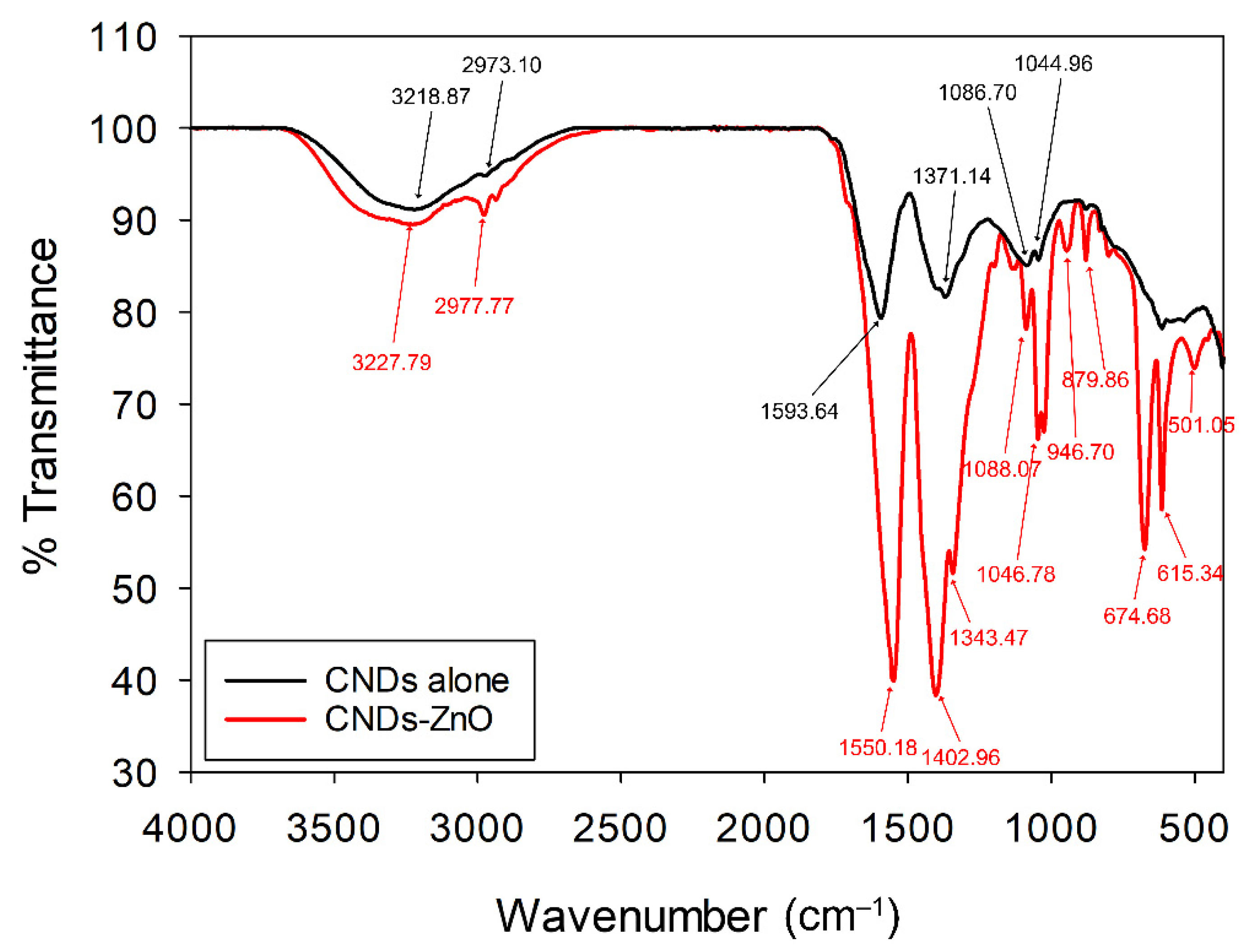
Figure 2 shows a comparison of the FT-IR spectra of CNDs alone and CNDs–ZnO. The spectrum of CNDs alone showed the O-H/N-H stretching vibration peaks at 3218.87 cm−1 with marginal C-H stretching vibration at 2973.10 cm−1. The peaks at 1593.64, 1371.14, 1086.70, and 1044.96 cm−1 were assigned to C=O stretching, C-H bending, C-N stretching, and C-O stretching, respectively. In the spectrum of CNDs–ZnO, the O-H/N-H stretching vibration at 3227.79 cm−1 was also present, and C-H stretching of aliphatic compounds was observed at 2977.77 cm−1. The bands at 1550.18, 1402.96, 1088.07, and 1046.78 cm−1 similarly corresponded to C=O stretching, C-H bending, C-N stretching, and C-O stretching, as with those observed in the spectrum of CNDs alone. However, vibration peaks according to O-H bending at 1343.47 cm−1 and =C-H bending at 879.86 and 946.70 cm−1 were observed. Moreover, the absorption bands at 501.05, 615.34, and 674.68 cm−1 represented the stretching vibrations of Zn-O and Zn-OH. XPS analysis was also performed to further evaluate the FT-IR assignments.
According to the XPS survey spectrum, C 1s (285.06 eV), N 1s (401.63 eV), O 1s (531.60 eV), and Zn 2p (1021.93 eV and 1044.96 eV) were confirmed (Figure 3). Although some peaks from residual elements such as Mg, Cl, and K were measured, they seemed to originate from salts that were incompletely washed away from S. horneri; these salts were also detected in the TEM images. The percentage of atomic concentrations in CNDs–ZnO is also displayed in Figure 3, with C = 54.78%, N = 2.95%, O = 32.40%, and Zn = 9.87%; by contrast, the atomic concentrations in CNDs alone were C = 68.79%, N = 8.70%, and O = 22.50%.
Figure 4a–g show the narrow spectra of C 1s, N 1s, O 1s, and Zn 2p of CNDs–ZnO in comparison to those of CNDs alone. In Figure 4a,b, three peaks such as sp2 C-C/C=C (284.54 eV), sp3 C-O/C-N (285.94 eV), and carbonyl C=O (288.46 eV) were observed in the C 1s spectrum of CNDs–ZnO. Compared to the C 1s spectrum of CNDs alone, which contained sp2 C-C/C=C (284.30 eV), sp3 C-O/C-N (285.69 eV), and carbonyl C=O (287.71 eV), the C 1s spectrum of CNDs–ZnO showed a positive shift in all peaks according to different formal oxidation states resulting from the formation of the nanocomposite structure with ZnO. In the N 1s spectra of CNDs–ZnO and CNDs alone, the negative peak shift in all peaks with pyrrolic N (from 399.27 eV to 398.45 eV) and Amino N (from 400.49 eV to 399.82 eV) was measured (Figure 4c,d). The high-resolution O 1s spectra of CNDs–ZnO and CNDs alone showed similar binding energies at 531.52 and 532.77 eV in CNDs–ZnO and at 531.14 and 532.41 eV in CNDs alone, as shown in Figure 4e,f, respectively. The changes in the O 1s spectra apparently resulted from the formation of the structures of oxide and carbonate with zinc in the nanocomposite. In the O 1s spectrum of the CNDs–ZnO, the peak centered at 531.52 eV represented the O2− state of oxygen vacancies or defects, involving the formation of nonstoichiometric ZnO, and another peak centered at 532.77 eV was assigned to the weak bonds of OH groups on the surface. In addition, the double peaks of Zn 2p corresponding to the Zn2+ oxidation state were detected at 1021.93 and 1044.96 eV, which are assigned to Zn 2p3/2 and Zn 2p1/2, respectively, with a separation of 23.03 eV (Figure 4g). The binding energies of Zn 2p also showed a positive shift compared to those of typical pure-zinc oxide nanostructures owing to the formation of the nanocomposite structure with carbon dots from S. horneri.
A TEM study of the CNDs alone and the CNDs–ZnO nanocomposite was performed to further analyze the structure and calculate the size distribution of the nanocomposite. The TEM image in Figure 5a,b shows various spherical-like carbon dots (CNDs alone) and the nanocomposite (CNDs–ZnO), where carbon dots are decorated on zinc oxide nanoparticles. Their morphological differences could be clearly identified in Figure 5a,b. Additionally, the elemental composition of the CNDs–ZnO was determined by EDS analysis. Figure S1 shows the elemental distribution with the atomic percentage. From the EDS data, the CND–ZnO consisted of carbon, oxygen, and zinc with residual Na, Mg, and Cl resulting from seawater that had not been thoroughly washed. The sizes of the CNDs–ZnO in Figure 5b were widely distributed from approximately 30 nm to 170 nm, with nanocomposites of 70–90 nm size occupying the largest proportion, as determined by counting 124 particles. Therefore, the marine brown algae S. horneri, which is considered useless, acts as a good reducing agent for synthesizing zinc oxide and as an excellent biomaterial for fabricating carbon dots.

3.3. Disk Diffusion Test for Antibacterial Activity

The antibacterial activities of CNDs–ZnO were determined by using a disk diffusion test with three Gram-negative and two Gram-positive bacteria. First, the CNDs–ZnO nanocomposites were fabricated with S. horneri from Buan-gun and Sinan-gun via the synthetic method described in Section 2.2. The as-synthesized CNDs–ZnO solutions with S. horneri from Buan-gun and Sinan-gun were denoted by CNDs–ZnO-1 and CNDs–ZnO-2, respectively. The two as-prepared pure ZnO NPs were used as experimental controls. After 30 μL of each CNDs–ZnO and pure ZnO was applied to paper disks on LB-agar plates containing the bacteria, the plates were incubated at 30 °C overnight, and then their inhibition zones were checked (Figure S2a). Clear inhibition zones were observed from both CNDs–ZnO-1 and CNDs–ZnO-2, whereas the pure ZnO samples showed poor inhibition zones. Because the antibacterial test for V. alginolyticus was additionally conducted solely, the photos are not shown. The results are summarized in Table 1.
Next, the antibacterial activities of the CNDs–ZnO-1 and CNDs–ZnO-2 were compared with those of CNDs alone synthesized based on different S. horneri samples collected from Buan and Sinan, respectively (Figure S2b). For the CNDs alone, no antibacterial activity and inhibition zones were observed; by contrast, CNDs–ZnO-1 and CNDs–ZnO-2 showed excellent antibacterial activities with obvious inhibition zones. After being powdered by lyophilization and re-dissolving in ultrapure water without ethanol, the CNDs–ZnO-1 and CNDs–ZnO-2 still showed clear inhibition zones for all bacterial plates, as shown in Figure S3a. In the figure, their antibacterial ability was maintained after reconstitution in ultrapure water. The solubility of zinc oxide nanoparticles is an important factor that influences the antibacterial property because it affects the distribution of Zn2+ ions in the media [53,54]. Because the CNDs–ZnO nanocomposites were well-dispersed even in ultrapure water, their antibacterial ability was retained. Furthermore, this antibacterial activity was still maintained even after storage at room temperature after sunlight was blocked for more than 6 months.
Finally, to compare the antibacterial ability of both CNDs–ZnO-1 and CNDs–ZnO-2 with that of a commercially available disinfectant, a 10% chlorine bleach solution (10% Clorox) was reacted with the bacteria. The bleach, which consists of sodium hypochlorite, is a known effective killing agent for bacteria and even the COVID-19 virus. In fact, the Korea Disease Control and Prevention Agency (KDCA) has provided guidelines for typical disinfection of surfaces by using sodium hypochlorite-containing Clorox at concentrations of 500 ppm to 1000 ppm as a precautionary measure to prevent COVID-19. Moreover, the use of 5000 ppm bleach solution to clean up the blood or body fluids of COVID-19 patients is encouraged. According to manufacturer information, the concentration of chlorine bleach is 50,000 ppm. Thus, the concentration of the chlorine bleach in this research was set to 5000 ppm through 1/10 dilution. Similar to the previous method, 30 μL of 1/10-diluted chlorine bleach was applied to the LB-agar plates on four selected bacteria, which were subsequently incubated at 30 °C overnight. Afterward, strongly apparent and large inhibition zones were detected in all bleach-treated plates (Figure S3b). The sizes of inhibition zones by CNDs–ZnO-1, CNDs–ZnO-2, and chlorine bleach are summarized in Table 1. Inhibition zones smaller than 8.5 mm in size were ignored and are expressed as negligible in the table. The two pure ZnO samples both showed marginal antibacterial activity, such as 17 ± 1 and 15 ± 1 mm in V. alginolyticus and 13 and 12 ± 2 mm in S. aureus, while its antibacterial effect against other bacteria was not noticeable. For CNDs alone based on S. horneri, no antibacterial activity against all bacteria was observed. By contrast, the two CNDs–ZnO nanocomposites exhibited a remarkable antibacterial effect against all bacteria. Except against B. cereus, both CNDs–ZnO presented excellent inhibition regions of 20 mm or larger against most bacteria; these inhibition zones were almost at a similar level or slightly inferior to those of 10% chlorine bleach. Interestingly, both CNDs–ZnO samples showed superior antibacterial activities against E. coli at 22 ± 3 and 21 ± 2 mm and against S. aureus at 25 ± 4 and 25 ± 4 mm compared to those of 10% chlorine bleach at 21 ± 2 and 23 ± 3 mm, respectively. Although the exact mechanism of antimicrobial activity of ZnO NPs is not fully revealed and remains controversial, the dominant principles are that ZnO NPs release Zn2+ ions or generate reactive oxygen species (ROS), which lead to the disruption of bacterial cell structures or an increase in oxidative stress triggered in preventing DNA replication or protein synthesis, respectively [55,56,57]. Based on the results of several studies, the antibacterial properties of CNDs–ZnO were also due to the same mechanisms mentioned above. Next, photo-induced antibacterial characteristics of the CNDs–ZnO were investigated under UV irradiation at 365 nm. The experimental procedures were identical, but the bacterial plates were incubated while exposed to UV light. The results are shown in Table 2 and compared with the existing CNDs–ZnO antibacterial results in Table 1. Under UV irradiation, the photo-enhanced antibacterial effect of the CNDs–ZnO against bacteria was detected except against E. coli and S. typhimurium. CNDs exhibit antibacterial activity owing to increased oxidative stress in bacteria based on producing ROS by light or UV irradiation [4,11]. In this paper, the photo-induced antibacterial properties of CNDs were similarly demonstrated. Interestingly, recent research has shown that CNDs exert an antibacterial effect by activating ROS in the absence of light [58]. Thus, the mechanism of the antibacterial effect of CND needs to be elucidated more in further studies.

3.4. MIC test and Growth Curve Inhibition Analysis

Results of the completed MIC test are shown in Table 3. The starting concentration was 5.8 mg/mL, which is numbered as 1 in the table. As described in Section 2.5, a total of 11 samples were prepared through 1/2 serial dilution. The as-prepared concentrations were 5.8 mg/mL, 2.9 mg/mL, 1.45 mg/mL, 725.0 μg/mL, 362.5 μg/mL, 181.25 μg/mL, 90.63 μg/mL, 45.31 μg/mL, 22.65 μg/mL, 11.33 μg/mL, and 5.66 μg/mL, which were numbered as 1, 2, 3, 4, 5, 6, 7, 8, 9, 10, and 11, respectively. To minimize the error between each well, bacteria that were not treated with CNDs–ZnO and the CNDs–ZnO-treated bacteria were simultaneously placed up and down. After incubation for 20 h, their turbidities were measured at OD600. As shown in Table 3, the minimum inhibitory concentrations for each bacteria were 362.5 μg/mL, 725.0 μg/mL, 362.5 μg/mL, 90.63 μg/mL, and 181.25 μg/mL for E. coli, B. cereus, S. typhimurium, S. aureus, and V. alginolyticus, respectively. In fact, the cell growth of V. alginolyticous was not observed visually until 362.5 μg/mL of CNDs–ZnO was added. Based on the MIC test results, the growth curve inhibition for CNDs–ZnO at 1/160-diluted concentration (362.5 μg/mL) against all bacteria in LB media was additionally achieved at OD600 during culture for 20 h. Commercial pure ZnO solution was prepared as an experimental control. Untreated bacteria were also cultivated together. All samples were prepared in 96-well plates. As shown in Figure 6a–e, except in B. cereus, CNDs–ZnO-treated bacteria noticeably showed growth inhibition compared to pure ZnO-treated or untreated bacteria. For B. cereus, the initial growth was delayed up to 10 h, but the bacteria grew to a certain extent afterward. However, the growth was significantly hindered by the CNDs–ZnO as B. cereus appeared to enter the stationary phase after approximately 18 h. In the results of the MIC test against B. cereus, some cell growth (turbidity value = 0.337 at OD600) was observed in the well added with 362.5 μg/mL of CNDs–ZnO after 20 h. This cell growth inhibition analysis demonstrated the consistency of the MIC test result.

3.5. Disk Diffusion Test for Antifungal Activity

The antifungal property of the CNDs–ZnO nanocomposites was also examined using the disk diffusion test (Figure S4). Similar to the mechanism of antibacterial activity of ZnO, the fungicidal mechanisms induced by ROS or mediation by zinc ions from ZnO are also considered to disrupt the cellular structure and anti-oxidative system of fungi [59,60,61]. In Figure S3, the inhibitory effect of CNDs–ZnO on fungal growth was more pronounced in R. mucilaginosa and A. niger with >20 mm of inhibition zone relative to that on other fungal species. Except for R. mucilaginosa and A. niger, they showed weak antifungal activity with <20 mm in halo size by CNDs–ZnO-1 and 2, and the measured inhibition zone is presented in Table 4. Especially, CNDs–ZnO-1 and CNDs–ZnO-2 showed the most effective antifungal activity against R. mucilaginosa, as indicated by inhibition zones of 24 ± 1 and 28 ± 2 mm in halo sizes, respectively. Although pure ZnO only showed weak antifungal activities against R. mucilaginosa and A. niger and no activity against other species, the inhibition sizes can be considered negligible (<10.5 mm in size). Overall, the inhibition zones of CNDs–ZnO-2 against all fungal species were 10% larger than those of CND-ZnO-1. This phenomenon may have been caused by an unidentified factor in nanocomposite synthesis due to physicochemical influences against S. horneri during drifting or after deposition on the beach. However, further investigation will be needed for a distinct cause of the assumption.

3.6. Toxicity Evaluation of the CNDs–ZnO

Five embryos of zebrafish were selected per batch, and each batch was exposed to chlorine bleach, ZnO NPs (commercial), and serially diluted CNDs–ZnO. The toxicity of CNDs–ZnO was assessed in terms of developmental rates of embryos, which are presented in Table 5. At 24 h post-fertilization, the embryos reared in EM without any exposure to the as-prepared solutions developed to over 60% of larvae. The eggs exposed to chlorine bleach, pure ZnO, and undiluted CNDs–ZnO-1 and CNDs–ZnO-2 failed to develop; furthermore, eggs reacted with chlorine bleach disappeared within 30 min. By contrast, embryos reacted with 1/10-diluted CNDs–ZnO-1 and CNDs–ZnO-2 survived and developed to 24 h successfully. Although the developmental rate of the embryos reacted with 1/10-diluted CNDs–ZnO-1 was 80%, the embryos upon 1/10-diluted CND–ZnO-2 showed a 60% survival rate. However, the 60% survival rate against CND–ZnO-2 was probably not solely attributed to the reaction with CNDs–ZnO-2, because the developmental rate of the embryos with no exposure to the nanocomposite solutions was up to 60%. In the case of a dilution factor of 1/100 or more, all embryos showed a 100% survival rate. In summary, the embryos reacted with CNDs–ZnO at a dilution ratio of 1/10 or more resulted in an 80% survival rate.

4. Conclusions

This study demonstrated and highlighted the applicability of S. horneri, which is currently treated as a nuisance, for the synthesis of an antimicrobial nanocomposite. The synthesis was designed to entail minimum available equipment, small cost, and simple procedure, and to be ecologically friendly. All fabricated CNDs–ZnO nanocomposites based on S. horneri collected from two different regions in Korea successfully exhibited antibacterial and antifungal activities. Because the nanocomposite with antimicrobial properties can be synthesized and well-dispersed in aqueous solution, the limitation of poor water solubility of the current widely used zinc oxide can be overcome; thus, this nanocomposite is expected to have a wide range of potential industrial applications. Moreover, under UV irradiation, the antibacterial effect was slightly improved, and the photo-induced ROS generation was possibly caused by the carbon nanodots produced from S. horneri on the nanocomposite. This work was a proof-of-concept study to use a hitherto unutilized or veiled marine species and demonstrated a valuable possibility of a wide application prospect using many underestimated marine organisms. However, some limitations of this study still existed. It is unclear by what kind of physicochemical influence the S. horneri used in this experiment had been exposed that could affect the synthesis of the CNDs–ZnO nanocomposites while floating in the sea. Further investigation will be required by comparison with the collection of S. horneri from various regions as well as distinct compositional analysis. In addition, environmental safety and human hazards of the CNDs–ZnO must be examined for the practical industrial applications through further study.

5. Patents

A Korean patent application has been filed for the contents described in this paper (patent number 10-2021-0067042).

Supplementary Materials

The following supporting information can be downloaded at: https://www.mdpi.com/article/10.3390/jmse10101546/s1, Figure S1: EDS spectra of the synthesized CNDs–ZnO and its atomic percentage; Figure S2: Plate images of disk diffusion antibacterial test for (a) CNDs–ZnO and Pure ZnO; (b) CNDs–ZnO and CNDs alone; Figure S3: Plate images of disk diffusion antibacterial test for (a) lyophilized CNDs–ZnO after re-dissolved in ultrapure water; (b) 10% Chlorine bleach solution; Figure S4: Plate images of disk diffusion antifungal test for CNDs–ZnO and Pure ZnO.

Author Contributions

Conceptualization, data curation, formal analysis, investigation, methodology, writing—original draft preparation, D.C., S.-H.J. and K.W.K.; validation, D.C., S.-H.J., Y.M.K., J.Y.H.K., K.B. and K.W.K.; resources, D.C., S.-H.J., Y.M.K., J.Y.H.K. and K.W.K.; writing—review and editing, D.C., S.-H.J., Y.M.K., J.Y.H.K., K.B. and K.W.K.; visualization, K.W.K.; supervision, K.B.; project administration, K.W.K. and K.B. All authors have read and agreed to the published version of the manuscript.

Funding

This work was funded by the National Marine Biodiversity Institute of Korea (MABIK) (grant number: 2022M00500).

Institutional Review Board Statement

Not applicable.

Informed Consent Statement

Not applicable.

Data Availability Statement

Not applicable.

Acknowledgments

We would like to thank Kim, H.S. for supporting some of the S. horneri collected in Sinan-gun.

Conflicts of Interest

The authors declare no conflict of interest.

References

  1. Da Costa, D.; Exbrayat-Héritier, C.; Rambaud, B.; Megy, S.; Terreux, R.; Verrier, B.; Primard, C. Surface charge modulation of rifampicin-loaded PLA nanoparticles to improve antibiotic delivery in Staphylococcus aureus biofilms. J. Nanobiotechnol. 2021, 19, 12. [Google Scholar] [CrossRef] [PubMed]
  2. Sánchez-López, E.; Gomes, D.; Esteruelas, G.; Bonilla, L.; Lopez-Machado, A.L.; Galindo, R.; Cano, A.; Espina, M.; Ettcheto, M.; Camins, A.; et al. Metal-based nanoparticles as antimicrobial agents: An overview. Nanomaterials 2020, 10, 292. [Google Scholar] [CrossRef] [PubMed] [Green Version]
  3. Al-Jumaili, A.; Alancherry, S.; Bazaka, K.; Jacob, M.V. Review on the antimicrobial properties of carbon nanostructures. Materials 2017, 10, 1066. [Google Scholar] [CrossRef]
  4. Ghirardello, M.; Ramos-Soriano, J.; Galan, M.C. Carbon dots as an emergent class of antimicrobial agents. Nanomaterials 2021, 11, 1877. [Google Scholar] [CrossRef] [PubMed]
  5. Alfei, S.; Schito, A.M. From nanobiotechnology, positively charged biomimetic dendrimers as novel antibacterial agents: A review. Nanomaterials 2020, 10, 2022. [Google Scholar] [CrossRef] [PubMed]
  6. Peng, Z.; Miyanji, E.H.; Zhou, Y.; Pardo, J.; Hettiarachchi, S.D.; Li, S.; Blackwelder, P.L.; Skromne, I.; Leblanc, R.M. Carbon dots: Promising biomaterials for bone-specific imaging and drug delivery. Nanoscale 2017, 9, 17533–17543. [Google Scholar] [CrossRef]
  7. Su, W.; Wu, H.; Xu, H.; Zhang, Y.; Li, Y.; Li, X.; Fan, L. Carbon dots: A booming material for biomedical applications. Mater. Chem. Front. 2020, 4, 821–836. [Google Scholar] [CrossRef]
  8. Liu, J.; Li, R.; Yang, B. Carbon dots: A new type of carbon-based nanomaterial with wide applications. ACS Cent. Sci. 2020, 6, 2179–2195. [Google Scholar] [CrossRef]
  9. Nocito, G.; Calabrese, G.; Forte, S.; Petralia, S.; Puglisi, C.; Campolo, M.; Esposito, E.; Conoci, S. Carbon dots as promising tools for cancer diagnosis and therapy. Cancers 2021, 13, 1991. [Google Scholar] [CrossRef]
  10. Chatzimitakos, T.; Stalikas, C. Nanotoxicity: Prevention and Antibacterial Applications of Nanomaterials; Elsevier BV: Amsterdam, The Netherlands, 2020; Volume 14, pp. 301–315. ISBN 978-0-12-819943-5. [Google Scholar]
  11. Dong, X.; Liang, W.; Meziani, M.J.; Sun, Y.P.; Yang, L. Carbon dots as potent antimicrobial agents. Theranostics 2020, 10, 671–686. [Google Scholar] [CrossRef]
  12. Wu, X.; Abbas, K.; Yang, Y.; Li, Z.; Tedesco, A.C.; Bi, H. Photodynamic Anti-Bacteria by Carbon Dots and Their Nano-Composites. Pharmaceuticals 2022, 15, 487. [Google Scholar] [CrossRef] [PubMed]
  13. Al Awak, M.M.; Wang, P.; Wang, S.; Tang, Y.; Sun, Y.P.; Yang, L. Correlation of Carbon Dots’ Light-Activated Antimicrobial Activities and Fluorescence Quantum Yield. RSC Adv. 2017, 7, 30177–30184. [Google Scholar] [CrossRef] [PubMed] [Green Version]
  14. Meziani, M.J.; Dong, X.; Zhu, L.; Jones, L.P.; LeCroy, G.E.; Yang, F.; Wang, S.; Wang, P.; Zhao, Y.; Yang, L.; et al. Visible-Light-Activated Bactericidal Functions of Carbon “Quantum” Dots. ACS Appl. Mater. Interfaces 2016, 8, 10761–10766. [Google Scholar] [CrossRef] [Green Version]
  15. Das, R.; Bandyopadhyay, R.; Pramanik, P. Carbon quantum dots from natural resource: A review. Mater. Today Chem. 2018, 8, 96–109. [Google Scholar] [CrossRef]
  16. Lin, X.; Xiong, M.; Zhang, J.; He, C.; Ma, X.; Zhang, H.; Kuang, Y.; Yang, M.; Huang, Q. Carbon dots based on natural resources: Synthesis and applications in sensors. Microchem. J. 2021, 160, 105604. [Google Scholar] [CrossRef]
  17. Bag, P.; Maurya, R.K.; Dadwal, A.; Sarkar, M.; Chawla, P.A.; Narang, R.K.; Kumar, B. Recent development in synthesis of carbon dots from natural resources and their applications in biomedicine and multi-sensing platform. ChemistrySelect. 2021, 6, 2774–2789. [Google Scholar] [CrossRef]
  18. Chaudhuri, S.K.; Malodia, L. Biosynthesis of zinc oxide nanoparticles using leaf extract of Calotropis gigantea: Characterization and its evaluation on tree seedling growth in nursery stage. Appl. Nanosci. 2017, 7, 501–512. [Google Scholar] [CrossRef]
  19. Mohd Yusof, H.; Abdul Rahman, N.; Mohamad, R.; Zaidan, U.H.; Samsudin, A.A. Biosynthesis of zinc oxide nanoparticles by cell-biomass and supernatant of Lactobacillus plantarum TA4 and its antibacterial and biocompatibility properties. Sci. Rep. 2020, 10, 19996. [Google Scholar] [CrossRef]
  20. Naseer, M.; Aslam, U.; Khalid, B.; Chen, B. Green route to synthesize zinc oxide nanoparticles using leaf extracts of Cassia fistula and Melia azadarach and their antibacterial potential. Sci. Rep. 2020, 10, 9055. [Google Scholar] [CrossRef]
  21. Chacko, L.; Poyyakkara, A.; Kumar, V.B.S.; Aneesh, P.M. MoS2-ZnO nanocomposites as highly functional agents for anti-angiogenic and anti-cancer theranostics. J. Mater. Chem. B 2018, 6, 3048–3057. [Google Scholar] [CrossRef]
  22. Dar, A.; Rehman, R.; Zaheer, W.; Shafique, U.; Anwar, J. Synthesis and characterization of ZnO-nanocomposites by utilizing aloe vera leaf gel and extract of Terminalia Arjuna nuts and exploring their antibacterial potency. J. Chem. 2021, 2021, 9448894. [Google Scholar] [CrossRef]
  23. El-Shafai, N.; El-Khouly, M.E.; El-Kemary, M.; Ramadan, M.; Eldesoukey, I.; Masoud, M. Graphene oxide decorated with zinc oxide nanoflower, silver and titanium dioxide nanoparticles: Fabrication, characterization, DNA interaction, and antibacterial activity. RSC Adv. 2019, 9, 3704–3714. [Google Scholar] [CrossRef] [PubMed] [Green Version]
  24. Mousavi-Kouhi, S.M.; Beyk-Khormizi, A.; Amiri, M.S.; Mashreghi, M.; Taghavizadeh Yazdi, M.E.T. Silver-zinc oxide nanocomposite: From synthesis to antimicrobial and anticancer properties. Ceram. Int. 2021, 47, 21490–21497. [Google Scholar] [CrossRef]
  25. Yu, H.; Zhang, H.; Huang, H.; Liu, Y.; Li, H.; Ming, H.; Kang, Z. ZnO/carbon quantum dots nanocomposites: One-step fabrication and superior photocatalytic ability for toxic gas degradation under visible light at room temperature. New J. Chem. 2012, 36, 1031. [Google Scholar] [CrossRef]
  26. Gao, D.; Zhao, P.; Lyu, B.; Li, Y.; Hou, Y.; Ma, J. Carbon quantum dots decorated on ZnO nanoparticles: An efficient visible-light responsive antibacterial agents. Appl. Organomet. Chem. 2020, 34, e5665. [Google Scholar] [CrossRef]
  27. Zhao, W.; Yan, L.; Gu, H.; Li, Z.; Wang, Y.; Luo, Q.; Yang, Y.; Liu, X.; Wang, H.; Ma, C.Q. Zinc oxide coated carbon dot nanoparticles as electron transport layer for inverted polymer solar cells. ACS Appl. Energy Mater. 2020, 3, 11388–11397. [Google Scholar] [CrossRef]
  28. Jana, J.; Chung, J.S.; Hur, S.H. ZnO-associated carbon dot-based fluorescent assay for sensitive and selective dopamine detection. ACS Omega 2019, 4, 17031–17038. [Google Scholar] [CrossRef] [Green Version]
  29. Atchudan, R.; Edison, T.N.J.I.; Mani, S.; Perumal, S.; Vinodh, R.; Thirunavukkarasu, S.; Lee, Y.R. Facile synthesis of a novel nitrogen-doped carbon dot adorned zinc oxide composite for photodegradation of methylene blue. Dalton Trans. 2020, 49, 17725–17736. [Google Scholar] [CrossRef]
  30. Bozetine, H.; Wang, Q.; Barras, A.; Li, M.; Hadjersi, T.; Szunerits, S.; Boukherroub, R. Green chemistry approach for the synthesis of ZnO-carbon dots nanocomposites with good photocatalytic properties under visible light. J. Colloid Interface Sci. 2016, 465, 286–294. [Google Scholar] [CrossRef]
  31. Efa, M.T.; Imae, T. Hybridization of carbon-dots with ZnO nanoparticles of different sizes. J. Taiwan Instig. Chem. Eng. 2018, 92, 112–117. [Google Scholar] [CrossRef]
  32. Halpern, B.S.; Longo, C.; Hardy, D.; McLeod, K.L.; Samhouri, J.F.; Katona, S.K.; Kleisner, K.; Lester, S.E.; O’Leary, J.; Ranelletti, M.; et al. An index to assess the health and benefits of the global ocean. Nature 2012, 488, 615–620. [Google Scholar] [CrossRef] [PubMed] [Green Version]
  33. Choudhary, B.; Chauhan, O.P.; Mishra, A. Edible seaweeds: A potential novel source of bioactive metabolites and nutraceuticals with human health benefits. Front. Mar. Sci. 2021, 8, 740054. [Google Scholar] [CrossRef]
  34. Rajapakse, N.; Kim, S.K. Nutritional and digestive health benefits of seaweed. Adv. Food Nutr. Res. 2011, 64, 17–28. [Google Scholar] [CrossRef] [PubMed]
  35. Sanjeewa, K.K.A.; Lee, W.W.; Jeon, Y.J. Nutrients and bioactive potentials of edible green and red seaweed in Korea. Fish Aquat. Sci. 2018, 21, 19. [Google Scholar] [CrossRef]
  36. Palmieri, N.; Forleo, M.B. The potential of edible seaweed within the western diet. A segmentation of Italian consumers. Int. J. Gastron. Food Sci. 2020, 20, 100202. [Google Scholar] [CrossRef]
  37. Byeon, S.Y.; Oh, H.J.; Kim, S.; Yun, S.H.; Kang, J.H.; Park, S.R.; Lee, H.J. The origin and population genetic structure of the ‘golden tide’ seaweeds, Sargassum horneri, in Korean waters. Sci. Rep. 2019, 9, 7757. [Google Scholar] [CrossRef] [Green Version]
  38. Choi, S.K.; Oh, H.J.; Yun, S.H.; Lee, H.J.; Lee, K.; Han, Y.S.; Kim, S.; Park, S.R. Population dynamics of the ‘golden tides’ seaweed, Sargassum horneri, on the Southwestern Coast of Korea: The extent and formation of golden tides. Sustainability 2020, 12, 2903. [Google Scholar] [CrossRef] [Green Version]
  39. Mukherjee, A.; Sarkar, D.; Sasmal, S. A Review of Green Synthesis of Metal Nanoparticles Using Algae. Front. Microbiol. 2021, 12, 693899. [Google Scholar] [CrossRef]
  40. Taherzadeh Soureshjani, P.; Shadi, A.; Mohammadsaleh, F. Algae-mediated route to biogenic cuprous oxide nanoparticles and spindle-like CaCO3: A comparative study, facile synthesis, and biological properties. RSC Adv. 2021, 11, 10599–10609. [Google Scholar] [CrossRef]
  41. Ramakrishna, M.; Rajesh Babu, D.; Gengan, R.M.; Chandra, S.; Nageswara Rao, G. Green synthesis of gold nanoparticles using marine algae and evaluation of their catalytic activity. J. Nanostruct. Chem. 2016, 6, 1–13. [Google Scholar] [CrossRef]
  42. Ponnuchamy, K.; Antony, J.J. Metal nanoparticles from marine seaweeds—A review. Nanotechnol. Rev. 2016, 5, 589–600. [Google Scholar] [CrossRef]
  43. Kim, K.W.; Choi, T.Y.; Kwon, Y.M.; Kim, J.Y.H. Simple synthesis of photoluminescent carbon dots from a marine polysaccharide found in shark cartilage. Electron. J. Biotechnol. 2020, 47, 36–42. [Google Scholar] [CrossRef]
  44. Kim, K.W.; Kwon, Y.M.; Kim, S.Y.; Kim, J.Y.H. One-pot synthesis of UV-protective carbon nanodots from sea cauliflower (Leathesia difformis). Electron. J. Biotechnol. 2022, 56, 22–30. [Google Scholar] [CrossRef]
  45. Song, W.C.; Kim, B.; Park, S.Y.; Park, G.; Oh, J.W. Biosynthesis of silver and gold nanoparticles using Sargassum horneri extract as catalyst for industrial dye degradation. Arab. J. Chem. 2022, 15, 104056. [Google Scholar] [CrossRef]
  46. Zeng, G.; Hong, C.; Ma, Y.; Du, M.; Zhang, Y.; Luo, H.; Chen, B.; Pan, X. Sargassum horneri-based carbon-doped TiO2 and its aquatic naphthalene photodegradation under sunlight irradiation. J. Chem. Technol. Biotechnol. 2022, 97, 1267–1274. [Google Scholar] [CrossRef]
  47. Nüsslein-Volhard, C.; Dahm, R. Zebrafish: A Practical Approach; Oxford University Press: Oxford, UK, 2002; ISBN 978-0-19-963809-3. [Google Scholar]
  48. Gerbreders, V.; Krasovska, M.; Sledevskis, E.; Gerbreders, A.; Mihailova, I.; Tamanis, E.; Ogurcovs, A. Hydrothermal synthesis of ZnO nanostructures with controllable morphology change. Cryst. Eng. Commun. 2020, 22, 1346–1358. [Google Scholar] [CrossRef]
  49. Ramimoghadam, D.; Bin Hussein, M.Z.; Taufiq-Yap, Y.H. Hydrothermal synthesis of zinc oxide nanoparticles using rice as soft biotemplate. Chem. Cent. J. 2013, 7, 136. [Google Scholar] [CrossRef] [Green Version]
  50. Kołodziejczak-Radzimska, A.; Jesionowski, T. Zinc oxide-from synthesis to application: A review. Materials 2014, 7, 2833–2881. [Google Scholar] [CrossRef] [Green Version]
  51. Shaba, E.Y.; Jacob, J.O.; Tijani, J.O.; Suleiman, M.A.T. A critical review of synthesis parameters affecting the properties of zinc oxide nanoparticle and its application in wastewater treatment. Appl. Water Sci. 2021, 11, 48. [Google Scholar] [CrossRef]
  52. Naik, G.G.; Alam, M.B.; Pandey, V.; Mohapatra, D.; Dubey, P.K.; Parmar, A.S.; Sahu, A.N. Multi-functional carbon dots from an ayurvedic medicinal plant for cancer cell bioimaging applications. J. Fluoresc. 2020, 30, 407–418. [Google Scholar] [CrossRef]
  53. Mendes, C.R.; Dilarri, G.; Forsan, C.F.; Sapata, V.M.R.; Lopes, P.R.M.; de Moraes, P.B.; Montagnolli, R.N.; Ferreira, H.; Bidoia, E.D. Antibacterial action and target mechanisms of zinc oxide nanoparticles against bacterial pathogens. Sci. Rep. 2022, 12, 2658. [Google Scholar] [CrossRef] [PubMed]
  54. Krishnamoorthy, R.; Athinarayanan, J.; Periyasamy, V.S.; Alshuniaber, M.A.; Alshammari, G.; Hakeem, M.J.; Ahmed, M.A.; Alshatwi, A.A. Antibacterial mechanisms of zinc oxide nanoparticle against bacterial food pathogens resistant to beta-lactam antibiotics. Molecules 2022, 27, 2489. [Google Scholar] [CrossRef] [PubMed]
  55. Tiwari, V.; Mishra, N.; Gadani, K.; Solanki, P.S.; Shah, N.A.; Tiwari, M. Mechanism of anti-bacterial activity of zinc oxide nanoparticle against carbapenem-resistant Acinetobacter baumannii. Front. Microbiol. 2018, 9, 1218. [Google Scholar] [CrossRef] [Green Version]
  56. Aydin Sevinç, B.; Hanley, L. Antibacterial activity of dental composites containing zinc oxide nanoparticles. J. Biomed. Mater. Res. B Appl. Biomater. 2010, 94, 22–31. [Google Scholar] [CrossRef] [PubMed] [Green Version]
  57. Sirelkhatim, A.; Mahmud, S.; Seeni, A.; Kaus, N.H.M.; Ann, L.C.; Bakhori, S.K.M.; Hasan, H.; Mohamad, D. Review on zinc oxide nanoparticles: Antibacterial activity and toxicity mechanism. Nanomicrol. Lett. 2015, 7, 219–242. [Google Scholar] [CrossRef] [Green Version]
  58. Liang, J.; Li, W.; Chen, J.; Huang, X.; Liu, Y.; Zhang, X.; Shu, W.; Lei, B.; Zhang, H. Antibacterial activity and synergetic mechanism of carbon dots against Gram-positive and -Negative bacteria. ACS Appl. Biol. Mater. 2021, 4, 6937–6945. [Google Scholar] [CrossRef]
  59. Lipovsky, A.; Nitzan, Y.; Gedanken, A.; Lubart, R. Antifungal activity of ZnO nanoparticles—The role of ROS mediated cell injury. Nanotechnology 2011, 22, 105101. [Google Scholar] [CrossRef]
  60. Arciniegas-Grijalba, P.A.; Patiño-Portela, M.C.; Mosquera-Sánchez, L.P.; Guerrero-Vargas, J.A.; Rodríguez-Páez, J.E. ZnO nanoparticles (ZnO-NPs) and their antifungal activity against coffee fungus Erythricium salmonicolor. Appl. Nanosci. 2017, 7, 225–241. [Google Scholar] [CrossRef] [Green Version]
  61. Sun, Q.; Li, J.; Le, T. Zinc oxide nanoparticle as a novel class of antifungal agents: Current advances and future perspectives. J. Agric. Food Chem. 2018, 66, 11209–11220. [Google Scholar] [CrossRef]
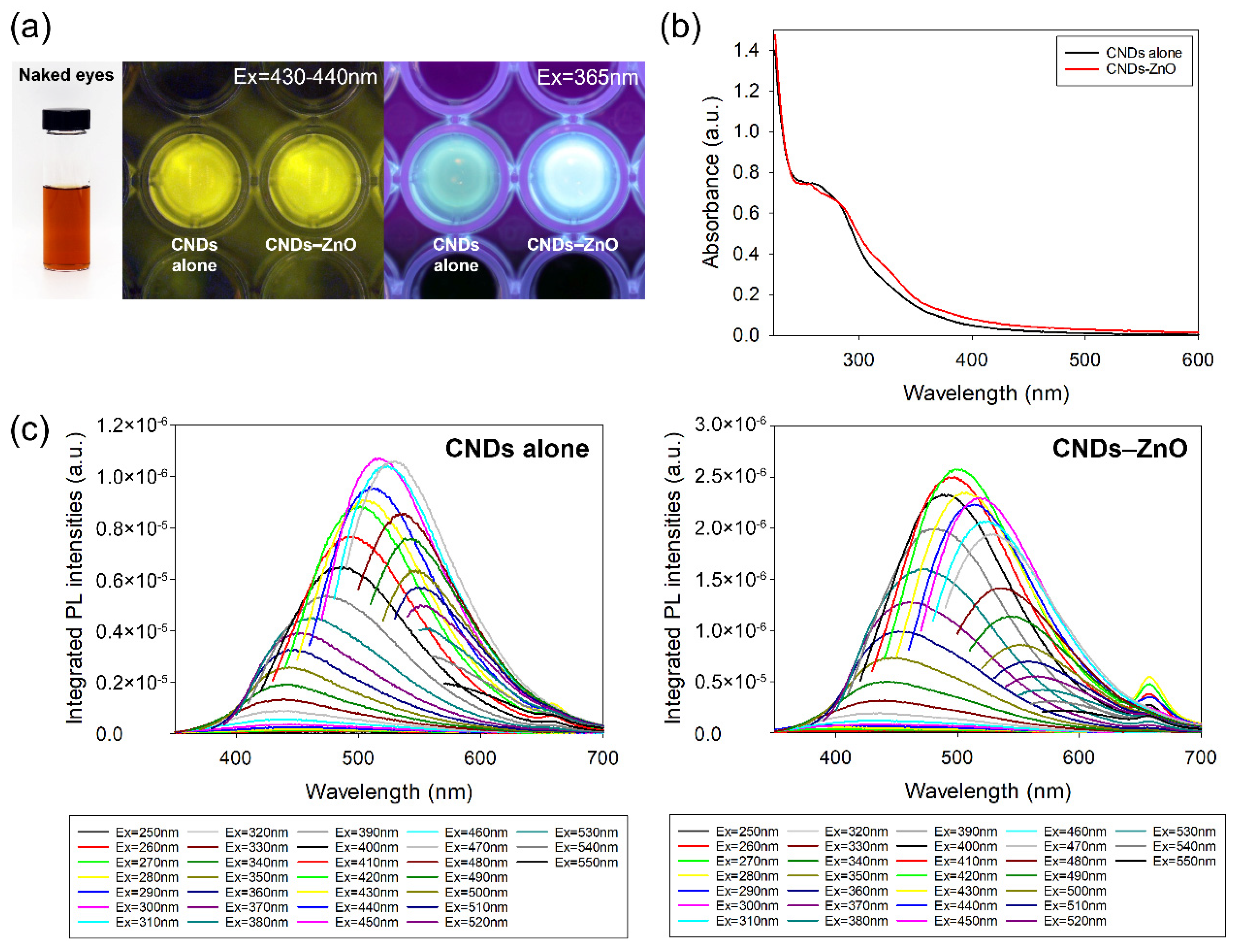
Figure 1. (a) Photo image of brown-colored CNDs–ZnO aqueous solution and fluorescent photo-images of both CNDs alone and CNDs–ZnO at specific excitation wavelengths; (b) UV–vis absorption spectra of CNDs alone and CNDs–ZnO; (c) PL emission spectra of CNDs alone and CNDs–ZnO under excitation wavelength change with 20 nm increments from 250 nm to 550 nm.
Jmse 10 01546 g001
Figure 2. FT-IR spectra comparison of CNDs alone and CNDs–ZnO.
Jmse 10 01546 g002
Figure 3. XPS survey spectra comparison of CNDs alone and CNDs–ZnO and their percentage of atomic concentrations.
Jmse 10 01546 g003
Figure 4. High-resolution XPS spectra of (a,b) C 1s, (c,d) N 1s, (e,f) O 1s, and (g) Zn 2p between CNDs alone and CNDs–ZnO.
Jmse 10 01546 g004
Figure 5. (a) TEM image of CNDs alone and (b) CNDs–ZnO nanocomposites; (c) size distribution of the as-fabricated CNDs–ZnO.
Jmse 10 01546 g005
Figure 6. Fitting curves of bacterial cell growth for (a) E. coli with pure ZnO and CNDs–ZnO; (b) B. cereus with pure ZnO and CNDs–ZnO; (c) S. typhimurium with pure ZnO and CNDs–ZnO; (d) S. aureus with pure ZnO and CNDs–ZnO; (e) V. alginolyticus with pure ZnO and CNDs–ZnO.
Jmse 10 01546 g006
Table 1. Size comparisons of inhibition zone for CNDs–ZnO against bacteria.
Bacteriamm of Inhibition Zone (Mean ± SD)
Pure ZnO
(Commercial)
Pure ZnO
(Synthesized)
CNDs AloneCNDs–ZnO-1CNDs–ZnO-210% Clorox
Gram-negativeE. coli10 ± 1Negligible-22 ± 322 ± 221 ± 2
S. typhimuriumNegligibleNegligible-21 ± 220 ± 225 ± 1
V. alginolyticus17 ± 115 ± 1-35 ± 635 ± 8>70
Gram-positiveB. cereusNegligibleNegligible-17 ± 216 ± 222 ± 2
S. aureus1312 ± 2-25 ± 425 ± 423 ± 3
Table 2. Enhanced antibacterial effect of CNDs–ZnO through UV irradiation.
Bacteriamm of Inhibition Zone (Mean ± SD)Photo-Enhanced Antibacterial Activity
UV onUV off
(From Table 1)
CNDs–ZnO-1CNDs–ZnO-2CNDs–ZnO-1CNDs–ZnO-2
Gram-negativeE. coli22 ± 321 ± 222 ± 322 ± 2-
S. typhimurium21 ± 221 ± 221 ± 220 ± 2-
V. alginolyticus36 ± 937 ± 1035 ± 635 ± 8Yes
Gram-positiveB. cereus18 ± 420 ± 217 ± 216 ± 2Yes
S. aureus26 ± 427 ± 325 ± 425 ± 4Yes
Table 3. MIC test of CNDs–ZnO upon five different bacteria.
Conc.ReactionBacteria
E. coliB. cereusS. typhimuriumS. aureusV. alginolyticus
MeanSDMeanSDMeanSDMeanSDMeanSD
1No treatment1.6950.0541.2050.1101.5820.0121.1780.0331.3890.009
with CNDs–ZnO0.3750.0190.3990.0980.3710.0500.4180.0560.1120.006
2No treatment1.5700.0211.1760.0611.5550.0561.0960.0121.6100.213
with CNDs–ZnO0.2430.0570.2290.0410.2850.0530.2500.0100.1130.004
3No treatment1.5310.0531.2070.1051.5650.0511.1120.0061.6280.268
with CNDs–ZnO0.1240.0050.1230.0050.1250.0160.1110.0050.1190.024
4No treatment1.5360.1261.1980.0891.5200.0091.1190.0121.6850.213
with CNDs–ZnO0.1110.0120.1050.0060.1070.0060.0910.0340.1140.017
5No treatment1.5170.0511.2170.0631.5050.0161.1140.0361.5820.199
with CNDs–ZnO0.0620.0130.3840.1090.0560.0050.0640.0180.1460.037
6No treatment1.4250.1171.1910.1161.4720.0121.1800.0441.5940.208
with CNDs–ZnO1.1350.1540.9400.1261.1620.1640.0470.0010.1500.040
7No treatment1.4770.1081.1970.0621.4980.0131.1260.0011.5780.221
with CNDs–ZnO1.3880.0551.0610.1161.3170.0570.0440.0030.2760.079
8No treatment1.4780.1011.2060.0611.4860.0141.1490.0241.5680.143
with CNDs–ZnO1.4850.0691.1750.0841.3860.0570.8020.0510.2680.070
9No treatment1.4560.1091.2890.1011.4620.0021.1310.0251.5220.132
with CNDs–ZnO1.3830.1061.1820.0641.4470.0011.1180.0040.9520.042
10No treatment1.4190.0921.2250.0591.4490.0021.1380.0521.4850.140
with CNDs–ZnO1.4010.0801.1720.0541.4700.0111.1500.0211.4290.051
11No treatment1.4390.0821.1990.0611.4570.0021.1280.0601.4280.106
with CNDs–ZnO1.4130.0751.2000.0681.4530.0181.1800.0091.5460.063
Table 4. Size comparisons of inhibition zone for CNDs–ZnO against fungal species.
Fungimm of Inhibition Zone (Mean ± SD)
Pure ZnO
(Commercial)
CNDs–ZnO-1CNDs–ZnO-2
MoldA. flavus-14 ± 115 ± 1
A. nigerNegligible22 ± 223 ± 2
A. terreus-12 ± 113 ± 1
YeastR. mucilaginosaNegligible24 ± 128 ± 2
S. cerevisiae-12 ± 116 ± 1
Table 5. Comparisons of developmental toxicity for CNDs–ZnO in zebrafish embryos.
TimeDevelopmental Rate of Zebrafish Embryos (%)
Chlorine BleachPure ZnODilution Ratio of CNDs–ZnO
CNDs–ZnO-1CNDs–ZnO-2
01:101:1001:10001:10,00001:101:1001:10001:10,000
0 h100100100100100100100100100100100100
0.5 h0100100100100100100100100100100100
24 h0008010010010008060100100
Publisher’s Note: MDPI stays neutral with regard to jurisdictional claims in published maps and institutional affiliations.