Article

Establishing Monoxenic Culture of Arbuscular Mycorrhizal Fungus Glomus sp. Through In Vitro Root Organ Culture and Swietenia macrophylla King In Vitro Cultures

by
Romero-Ceciliano Marysol
1,
Andrade-Torres Antonio
2,*,
Artavia-Salazar Evelyn
1 and
Solís-Ramos Laura Yesenia
1,*
1
Biotecnología de Plantas y Hongos Micorrícicos Arbusculares (Biotec-PYHMA), Escuela de Biología y Centro de Investigación en Biodiversidad y Ecología Tropical (CIBET), Universidad de Costa Rica, San Pedro de Montes de Oca 11501-2060, San José, Costa Rica
2
Biotecnología y Ecología de Organismos Simbióticos, CAUV-173 Ecología y Manejo de la Biodiversidad, INBIOTECA (Instituto de Biotecnología y Ecología Aplicada), Universidad Veracruzana, Av. de las Culturas Veracruzanas No. 101, Col. E. Zapata, Xalapa 91090, Veracruz, Mexico
*
Authors to whom correspondence should be addressed.
Agriculture 2025, 15(7), 673; https://doi.org/10.3390/agriculture15070673
Submission received: 31 December 2024 / Revised: 14 February 2025 / Accepted: 18 February 2025 / Published: 21 March 2025

Abstract

:
In vitro cultivation of arbuscular mycorrhizal fungi (AMF) is challenging due to their biotrophic symbiosis. The principal aim of this study was to demonstrate the effect of establishing in vitro dual cultures of arbuscular mycorrhizal fungi (AMF) inoculated on Swietenia macrophylla (mahogany) roots on plant growth. Furthermore, it was sought to demonstrate that plant colonization by Glomeromycota can be achieved with a replicable protocol. This study established monoxenic cultures of carrot (Daucus carota) Ri T-DNA ROC inoculated with Glomus sp. on two-compartment plates. At 75 days, hyphal growth reached 223.93 mm in the root compartment and 103.71 mm in the hyphal compartment. Spores produced in vitro measured 26.14 ± 1.70 µm, smaller than ex vitro spores (101.2 ± 4.22 µm). Rhodotorula mucilaginosa was isolated from cultures and appeared to stimulate hyphal growth and root–fungal contact. From these cultures, a dual culture of mahogany inoculated with Glomus sp. was established. No significant differences were observed between inoculated and non-inoculated plants in stem length, root length, root number, or leaf number at 30 days. Spore production ranged from 10,166 to 27,696 per plate, averaging 14,795 ± 3301, with hyphal lengths of 3655.46 ± 308.75 mm. Hyphal development included running and branching patterns, with solitary and clustered spores. Spore diameter averaged 27.68 ± 3.85 µm. Arbuscular colonization reached 41.49% at 30 days and 52.13% at 75 days, exceeding rates reported for other culture systems. Monoxenic cultures are a reliable, aseptic source of high-quality inoculum, supporting biofertilizer production and biotechnological applications. These methods provide valuable tools for studies involving AMF, such as those demonstrated with mahogany.

1. Introduction

Large-scale production of Arbuscular Mycorrhizal Fungi (AMF) consortia is a crucial milestone in harnessing their potential for sustainable agriculture and plant growth enhancement [1]. Arbuscular mycorrhizal (AM) symbioses have an important role in ecosystems, including modification of the soil environment and plant interactions with other living organisms, and uptake and transfer of nutrients [2]. AMF application in the tropics is potentially effective for improving yields [3]. However, in tropical soils, AMF isolates are not present in the numbers required to effectively improve plant health. Bulk inoculum production is a promising solution to this problem [4]. The most common methods comprise in vivo, substrate-based or aeroponic cultures, and in vitro, in which transformed roots are cultivated under aseptic conditions [5].
Due to the biotrophic nature of these fungi it is impossible to propagate them massively without the presence of a host [6]. The establishment of AMF with root cultures in laboratory conditions was first reported by White [7], then Mosse and Hepper proposed the use of excised roots as a host partner in AM symbiosis [8]. Most recently, carrot roots transformed with Ri T-DNA have become the primary medium for the production of AMF in vitro [9]. Few AMF species have been established successfully in root organ culture (ROC) [10,11,12,13]. This technique offers sterile, pure, bulk, and contaminant-free propagules, hitherto not obtained by the conventional methods of pot culture, hydroponic, or aeroponics techniques. Moreover, this technique has advantages over other conventional modes of mass production, achieving a several-fold increase in spore/propagule production using less time and space [14]. This alternative not only maintains the quality of AMF propagules, but also can serve as a cost-effective process to propagate fungi massively [6]. This co-culture technique allows us to evaluate various phenomena such as interaction, competition, and dominance occurring between different AMF genera in nature, which would otherwise be difficult to understand due to the lack of pure and sterile systems [4]. In vitro systems have proven to be useful for the cultivation and preservation of a large number of AM fungal species and isolates [15]. In addition, it is a useful technique to obtain axenic AMF cultures for germplasm collections. Mycorrhizal fungal preservation biobanks aim to preserve and provide viable and pure AM fungal starter cultures to researchers. Biobanks can contribute to biodiversity conservation and biotechnological innovation and the enhancement of AM fungi in sustainable agriculture [16].
The annual transformation of more than 5 million hectares of tropical forest into poorly managed and degraded forests due to timber harvesting has significant consequences for the sustainable extraction of timber and the global environment [17]. The big-leaf mahogany (Swietenia macrophylla King, Meliaceae) is one of the world’s most high-valued timber species. S. macrophylla is subject to international trade and intense exploitation, therefore its population size declines while its population fragmentation increases in several areas of natural distribution [18]. As a result, the population of this species globally has declined by at least 60% over the last three generations (180 years) and it continues to diminish due to logging and conversion of the natural habitat into farmlands, further reducing the possibility of regeneration; for this reason, it has been assessed as an endangered species [19]. Stand regeneration of tropical hardwood tree species for sustainable harvesting requires seedlings with high survival probabilities; one way to strengthen plants for outplanting is through the pre-colonization of their roots with AMF [20]. The objective of this research was to establish monoxenic cultures through root organ cultures (ROC) of Ri T-DNA-transformed carrots associated to arbuscular mycorrhizal fungi (AMF) and also evaluate the effect of an in vitro mycorrhization system adapted to seedlings of S. macrophylla.

2. Materials and Methods

The experiments were conducted between the years 2023 and 2024.

2.1. Biological Materials

Rhizospheric soil of Cedrela odorata L. (cedar) was collected from the Rodrigo Facio University Campus at the University of Costa Rica. Four soil sub-cores were taken per tree at a depth of 15 to 20 cm, 1 m away from the base of the tree. Soil samples, including fragments of roots, were stored in tagged plastic bags and transferred to our laboratory of Plant Biotechnology and Arbuscular Mycorrhizal Fungi (Biotec-PYHMA) at School of Biology, University of Costa Rica, San Pedro Montes de Oca, Costa Rica, where roots were separated and placed in 70% ethanol. The soil was processed for the extraction of spores. Spores were isolated from the rhizosphere of C. odorata, a forest species belonging to the family Meliaceae, to which S. macrophylla belongs; both share an AMF community with some species abundantly present in the rhizosphere of mahogany ([21,22,23], Biotec-PYHMA lab data).
Seeds of S. macrophylla were obtained from the forest seed bank of the Centro Agronómico Tropical de Investigación y Enseñanza (CATIE, Turrialba, Costa Rica).

2.2. Extraction and Identification of Glomerospores

Isolation of spores from the rhizosphere was carried out using the modified wet sieving and decanting method of Blaszkowski [24]. First, 100 g of soil was weighed, taking equal parts from the four cores of each tree to have a representative sample of the soil. Then, 1 L of tap water was added and the mix was stirred for 1 min. Ten seconds after that, the supernatant was poured through sieves in tandem, from the largest to the smallest pore diameter (500, 125 and 37 µm). One liter of water was added to the same sample and the following steps were repeated twice more.
The sediment was collected in conical tubes of 50 mL using a pipette with water, then was centrifuged for 3 min at 3000 rpm. The supernatant was discarded and 25 mL of 70% sucrose solution was added. The tubes were manually shaken until the precipitate was resuspended and centrifuged (Thermo Scientific ST8R, Salisbury, UK) at 1000 rpm for 1 min. The supernatant from all tubes of same sample passed through a 37 µm sieve. This step, adding sucrose, and centrifugation were repeated twice more. The sample was carefully rinsed with distilled water and the sediment collected in Petri dishes. Using a stereoscope, spores were extracted with needles and micropipettes. Finally, they were stored in distilled water at 4 °C. Taxonomic identification was based on specialized literature [24,25].

2.3. Spore Sterilization

Tween 20 dilution 0.005% was added to the sample. Afterwards, the sample was subject to mixing with 70% (v/v) ethanol solution for 1 min and two rinses with sterile water. Next, a dilution of 2% chloramine T (Merk, Darmstadt, Germany) and 1 µL of Tween 20 were added for 10 min of agitation and three rinses with sterile distilled water. Then, spores were left in a mixture of 0.02% streptomycin (Sigma-Aldrich, Saint Louis, MO 63103, USA) + 0.01% gentamicin (Phytotech Labs, Shawnee Mission, KS, USA) for 24 h. These were inoculated on MSR medium [26] and incubated in the dark. Repeat disinfection was performed with a first wash with 0.05% (v/v) Tween 20 for 1 min. This was followed by a wash with 2% chloramine T for 10 min and the above antibiotic mixture for 10 min. These were incubated in MSR medium in the dark.

2.4. MSR Medium [26]

The medium contained 731 mg MgSO4·7H2O; 80 mg KNO3; 65 mg KCl; 4.8 mg KH2PO4; 288 mg Ca (NO3)2·4H2O; 0.5 mg nicotinic acid; 0.1 mg pyridoxine clohidrate; 0.1 mg thiamine clohidrate; 8 mg Na Fe (III)-EDTA; 6 mg MnCl2·4H2O; 2.65 mg ZnSO4·7H2O; 1.5 mg H3BO3; 0.13 mg CuSO4·5H2O; 0.0024 mg Na2MoO4·2H2O; 0.75 mg KI; 3 mg glycine; 0.1 mg pyridoxine; 0.1 mg thiamine; 0.5 mg nicotinic acid; 50 mg myo-inositol; and 10,000 mg sucrose, solidified with 4000 mg Phytagel in 1 L distilled water. All chemicals were provided by Sigma or Sigma-Aldrich (Burlington, MA, USA) and phytagel gelzan CM (Phytotech Labs, Shawnee Mission, KS, USA). The pH was adjusted to 5.6 before adding phytagel. After that, the medium was sterilized at 1 atmosphere (101.325 kPa) for 15 min 121 °C.

2.5. Bacterial Strain Propagation and Carrot Hairy Root Culture (Modified from [10,27,28])

Agrobacterium rhizogenes strain 15,834 (provided by Dr. Gatica Arias’ lab from the University of Costa Rica) was used and grown for 35 h at 28 °C on Luria–Bertani (LB) (Phytotech Labs, Shawnee Mission, KS, USA) medium gelled with 15 g of agar spiked with 100 mg/L rifampicin (Sigma-Aldrich, Saint Louis, MO 63103, USA) and 200 µM acetosyringone (Merk, Darmstadt, Germany). A whole carrot (D. carota) was taken and washed with soap and water three times (5 min each) and finally with sterile distilled water. The peel was then removed and the carrot was immersed in 70% ethanol for 10 s. Subsequently, it was submerged in 1% sodium hypochlorite for 15 min and then rinsed with sterile distilled water. Slices of 5 mm thickness (17 pieces) were sectioned and sown in 1% water agar medium distal side up on MS medium [29]. A slice of the previously cultured bacteria was rubbed onto each piece. These were stored in the dark at room temperature and, after 55 days, the roots that had emerged from the carrot were subcultured on White or MSR medium [10,26], supplemented with 500 mg/L chloramphenicol (Sigma-Aldrich, Saint Louis, MO 63103, USA) or carbenicillin (Sigma-Aldrich, Saint Louis, MO 63103, USA) in Petri dishes of 9 cm diameter. After 15 days, a second subculture was performed on the same medium with 5 replicates each time. The plates were incubated in the dark at 25 ± 2 °C until the roots grew.

2.6. Spore Inoculation and Culture Development on Bi-Compartmented Petri Dishes (Modified from [30,31])

The bi-compartment system is composed of a proximal section, a sucrose-enriched section, and a paper bridge system. Rectangular pieces of filter paper (Whatman #2, 150 mm) were cut 1 cm wide, 3 cm long, folded in the middle, and autoclaved at 1 atmosphere (101.325 kPa) for 15 min. Sterilized paper folds were placed over the median Petri dish wall. Bi-compartment Petri dishes (9 cm diameter) contained 20 mL MSR medium [26] in the root compartment (RC root compartment), and on MSR medium without sucrose in the root-free, hyphal compartment (HC). One segment of carrot Ri T-DNA ROC (5 cm length) (D. carota L.) was placed in the RC compartment. The carrot Ri T-DNA ROC were inoculated with 5 AMF spores from healthy, uncontaminated cedar rhizosphere, previously disinfected 6 days before inoculation (procedure indicated above). Spores were placed near the carrot hairy root in each plate. Petri dishes were sealed (Parafilm TM) and incubated in darkness, upside-down at 25 ± 2 °C, with 5 replicates each time. Hyphal growth and spore production were subcultured onto new two-compartment plates. A new segment of carrot Ri T-DNA ROC was placed with 5 segments of medium (MSR), and spores and mycelium (4 × 4 mm) obtained from the previous cultures were inoculated close to the root (RC), with 5 replicates each time.
To assess mycelial growth, a grid of 3 × 3 mm squares was laid out and the number of times a hyphae intercepted the grid lines was counted under the stereoscope at 2X magnification. The total number was entered into the Tennant [32] equation to estimate mycelial length in millimeters (Equation (1)):
Root length (R) = 11/14 × number of intercepts (N) × grid unit (3 cm)
The observations took into account the presence of mycorrhizal structures such as running and branching hyphae. Running hyphae are those that form the main structure and are thick [33] or absorbing branched structures (BAS) [13]. These give rise to second-order hyphae, which in turn give rise to higher orders and become thinner [33].

2.7. Disinfection and Germination of Seeds

Initially, the seeds were manually scarified prior to the disinfection protocol using a brush. Next, they were immersed in a 70% sodium hypochlorite solution (3.5% active ingredient) for 10 min under constant agitation. Then, they were washed thrice using sterilized distilled water [34]. Seeds were dried on sterile paper towel and subsequently sown horizontally on culture medium Basal Medium MS [29] with vitamins (M519, Phytotech LABS), 0.001 g/L gibberellic acid, 30 g/L sucrose, and 6 g/L agar, and adjusted to pH 5.7 ± 0.1. They were kept in darkness at room temperature and, when germinated, were switched to a photoperiod of 12 h light/12 h dark at 25 ± 2 °C, with a light intensity of 100 μE/m 2 s provided by cool-white fluorescent lights.

2.8. Monoxenic Culture of AM Inoculum Production by Root Organ Culture (ROC) (Modified from [35])

MSR medium (70 mL) was set in 120 × 120 mm plates [26]. Once solidified, the medium was cut with a scalpel, leaving a rectangle of 5 by 12 cm with medium and 7 by 12 cm without medium. Mahogany plants were taken with the presence of radicle 7 days after germination. Culture medium attached to the roots was removed with forceps, and the plant was set on the upper limit of the solidified culture medium, placing the radicle into the medium. The bulk was obtained by cutting squares of medium at random from an inoculated Ri T-DNA ROC carrot plate, which contained 111 ± 50 spores, varying in shape and size, plus external fungal mycelium. Three segments of 4 × 4 mm each were placed in an arc between 0.5–1 cm from the radicle. Five replicates were used each time with and without inoculum. To protect roots from light while allowing the shoots to receive light for photosynthesis, the plants were incubated in an inverted position in a photoperiod of 12 h light/12 h dark, at 25 ± 2 °C, under a light intensity of 100 μE/m 2 s provided by cool-white fluorescent lights.

2.9. Assessment of Contamination and Spore Production

Spore formation was monitored each week using a Stereo Zoom Microscope Nikon SMZ800N (Nikon Instruments, Melville, N.Y, USA) and the plates were checked for contamination with other microorganisms. Each culture system was considered as an experimental unit in the experiment. In the case of plates that were contaminated, the number of AMF spores was estimated and then these plates were discarded to reduce the risk of contamination among the cultures.
In the culture experiment with AMF-inoculated mahogany, stem height, length of the main root of the plant, and total numbers of roots and leaves were measured every week for one month.
A 1 mm grid was marked on the bottom of each Petri dish to facilitate counting of spores. The evaluation method proposed by Rosikiewicz et al. [36] was used. In the first week, an area of 4 × 5 cm (20 cm2) was taken for random sampling and divided into 20 squares of 1 cm2, then numbered, and five were chosen at random and counted. For two of the replicates, the total number of spores was measured in the first week, and for all replicates it was estimated by counting in the five pre-selected squares.
In the second and fourth weeks, spore number and mycelial length were measured. For two of the replicates, the total number of spores was measured, and for all replicates it was estimated by counting in five randomly selected 1 × 1 cm squares pre-selected for all measurements. This number was used to extrapolate the total number of spores on the plate. To assess mycelial growth, a grid with 4 × 4 mm squares was placed and used to count the horizontal and vertical intersections of hyphae with the grid, under a Stereo Zoom Microscope Nikon SMZ800N (Nikon Instruments, Melville, N.Y, USA) at 2X magnification, counting the times a hypha intersected the grid lines. This was repeated in the second and fourth weeks. To obtain the mycelium length, Tennant’s equation [32] was used as follows (Equation (2)). Photographic recordings were made with the AmScope MU1803 Microscope Camera (United Scope LLC, CA, USA). To quantify the sporulation area, the same grid was used and the number of squares where spores were present was counted. This was done in the second and fourth weeks. Spore morphometry was performed using micrographs and the ImageJ (1.38e application).
Root length = 11/14 × number of intercepts × grid unit (4 cm)
Final measurements were analyzed by T-Student tests and Wilcoxon tests in order to verify differences between treatments, when assumptions were not met. Analyses were performed in R Studio (2024-12-1) [37].

2.10. Thinning and Staining of Roots

The modified methodology of Philips and Hayman [38] was used. Roots were taken and washed by placing them in a strainer. The roots were placed in a glass jar or a centrifuge tube. KOH 10% was added until the sample was covered, then samples were placed in a water bath at 90 °C for 30 min; after decanting the solution used, 10 mL KOH 10% + 3 mL hydrogen peroxide 3% was added and the sample remained in the water bath until depigmented. The liquid was then discarded and acetic acid 4% v/v (commercial white vinegar) was added until the roots were covered for 1 h at 50 °C. The vinegar was discarded and trypan blue was added for 20 min at 60 °C or until well stained. The roots were observed under a microscope at 40× magnification to search for hyphae, arbuscules, or vesicles. To calculate the colonization percentage, a total of 100 randomly selected fields in the roots were examined. The presence of stained hyphae, arbuscules, or vesicles was recorded, and the percentage was calculated.

2.11. Culture and Molecular Identification of Yeast

A mucilaginous sample was taken from culture media using a sterile stick; it appeared on the bi-compartment plates and was cultured on PDA (Papa dextrose agar) medium. The plates were incubated in the growth room at 25 °C in a 12 h light/12 h dark photoperiod. After one week, new growth was collected with a sterile toothpick and placed in a sterile microtube. They were stored at 4 °C until DNA extraction using the CTAB method.
For DNA extraction, 500 μL of CTAB buffer and mercaptoethanol (0.2%) were added to each sample and macerated with sterile pistil. The sample was vortexed and incubated at 65 °C for 30 min in thermoblock with inversion shaking every 10 min. A total of 500 μL of chloroform:octanol (24:1) was added and shaken by inversion. The mixture was centrifuged for 6 min at 13,000 rpm. The supernatant was transferred to a new tube and an equal part of cold isopropanol was added. The supernatant was shaken by inversion 5 times and frozen for 1 h at −20 °C. It was centrifuged for 6 min at 13,000 rpm at 4 °C. The supernatant was discarded and 500 μL of cold 70% EtOH were added to wash. Again, it was taken to the freezer for 5 min and centrifuged for 2 min at 13,000 rpm at 4 °C. The supernatant was discarded and centrifuged again for 1 min at 13,000 rpm to remove as much dissolvent as possible. The thermoblock was placed at 45 °C to dry the button. A total of 50 μL of buffer TE 1X pH 8.0 + 1 μL of RNAase were added per sample. The samples were vortexed and incubated for 30 min at 37 °C to resuspend the DNA. To amplify the extracted DNA previously, the following mixture was used for a 25 μL reaction: 2.5 μL DreamTaq Buffer, 0.5 μL 0.2 mM dNTPs, 1.25 μL ITS1-F primer (5′-CTT GGT CAT TTA GAG GAA GTA A-3′), 1.25 μL ITS4 primer (TCCTCCGCTTATTGATATGC), 1.25 μL bovine serum albumin, 1.25 μL dimethyl sulfoxide, 0.5 μL Dream Taq polymerase, 14.5 μL water, and 2 μL DNA. Measurements were made in the thermocycler program: 1 cycle 98 °C (3 min), 35 cycles 94 °C (1 min), 55 °C (45 s), 72 °C (1 min), and 1 cycle 72 °C (5 min). To verify amplification, 1.2% agarose gel electrophoresis was run, loading 3 μL of PCR product + 1 μL of loading dye per sample and 1 μL of PM 1 kB Plus DNA Ladder marker. Electrophoresis was run at 100 V for 40 min.
The amplified samples were sent to Macrogen (Seul, Korea) for sequencing by the Sanger method. The sequences obtained were analyzed with reference to the NCBI database in order to identify the microorganisms according to percentages of similarity.

3. Results

Morphological identification of the isolated spores showed that one abundant species belonged to genus Glomus sp., and this species was used in the cultures and experiments.
In dual cultures, carrot segments transformed with A. rhizogenes showed 58% tumor growth and 6% root growth, after 20 days of inoculation. After 30 days, 70% of the carrot segments showed root growth and no contamination. Subsequently, carrot Ri T-DNA ROC were subcultured on White or MSR medium in bi-compartment plates and, after 10 days, an average growth of 1.09 ± 0.19 cm was obtained in 83.3% of the roots. In cultures close to the root, 16.67% yeast contamination was obtained. In two subcultures on White or MSR medium with antibiotic, root proliferation was successful, free of contamination, and the root culture was established as a stock for further inoculation experiments.
Segments of carrot Ri T-DNA ROC (5 cm long) were inoculated with AMF on two-compartment plates and, after one month of culture, a kind of mucilage started to appear on one of the plates. The results of molecular analysis of the extracted mucilage identified it as the yeast Rhodotorula mucilaginosa (A. Jörg.) F.C. Harrison [39]. This identification matched similarity percentages of more than 98.05% to type sequences in NCBIn.
At 75 days, in one of the plates inoculated with AMF, mycelial growth and spore production was observed in both RC and HC compartments (Figure 1); however, the yeast continued to develop in association with the root (RC) mainly. At the same time, by means of the filter paper bridge, mycelium started to cross successfully to the hyphal compartment (HC) side, with an average length of 223.93 mm and 103.71 mm on the RC and HC side respectively. Spore size average diameter was 26.14 ± 1.70 µm, which is smaller compared to the ex vitro measurement of 101.2 ± 4.22 µm on average diameter.
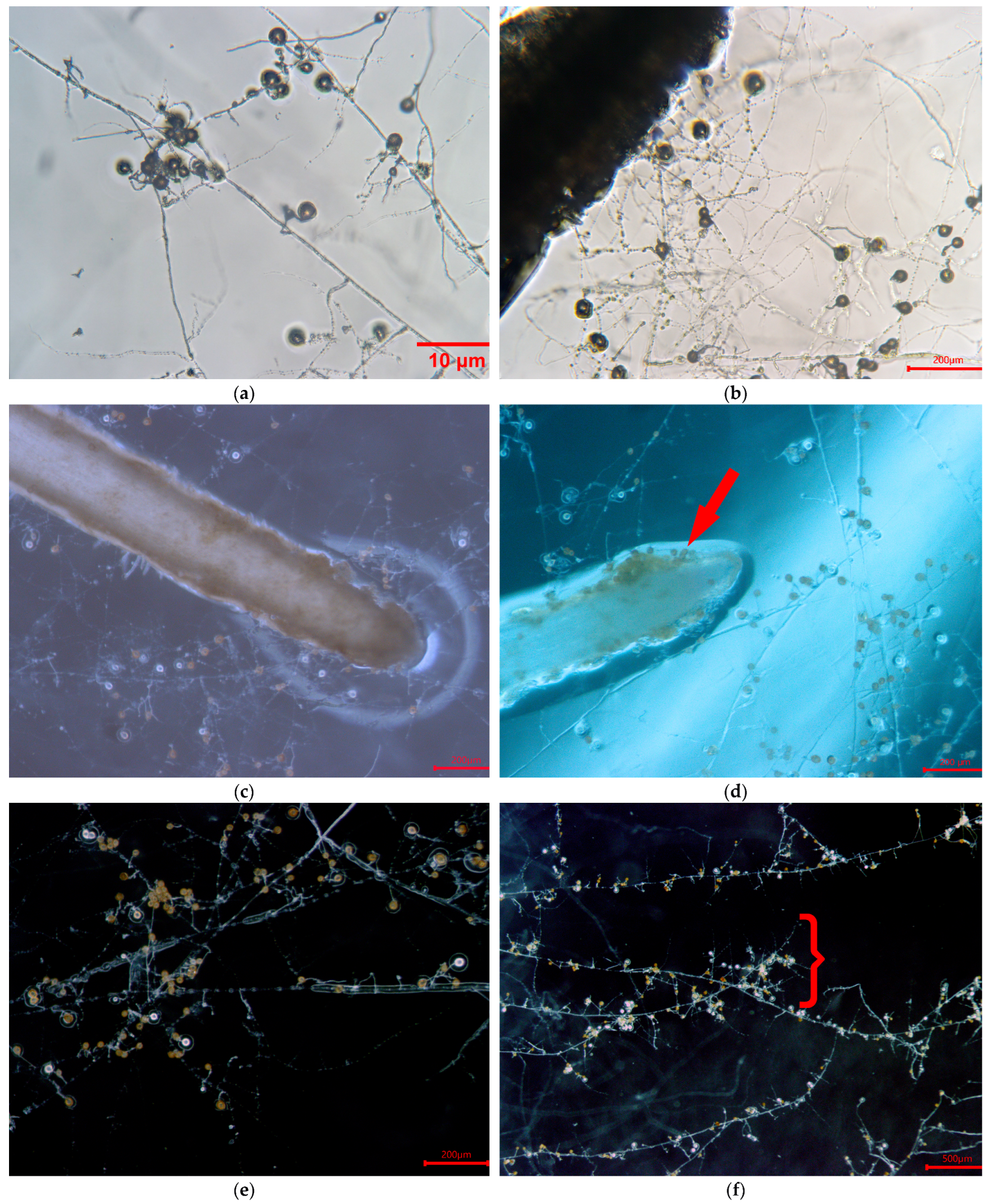
From that culture, three new monoxenic cultures were established with AMF inoculated roots and, after 7 days, there was an average hyphal growth of 1719.93 ± 525.48 mm in RC and 971.93 ± 486.14 mm in HC, in addition to the production of spores, running hyphae, branched hyphae, and hyphae coming out of the root (Figure 2).
Typical structures of arbuscular mycorrhiza, helper “mycorrhiza-like structures” (ALS), or “branched absorbing structures” (BAS) were observed (Figure 2). Yeast continued growing in the RC compartment of the plates, in close proximity to the carrot root. The solution was to subculture only the mycelium and spores from the hyphal compartment (HC), which had no yeast, and aseptic cultures were established after 15 days.
Mahogany (S. macrophylla) seeds were disinfected and successfully germinated in vitro. The 7-day-old germinated plants were placed on plates containing MSR medium and subsequently transferred to MSR medium supplemented with AMF spores and mycelium obtained from monoxenic cultures. In the third week, one plate showed contamination of white filamentous fungus. It was observed that no other microorganisms exhibited signs of growth on any of the control plates. Furthermore, it was recorded that all plants survived until the conclusion of the experiment. In mahogany plants from cultures with and without AMF inoculum, no significant statistical differences were observed in any of the variables at 30 days: stem length (t = 2.34, df = 4.99, p = 0.06), root length (t = 0.41, df = 6.37, p = 0.70), number of roots (t = −0.50, df = 6.50), and number of leaves (W = 14, p = 0.24) (Table 1).
Regarding the variables related to AMF growth, the development of spores and hyphae was observed throughout the culture medium from the first week in the inoculated plants. Total spore counts in the monoxenic cultures were 8736 and 12,457 per plate after seven days of cultivation. At 30 days of culture, the spore range was from 10,166 to 27,696, with an average of 14,795 ± 3301. Hyphae rapidly covered the Petri plate, forming a dense hyphal network showing hyphae going through in and out the roots. Hyphal length ranged from 2698.93 mm to 4219.28 mm, with an average of 3655.46 ± 308.75 mm. Extraradical mycelium development (running and branching hyphae), spore development (solitary and in clusters), as well as contact points between hyphae and root were observed (Figure 3). Final measurements exhibit that spores had reached an average diameter of 27.68 ± 3.85 µm.
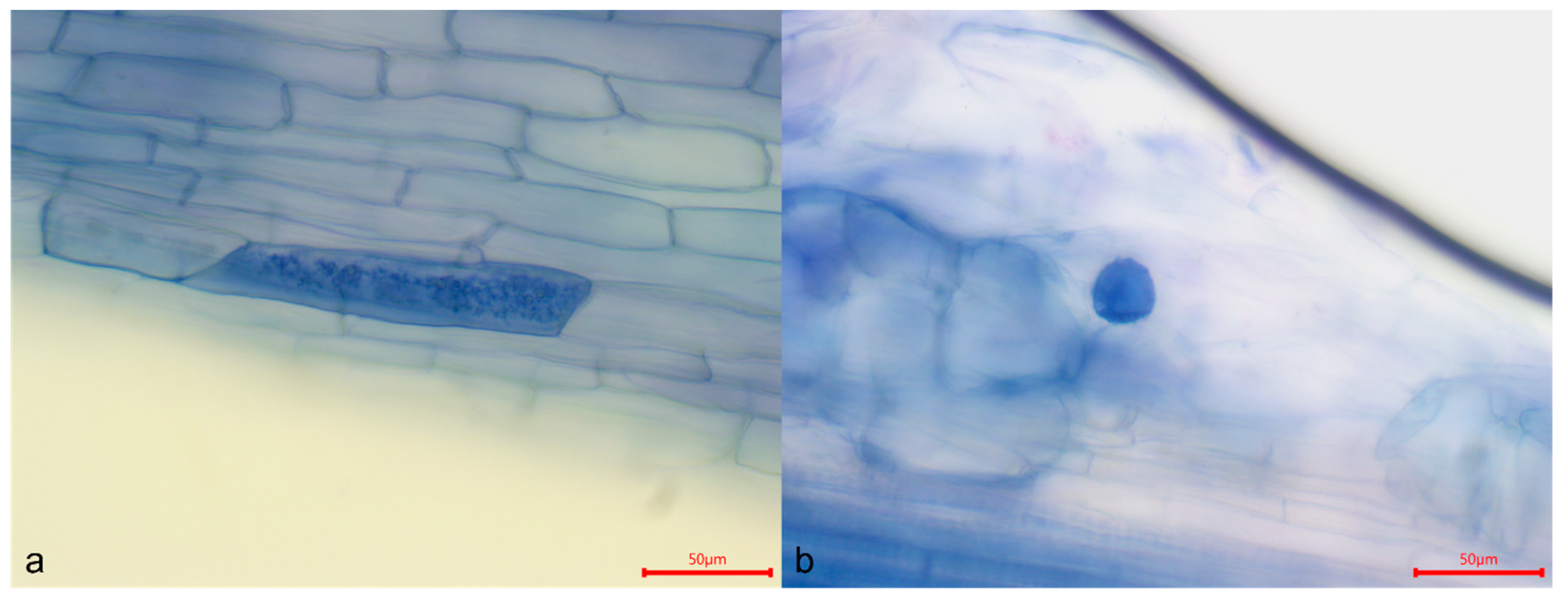
In terms of intraradical structures, arbuscules were observed from 30 days after inoculation, with a colonization rate of 41.49%. At 75 days, this percentage increased to 52.13% and colonization by vesicles was 1.06% (Figure 4). Arbuscules at 75 days were larger and more conspicuous than at 30 days. No hyphae were detected on any of the roots.

4. Discussion

The monoxenic cultures of AM fungi can be separated into four main steps: (1) selection of the adequate AM fungal propagules; (2) sampling: disinfection and incubation of the propagules on a suitable growth medium; (3) association of the propagules with a suitable host root; and (4) subcultivation of the AM fungi [40]. The use of bi-compartmental plates is a viable alternative for AMF production; the compartment called the root compartment (RC) contains roots transformed with A. rhizogenes. The RC is inoculated with AMF that colonizes the carrot roots. Fungal hyphae can grow from the RC to a second compartment, called the fungal compartment (HC), where mycelium proliferates and produces spores [36].
We selected the most abundant morphotype (species) present in the rhizosphere to work with it. The spore disinfection method used was effective by introducing AMF spores from cedar rhizosphere in vitro. Chloramine T to 2% is the most commonly used disinfectant for AMF spore disinfection, being efficient at different levels to remove surface contaminants [30,41,42,43,44]. Streptomycin and gentamicin are the most commonly used antibiotics and a 24 h lapse worked well to remove microorganisms in our spores, at least in the first days [30,41,42].
Due to symbiotic nature of this biotroph, in the absence of a suitable host plant, it is quite impossible to propagate these fungi at mass level [6]. For this reason, it is necessary to establish of pure and suitable carrot root cultures such as those obtained from carrot segments transformed with A. rhizogenes. Root organ culture has obvious advantages over traditional systems, allowing production of contaminant-free propagules [45]. Hairy roots are biochemically and genetically stable, and can grow in hormone-free media [46]. Several publications have used this type of culture to inoculate AMF [13,15,47].
Bi-compartmental plates were used and a segment of ROC was placed in one compartment and inoculated with Glomus sp. spores isolated from cedar rhizosphere. After 7 days of culture, more hyphal growth was observed in the RC than in the HC. It is relevant that on the bi-compartmental plates in the RC, growth of a yeast molecularly identified as Rhodotorula mucilaginosa (A. Jörg.) F.C. Harrison [39] was observed; however, on the HC side this yeast did not appear. A previous study reported that R. mucilaginosa or its exudates stimulates AM hyphal growth; this might increase the chance of contact between fungal hyphae and plant roots, and, consequently, improve mycorrhizal establishment [48]. Plant growth upgrade has been reported after inoculation with soil yeasts, which may be related to their P solubilizing traits and production of plant growth regulating hormones [49,50]. The capacity of yeasts or their exudates to increase the positive effects of Funneliformis mosseae (T.H. Nicolson & Gerd.) C. Walker & A. Schüßler (Glomus mosseae T.H. Nicolson & Gerd.-Gerd. & Trappe) on soybean dry matter might be exploited to improve the use and efficiency of this fungus in agriculture [51]. The colonization of Rhizoglomus intraradices (N.C. Schenck & G.S. Sm.) Sieverd., G.A. Silva & Oehl was able to positively regulate sugar concentrations, plant growth, root morphology, soil nutrient absorption, leaf gas exchange, structure, and stability of host plants, where the improvement in soil aggregate distribution and stability was not dependent on the plant species [52]. Regarding dual cultures with mycelial growth and spore production, three MSR medium segments with mycelium and spores (average 111 ± 50 spores per 1 × 1 mm) were taken and inoculated proximal to the carrot Ri T-DNA ROC segment. During the first week, the first contact points between roots and hyphae were obtained, which agrees with studies by Voets et al. [53] and Perera-García et al. [54], who observed germination of R. intraradices in less than seven days. In addition, the characteristic extraradical structures of the establishment of the symbiosis were observed, such as the runner hyphae, which are the most numerous and produce higher order hyphae [33].
High numbers of typical structures of arbuscular mycorrhiza, helper cells, “mycorrhiza(arbuscular)-like structures” (ALS), or “branched absorbing structures” (BAS) were observed [13], whose function seems to enhance nutrient uptake and spore proliferation, because ALS/BAS expands the substrate–fungus contact surface [13,33]. Changes in thickness and appearance of hyphae in the culture could demonstrate that an active movement of substances was taking place, so mycelium was growing functionally [12].
Colonization rates of 41% and 52.13% were obtained at 30 and 75 days of culture, which is contrary to the literature that indicates more time is required to establish AMF colonization in vitro. For example, in R. intraradices on Medicago truncatula roots, researchers reached 50% colonisation at 56 days [53], and for Rhizoglomus irregulare (Blaszk., Wubet, Renker & Buscot) Sieverd., G.A. Silva & Oehl (Rhizophagus irregularis-Blaszk., Wubet, Renker & Buscot-C. Walker & A. Schüßler), F. mosseae, and Entrophospora etunicata (W.N. Becker & Gerd.) Blaszk., Niezgoda, B.T. Goto & Magurno (Claroideoglomus etunicatum-W.N. Becker & Gerd.-C. Walker & A. Schüßler), they obtained at 3 months a colonization percentage of 35.5, 29.1, and 12.7, respectively, on Tectona grandis roots in vitro [20]. The low percentage of colonized root tissue obtained (usually <20%) is one of the major drawbacks found when using in vitro cultures of AM, and, more relevant, the long period (approximately 8 weeks) needed to reach it [55]. Our research team obtained a percentage of colonization of more than 20% after 30 days of culture.
Spores produced in vitro are smaller in size compared to those collected in the field, consistent with findings from other studies [13], suggesting that this may be due to nutrient or carbon depletion in the RO culture [15,56]. Spore production in our study was evident from the first week. Organisms like Glomus spp. invest their energy in reproduction, causing them to become more adapted to the in vitro culture system than k-organisms, which allocate most of their resources to growing and surviving [9].
The hyphal length obtained was greater than that reported in cultures of R. irregulare (R. irregularis) on flax plants [57] and of Glomus sp. on M. trunculata [58]. However, it is lower than that previously reported by Tiwari and Adholeya [59] of R. intraradices (G. intraradices) on carrot roots and on M. truncatula roots [53]. Similar results at 10 weeks were found in dual cultures of date palm with R. irregulare (R. irregularis) [60]. The absence of sucrose, physical exclusion of diffusible exudates from the host roots, or thinner growth medium (easier diffusion of gas) are stimulatory factors involved in the enhancement of hyphal and spore densities in the distal compartment [30].
In dual cultures on bi-compartment plates, AMF mycelial growth (ERM) from RC to HC through the “bridge” was observed from the first week. Initially, extraradical mycelium was observed spreading rapidly in RC and HC through the bridge (filter paper), completely covering at 75 days. Implementing a paper bridge ensured hyphae crossed over the media wall and accelerated this, with a consequent increase in the successful establishment of root-free AM fungal material [31]. The extraradical mycelium of AM is the functional organ for nutrient uptake and translocation; it has a dimorphic hyphal nature, with coarse thick-walled hyphae and fine thin-walled hyphae [33].
In this study, the effect of inoculation (MSR segments with mycelium and spores from axenic ROC cultures) of Glomus sp. on the growth of mahogany plants germinated in vitro was analyzed. The selected inoculum proved to be effective for spore production, as it ranged from 10,166 to 27,696 after 30 days of culture. This was far greater than in previous reports with R. irregulare (R. irregularis), in which an average of 8500 to 9000 spores were obtained from 10–15 parent spores [28], the number of Glomus sp. spores formed after 5 months of dual culture averaged 9500 per Petri dish [61], and 7300 ± 1600 spores were produced in M. trucatula seedlings associated with extraradical mycelium of R. intraradices (G. intraradices) at 12 days [53]. Mass production of spores (i.e., approximately 12,000 in 22 weeks) was obtained with potato [62]. Artificially injured extraradical hyphae were able to re-grow, repair, explore the surrounding environment, and colonize new roots [63].
Constant spore production depends on the biomass of the extraradical mycelium and allocation of resources from both the intraradical mycelium and the auxiliary cells through the hyphal network [64].
The presence of the characteristic intraradical structures of AMF, mainly arbuscules, was evidenced, inducing metabolic exchange between the host plant and the symbiotic fungus. The percentage of colonization by arbuscules found at 11 weeks is higher than that reported for the rubber tree at 13 weeks [65]. It is also higher than the percentage found in systems with plants such as M. truncatula at 10 weeks [58] and with argan plants at 11 weeks [66]. The percentage of vesicles is variable compared to other studies. There are systems with similar, lower, or higher percentages [65,66]. The presence of these structures points out that the fungus is carrying out its life cycle favorably [59,65], using the mahogany plant as host.
AM fungi associated with a root organ provide vigorous and uniform fungal mycelium capable of faster root colonization than isolated germinated spores or surface-sterilized AM colonized root fragments [67]. This type of AM inoculum production would significantly increase the number of spore loads to be inoculated in field, greatly influencing the production of agricultural and horticultural crops [28]. This demonstrated that an AMF symbiotically attached to an autotrophic donor plant is a powerful source of inoculum, allowing fast and heavy in vitro mycorrhization of seedlings [53].
After 30 days of cultivation, the variables evaluated for mahogany plants showed no significant differences between control and AMF-inoculated plants. This is in agreement with a study in which no differences were found in the shoot weight, number of leaves, and root wet weight between inoculated and uninoculated plantlets; this likely resulted from the optimal conditions and availability of nutrients (especially P, which could flow throughout the gel) in the in vitro culture experiment [20]. It has been reported that commercial inoculants available on international markets do not always successfully establish arbuscular mycorrhizal symbiosis [68]. Organic manures and bioinoculants are less expensive and achieve high productivity without harming the environment, contrary to chemical fertilizers [28]. So, in sustainable agriculture, AMF has great potential in crop production and environment preservation and, by using AMF in farming as biofertilizer, employment of toxic chemical pesticides and synthetic fertilizers can be minimized [69]. The fungus has been produced biotechnologically in an in vitro system that affords many advantages [3]. Rhizosphere yeasts and AMF are important to consider when integrating such root-associated microorganisms as biofertilizers in agroecosystems [50]. R. irregulare (R. irregularis) has the capacity to complete its life cycle with adequate spore (and/or other fungal infective propagules) production, and the ability to be continuously cultured under monoxenic conditions; both are prerequisites for inclusion in monoxenic culture collections, according to criteria for other AMF species (described by Fernández et al. [9], Declerk et al. [70]).

5. Conclusions

In the present study, monoxenic cultures were established on Glomus sp. carrot Ri T-DNA ROC, from which elevated levels of mycelial and spore production were obtained. The inoculation of Glomus sp. was conducted in vitro on the roots of mahogany plants that had previously undergone in vitro germination. The results demonstrated high mycelial growth, spore production, and root colonization rates of 41% and 52.13% at 30 and 75 days of culture, respectively. These values significantly exceed those reported for monoxenic cultures. As far as we know, this is the first report of an in vitro system using mahogany plants inoculated with AMF. Monoxenic cultures are a reliable, aseptic, properly identified and preserved source of inoculum in sufficient quantity and quality to produce a biofertilizer, as well as for other studies and biotechnological applications. More research is needed to investigate other variables that can alter the effects of mycorrhiza in mahogany in vitro. In addition, it is imperative to protect genetic resources of the native arbuscular mycorrhizal fungi preserving these species in culture collections and stimulating their cultivation under artificial conditions.

Author Contributions

Conceptualization: S.-R.L.Y.; funding acquisition: S.-R.L.Y. and A.-T.A.; investigation: S.-R.L.Y. and R.-C.M.; methodology: S.-R.L.Y., R.-C.M. and A.-S.E.; project administration: S.-R.L.Y. and A.-S.E.; resources: S.-R.L.Y.; supervision: S.-R.L.Y. and A.-T.A.; writing—original draft preparation: S.-R.L.Y., R.-C.M. and A.-T.A.; writing—review and editing: S.-R.L.Y., R.-C.M., A.-S.E. and A.-T.A. All authors have read and agreed to the published version of the manuscript.

Funding

This research was supported by the University of Costa Rica through projects C0057, C1099, and C4764, and the funding support for the Trabajos Finales de Graduación (2023). The APC was funded by the Universidad de Costa Rica, Costa Rica and Universidad Veracruzana, Mexico.

Institutional Review Board Statement

Not applicable.

Data Availability Statement

The data presented in this study are available on request from the corresponding author.

Acknowledgments

Special thanks to students Daniel Cordero-Orozco and Priscilla Solera-Rodríguez for their assistance with the disinfection and germination of mahogany seeds. We are also grateful to Andres Gatica (Laboratorio de Biotecnología Vegetal, Escuela de Biología, UCR) for providing the Agrobacterium rhizogenes strain and to Sebastian Fracchia (CRILAR-CONICET, Argentina) for his invaluable advice.

Conflicts of Interest

The authors declare no conflict of interest.

Abbreviations

AMF, Arbuscular mycorrhizal fungi; AM, Arbuscular mycorrhizal; M media, minimal mineral media; MSR media, modified Strullu and Romand media; Ri, root inducing; T-DNA, transfer DNA; LB, Luria–Bertani; YMB, yeast mannitol broth; YMA, yeast mannitol agar; YEP, yeast extract peptone; MS, Murashige and Skoog; MW medium, modified White’s medium. ROC, root organ culture.

References

  1. Ghorui, M.; Chowdhury, S.; Das, K.; Sunar, K.; Prakash, B. Optimizing factors for large-scale production of arbuscular mycorrhizal fungi consortia using root organ cultures. J. Biol. Methods 2023, 10, e99010006. [Google Scholar] [CrossRef]
  2. Powell, J.R.; Rilling, M.C. Biodiversity of arbuscular mycorrhizal fungi and ecosystem function. Tasley review. New Phytol. 2018, 220, 1059–1075. [Google Scholar] [CrossRef] [PubMed]
  3. Ceballos, I.; Ruiz, M.; Fernández, C.; Peña, R.; Rodríguez, A.; Sanders, I.R. The in vitro mass-produced model mycorrhizal fungus, Rhizophagus irregularis, significantly increases yields of the globally important food security crop cassava. PLoS ONE 2013, 8, e70633. [Google Scholar] [CrossRef]
  4. Tiwari, P.; Adholeya, A. In vitro co-culture of two AMF isolates Gigaspora margarita and Glomus intraradices on Ri T-DNA transformed roots. FEMS Microbiol. Lett. 2002, 206, 39–43. [Google Scholar] [CrossRef] [PubMed]
  5. IJdo, M.; Cranenbrouck, S.; Declerck, S. Methods for large-scale production of AM fungi: Past, present, future. Mycorrhiza 2011, 21, 1–16. [Google Scholar] [CrossRef] [PubMed]
  6. Akhtar, M.S.; Abdullah, S.N.A. Mass production techniques of arbuscular mycorrhizal fungi: Major advantages and disvantages: A Review. Biosci. Biotechnol. Res. Asia 2014, 11, 1199–1204. [Google Scholar] [CrossRef]
  7. White, P.R. The Cultivation of Animal & Plant Cells, 2nd ed.; Ronald Press: New York, NY, USA, 1963. [Google Scholar]
  8. Mosse, B.; Hepper, C.M. Vesicular-arbuscular infections in root organ cultures. Physiol. Plant Pathol. 1975, 5, 215–223. [Google Scholar] [CrossRef]
  9. Fernández, L.B.; Pergola, M.; Silvani, V.; Colombo, R.; Bompadre, J.; Godeas, A. Continuous and long-term monoxenic culture of the arbuscular mycorrhizal fungus Gigaspora decipiens in root organ culture. Fungal Biol. 2012, 116, 729–735. [Google Scholar]
  10. Bécard, G.; Fortin, J.A. Early events of vesicular arbuscular mycorrhizal formation on Ri T-DNA transformed roots. New Phytol. 1988, 108, 211–218. [Google Scholar] [CrossRef] [PubMed]
  11. Declerck, S.; Strullu, D.G.; Plenchette, C. Monoaxenic culture of the intraradical forms of Glomus sp. Isolated from a tropical ecosystem: A proposed methodology for germplasm collection. Mycologia 1998, 90, 579–585. [Google Scholar] [CrossRef]
  12. Karandashov, V.; Kuzovkina, I.; Hawkins, H.J.; George, E. Growth and sporulation of the arbuscular mycorrhizal fungus Glomus caledonium in dual culture with transformed carrot roots. Mycorrhiza 2000, 10, 23–28. [Google Scholar] [CrossRef]
  13. Araújo Costa, F.; Meira Haddad, L.; Megumi Kasuya, M.; Campos Oton, W.; Dutra Costa, M.; Chaer Borge, A. In vitro culture of Gigaspora decipiens and Glomus clarum in transformed roots of carrot: The influence of temperature and pH. Acta Sci. Agron. 2013, 35, 315–323. [Google Scholar]
  14. Adholeya, A.; Tiwari, P.; Singh, R. Large-Scale Inoculum Production of Arbuscular Mycorrhizal Fungi on Root Organs and Inoculation Strategies. In In Vitro Culture of Mycorrhizas; Springer: Berlin/Heidelberg, Germany, 2005; pp. 315–333. [Google Scholar]
  15. Fortin, J.A.; Bécard, G.; Declerck, S.; Dalpe, Y.; St-Arnaud, M.; Coughlan, P.; Piche, Y. Arbuscular mycorrhiza on root-organ cultures. Can. J. Bot. 2002, 80, 1–20. [Google Scholar] [CrossRef]
  16. Drazhnikova, A.; Andrlanova, T.V. Strategy for conservation of arbuscular mycorrhizal fungi. In Proceedings of the Material of the International Scientific Practical Conference, Ternopil, Ukraine, 4–5 June 2020. [Google Scholar]
  17. Kobayashi, S. Landscape rehabilitation of degraded tropical forest ecosystems, case study of the CIFOR/Japan project in Indonesia and Peru. For. Ecol. Manag. 2004, 201, 13–22. [Google Scholar] [CrossRef]
  18. Navarro, C.; Hernández, G. Progeny test analysis and population differentiation of Mesoamerican Mahogany (Swietenia macrophylla). Agron. Costarric. 2004, 28, 37–51. [Google Scholar]
  19. Barstow, M.; Negrão, R. Swietenia macrophylla. The IUCN Red List of Threatened Species 2023: e.T32293A68104718. 2023. Available online: https://dx.doi.org/10.2305/IUCN.UK.2023-1.RLTS.T32293A68104718.en (accessed on 17 April 2024).
  20. Chaiyasen, A.; Douds, D.D.; Gavinlertvatana, P.; Lumyong, S. Diversity of arbuscular mycorrhizal fungi in Tectona grandis Linn.f. plantations and their effects on growth of micropropagated plantlets. New For. 2017, 48, 547–562. [Google Scholar] [CrossRef]
  21. Rodríguez-Morelos, V.H.; Soto-Estrada, A.; Pérez-Moreno, J.; Franco-Ramírez, A.; Díaz-Rivera, P. Arbuscular mycorrhizal fungi associated with the rhizosphere of seedlings and mature trees of Swietenia macrophylla (Magnoliophyta: Meliaceae) in Los Tuxtlas, Veracruz, México. Rev. Chil. De Hist. Nat. 2014, 87, 9. [Google Scholar] [CrossRef]
  22. Polo-Marcial, M.H.; Lara-Pérez, L.A.; Goto, B.T.; Noa-Carrazana, J.C.; Díaz-Fleischer, F.; Andrade-Torres, A. Tropical deciduous species under different land use retain a high glomerospores diversity and arbuscular and septate endophyte colonization. Nova Hedwig. Band 2022, 115 Heft 3–4, 487–517. [Google Scholar] [CrossRef]
  23. Polo-Marcial, M.H.; Solís-Ramos, L.Y.; Murillo-Cruz, R.; Ávila-Arias, C.; Andrade-Torres, A. Mycorrhizal and endophytic richness and colonization in Cedrela odorata L., in agroforestry systems and secondary forest from southeastern Costa Rica. Agrofor. Syst. 2023, 97, 647–658. [Google Scholar] [CrossRef]
  24. Błaszkowsk, J.; Glomeromycota, W. Szafer Institute of Botany; Polish Academy of Sciences: Warsaw, Poland, 2012. [Google Scholar]
  25. Schenck, N.C.; Pérez, Y. Manual for Identification of Vesicular Arbuscular Mycorrhizal Fungi. (INVAM). Synergistic; University of Florida: Gainesville, FL, USA, 1990. [Google Scholar]
  26. Strullu, D.G.; Romand, C. Méthode d’obtention d’ endomycorhizes a vesicules et arbuscules en condition axeniques. Comptes Rendus De Académie Sci. 1986, 303, 245–250. [Google Scholar]
  27. Mugnier, J.; Mosse, B. Vesicular–arbuscular infections in Ri T-DNA transformed roots grown monoxenically. Phytopathology 1987, 77, 1045–1050. [Google Scholar] [CrossRef]
  28. Srinivasan, M.; Kumar, K.; Kumutha, K.; Marimuthu, P. Establishing monoxenic culture of arbuscular mycorrhizal fungus Glomus intraradices through root organ culture. J. Appl. Nat. Sci. 2014, 6, 290–293. [Google Scholar] [CrossRef]
  29. Murashige, T.; Skoog, F. A revised medium for rapid growth and bioassays with tobacco tissue cultures. Plant Physiol. 1962, 15, 473–497. [Google Scholar] [CrossRef]
  30. St-Arnaud, M.; Hamel, C.; Vimard, B.; Caron, M.; Fortin, J.A. Enhanced hyphal growth and spore production of the arbuscular mycorrhizal fungus Glomus intraradices in an in vitro system in the absence of host roots. Mycol. Res. 1996, 100, 328–332. [Google Scholar] [CrossRef]
  31. Dalpé, Y.; Seguin, S. A “paper bridge” system to improve in vitro propagation of arbuscular mycorrhizal fungi. Botany 2010, 88, 617–620. [Google Scholar] [CrossRef]
  32. Tennant, D. A Test of a Modified Line Intersect Method of Estimating Root Length. J. Ecol. 1975, 63, 995–1001. [Google Scholar] [CrossRef]
  33. Bago, B.; Azcón-Aguilar, C.; Piché, Y. Architecture and developmental dynamics of the external mycelium of the arbuscular mycorrhizal fungus Glomus intraradices grown under monoxenic conditions. Mycologia 1998, 90, 52–62. [Google Scholar] [CrossRef]
  34. Marturet, G.; Salazar, R.V.; Regales, B.; Betancourt, C. Efecto de los reguladores de crecimiento en la morfogénesis in vitro de caoba (Swietenia macrophylla king). Rev. Pittieria. 2016, 40, 164–173. [Google Scholar]
  35. Pérez-Moncada, U.A.; Ramírez-Gómez, M.M.; Núñez-Zarante, V.M.; Franco-Correa, M.; Roveda-Hoyos, G. Evaluación de un sistema para la micorrización in vitro en plantas de mora de castilla (Rubus glaucus, Benth). Univ. Sci. 2012, 17, 140–151. [Google Scholar] [CrossRef]
  36. Rosikiewicz, P.; Bonvin, J.; Snaders, I.R. Cost-efficient production of in vitro Rhizophagus irregularis. Mycorrhiza 2017, 27, 477–486. [Google Scholar] [CrossRef]
  37. R Core Team. R: A Language and Environment for Statistical Computing; R Foundation for Statistical Computing: Vienna, Austria, 2023; Available online: https://www.R-project.org/ (accessed on 21 July 2024).
  38. Philips, J.M.; Hayman, D.S. Improved procedures for clearing roots and staining parasitic and vesicular-arbuscular mycorhizal for rapid assessement of infection. Trans. Br. Mycol. Soc. 1970, 55, 156–161. [Google Scholar] [CrossRef]
  39. Harrison, F.C. Rhodotorula truncatula, a new species of yeast-like organism. Can. J. Res. 1927, 2, 347–348. [Google Scholar]
  40. Danesh, Y.R.; Tufenkci, S. In vitro culturing of mycorrhiza and mycorrhiza like fungi. Int. J. Agric. Technol. 2017, 13, 1675–1689. [Google Scholar]
  41. Cranenbrouck, S.; Voets, L.; Bivort, C.; Renard, L.; Strullu, D.G.; Declerck, S. Methodologies for in Vitro Cultivation of Arbuscular Mycorrhizal Fungi with Root Organs. In In Vitro Culture of Mycorrhizas; Declerck, S., Fortin, J.A., Strullu, D.-G., Eds.; Springer: Berlin/Heidelberg, Germany, 2005; Volume 4, pp. 341–375. [Google Scholar] [CrossRef]
  42. Eskandari, A.; Danesh, Y.R. Study on life cycle of arbuscular mycorrhizal fungus Glomus intraradices using in vitro culturing technique. J. Phytol. 2010, 2, 69–75. [Google Scholar]
  43. Mosse, B. The Establishment of Vesicular-Arbuscular Mycorrhiza under Aseptic Conditions. Microbiology 1962, 27, 509–520. [Google Scholar] [CrossRef]
  44. Raghavendra, K.; Nirmalnath, J.; Jagadeesh, K.S. Axenic Germination of Glomus intraradices in vitro. Int. J. Curr. Microbiol. Appl. Sci. 2020, 9, 2387–2396. [Google Scholar] [CrossRef]
  45. Aryal, H.P. A Protocol on in vitro Propagation of Arbuscular Mycorrhizal Fungi using Root Organ Culture Technique. Tribhuvan Univ. J. 2017, 31, 17–24. [Google Scholar] [CrossRef]
  46. Sawsan, A.E.E.; Abdullah, M.; Ali Hoda, H.; Senousy; Elsayed, S.; Razik, A. Production of Arbuscular Mycorrhizal Fungi using in vitro Root Organ Culture and Phenolic Compounds. J. Pure Appl. Microbiol. 2019, 13, 1985–1994. [Google Scholar]
  47. Zhang, L.; Fan, J.; Feng, G.; Declerck, S. The arbuscular mycorrhizal fungus Rhizophagus irregularis MUCL 43194 induces the gene expression of citrate synthase in the tricarboxylic acid cycle of the phosphate-solubilizing bacterium Rahnella aquatilis HX2. Mycorrhiza 2019, 29, 69–75. [Google Scholar] [CrossRef] [PubMed]
  48. Fracchia, S.; Godeas, A.; Scervino, J.M.; Sampedro, I.; Ocampo, J.A.; García-Romera, I. Interaction between the soil yeast Rhodotorula mucilaginosa and the arbuscular mycorrhizal fungi Glomus mosseae and Gigaspora rosea. Soil Biol. Biochem. 2003, 35, 701–707. [Google Scholar] [CrossRef]
  49. Nakayan, P.; Hameed, A.; Singh, S.; Young, L.; Hung, M.; Young, C. Phosphate-solubilizing soil yeast Meyerozyma guilliermondii CC1 improves maize (Zea mays L.) productivity and minimizes requisite chemical fertilization. Plant Soil 2013, 373, 301–315. [Google Scholar]
  50. Sarabia, M.; Cornejo, P.; Azcón, R.; Carreón-Abud, Y.; Larsen, J. Mineral phosphorus fertilization modulates interactions between maize, rhizosphere yeasts and arbuscular mycorrhizal fungi. Rhizosphere 2017, 4, 89–93. [Google Scholar] [CrossRef]
  51. Sampedro, I.; Aranda, E.; Scervino, J.M.; Fracchia, S.; García-Romero, I.; Ocapo, J.A.; Godeas, A. Improvement by soil yeasts of arbuscular mycorrhizal symbiosis of soybean (Glycine max) colonized by Glomus mosseae. Mycorrhiza 2004, 14, 229–234. [Google Scholar] [CrossRef] [PubMed]
  52. Wu, W.J.; Zou, Y.N.; Hashem, A.; Avila-Quezada, G.D.; Abd Allah, E.F.; Wu, Q.S. Rhizoglomus intraradices Is More Prominent in Improving Soil Aggregate Distribution and Stability Than in Improving Plant Physiological Activities. Agronomy 2023, 13, 1427. [Google Scholar] [CrossRef]
  53. Voets, L.; De la Providencia, I.E.; Fernández, K.; IJdo, M.; Cranenbrouck, S.; Declerck, S. Extraradical mycelium network of arbuscular mycorrhizal fungi allows fast colonization of seedlings under in vitro conditions. Mycorrhiza 2009, 19, 347–356. [Google Scholar] [CrossRef] [PubMed]
  54. Perera García, S.S.; Fernández Suárez, K.; Pérez Ortega, E.J.; Mujica Pérez, Y.; Pérez-Pérez, R.; Rodríguez Yon, Y.; Haesaert, G.; Perera García, S.S.; Fernández Suárez, K.; Pérez Ortega, E.J.; et al. In vitro propagation of a cuban Arbuscular Mycorrhizal Fungal strain and factible bacteria’s association. Rev. Colomb. De Biotecnol. 2022, 24, 36–45. [Google Scholar] [CrossRef]
  55. Cano, C.; Dickson, S.; González-Guerrero, M.; Bago, A. In vitro cultures open new prospects for basic research in arbuscular mycorrhizas. In Mycorrhiza; Varma, A., Ed.; Springer: Berlin/Heidelberg, Germany, 2008; pp. 627–654. [Google Scholar]
  56. Calvet, C.; Camprubi, A.; Pérez-Hernández, A. Plant growth stimulation and root colonization potential of in vivo versus in vitro arbuscular mycorrhizal inocula. HortScience 2013, 48, 897–901. [Google Scholar] [CrossRef]
  57. Kokkoris, V.; Hart, M.M. The role of in vitro cultivation on symbiotic trait and function variation in a single species of arbuscular mycorrhizal fungus. Fungal Biol. 2019, 123, 732–744. [Google Scholar] [CrossRef]
  58. De Boulois, H.D.; Voets, L.; Delvaux, B.; Jakobsen, I.; Declerck, S. Transport of radiocaesium by arbuscular mycorrhizal fungi to Medicago truncatula under in vitro conditions. Environ. Microbiol. 2006, 8, 1926–1934. [Google Scholar] [CrossRef]
  59. Tiwari, P.; Adholeya, A. Host dependent differential spread of Glomus intraradices on various Ri T-DNA transformed roots in vitro. Mycol. Prog. 2003, 2, 171–177. [Google Scholar] [CrossRef]
  60. El Hilali, R.; Bouamri, R.; Crozilhac, P.; Calonne, M.; Symanczik, S.; Ouahmane, L.; Declerck, S. In vitro colonization of date palm plants by Rhizophagus irregularis during the rooting stage. Symbiosis 2021, 84, 83–89. [Google Scholar] [CrossRef]
  61. Declerck, S.; Strullu, D.G.; Plenchette, C. In vitro mass-production of the arbuscular mycorrhizal fungus, Glomus versiforme, associated with Ri T-DNA transformed carrot roots. Mycol. Res. 1996, 100, 1237–1242. [Google Scholar] [CrossRef]
  62. Voets, L.; Dupré de Boulois, H.; Renard, L.; Strullu, D.G.; Declerck, S. Development of an autotrophic culture system for the in vitro mycorrhization of potato plantlets. FEMS Microbiol. Lett. 2005, 248, 111–118. [Google Scholar] [CrossRef] [PubMed]
  63. de la Providencia, I.E.; Fernandez, F.; Declerck, S. Hyphal healing mechanism in the arbuscular mycorrhizal fungi Scutellospora reticulata and Glomus clarum differ in response to severe physical stress. FEMS Microbiol. Lett. 2007, 268, 120–125. [Google Scholar] [CrossRef]
  64. Declerck, S.; Bivort, C.; D’Or, D.; de Souza, F.A. Development of extraradical mycelium of Scutellospora reticulata under root-organ culture: Spore production and function of auxiliary cells. Mycol. Res. 2004, 108, 84–92. [Google Scholar] [CrossRef] [PubMed]
  65. Sosa-Rodríguez, T.; Dupré de Boulois, H.; Granet, F.; Gaurel, S.; Melgarejo, L.M.; Carron, M.P.; Declerck, S. In vitro mycorrhization of the rubber tree Hevea brasiliensis Müll Arg. Vitr. Cell. Dev. Biol. Plant 2013, 49, 207–215. [Google Scholar] [CrossRef]
  66. Ganoudi, M.; Calonne-Salmon, M.; Ibriz, M.; Declerck, S. In vitro mycorrhization of Argania spinosa L. using germinated seeds. Symbiosis 2021, 85, 57–68. [Google Scholar] [CrossRef]
  67. Bécard, G.; Piché, Y. Establishment of vesicular-arbuscular mycorrhiza in root organ culture: Review and proposed methodology. Method Microbiol. 1992, 24, 89–108. [Google Scholar]
  68. Vosátka, M.; Látr, A.; Gianinazzi, S.; Albrechtova, J. Development of arbuscular mycorrhizal biotechnology and industry: Current achievements and bottlenecks. Symbiosis 2012, 58, 29–37. [Google Scholar] [CrossRef]
  69. Oruru, M.B.; Njeru, E.M. Upscaling arbuscular mycorrhizal symbiosis and related agroecosystems services in smallholder farming systems. BioMed. Res. Int. 2016, 2016, 4376240. [Google Scholar] [CrossRef]
  70. Declerck, S.; Strullu, D.G.; Fortin, J.A. In Vitro Culture of Mycorrhizas; Springer: Berlin/Heidelberg, Germany, 2005. [Google Scholar]
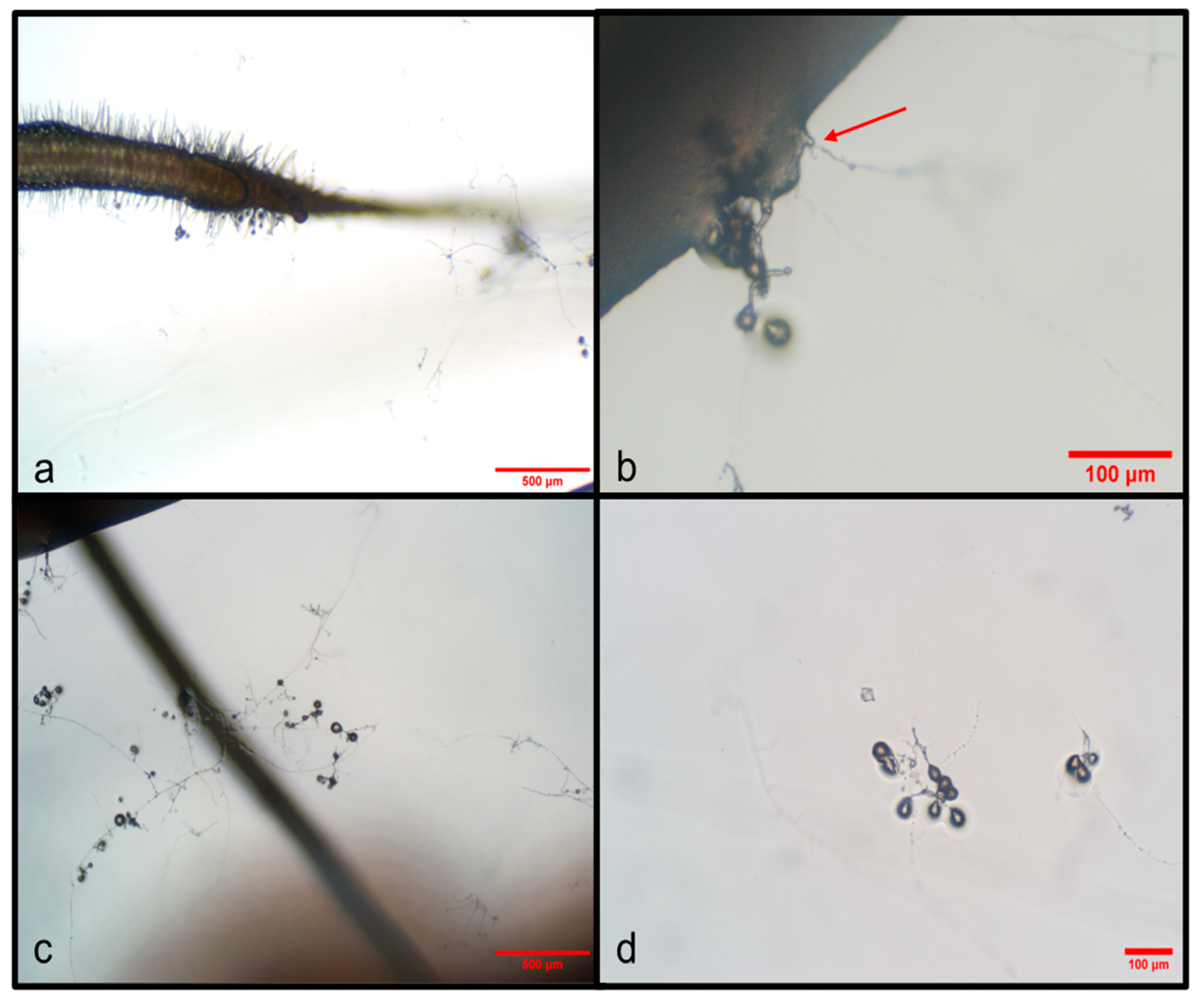
Figure 1. AMF growth on carrot (D. carota) roots transformed with A. rhizogenes and inoculated with spores of Glomus sp. at 75 days in monoxenic cultures. (a) Small spores and mycelium close to the root in the root compartment (RC). (b) The arrow shows the point of hyphal entry into the root in the root compartment. (c) Developing mycelium and spores in the root compartment. (d) Developing mycelium and spores in the hyphal compartment. Each photograph indicates the scale.
Agriculture 15 00673 g001
Figure 2. Monoxenic cultures of carrot (D. carota) transformed with A. rhizogenes and inoculated with spores of Glomus sp. after seven days. (a) Root with presence of hyphae. (b) Running hyphae with small developing spores. (c) Root apex growing with hyphae. (d) Branching hyphae producing structures called mycorrhiza-like structures (ALS) or absorbing branched structures (BAS). Each photograph indicates its scale.
Agriculture 15 00673 g002
Figure 3. Extraradical structures developed in dual culture of AMF with mahogany plants. (a,b) Spore development. (c,d) Hyphae and spores close to the root with contact points to the root; the arrow points to spores growing on the root. (e) Hyphae and spores; a change in hyphal thickness can be seen. (f) Mycorrhiza-like structures (ALS) or absorbing branched structures (BAS) (marked with red key) with spore development.
Agriculture 15 00673 g003
Figure 4. Intraradical structures in mahogany roots in vitro after 75 days of inoculation with AMF isolated from cedar rhizosphere and propagated in monoxenic carrot root culture. (a) Arbuscule in cortical cell. (b) Vesicle in root cells.
Agriculture 15 00673 g004
Table 1. Growth responses of mahogany (S. macrophylla) plantlets after 1 month of in vitro inoculation with Glomus sp.
TreatmentStem Length
(cm)
Root Length (cm)Number of LeavesNumber of Roots% Colonization
Control (uninoculated)12.4 ± 0.414.7 ± 3.02 ± 0.046 ± 4.00.0
Glomus sp. colonized root13.6 ± 0.216.3 ± 2.53 ± 0.042 ± 730%
Disclaimer/Publisher’s Note: The statements, opinions and data contained in all publications are solely those of the individual author(s) and contributor(s) and not of MDPI and/or the editor(s). MDPI and/or the editor(s) disclaim responsibility for any injury to people or property resulting from any ideas, methods, instructions or products referred to in the content.