Review

Melatonin: An Overview on the Synthesis Processes and on Its Multiple Bioactive Roles Played in Animals and Humans

by
Vasile-Cosmin Andronachi
,
Cristina Simeanu
*,
Mădălina Matei
,
Răzvan-Mihail Radu-Rusu
and
Daniel Simeanu
“Ion Ionescu de la Brad” University of Life Sciences, 3 Mihail Sadoveanu Alley, 700489 Iasi, Romania
*
Author to whom correspondence should be addressed.
Agriculture 2025, 15(3), 273; https://doi.org/10.3390/agriculture15030273
Submission received: 20 December 2024 / Revised: 20 January 2025 / Accepted: 24 January 2025 / Published: 27 January 2025
(This article belongs to the Special Issue Farming Factors’ Influence on Animal Productions)

Abstract

:
Melatonin is a natural hormone synthesized mainly by the pineal gland of vertebrates, and, secondarily, by other tissues and organs as well. It is deemed a bioactive molecule due to the multiple roles and functions it performs in animals and humans. Research conducted up to 2024 has reported the presence of melatonin in a wide variety of plants and bacteria, as well. This review aims to collect some of the scientific data to identify and describe the main sources of melatonin, and to document the functions and roles it plays in animal organisms. It also includes a description of the main technological and nutritional factors that can positively or negatively influence the synthesis and secretion process of melatonin, which is subsequently transported from the animal body into some food products, such as milk. This paper also includes information on the interaction between melatonin and other bioactive compounds present in animal and human bodies, with the aim of identifying what other functions and roles this hormone performs, and whether it interacts with other substances present in the vertebrate organism.

1. Introduction

Melatonin, also known by its chemical name, N-acetyl-5-methoxytryptamine [1], is a natural hormone synthesized and secreted generally by the pineal gland of mammals [2], derived from the limiting essential amino acid tryptophan [3,4], which plays multiple roles and functions in both animal and human organisms. Melatonin is an endogenous idolamine [5] first discovered in 1958, in the pineal gland of cattle [6]; it was first isolated in 1960 [7], and is known as the hormone of darkness or the sleep hormone due to the fact that melatonin secretion occurs as an automatic response of organisms to the lack of light [8,9].
The pineal gland is an endocrine gland located on the third ventricle of the brain of all vertebrates, and its basic function is the production and secretion of melatonin [10,11,12].
The determination of circadian rhythms in vertebrates is carried out by an internal biological clock, which consists of the retina, hypothalamus, and pineal gland. Through the retinal, encephalic, and pineal photoreceptors, these three components (the retina, hypothalamus, and pineal gland) are synchronized with light cycles [13]. The pineal gland is an organ characteristic of vertebrates, being present in the animal body of all mammals, including in the human body [14], as well as in birds [15] and fish [16]. Through specialized studies, melatonin has been detected in other life forms that do not have a pineal gland, and its presence has also been reported in some species of microorganisms [17].
Melatonin is a bioactive molecule that acts in the regulation of sleep [18] and of the circadian rhythm [19], while also performing antioxidant [20], anti-inflammatory [21,22], immunomodulatory [23], anti-aging, anti-carcinogenic [24,25] and anti-apoptotic functions, being able to regulate apoptosis [26,27,28], and improving immune activity in organisms [29].
The term bioactive molecule (bioactive compound) is a notion whose definition is still being discussed and debated in 2024, because the opinions of different authors are divided into two spheres of classification of these substances [30]. Some researchers define bioactive molecules as substances with a positive or negative biologically active role, influenced by the nature of the bioactive compound, and by the quantity of molecules available in the sources of supply of these biologically active substances (the uptake of dietary bioactive molecules). Other authors strictly emphasize that a substance can be considered a bioactive molecule only if it fulfills an exclusively positive role on the organism in which it exerts its action [31,32].
Based on the literature survey, we have defined bioactive molecules as substances of dietary origin, with a biologically active role, fulfilling multiple roles and functions in the animal and human body, such as regulating the circadian rhythm, the normal and harmonious development of the physiological processes of growth and development of the body, maintaining the health of vertebrates, etc. These substances with bioactive role are widely distributed in nature, found in a multitude of sources, including raw materials and finished food products. They are classified from a chemical point of view as compounds different from the nutrients found in food. From a biological point of view, these substances fulfill an exclusively positive role in the animal and/or human body, acting either individually or in a synergistic relationship with other molecules present in the vertebrate organism [30,31,32].

2. Melatonin Synthesis and Secretion

Melatonin, with the chemical formula C13H16N2O2, can be synthesized and secreted in vertebrates by hydroxylation, decarboxylation, acetylation, and methoxylation processes [4,5]. Physically, melatonin in its pure state is a colorless powder with a whitish hue. The density of melatonin is 1.175 g/cm3, with a molar mass of 232.28 g/mol and a boiling point reached at a temperature of +512.8 °C. The melting point of melatonin is within the limits of the thermal range of +116.5–+118 °C [7].
From a physiological point of view, melatonin can be secreted in the animal and human body via the pineal route (via the pineal gland) and via the extrapineal route in other organs and tissues of the body. In the vertebrate organism a much higher amount of melatonin is secreted at the extrapineal level, compared to the level of melatonin secreted exclusively by the pineal gland. However, it was observed that the quantitative level of melatonin secreted extrapineally cannot compensate for or replace the role played by melatonin secreted by the pineal gland, in terms of regulating the circadian rhythm and improving sleep quality. Compared to pineal melatonin, which has a well-established circadian rhythm, the melatonin secreted at the extrapineal level is not released into the blood, acting only locally, at the level of the tissues and organs that produce it, and does not have a circadian secretion pattern [1]. With the exception of intrapineal and extrapineal sources of melatonin secretion, the vertebrates’ also have two other sources of this hormone, namely: gut microbiota and the dietary uptake [4]. Thus, by consuming food products rich in melatonin obtained through synthesis and natural secretion, the level of circulating melatonin in the body can be increased, leading to the improvement of people’s health by manifesting the multiple roles and functions that melatonin can perform in the human body.
Pineal melatonin and extrapineal melatonin have the same chemical structure and perform similar roles in the animal and human body, but have different sources of origin (Figure 1). In the animal and human body, both pineal melatonin and extrapineal melatonin have an antioxidant role by eliminating free radicals [33], also performing functions in modulating inflammatory responses at the intestinal level [34].
Acuña-Castroviejo et al. (2014) [33] suggested that the absence of photoperiod-induced variations in the synthesis and secretion process of extrapineal melatonin is due to the existence of different synthetic pathways for this hormone at the extrapineal level, compared to melatonin synthesized and secreted exclusively by the pineal gland. These differences observed in the synthesis process of extrapineal melatonin may be due to states of adaptability in the organism, involved in cell survival. An example suggested by Acuña-Castroviejo et al. (2014) [33] is the antioxidant effect of melatonin, achieved through the neutralization of reactive oxygen species (ROS) and reactive nitrogen species (RNS). The production of ROS and RNS mainly occurs during phases of metabolic, motor, and neural activity, when oxygen consumption is at its maximum in both animal and human organisms [33]. Thus, in the case of diurnal animal species, which consume the largest amount of oxygen during the day, the organism’s synchronization with environmental conditions and with physiological processes that are more intense during the day, leading to the production of large amounts of ROS and RNS, could represent an evolutionary factor that determined the adaptation of the organism through the establishment of an extrapineal melatonin synthesis process that is not dependent on day–night variations, and which provides a strong protective mechanism for cellular survival.
Melatonin has a very low toxicity, and in relatively high doses, due to its optimal dimensions, it is able to easily cross physiological barriers. N-acetyl-5-methoxytryptamine (melatonin) has an amphiphilic character, being partially soluble in water and highly soluble in lipids [35].

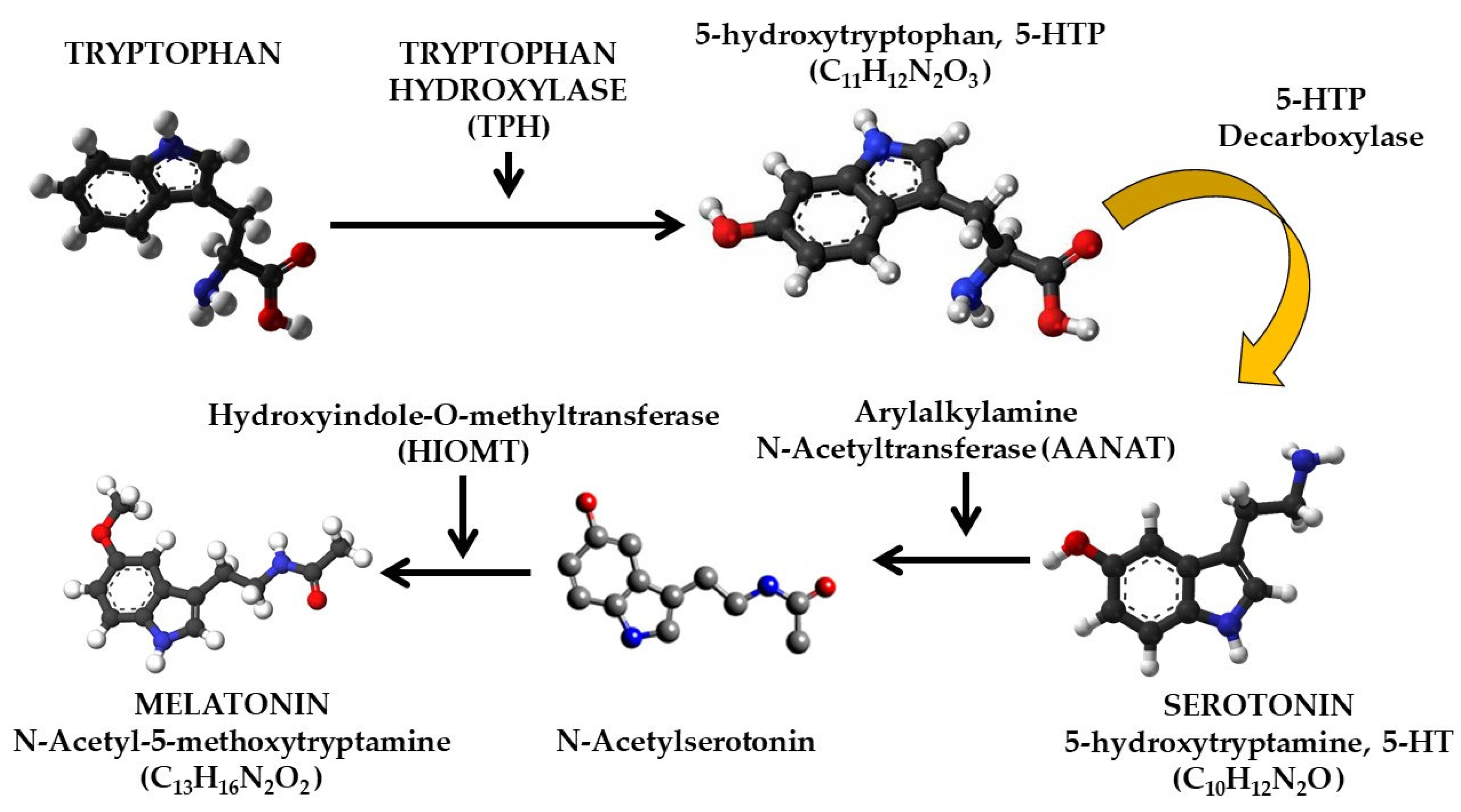
2.1. Pineal-Originated Melatonin

The pineal synthesis and secretion of melatonin begins with the help of noradrenaline, which is the main neurotransmitter involved in the activation of the pineal enzyme group, especially N-acetyltransferase. This activation of the pineal enzyme group is due to the cAMP (cyclic adenosine monophosphate) and cGMP (cyclic guanosine monophosphate) pathways that contributes to the activation of alpha1 (α1), alpha2 (α2) and beta1 (β1) receptors, located on the pinealocyte membrane [36].
Melatonin synthesis (Figure 2) is a sequential process, consisting of four phases/steps [37], and begins in the first phase with the transformation of tryptophan into 5-hydroxytryptophan, a transformation that occurs due to the action of the enzyme tryptophan hydroxylase (TPH). Subsequently, 5-hydroxytryptophan is transformed into serotonin (5-HT), and after transformation, serotonin undergoes the process of N-acetylation as a result of the action of arylalkylamine N-acetyltransformase (AANAT), forming NAS (N-acetylserotonin). In the last step, N-acetylserotonin is transformed into melatonin, and this process of transformation of N-acetylserotonin into melatonin is facilitated by the enzyme hydroxyindole-O-methyltransferase (HIOMT) [38].
Due to the fact that the enzyme arylalkylamine N-acetyltransformase (AANAT), which plays the role of catalyzing the conversion of serotonin into N-acetylserotonin, has minimal activity in the organism during the day, the melatonin production process is limited, thus favoring the accumulation of serotonin in pinealocytes, while melatonin synthesis at night is due to the increased activity of the AANAT as a result of the onset of darkness [39].
Melatonin synthesis in the pineal gland is rhythmically regulated by the body’s “master biological clock” located in the hypothalamic suprachiasmatic nucleus (SCN). Melatonin synthesis begins with the conversion of the essential amino acid tryptophan (which is of dietary origin) into serotonin [40]. Furthermore, some studies have reported that mitochondria contain high levels of melatonin [41].
The process of capturing and transmitting light information from the retina to the pineal gland begins with photoreceptor cells in the retina, which receive light signals (the retina is responsible for transforming optical signals into biological signals), and transmit the information to the hypothalamic suprachiasmatic nucleus (SCN) via the retinohypothalamic tract. The main cells on the surface of the retina that receive light information are the photosensitive ganglion cells (ipRGC), which are highly sensitive to blue light. The suprachiasmatic hypothalamic nucleus (SCN) is the body’s main biological clock, and through the information it receives from the retina, it determines whether it is day or night outside, depending on the intensity of light that has penetrated the surface of the retina. In turn, the suprachiasmatic hypothalamic nucleus sends the information received through the sympathetic preganglionic neurons that are located in the brainstem (especially in the modular area or in the lower part of the brainstem) to the superior cervical ganglia (SCG) [42].
In humans, melatonin synthesis is initiated immediately after sunset, reaching a peak in secretion in the middle of the night, and gradually decreasing in the second half of the night. About 80% of the melatonin present in the human body over a 24 h period is synthesized during the night. Serum melatonin levels during the night in humans reach values between 80 pg/mL and 120 pg/mL, respectively, and during the day the amount of melatonin decreases drastically to values of about 10–20 pg/mL [42,43].

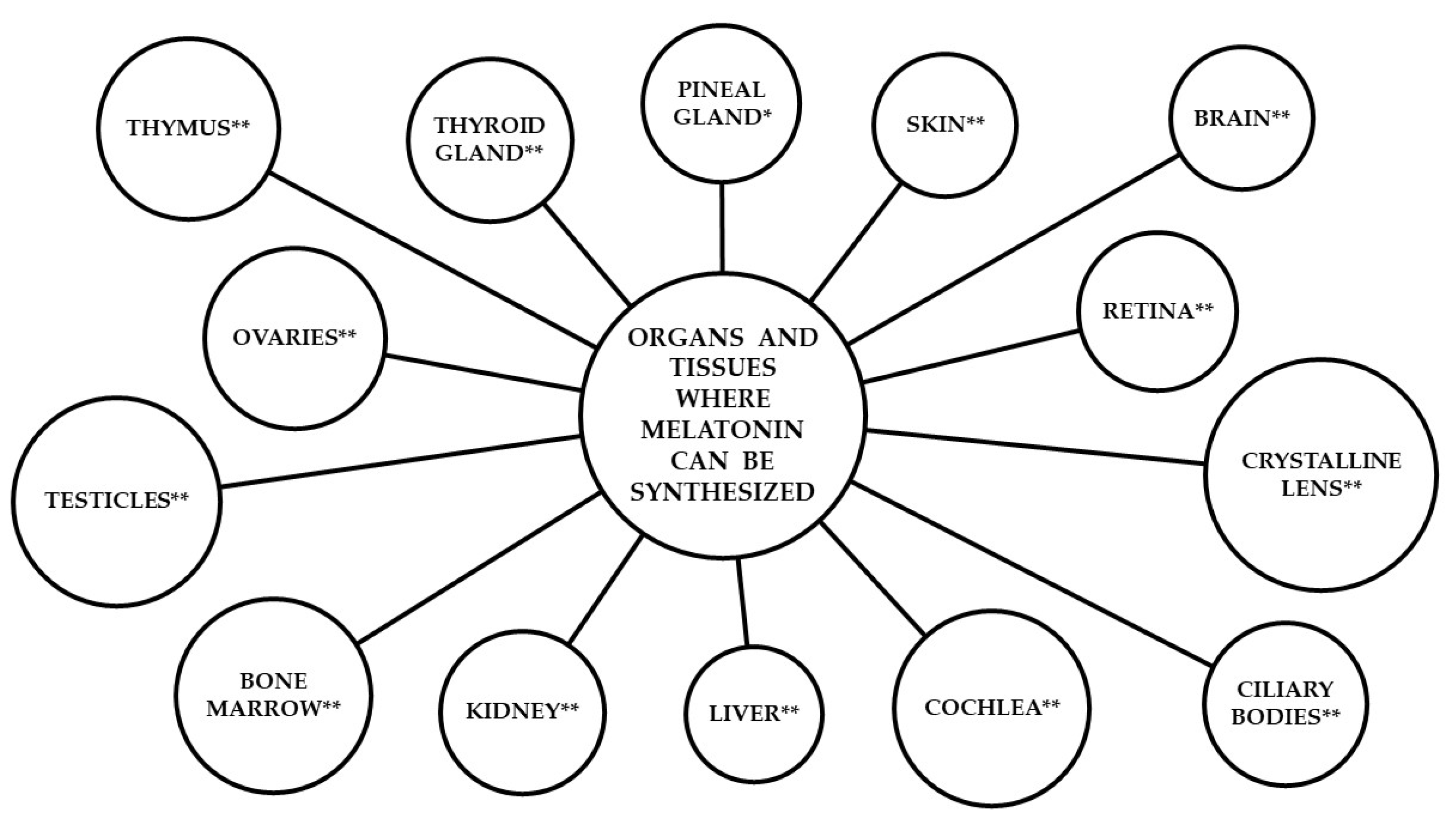
2.2. Extra-Pineal-Originated Melatonin

Melatonin can also be synthesized and secreted in the animal and human body, and in other tissues and organs such as the skin [44,45,46], the retina [47,48], certain brain regions [49], the liver, kidneys, and female reproductive organs [33], the thyroid gland, the lens and bone marrow [50], the ciliary bodies [51], the thymus [52], and the cochlea [53], and is also present in the gastrointestinal tract [54,55].
It has been found that melatonin synthesized and secreted in other tissues and organs fulfills other roles in living organisms, compared to the ones played by the pineal melatonin, depending on the tissue or organ in which it is synthesized and secreted. Another characteristic of extrapineal melatonin is that it is not transported in the body through the blood and acts only locally, in the area where it was secreted. For instance, it was discovered that extrapineal melatonin synthesized in the skin protects the tissue against reactive oxygen species (ROS) and reactive nitrogen species (RNS) that are induced as a result of skin exposure to chemical toxins or ultraviolet radiation [4,56,57].
Extrapineal melatonin secreted in the skin is not able to directly neutralize ROS and RNS without assistance [1]. Thus, it has been demonstrated that when melatonin donates an electron in order to inactivate a radical species, it is transformed into another radical scavenger, called 3-hydroxymelatonin. This phenomenon of melatonin transformation into other derivatives with a role in the capture of reactive species is generally called the melatonin antioxidant cascade, or the free radical elimination cascade [4,55]. This transformation of melatonin into different radical scavengers includes the following forms: N-acetylserotonin (NAS), 5-methoxytryptamine (5-MT), cyclic 3-hydroxylated melatonin (c3OHM), N1-acetyl-N2-formyl-5-methoxykynuramine (AFKM), N1-acetyl-5-methoxykynuramine (AMK), 6-Hydroxymelatonin (6-OHM), 4-Hydroxymelatonin (4-OHM), and 2-Hydroxymelatonin (2-OHM) [58].

3. The Role of Melatonin

Multiple studies conducted in the field of knowledge of the mode of action of melatonin on vertebrate organisms have highlighted a series of roles and functions that this hormone performs in the animal and human body. Thus, depending on the nature of melatonin (pineal or extrapineal), and depending on the tissue or organ in which it is synthesized and secreted, this hormone performs different roles and functions at the level of each individual organism.

3.1. The Role of Melatonin in the Animal Body

In the animal body, melatonin plays a role in regulating the circadian rhythm [19], improves sleep quality by regulating the sleep–wake rhythm [59], increases the quality of milk in mammals, mediates seasonal reproductive changes [60,61], modulates energy metabolism [62], regulates cellular redox homeostasis [63], and plays a role in domestic species’ reproduction [19].
Melatonin is rhythmically secreted in the animal body, as a result of the photostimulatory action caused by darkness, or by suppressing synthesis by exposing the eyeball to natural and/or artificial light [7]. After synthesis, pineal melatonin is released into the cerebrospinal fluid of the third ventricle, after which it is distributed to different regions of the brain in order to signal photoperiodic changes in the environment [64,65].
A study conducted on rats has shown that melatonin has the ability to inhibit the growth and development of certain cancerous tumors. Two groups of tumor-bearing rats were analyzed, infused with blood from human donors. The rats in the first group were infused with blood that had melatonin concentrations specific to the night period, and the rats in the second group were infused with blood that had melatonin levels specific to the day period. The results showed that in the case of rats infused with melatonin-rich blood, tumor growth was inhibited, while perfusion with melatonin-deficient blood led to tumor growth in the studied rats [66].
The involvement of melatonin in the reproductive process of animals is a well-known and documented function of this hormone. Melatonin primarily acts through the MT1 and MT2 receptors, whose presence has been reported in several specialized studies, in numerous brain and peripheral tissues, including the testes and ovaries. Specialized studies have reported that the melatonin receptor MT1 is more widely distributed in brain regions and endocrine tissues, compared to the MT2 receptor, which appears to be generally absent in the pituitary gland and hypothalamus. The brain regions and endocrine tissues are the main response areas for the circadian and physiological effects induced by N-acetyl-5-methoxytryptamine (melatonin), and the presence of the MT1 receptor in these organs indicates that this receptor (MT1) plays a major role in the physiological reproductive processes of mammals that are modulated by melatonin [67]. Melatonin is a hormone involved in the modulation of the hypothalamo–pituitary–gonadal (HPG) axis, which serves as a regulatory center for the reproductive process in both seasonally reproducing animals and non-seasonal reproducers (including humans) [68].
The influence of melatonin-rich diets on reproductive performance in rams was studied by Peña-Delgado et al. (2023) [69], who conducted a study in Spain on a group of 16 rams of the Aragonesa breed. In that study, the animals were divided into two groups, each consisting of 8 rams: a control group that received 500 g of a commercial diet, and an experimental group that received a modified diet, also administered at a rate of 500 g per day, but consisting of 20% agro-food by-products rich in phytomelatonin (plant melatonin), with the remaining 80% being the same commercial diet given to the control group. The study was conducted over a period of 5 months, from February to July, representing the non-reproductive season for the rams, and the animals were fed ad libitum with straw throughout the entire duration of the research. The phytomelatonin-rich by-products introduced into the diet of the experimental group consisted of pomegranate pomace with a melatonin content of 35.81 ± 0.4 ng/g, tomato pomace with a melatonin content of 23.76 ± 1.37 ng/g, and grape pulp with a melatonin content of 45.94 ± 4.19 ng/g. The plant components that made up the modified ration for the experimental group were mixed in equal proportions, and the melatonin levels in the plant by-products were determined by the authors using the HPLC-ESI-MS/MS technique (High Performance Liquid Chromatography–Electrospray Ionization–Tandem Mass Spectrometry). The authors of that study reported in their findings that a diet rich in phytomelatonin increased melatonin levels in seminal plasma and improved sperm viability and morphology. In the context of the same research, the authors highlighted that the introduction of plant by-products rich in phytomelatonin into the diets of farm animals offers economic benefits for both the agro-food industry and animal husbandry, as the reuse of plant materials, derived from certain production processes, reduces the waste levels resulting from the food and beverage processing processes, while the presence of a higher amount of melatonin in the animal’s body exhibits protective effects against oxidative damage to sperm cells by reducing intracellular levels of reactive oxygen species [69].

3.2. The Role of Melatonin in the Human Body

Melatonin, a hormone generally synthesized by the pineal gland under the influence of light intensity, and whose synthesis and secretion are carried out in the highest quantities in the time interval 01:00–04:00 in the morning, as specified by some authors [70], fulfills multiple roles in the human body, some of which are similar to the roles performed in the animal body. The most studied roles and functions that melatonin fulfills in the human body are represented by the regulation of the circadian rhythm and the improvement of sleep quality [18], the reduction of oxidative stress at the level of the whole body [71,72], the intervention in the regulation of the immune system [73], the regulation of the functions of the cardiovascular system [74] and the nervous system [75], the participation in the regulation of the biological rhythm [76], the stimulation of immune cells, and the regulation of cytokine production [77].
Under optimal conditions of human activity, performed within 24 h, the share allocated to rest through quality sleep should represent one third of the duration of a circadian cycle [78]. Sleep-related disorders are situations encountered in all age groups among the human population, and various studies have demonstrated that a low quality of sleep achieved during a 24 h day of activity has multiple negative effects on the general health of the human body, such as fatigue, low performance in carrying out daily activities, and others [79,80].
According to some clinical research, it has been found that in the first months of a newborn’s life, the pineal gland is not able to start a natural and individual synthesis and secretion of melatonin, so that infants must benefit exclusively from melatonin from external sources, such as breast milk [81,82]. The secretion and synthesis of melatonin, as well as the development of circadian sleep–wake rhythms, begin to manifest in infants only after the second to sixth month of life of newborns, or even after the sixth month in certain situations [81]. Due to these observations, the need for a more focused study of the way melatonin is secreted in cow’s milk is justified, in order to allow the production of milk and dairy products derived from it, which can support the population suffering from sleep disorders due to melatonin deficiency.
According to other studies, melatonin secretion in human milk also exhibits a circadian rhythm [83], with a maximum melatonin hormonal content of 46.9 ± 4.2 pg/mL determined in milk collected at midnight, and very low levels (undetectably low) in milk collected during the day, when the retina was exposed to the influence of natural and/or artificial light [84].
An advantageous aspect of melatonin is that this hormone does not present toxicity in the human body when administered in high doses. Thus, it has been demonstrated through various clinical studies that daily oral administration of melatonin, even in high doses (between 1 and 300 g or between 4.3 and 1291.5 μmol), did not produce negative effects on the health of human patients who benefited from melatonin treatment [35], an aspect that may encourage the consumption of foods with a high content of melatonin, especially due to the simple fact that in milk and other food products, melatonin is found in small quantities (expressed in pg) compared to the levels of melatonin administered to human patients in clinical studies.
Some researchers have reported through their studies that melatonin has an inhibitory effect on intrinsic apoptotic pathways in neurodegenerative diseases, especially in Alzheimer’s disease, Huntington’s disease, Parkinson’s disease, vascular accidents, and amyotrophic lateral sclerosis [85].
Melatonin is synthesized and secreted in the human body depending on the age of each individual. A circadian rhythm is an oscillation between the light period and the dark period, carried out within approximately 24 h. In the vertebrate body, the suprachiasmatic nucleus (SCN) determines the synchronization of circadian rhythms by regulating body temperature, through various hormonal signals and by regulating neuronal activity. According to research conducted up to 2024, aging of people leads to various changes in sleep patterns, such as daily synchronization of rest hours, sleep duration, high latency of sleep onset, greater susceptibility to awakenings, sleep fragmentation manifested by periodic awakenings during a rest cycle, reduction in the quality of deeper sleep, amplification of periods spent in lighter stages of sleep, etc. Thus, it has been found that starting at the age of 50, the amount of melatonin secreted in the human body begins to decrease, and after the age of 70, the process of synthesis and secretion of melatonin naturally seems to be almost completely absent [86,87,88].
Milagres et al. (2013) [89] reported that the administration of cow’s milk collected at 02:00 AM, which was rich in melatonin obtained through both synthetic production and natural secretion, increased plasma melatonin levels by 26.5% in adult Wistar rats, compared to plasma melatonin levels detected in adult Wistar rats that consumed daytime milk (cow’s milk collected at 15:00 PM). In the same study, it was determined that the addition of tryptophan to nighttime milk increased plasma melatonin levels by 35.6% in adult Wistar rats that consumed this type of milk enriched with natural melatonin and added tryptophan [89]. Similarly, beneficial effects of consuming melatonin-rich milk have been reported in humans, through the improvement of sleep quality, which was manifested by greater satisfaction with the rest period achieved through sleep and by the improvement in the performance of daily activities [59].

4. Melatonin in Milk

Milk is a liquid mixture, formed of water and dry matter (the proportions of the two components being approximately 87.5% water and 12.5% dry matter, respectively). Milk is defined as a homogeneous and opalescent liquid, white in color and free of foreign bodies suspended in the liquid volume, being secreted by the mammary gland of female mammals [90].
The dry matter in milk is in turn represented by several nutritional components (proteins, fats, lipids, etc.) with a role in providing the body with the energy necessary to support healthy and harmonious growth and development [91], and by a series of molecules with a bioactive role in the animal and human body (vitamins, hormones, minerals). Due to the chemical composition of cow’s milk, which is considered to be complex and complete, and due to the high nutritional value of this liquid, milk is considered to be one of the most important products of animal origin [92]. Naturally, raw milk has in its chemical composition several bioactive molecules (including free oligosaccharide structures, various hormones, peptides, lipids, etc.), which fulfill multiple active biological roles in the animal and human body with a different metabolic impact compared to the nutritional value of milk [93,94].
The natural secretion of melatonin is a process that occurs in the animal and human body during the night, so melatonin is also called the sleep hormone. Melatonin is naturally secreted first in the blood and in the cerebrospinal fluid of the third ventricle of the vertebrate brain [1], and in the case of female mammals, melatonin is subsequently released into the milk. For this reason, any variation in nutrients and bioactive molecules in the blood (including melatonin) will directly influence the chemical composition of milk, in terms of the milk’s content in nutrients and bioactive molecules. Due to the nocturnal nature of melatonin secretion, numerous researchers have initiated the idea of harvesting animal milk at night, so as to obtain milk with a higher melatonin content, thus giving rise to the concept of daytime milk (milk harvested during the day) and nighttime milk (milk harvested at night).
Studies conducted in the zootechnical field on the factors that can influence the secretion of melatonin in cow’s milk (but also in other species of animals of zootechnical interest), have demonstrated that the process of melatonin release in milk is a phenomenon influenced by a series of technological factors (breed and species of animals, animal health, conditions in shelters, photoperiods, the time when milk is harvested, milking frequency, environmental conditions in which animals are raised, etc.), and nutritional factors (animal nutrition and feeding).
According to the specialized literature and experiences in zootechnical research, it has been highlighted that the health status of animals (cattle, sheep, goats, birds, etc.), as well as the conditions of raising and care (generally exposure to stress factors) directly influence the quality and quantity of productions made [95].
Some authors reported in a study conducted in Switzerland, on a herd of 125 cows from eight farms with automatic milking systems, a correlation between the increase in the number of milkings performed in a night and the low content of salivary melatonin detected [96]. In this review, we will analyze and describe the influence of the main technological and nutritional factors that can significantly influence (positively or negatively) the natural secretion of melatonin in cow’s milk, according to the information available in the specialized literature.

4.1. Farming Technology Factors

The most studied technological factors that can influence the melatonin content in cow’s milk are represented by the species and breed of animals, environmental conditions, the daily milk production of the animals, the frequency of milking, the lighting conditions, and the intensity of artificial light in the animal shelters [5,97,98].

4.1.1. Species and Breed

Animal species and breed are two factors that can directly influence the melatonin content in the milk of cows and other animals raised for dairy production. Studies on cattle and sheep have shown that there are major differences between the melatonin content released in milk collected during the day and the melatonin content released in milk collected at night, as well as between milk collected individually from the two animal species studied, according to the data presented in Table 1 [99,100]. These differences are mainly due to the circadian rhythm of melatonin synthesis, as well as to genetic differences between the two animal species.
In sheep [100], it was demonstrated that keeping the animals for a period of 16 h in the dark and 8 h in the light did not influence the chemical composition of the milk in protein, fat, lactose, and salt, but led to the obtaining of milk with a higher melatonin content in the case of samples analyzed from milk collected at night (11.06 ± 7.24 pg/mL), compared to milk samples collected during the day (6.12 ± 4.55 pg/mL). The results presented in Table 1 highlight the nocturnal nature of melatonin secretion in bovine and ovine milk, due to the obtaining of higher melatonin values in nocturnal milk, compared to the results obtained in the case of daytime milk collected from both species.

4.1.2. Environmental Conditions

Environmental conditions are other important factors in obtaining milk with a high melatonin content. Significant differences in the melatonin content of cow milk were also recorded in the milk of the same breed (Holstein) but located in different geographical regions, according to the data presented in Table 2.
The significant differences between the values of melatonin levels in the milk of Holstein cows are mainly due to the conditions in which the experiments were carried out. Boztepe et al. [99] carried out their study in January (with a photoperiod of 11 h of natural light and 13 h of darkness) and with an artificial light intensity measured at eye level during the night of 150 lx. In that study, milk was collected in two different time intervals (between 15:00 and 17:00 for daytime milk, and between 03:00 and 05:00 in the morning for nighttime milk). In another study, Şahin et al. [101] collected milk from cows three times a day at 07:00 in the morning, 16:00 in the afternoon, and 01:00 in the morning. Milagres et al. [89] conducted a study on the differences in melatonin detected in the milk of Holstein cows during the summer season (between 2 and 16 June) for a period of 15 days, collecting milk at 02:00 in the morning for nighttime milk and 15:00 in the afternoon for daytime milk.
In the context of the study conducted by Romanini et al. [102], nighttime milk was collected between 05:00 and 06:00 in the morning, and daytime milk was collected during the day, when the animals were milked according to the milk collection schedule applied in the farm where the research was carried out.
The research conducted by Asher et al. [103] was carried out during the period of 1–15 November of the year, with a photoperiod duration of 10.4 h of natural light and 13.6 h of darkness. The study involved the formation of two experimental group that were subjected to different artificial lighting conditions during the night. In the case of cows in the Dark-Night batch, the lighting conditions applied in the animal shelters during the night were 648 ± 5.12 nm and 5.08 ± 0.04 lx, and for cows in the Night-Illumination group, lighting conditions of 462 ± 5.12 nm and 105 ± 3.91 lx were applied.
Teng et al. [104] conducted a study on the melatonin content of cow’s milk at the end of August, and the milk was collected at 03:00 PM, representing daytime milk, and at 05:00 AM, representing nighttime milk.
The correlation of the data presented in Table 2, in relation to all the experimental conditions that were applied in the studies presented in this article, shows the high degree of variability of the melatonin content found in bovine milk. The experimental data presented in Table 2 indicate different values of the melatonin content range determined in both daytime and nighttime milk of cows. The presence of a higher level of melatonin in nighttime milk demonstrates the nocturnal nature of biosynthesis and secretion of this pineal hormone in the bovine body. The existence of a higher concentration of melatonin in nighttime milk, compared to daytime milk, indicates that a higher synthesis and secretion of melatonin achieved in the cow’s body will lead to a subsequent release of this hormone in a larger quantity in milk, due to the nature of melatonin as a circulating molecule.
The large fluctuations in the minimum and maximum limits of melatonin found in both daytime milk (2.912–103.7 pg/mL) and nighttime milk of cattle (11.314–163.13 pg/mL), as well as the significant differences in melatonin content determined by different researchers (data presented in Table 2), are most likely due to the different experimental conditions (length of photoperiods, intensity of artificial light in animal shelters, and times at which milk samples were collected) in which the research was conducted [89,99,101,102,103,104]. Among the environmental conditions that represent technological factors that can influence the synthesis and secretion of melatonin, and the subsequent release of this hormone in cow’s milk, are photoperiods (in the southern parts of the globe, the days are longer compared to the northern parts, which increases the period of exposure of the eyeball to natural light, thus inhibiting the synthesis and secretion of melatonin), and ambient temperature (the heat stress of animals is a factor that can influence the productivity of cattle).
Heat stress in dairy cows is a phenomenon that can lead to a decrease in the milk yield and to a decrease in the quality of the product obtained. It is well documented that the vertebrate organism must receive certain specific and distinct signals (the absence of light and the relatively low temperature of the environment) to initiate the process of entering rest. Cattle are animals tolerant to low temperatures (cows can also withstand temperatures in the thermal range of 0 ÷ +5 °C), and begin to show behavioral and physiological changes when they are exposed for a longer period of time to the influence of too high or even too low temperatures. The occurrence of heat stress in cattle is a phenomenon that can be regulated to certain extent through the animal’s nutrition. In situations of heat stress, cattle will use more energy for thermoregulation, an aspect that will depreciate milk production from a qualitative and quantitative point of view, as a result of the manifestation of an energy unavailability at the animal’s body level. This situation can also be accentuated by the low dry matter intake in forage, as a result of the unavailability of nutrients that should be assimilated by the animal’s body [105].
It has been demonstrated (according to the data presented in Table 2) that milk has in its composition, different amounts of melatonin, depending on the environmental conditions to which the animals are exposed. Thus, for Holstein cows, in Konya, Turkey, melatonin amounts were recorded that varied in daytime milk between the limits of 2.912 pg/mL [99] and 103.7 pg/mL [101], and in nighttime milk, between the limits of 11.314 pg/mL [99] and 163.13 pg/mL [101]. In other studies conducted in different geographical regions of Brazil (Viçosa and Castro), the degree of variability of the melatonin content secreted in the milk of Holstein cattle was highlighted [89,102], with cattle from the Viçosa area recording a melatonin content of 4.03 pg/mL in daytime milk and 39.43 pg/mL in nighttime milk [89], while in the study conducted in the Castro region, the melatonin content level was 6.98 ± 3.05 pg/mL in daytime milk and 14.87 ± 7.69 pg/mL in nighttime milk [102].
In other studies conducted in Israel [103] and China [104], variations in melatonin content were also reported both between daytime and nighttime milk of cattle (highlighting once again the nocturnal nature of melatonin synthesis and secretion), and between milk collected from the experimental groups analyzed in the two works (highlighting the impact of environmental conditions to which the animals are exposed on the melatonin content in milk).
In the study conducted in Israel [103], milk samples were collected from two groups of animals kept under different lighting conditions throughout the rest period (night period). One group of cows was kept in dark conditions throughout the rest period, and the other group was kept under lighting conditions throughout the night. Differences were recorded both in terms of individual comparison (comparison made at group/lot level) and in terms of the common comparison made between the milk collected from the two groups of cattle studied. According to the results obtained by Asher and his collaborators, a higher melatonin content was recorded for both experimental groups studied, in the case of nighttime milk (30.7 ± 1.79 pg/mL for the group kept in dark conditions, and 17.81 ± 0.33 pg/mL for the group kept in light conditions), compared to the melatonin values detected in the daytime milk (5.36 ± 0.33 pg/mL for the group kept in dark conditions, and 3.3 ± 0.18 pg/mL for the group kept in light conditions).
The common comparison made on the research conducted by Asher et al. [103] was established between the results of the melatonin content determined in the daytime milk and in the nighttime milk, which was collected from the two groups/lots of animals studied. Thus, through the data presented in Table 2, it is observed that the melatonin level determined in the milk of cows was higher in the case of animals kept in the dark during the rest period, in the case of both types of milk (day and night). In the case of milk collected from cattle in the dark group, the melatonin level was 5.36 ± 0.33 pg/mL in daytime milk, and 30.7 ± 1.79 pg/mL in nighttime milk, compared to the melatonin level determined in milk collected from animals kept under light conditions (3.3 ± 0.18 pg/mL of melatonin determined in daytime milk, and 17.81 ± 0.33 pg/mL of melatonin determined in nighttime milk). These results demonstrate the nocturnal nature of melatonin synthesis and secretion, as well as the influence of artificial light as an inhibitory factor in the pineal gland action process. Due to the fact that differences were also recorded between the melatonin content in the daytime milk collected from the two groups of animals studied (5.36 ± 0.33 pg/mL of melatonin determined in the daytime milk of the animals in the group kept in dark conditions, and 3.3 ± 0.18 pg/mL of melatonin determined in the daytime milk of the animals in the group kept in light conditions), it can be deduced that the exposure of cattle to lighting conditions during the night (which should be intended for rest through sleep), can inhibit the synthesis and secretion of melatonin during the day, which will lead to circadian rhythm disorders, decreased sleep quality, and possible occurrence of health conditions in the animal body.
Data found in specialized literature indicate that the levels of melatonin that can be found in milk show a very high degree of variation, which can be explained by the management of the farms where the studied animals were raised and maintained.

4.1.3. Animals’ Productivity

Many authors have reported in their studies that they have recorded a much higher melatonin content in milk collected at night, and during winter periods. This phenomenon can be explained by the longer duration of winter nights, compared to other seasons, a factor that reduces the duration of exposure of animals to natural light conditions. At the same time, the shorter duration of winter days is also correlated with the lower milk production that animals have during the cold season. Thus, by extending the duration of the animals’ exposure to darkness, a greater amount of melatonin is obtained in milk, and by obtaining a small amount of milk, the ratio of melatonin dissolved in the total volume of liquid increases. For these reasons, the amount of milk that the animal produces in a day is another factor that must be taken into account when it comes to obtaining milk rich in melatonin, because in the case of a high milk production, the amount of melatonin secreted will be diluted in a larger volume of liquid, compared to the situation in which a smaller amount of milk is obtained, in which case the melatonin content will be diluted in a smaller volume of liquid [102].

4.1.4. Frequency of Milking

The frequency of milking is another important factor that influences the hormonal level of melatonin secreted in cow’s milk. Some studies have shown that milk milked in the morning (recommended at 04:30) contains the highest amount of melatonin, and other authors have reported that the highest melatonin secretion in the animal organism reaches its maximum values at 00:00 [103]. Thus, by performing a milking process in the morning, a milk rich in melatonin can be obtained, as a result of the hormonal accumulation of N-acetyl-5-methoxytryptamine (melatonin) in the mammary gland fluid throughout the whole night.
From the point of view of productivity, the number of milkings performed in a day must also be taken into account. Under optimal management conditions of a dairy farm, two milkings per day are usually applied, although there are also situations in which this number can vary in an increasing or decreasing direction. Thus, some authors have reported that performing a single milking per day can reduce part of the farm’s operating expenses, but it also reduces milk production in terms of quantity [106], and by increasing the number of milkings, from two to four milkings per day, milk production can be increased by modifying the expression of genes in the mammary gland [107], but it still remains debatable whether the milk loses its quality or not if a greater number of milkings per day is performed.
Harvesting milk twice a day (once during the day before sunset, and once in the early morning hours), and storing the harvested milk in different storage tanks, is a practice that can facilitate the obtaining and marketing of milk with a high content of melatonin that has been eliminated in the milk naturally.

4.1.5. Lighting Conditions

Organizing a nighttime milk collection program is a process that would facilitate obtaining milk with a high melatonin content, but the involvement of the stress factor of animals subjected to numerous sleep interruption processes, as a result of the need to collect milk, must also be taken into account.
An inhibitory factor of the synthesis and secretion process of melatonin is represented by the intensity of light in the milk collection space. In order to carry out the milking stage, the existence of light in the collection room is necessary in order to facilitate the milking process in good conditions. This aspect could depreciate the quality of the next volume of milk collected, as a result of the inhibition of melatonin secretion, inhibition resulting from the penetration of light into the retina of the animals. For this reason, both the type of light used and the intensity of artificial light in the resting areas of the cows and in the milk collection spaces must be taken into account.
Numerous studies have demonstrated that the highest amount of melatonin in cow and sheep milk is secreted at night, in dark conditions, when the intensity of natural light is very low [5,108].

4.1.6. Type and Intensity of Artificial Lighting

The intensity of artificial light that enters the retina is a very important factor for stimulating or inhibiting the synthesis and secretion of melatonin in the animal body. The high intensity of natural light throughout the day inhibits the secretion of melatonin and stimulates the secretion of serotonin, and the absence of light at night stimulates the secretion of melatonin and the release of this hormone into the blood and subsequently into the milk. Some studies have shown that the highest amount of melatonin can be obtained from cow’s milk during the night, if the influence of the light intensity in the animal’s shelters is suppressed to a minimum. At the same time, the duration of photoperiods directly influences the natural secretion of melatonin in milk, so that the highest quality milk, in terms of melatonin content, can be obtained from cows during the winter, as a result of the animals being exposed to darkness for a longer period of time. This longer exposure of animals to darkness is due to longer winter nights compared to the rest of the seasons, and as a result of obtaining a smaller amount of milk, which leads to the dilution of the melatonin content in a smaller volume of liquid [5,102].
Studies conducted to determine the impact of artificial light in cow sheds on the quality of raw milk have shown that exposing animals for a longer period of time to artificial light can increase milk production, but decreases the melatonin content in the body and in the milk. Thus, an effective method to stimulate the synthesis and secretion of melatonin in the body of cows, which will subsequently lead to the release of this hormone in the milk of cattle, is represented by increasing the photoperiod of darkness, and by using low-intensity light sources in animal sheds [5,108,109].
From a qualitative point of view, it has been demonstrated that the use of artificial light in cattle housing does not influence the chemical composition of milk in terms of dry matter, protein, fat, and lactose [104,108], but excessive use of artificial light in animal housing inhibits melatonin secretion and implicitly the release of this hormone in milk [5]. Due to the fact that the implementation of a nighttime milk collection program also requires the use of artificial light sources, it is recommended to use a certain type of artificial light and with a certain light intensity, which does not greatly inhibit melatonin synthesis and secretion. Research has been carried out and focused on studying the influence of the intensity and color of LED light on the melatonin content found in the milk of cows that were exposed during rest periods to artificial lighting conditions, with the application of different types of lights of different intensities and colors. It seems that LED light is the most useful artificial source of light propagation when it comes to the effects it has on the animal body, because LEDs have the ability to disperse light evenly over the entire area of action and to imitate natural light. The type and intensity of artificial light used in animal resting areas are two key factors in stimulating or inhibiting melatonin secretion, with the use of blue artificial light being found to produce milk with lower melatonin content than red or yellow light [110,111]. Other studies have also shown that keeping animals in natural darkness throughout the resting period stimulates melatonin secretion and reduces the number of somatic cells in milk, and by reducing the number of somatic cells, qualitative milk is obtained, the risk of mastitis is reduced, the health of the animals is improved, and stress in the cows is reduced by inducing a state of well-being [5].
Some studies have shown that LED lights with different color shades and wavelengths can inhibit to a greater or lesser extent the synthesis and secretion of melatonin in the animal body. The use of short wavelength (465–485 nm) blue light applied over a long period of time can quantitatively increase milk production but inhibits melatonin synthesis and secretion [110,111].
According to research conducted up to 2024, it has been demonstrated that in order to inhibit melatonin secretion from cow’s milk to the baseline melatonin values recorded in daytime milk, it is necessary for cattle to be kept in shelters with artificial white light and a light intensity of at least 400 lx applied to both eyes of the animals, and in the case of using blue light, but applied to only one eye, to inhibit melatonin secretion, it is necessary to apply a light intensity of only 225 lx [109,110]. However, more concrete studies are needed in which blue light penetrates both eyes of the animals to determine how different shades of artificial light, applied under the same conditions, influence the level of melatonin secreted in the animal’s body and subsequently released into the milk.

4.2. Nutritional Factors

Nutritional factors directly influence the chemical composition of milk secreted by the mammary gland of cows, through the intake of feed nutrients assimilated by the animal body. Any nutritional deficiency in the feed rations administered to animals has direct negative effects on the health of the animal body and on the quality of the production obtained, as a result of the non-assimilation of some chemical components with an energetic and bioactive role, necessary for the proper functioning of physiological and production processes [112].
Nutritional factors that can influence the melatonin content found in cow’s milk include animal nutrition and feeding. Some studies have highlighted the fact that one of the sources of melatonin procurement by the body is represented by each individual’s own diet [4], so that, also related to the process of synthesis and natural secretion of melatonin in the vertebrate body, the level of circulating melatonin can be increased, which can subsequently be eliminated in cow’s milk by administering feeds rich in melatonin and/or tryptophan, as well as by administering rumen-protected tryptophan in the animal’s rations.

4.2.1. Feeding Melatonin Rich Feedstuffs

Some studies have reported the existence of melatonin in plant organisms [113], thus there is the possibility that by administering feeds containing plants with a high level of melatonin, a higher quality milk can be obtained, in terms of the content in some bioactive molecules, brought into the animal body through the diet. Melatonin from plants has been identified in the highest quantities in roots, stems, flowers, and leaves [114,115].
An important aspect that must be taken into account in the process of designing feed recipes is represented by the fact that animal rations must be calculated in such a way that they can cover the daily nutrient requirements that ensure the growth and development of the body in optimal conditions [112].
In mammals, it has been reported that the gastrointestinal tract contains higher levels of melatonin than the pineal gland, with melatonin in the rumen originating from the food consumed by the animals, from ruminal microorganisms, and from the ruminal wall [116].
According to a study conducted in the zootechnical field, regarding the influence of feed rations administered to cows on the hormonal secretion of melatonin in milk, it was found that supplementing a ration with ruminally protected B complex vitamins (D-pantothenic acid, pyridoxine, biotin, folic acid, cyanocobalamin) and ruminally unprotected vitamins A, D3, E, and B3, has no significant influence on the melatonin content in daytime milk, but negatively influences melatonin secretion in nighttime milk, with a melatonin content approximately 40.55% lower in nighttime milk collected from cows that consumed vitamin-supplemented feed, compared to the optimal melatonin values determined in the milk of cows that did not consume a vitamin supplement [117].
Table 3 presents the melatonin contents determined in various raw materials of plant origin, materials that can represent a feed base in the design of rations for dairy cows.

4.2.2. Feeding Ruminally Protected L-Tryptophan

Tryptophan, with the chemical formula C11H12N2O2, and a molar mass of 204.22 g/mol, is an essential amino acid, introduced into the animal and human body through the diet. Tryptophan, along with phenylalanine and tyrosine, are amino acids that contain at least one six-membered benzene ring in their side chain [119].
Tryptophan is an amino acid that must be supplemented in the body through food, being the first precursor in the synthesis and secretion process of melatonin. From a biochemical point of view, the synthesis and secretion of melatonin begins with the transformation of the essential amino acid tryptophan into 5-hydroxytryptophan [38].
Supplementing dairy cow rations with different feeds containing high levels of tryptophan may be a useful method for stimulating melatonin secretion and synthesis. Supplementing feed rations with a higher intake of tryptophan in the animal body, which is absorbed and transported to the brain, can lead to the synthesis and secretion of a greater amount of serotonin, which subsequently, by ensuring optimal dark conditions, is transformed into melatonin, thus providing the body with much higher circulating melatonin hormonal amounts, compared to the situation in which the animals would not benefit from a tryptophan supplementation of the rations.
Various plant products rich in tryptophan (Table 4), which can be administered in the rations of cows intended for milk production, are represented by soybeans, soybean cake, alfalfa hay, oat hay, and wheat bran.
Some studies that had as their main purpose the stimulation of the hormonal secretion of melatonin in cow’s milk, or the increase of the protein content of milk, by the administration of L-tryptophan, have highlighted that the supplementation of feed rations with the amino acid L-tryptophan can influence the melatonin content in the body only under certain conditions [105,122,123].
Studies conducted up to the year 2024 have highlighted that the administration of ruminally protected L-tryptophan in different amounts (20, 30, 50, 100, and 125 g) had different positive effects on animal productivity. The administration of 20 g of ruminally protected L-tryptophan had the effect of decreasing the amount of food consumed by each animal, increasing milk production, decreasing the plasma cortisol concentration by reducing the thermal stress to which the animals are subjected, and increasing the amount of melatonin in milk [122].
Supplementation of Holstein cows with 30 g of rumen-protected L-tryptophan increased milk production, altered the ratio of basic milk chemical composition (increased dry matter and reduced water content), and increased milk protein content [105]. Liu et al. [123] observed that supplementation of the rations with 50 g and 100 g of rumen-protected L-tryptophan, respectively, did not appear to influence the melatonin and tryptophan content of the milk or the tryptophan content of the blood of Holstein cows, but did appear to increase the circulating melatonin level in the blood of the animals. In the study conducted by Liu et al. (2024) [123], it was reported that supplementing the diet with rumen-protected L-tryptophan in Holstein cows during the preparation period had positive effects on reproductive performance and postpartum lactation, as a result of the increased serum concentrations of FSH with 100 g of rumen-protected L-tryptophan supplementation, and the increased serum LH content with 50 g of rumen-protected L-tryptophan supplementation, compared to the control group. FSH (follicle-stimulating hormone) and LH (luteinizing hormone) are two hormones involved in the regulation of reproductive functions in both males and females [123].
A comparative study conducted in Germany on a herd of 12 non-pregnant Brown Swiss heifers weighing 536 ± 13 kg and aged 22 ± 3 months and on a herd of 12 adult cows (also Brown Swiss) aimed to determine the effects of rumen-protected tryptophan supplementation at a dose of 125 g/day on plasma tryptophan content and hormonal levels in the heifers. The study showed that plasma levels of tryptophan (in heifers and cows) and melatonin (in heifers only) increased in response to dietary tryptophan supplementation [124]. An increase in the content of melatonin in the blood of cows was also observed in a study conducted in Viçosa, Brazil on an experimental batch of Holstein cows whose diet was supplemented with tryptophan [89].
The correlation of technological and nutritional factors that can influence the content of melatonin in the milk secreted by the mammary gland of cows is most likely a main reason why different authors have recorded different levels of melatonin in the milk of cows located in different geographical regions, both in daytime milk (between 2.912–103.7 pg/mL) and in nighttime milk (between 11.314–163.13 pg/mL), according to the results presented in Table 2 [99,101].
The management of livestock farms is the main starting point that determines the quality of the productions obtained, so that poor management of livestock units will have a negative impact on the welfare of the animals, as well as the quantity and quality of the resulting productions. Melatonin is a molecule that has been detected in cow’s milk, in a very wide range of variation, mainly due to the lack of correlation of factors that can influence the synthesis and secretion of this hormone. The studies available in the specialized literature up to 2024, which focused both on determining the melatonin content in cow’s milk and on the factors that can influence the quantitative variations of this hormone present in the mammary gland fluid, were carried out under particular and different conditions, so that we identified the following two situations: (1) different researchers collected milk samples from farms only to determine the melatonin content of that milk, without intervening in the management system of the livestock units from which they took the milk samples, and (2) other authors applied certain conditions for raising and maintaining the animals, to determine the degree of inhibition of melatonin synthesis and secretion, related to the evaluation of one or more factors, a situation in which it was not taken into account whether the hormonal secretion of melatonin can be stimulated or inhibited by correlating technological and nutritional factors. Thus, conducting a larger number of researches focused on the interrelationships that may arise between the application of different technological and nutritional factors on the processes of melatonin synthesis and secretion in the cow’s body is a necessity for a deeper understanding of this subject.

5. Sources of Melatonin in Human Nutrition

Obtaining milk with a high content of naturally secreted melatonin is a complex process, influenced by a series of technological and nutritional factors, the correct management of which can lead to the desired result. However, in the milk processing process, in order to obtain derived food products with a high content of melatonin, two important factors intervene that must be taken into account and on which more studies must be carried out. These two factors are represented by the technological parameters applied during the raw milk processing process, and by the conditions for the development and obtaining of certain categories of dairy food products.
From a technological point of view, the possible influences that the work processes performed and the technological parameters used to obtain finished dairy products may have should be studied. Among these processes, greater attention should be paid to the stages of milk heat treatment (pasteurization and sterilization), homogenization, standardization/normalization, defatting, and concentration.
Heat treatment is a critical point in the technological flow of milk processing, because any variation in the technological parameters of pasteurization/sterilization can have irreversible repercussions on the finished product (for example, the appearance of the effect of over-pasteurization of milk). The impact that the temperature–time–thermal shock ratio can have on the melatonin concentration in the finished product should be studied. At the same time, the influences that the pressures used during the homogenization process, the milk centrifugation processes with the aim of separating a certain amount of fat from the liquid, and the milk filtration concentration processes can have on the melatonin content found in the obtained dairy products should also be studied.
The type of dairy product is another factor that should be taken into account when it is desired to process a milk rich in melatonin, in order to obtain finished dairy products with a similar melatonin content, compared to the melatonin content found in the raw material. Thus, greater attention should be paid to the influence that the acidic environment of fermented dairy products (yogurt, kefir, sana, acidophilus milk, buttermilk) can have on the melatonin content found in finished products.
Studies conducted up to 2024 on the amount of melatonin determined in different finished dairy products are few, which is why it would be useful to pay greater attention to this area. Table 5 presents the data available from specialized literature regarding the melatonin content found in some finished dairy products.
The lack of melatonin content in kefir [126] may be due to one of the following two causes (or even both of these related situations):
  • The use in the kefir production process of raw milk with a very low, almost non-existent, melatonin hormonal content, which would explain the absence of melatonin in the finished product;
  • The type of double fermentation (lactic + alcoholic) characteristic to kefir could be a factor in the depreciation of the melatonin content in the finished product.
A study conducted on determining the difference between the melatonin content in milk collected from temporary storage tanks, milk collected from cows individually, and milk heat-treated by UHT pasteurization process highlighted the fact that no major differences were recorded between the melatonin content determined in milk collected from storage tanks of different farms and milk heat-treated by UHT process. The study was conducted in Brazil (Castro), and the milk samples were collected from the same geographical area, as follows: milk samples collected from tanks were collected from 16 temporary raw material storage tanks, individual milk samples were collected from 30 Holstein cows, and UHT milk samples were collected commercially from 12 brands from different manufacturers. According to the results presented by the authors, there was no significant difference between milk collected from tanks and milk processed by the UHT heat treatment technique. Thus, for milk collected individually from Holstein cows, a melatonin content of 5.24 pg/mL was determined, for milk collected from raw material storage tanks, the melatonin content recorded was 4.08 pg/mL, and for UHT milk, a melatonin content of 4.16 pg/mL was recorded [102].
These results may suggest that the heat treatment by UHT pasteurization process does not negatively influence the melatonin content of milk. Romanini and his collaborators [102] collected milk from temporary storage tanks and from 30 Holstein cows in two different seasons, summer and winter. This aspect suggests that the collection of milk over a longer period of time, and from the same geographical area, correlated with the milk samples heat-treated by UHT process and purchased from the market, could strengthen the idea that the high temperatures of UHT pasteurization and the thermal shock to which the milk was subjected during sudden cooling, would not significantly influence the melatonin content of the finished product. However, it should also be taken into account that the samples analyzed did not follow a tradability flow, since the UHT milk samples collected from the market were not part of the same batch of milk with the samples collected from the tanks, or with the samples collected individually from each animal. Thus, there is a probability that to obtain the UHT milk analyzed in that study, a raw material milk with a higher melatonin content was used, and the heat treatment process negatively influenced the hormonal melatonin content of the finished product. This statement is also supported by the higher melatonin content found by the authors in the milk collected individually from Holstein cows (5.24 pg/mL), compared to the melatonin level found in the milk collected from the tanks (4.08 pg/mL). To explain this phenomenon, the authors proposed the idea that in the case of individually collected milk, there was a much better and more efficient farm management regarding the conditions of melatonin secretion in cow’s milk, compared to the farms from which the samples were collected from the tanks.
Due to the fact that the respective research was not focused on studying the melatonin content in a volume of milk that would follow an optimal transability of the production flow (from farm to factory, and from factory to trade), we propose to carry out more precise research in which the level of melatonin found in raw milk, entirely obtained in livestock farms, would be compared with the finished milk/dairy products obtained from milk received from livestock farms and subjected to various technological processes (homogenization, centrifugal separation, concentration, thermal tartarization, seeding using production cultures, etc.). In this way, more precise results can be obtained that provide more concrete and realistic information.
Obtaining natural milk with a high melatonin content by managing technological and nutritional factors that can influence the process of melatonin synthesis and secretion in the animal body is a research topic the results of which would bring multiple benefits to farmers (by increasing the welfare of farm animals and by obtaining higher quality milk, with the same production costs, but with a higher selling price, compared to raw, whole milk that does not contain a surplus of bioactive molecules naturally existing in the liquid volume), processors (by marketing higher quality products at a better price), and end consumers (through the existence of food products with beneficial effects on the body).
Milk and dairy products are important sources of melatonin for the human body; however, the presence of N-acetyl-5-methoxytryptamine has been reported in varying amounts in other raw materials and finished food products of both plant and animal origin [29]. In cereals, large amounts of melatonin have been reported in black rice (182.04 ± 1.62 ng/g dry weight) and in red rice (212.01 ± 1.37 ng/g dry weight); in fruits, large amounts of melatonin have been determined in strawberries Fragaria ananassa L. cv. Festival (11.26 ± 0.13 ng/g fresh weight) [29]; and in vegetables, melatonin has been found in large amounts in tomatoes Lycopersicon esculentum cv. Gordala (17.1 ± 1.21 ng/g fresh weight) and Lycopersicon esculentum cv. Marbonea (18.13 ± 2.24 ng/g fresh weight) [127]. Melatonin is present in varying amounts in animal-origin products, such as lamb meat (1.6 ± 0.14 ng/g), beef (2.1 ± 0.13 ng/g), pork (2.5 ± 0.18 ng/g), chicken meat and skin (2.3 ± 0.23 ng/g), fish meat from salmon (3.7 ± 0.21 ng/g), and in whole eggs, where melatonin is found in a concentration of 1.54 ng/g [29].
The difficulties related to insomnia and the decrease in the amounts of melatonin naturally synthesized in the human body due to aging make the presence of melatonin in plant and animal-origin raw materials a useful technique through which the human body can benefit from exogenous melatonin derived from natural sources. The multiple roles and bioactive functions that melatonin performs in the human body, the evidence reported through clinical studies demonstrating that the presence of higher levels of melatonin in the human body does not appear to have negative effects on human health, and the fact that, up until 2024, no cases have been reported where the consumption of melatonin-rich foods has endangered the health of consumers, make N-acetyl-5-methoxytryptamine an ideal candidate for maintaining human health and improving the performance of physiological processes in the human body.

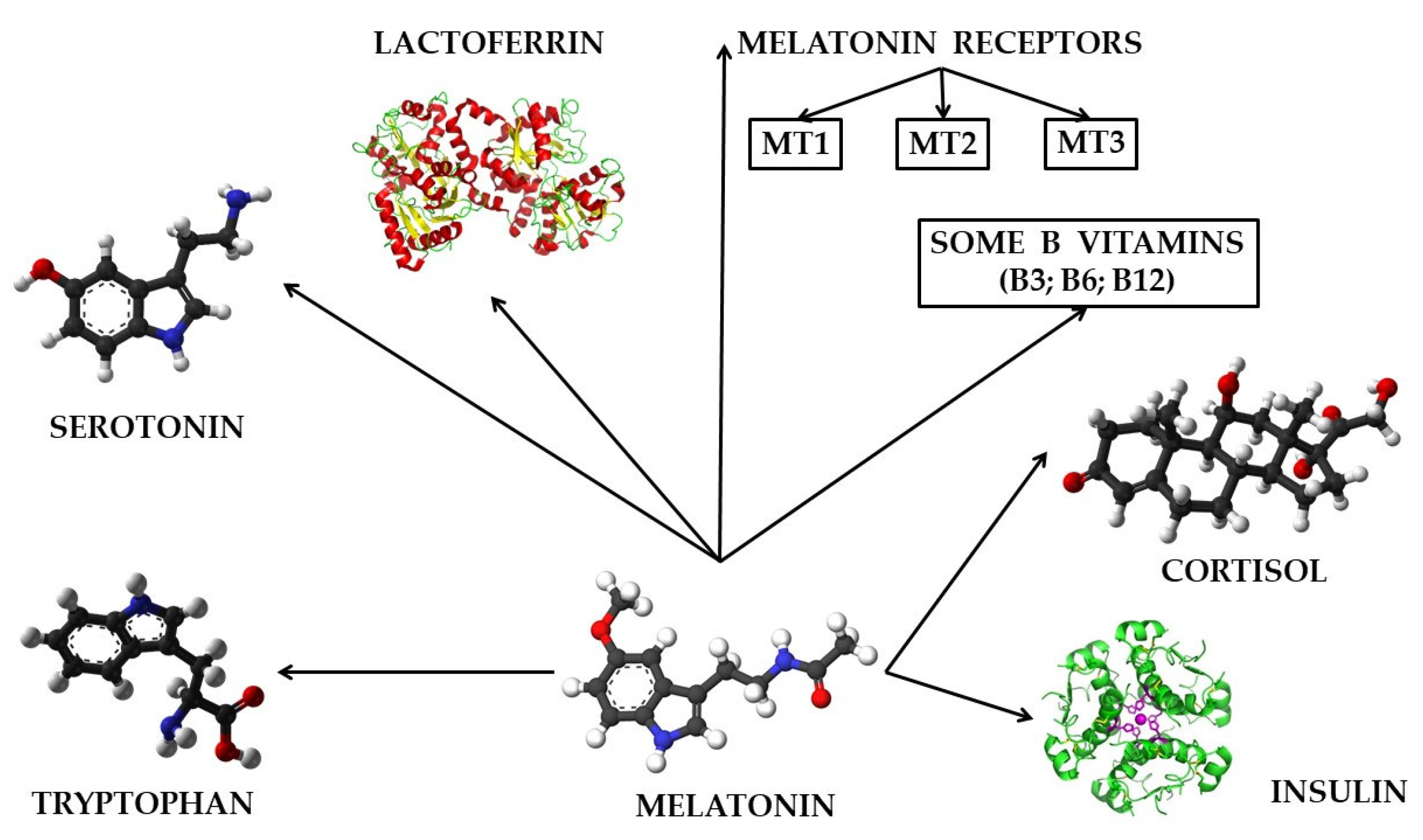
6. The Role of Melatonin in Relation to Other Bioactive Molecules on the Animal and Human Body

Numerous studies have demonstrated that melatonin as a hormone secreted by the pineal and extrapineal pathways, as well as melatonin originating from the body’s microflora or brought through the diet of each individual, interacts with a series of other molecules with a biologically active role, to ensure the achievement of various functions and roles in the animal and human body. Studies conducted in this field have reported that melatonin can interact in the animal and human body with serotonin [128], with melatonin receptors MT1, MT2, and MT3 [42,129,130], with lactoferrin [131,132,133,134,135,136,137], with vitamins B3, B6, and B12 [138,139], with the stress hormone cortisol [5,140], and with insulin [141,142,143,144,145], as shown in Figure 3.
Serotonin, also known as 5-hydroxytryptamine or 5-HT, acts as a neurotransmitter and a peripheral hormone. Serotonin synthesis is carried out in two steps from the essential amino acid tryptophan. In the first step, tryptophan hydroxylase (TPH) hydrolyzes tryptophan to produce 5-hydroxytryptophan, and in the second step of serotonin synthesis, decarboxylation of L-aromatic amino acids and conversion to 5-hydroxytryptamine are carried out [128]. Any variation in tryptophan and/or serotonin in the body will directly influence the melatonin content in the body and in the milk of mammals.
The MT1 (MTNR1A) and MT2 (MTNR1B) receptors are two membrane receptors for melatonin. These two receptors belong to the G protein-coupled receptor superfamily [119]. Activation of MT1 or MT2 receptors by melatonin leads to inhibition of PKA (kinase A) activity, since melatonin activation of these two receptors decreases the amount of cyclic adenosine monophosphate (cAMP) [42]. In terms of interaction with melatonin, the MT3 receptor shows a lower affinity for this hormone [130].
Lactoferrin (Lf) is a multifunctional glycoprotein, which belongs to the transferrin family and plays a role in iron binding in the animal and human body [131].
Milk proteins are of two types: casein (the main protein in milk, accounting for about 80% of total milk proteins) and serum proteins (accounting for about 20%), which are proteins that pass into whey and buttermilk after milk processing to obtain certain categories of dairy products [132].
Lactoferrin, along with alpha-lactalbumin, beta-lactoglobulin, immunoglobulins, bovine serum albumin, glycomacropeptides, lactoperoxidase, and lysozyme, are part of the serum protein category [133].
Numerous studies have highlighted the role of lactoferrin as an antioxidant, antibacterial, and antiviral factor [134], antimicrobial and anticancer [135], and antiparasitic and antifungal [133], as well as a role in maintaining intestinal health [136].
Lactoferrin performs an antibacterial role in the body (being also the first discovered function of this protein) through two different mechanisms. A first mechanism for achieving the antibacterial function of lactoferrin involves sequestering free iron, thus depriving bacteria of an essential substrate necessary for the growth and development of these microorganisms. The second mechanism for fulfilling the antibacterial role involves the binding of lactoferrin to the lipopolysaccharide that enters the structure of the bacterial cell walls, thus degrading the bacteria by forming peroxides catalyzed by iron (III) ions bound to lactoferrin. Thus, the permeability of the bacterial membrane is affected, resulting in bacterial cell lysis [137].
Bovine lactoferrin (bLF) has the ability to control the production of reactive oxygen species (ROS) and the rate of their elimination by sequestering iron [131].
Pyridoxine (vitamin B6) acts as a coenzyme in the synthesis of melatonin, and any deficiency in this vitamin will inevitably lead to sleep disorders [138,139], and therefore to various conditions with a negative impact, caused by the lack of melatonin in the body.
Some studies have reported that vitamin B3 (niacin) can have a tryptophan-sparing effect and that vitamin B12 (cobalamin) contributes directly to the secretion process of melatonin [139].
Cortisol is a glucocorticoid hormone that is produced by the adrenal glands, and the release of this hormone in the body follows a circadian rhythm regulated by the internal clock located in the suprachiasmatic nucleus [140]. Melatonin can regulate the secretion of certain hormones, in particular inhibiting the release of corticotropin (CRH) from the hypothalamus. By inhibiting CRH secretion by melatonin, it results in decreased levels of adrenocorticotropic hormone (ACTH) and cortisol during the night [5].
Insulin is a polypeptide hormone, composed of 51 amino acids, and secreted mainly by β cells located in the islets of Langerhans of the pancreas. The main role of this hormone in the body is to modulate blood glucose levels, also having a role in glucose homeostasis, metabolism and cell growth [141]. Some clinical studies have highlighted the role that melatonin MT1 and MT2 receptors have on the process of insulin secretion [142].
Research carried out in the field of knowledge of the interactions that can be achieved between melatonin and insulin has demonstrated that there is a direct and inversely proportional relationship between the amount of melatonin synthesized in the body, and the processes of inhibition or stimulation (as appropriate) of insulin synthesis and secretion. Thus, it has been demonstrated that a large amount of melatonin secreted in the body can inhibit insulin secretion, through melatonin receptors located on pancreatic β cells. Melatonin receptors are coupled to three different signaling pathways (cAMP, cGMP, and IP3) and have their own unique and different influences on insulin secretion [143,144,145]. Binding of melatonin to the MT1 receptor can lead to inhibition of insulin secretion by decreasing cAMP. This phenomenon occurs due to MT1 receptors binding to Gi (inhibitory G) proteins, which in turn leads to decreasing cAMP (cyclic adenosine monophosphate) and inhibiting PKA activity [145].
Melatonin may also influence the cGMP (cyclic guanosine monophosphate) signaling pathway by stimulating vasodilator signals via nitric oxide (NO). The cGMP signaling pathway may promote insulin secretion, as it may increase pancreatic blood flow by activating protein kinase (which is dependent on the cGMP signaling pathway). Thus, NO may activate guanylate cyclase, thereby increasing cGMP levels and stimulating insulin secretion. The IP3 (inositol triphosphate) pathway plays a role in regulating intracellular calcium concentration, and can be influenced by the presence of melatonin that binds to MT1 and MT2 receptors, receptors that can activate Gq-type G proteins. Thus, when the two melatonin receptors (MT1 and MT2) are activated, they allow the Gq protein to which they are bound to activate PLC (phospholipase C). Subsequently, PLC catalyzes the decomposition of PIP2 (phosphatidylinositol 4,5-bisphosphate) into two other secondary messengers (IP3 and diacylglycerol). Finally, the IP3 pathway stimulates the release of Ca2+ ions from the endoplasmic reticulum into the cytoplasm, thus favoring insulin secretion, due to the increase in intracellular calcium levels [145].
Therefore, melatonin can play a role in modulating insulin synthesis and secretion, through the MT1 and MT2 receptors to which melatonin binds. The presence of melatonin in the vertebrate body, and its interaction (of melatonin) with the MT1 and MT2 receptors can modulate insulin synthesis and secretion, through stimulation or inhibition, due to the three signaling pathways (cAMP, cGMP, and IP3) through which melatonin works.

7. Conclusions

The multiple roles and functions that melatonin performs in the animal and human body, both individually and in relation to other bioactive molecules, make this hormone a valuable compound that actively participates in improving living conditions by maintaining optimal health of the body.
The proper management of technological and nutritional factors, which can positively influence the melatonin content in cow’s milk, has major implications for the implementation of high-quality agricultural and zootechnical practices, due to a higher level of farm animal welfare. This level is achieved by regulating the circadian rhythm, improving sleep quality, and regulating basic physiological processes such as reproduction.
This paper contains valuable information regarding the synthesis and secretion processes of melatonin in vertebrate organisms, its multiple roles and functions in both animal and human organisms, and the main factors that can directly influence melatonin synthesis in the bovine organism, as well as the subsequent release of this hormone into the milk of cows. Specialized studies conducted in this field up until 2024 have provided valuable information, resulting in a deeper understanding of melatonin as a bioactive molecule. However, it is necessary to conduct more experiments focused on studying the impact of cow maintenance conditions on the melatonin content in milk, and some future research should also address the influence of milk processing technological processes on the melatonin content found in finished dairy products.

Author Contributions

Conceptualization, V.-C.A. and D.S.; methodology, C.S. and V.-C.A.; software, M.M. and R.-M.R.-R.; writing—original draft preparation, V.-C.A., C.S. and R.-M.R.-R.; writing—review and editing, V.-C.A., M.M. and R.-M.R.-R.; visualization, V.-C.A. and C.S.; supervision, D.S. All authors have read and agreed to the published version of the manuscript.

Funding

This research received no external funding.

Conflicts of Interest

The authors declare no conflicts of interest.

References

  1. Reiter, R.J.; Sharma, R.; Tan, D.-X.; Chuffa, L.G.d.A.; da Silva, D.G.H.; Slominski, A.T.; Steinbrink, K.; Kleszczynski, K. Dual sources of melatonin and evidence for different primary functions. Front. Endocrinol. 2024, 15, 1414463. [Google Scholar] [CrossRef]
  2. Chlubek, D.; Sikora, M. Fluoride and Pineal Gland. Appl. Sci. 2020, 10, 2885. [Google Scholar] [CrossRef]
  3. Tan, D.-X.; Zheng, X.; Kong, J.; Manchester, L.C.; Hardeland, R.; Kim, S.J.; Xu, X.; Reiter, R.J. Fundamental issues related to the origin of melatonin and melatonin isomers during evolution: Relation to their biological functions. Int. J. Mol. Sci. 2014, 15, 15858–15890. [Google Scholar] [CrossRef] [PubMed]
  4. Tan, D.-X.; Reiter, R.J.; Zimmerman, S.; Hardeland, R. Melatonin: Both a Messenger of Darkness and a Participant in the Cellular Actions of Non-Visible Solar Radiation of Near Infrared Light. Biology 2023, 12, 89. [Google Scholar] [CrossRef]
  5. Andrani, M.; Dall’Olio, E.; De Rensis, F.; Tummaruk, P.; Saleri, R. Bioactive Peptides in Dairy Milk: Highlighting the Role of Melatonin. Biomolecules 2024, 14, 934. [Google Scholar] [CrossRef]
  6. Lerner, A.B.; Case, J.D.; Takahashi, Y.; Lee, T.H.; Mori, W. Isolation of melatonin, the pineal gland factor that lightens melanocytes. J. Am. Chem. Soc. 1958, 80, 2587. [Google Scholar] [CrossRef]
  7. Mannino, G.; Pernici, C.; Serio, G.; Gentile, C.; Bertea, C.M. Melatonin and Phytomelatonin: Chemistry, Biosynthesis, Metabolism, Distribution and Bioactivity in Plants and Animals—An Overview. Int. J. Mol. Sci. 2021, 22, 9996. [Google Scholar] [CrossRef]
  8. Masters, A.; Pandi-Perumal, S.R.; Seixas, A.; Girardin, J.L.; McFarlane, S.I. Melatonin, the Hormone of Darkness: From Sleep Promotion to Ebola Treatment. Brain Disord. Ther. 2014, 4, 1000151. [Google Scholar] [CrossRef]
  9. Srinivasan, V.; Spence, W.D.; Pandi-Perumal, S.R.; Zakharia, R.; Bhatnagar, K.P.; Brzezinski, A. Melatonin and human reproduction: Shedding light on the darkness hormone. Gynecol. Endocrinol. 2009, 25, 779–785. [Google Scholar] [CrossRef]
  10. Patel, S.; Rahmani, B.; Gandhi, J.; Seyam, O.; Joshi, G.; Reid, I.; Smith, N.L.; Waltzer, W.C.; Khan, S.A. Revisiting the pineal gland: A review of calcification, masses, precocious puberty, and melatonin functions. Int. J. Neurosci. 2020, 130, 464–475. [Google Scholar] [CrossRef] [PubMed]
  11. Tan, D.-X.; Manchester, L.C.; Esteban-Zubero, E.; Zhou, Z.; Reiter, R.J. Melatonin as a Potent and Inducible Endogenous Antioxidant: Synthesis and Metabolism. Molecules 2015, 20, 18886–18906. [Google Scholar] [CrossRef]
  12. Manchester, L.C.; Coto-Montes, A.; Boga, J.A.; Andersen, L.P.H.; Zhou, Z.; Galano, A.; Vriend, J.; Tan, D.-X.; Reiter, R.J. Melatonin: An ancient molecule that makes oxygen metabolically tolerable. J. Pineal Res. 2015, 59, 403–419. [Google Scholar] [CrossRef] [PubMed]
  13. Horodincu, L.; Solcan, C. Influence of Different Light Spectra on Melatonin Synthesis by the Pineal Gland and Influence on the Immune System in Chickens. Animals 2023, 13, 2095. [Google Scholar] [CrossRef] [PubMed]
  14. Polakovičová, S.; Líška, J.; Varga, I.; Gálfiová, P. Morphology of the Human Pineal Gland Studied by Freeze-Fracturing in Scanning Electron Microscopy. Life 2024, 14, 1617. [Google Scholar] [CrossRef] [PubMed]
  15. Cassone, V.M. Avian circadian organization: A chorus of clocks. Front. Neuroendocrinol. 2014, 35, 76–88. [Google Scholar] [CrossRef] [PubMed]
  16. Esteban, M.Á.; Cuesta, A.; Chaves-Pozo, E.; Meseguer, J. Influence of Melatonin on the Immune System of Fish: A Review. Int. J. Mol. Sci. 2013, 14, 7979–7999. [Google Scholar] [CrossRef]
  17. Bisquert, R.; Planells-Cárcel, A.; Alonso-del-Real, J.; Muñiz-Calvo, S.; Guillamón, J.M. The Role of the PAA1 Gene on Melatonin Biosynthesis in Saccharomyces cerevisiae: A Search of New Arylalkylamine N-Acetyltransferases. Microorganisms 2023, 11, 1115. [Google Scholar] [CrossRef] [PubMed]
  18. Zisapel, N. New perspectives on the role of melatonin in human sleep, circadian rhythms and their regulation. Br. J. Pharmacol. 2018, 175, 3190–3199. [Google Scholar] [CrossRef] [PubMed]
  19. Li, Z.; Zhang, K.; Zhou, Y.; Zhao, J.; Wang, J.; Lu, W. Role of Melatonin in Bovine Reproductive Biotechnology (Review). Molecules 2023, 28, 4940. [Google Scholar] [CrossRef]
  20. Kopustinskiene, D.M.; Bernatoniene, J. Molecular Mechanisms of Melatonin-Mediated Cell Protection and Signaling in Health and Disease. Pharmaceutics 2021, 13, 129. [Google Scholar] [CrossRef] [PubMed]
  21. Mauriz, J.L.; Collado, P.S.; Veneroso, C.; Reiter, R.J.; Gonzalez-Gallego, J. A review of the molecular aspects of melatonin’s anti-inflammatory actions: Recent insights and new perspectives. J. Pineal Res. 2013, 54, 1–14. [Google Scholar] [CrossRef] [PubMed]
  22. Hardeland, R. Aging, Melatonin, and the Pro- and Anti-Inflammatory Networks. Int. J. Mol. Sci. 2019, 20, 1223. [Google Scholar] [CrossRef]
  23. Mendes, L.; Queiroz, M.; Sena, C.M. Melatonin and Vascular Function. Antioxidants 2024, 13, 747. [Google Scholar] [CrossRef] [PubMed]
  24. Asghari, M.H.; Moloudizargari, M.; Ghobadi, E.; Fallah, M.; Abdollahi, M. Melatonin as a Multifunctional Anti-Cancer Molecule: Implications in Gastric Cancer. Life Sci. 2017, 185, 38–45. [Google Scholar] [CrossRef]
  25. Di Bella, G.; Mascia, F.; Gualano, L.; Di Bella, L. Melatonin Anticancer Effects: Review. Int. J. Mol. Sci. 2013, 14, 2410–2430. [Google Scholar] [CrossRef]
  26. Fernández, A.; Ordóñez, R.; Reiter, R.J.; González-Gallego, J.; Mauriz, J.L. Melatonin and endoplasmic reticulum stress: Relation to autophagy and apoptosis. J. Pineal Res. 2015, 59, 292–307. [Google Scholar] [CrossRef]
  27. Zhi, S.M.; Fang, G.X.; Xie, X.M.; Liu, L.H.; Yan, J.; Liu, D.B.; Yu, H.Y. Melatonin reduces OGD/R-induced neuron injury by regulating redox/inflammation/apoptosis signaling. Eur. Rev. Med. Pharmacol. Sci. 2020, 24, 1524–1536. [Google Scholar] [CrossRef] [PubMed]
  28. Lee, J.H.; Yoon, Y.M.; Song, K.-H.; Noh, H.; Lee, S.H. Melatonin suppresses senescence-derived mitochondrial dysfunction in mesenchymal stem cells via the HSPA1L-mitophagy pathway. Aging Cell 2020, 19, e13111. [Google Scholar] [CrossRef]
  29. Meng, X.; Li, Y.; Li, S.; Zhou, Y.; Gan, R.-Y.; Xu, D.-P.; Li, H.-B. Dietary Sources and Bioactivities of Melatonin. Nutrients 2017, 9, 367. [Google Scholar] [CrossRef]
  30. Guaadaoui, A.; Bellaoui, M.; Elmajdoub, N.; Bellaoui, M.; Hamal, A. What is a bioactive compound? A combined definition for a preliminary consensus. Int. J. Nutr. Food Sci. 2014, 3, 174–179. [Google Scholar] [CrossRef]
  31. Vilas-Boas, A.A.; Pintado, M.; Oliveira, A.L.S. Natural Bioactive Compounds from Food Waste: Toxicity and Safety Concerns. Foods 2021, 10, 1564. [Google Scholar] [CrossRef]
  32. Sorrenti, V.; Burò, I.; Consoli, V.; Vanella, L. Recent Advances in Health Benefits of Bioactive Compounds from Food Wastes and By-Products: Biochemical Aspects. Int. J. Mol. Sci. 2023, 24, 2019. [Google Scholar] [CrossRef] [PubMed]
  33. Acuña-Castroviejo, D.; Escames, G.; Venegas, C.; Díaz-Casado, M.E.; Lima-Cabello, E.; López, L.C.; Rosales-Corral, S.; Tan, D.X.; Reiter, R.J. Extrapineal melatonin: Sources, regulation, and potential functions. Cell Mol. Life Sci. 2014, 71, 2997–3025. [Google Scholar] [CrossRef]
  34. Markus, R.P.; Sousa, K.S.; da Silveira Cruz-Machado, S.; Fernandes, P.A.; Ferreira, Z.S. Possible Role of Pineal and Extra-Pineal Melatonin in Surveillance, Immunity, and First-Line Defense. Int. J. Mol. Sci. 2021, 22, 12143. [Google Scholar] [CrossRef] [PubMed]
  35. Bonnefont-Rousselot, D.; Collin, F. Melatonin: Action as antioxidant and potential applications in human disease and aging. Toxicology 2010, 278, 55–67. [Google Scholar] [CrossRef] [PubMed]
  36. Lumsden, S.C.; Clarkson, A.N.; Cakmak, Y.O. Neuromodulation of the Pineal Gland via Electrical Stimulation of Its Sympathetic Innervation Pathway. Front. Neurosci. 2020, 14, 264. [Google Scholar] [CrossRef]
  37. Lee, K.; Back, K. Functional Characterization of the Ciliate Stylonychia lemnae Serotonin N-Acetyltransferase, a Pivotal Enzyme in Melatonin Biosynthesis and Its Overexpression Leads to Peroxidizing Herbicide Tolerance in Rice. Antioxidants 2024, 13, 1177. [Google Scholar] [CrossRef] [PubMed]
  38. Xie, X.; Ding, D.; Bai, D.; Zhu, Y.; Sun, W.; Sun, Y.; Zhang, D. Melatonin biosynthesis pathways in nature and its production in engineered microorganisms. Synth. Syst. Biotechnol. 2022, 7, 544–553. [Google Scholar] [CrossRef] [PubMed]
  39. Hwang, O.J.; Back, K. Functional Characterization of Arylalkylamine N-Acetyltransferase, a Pivotal Gene in Antioxidant Melatonin Biosynthesis from Chlamydomonas reinhardtii. Antioxidants 2022, 11, 1531. [Google Scholar] [CrossRef] [PubMed]
  40. Zhao, D.; Yu, Y.; Shen, Y.; Liu, Q.; Zhao, Z.; Sharma, R.; Reiter, R.J. Melatonin Synthesis and Function: Evolutionary History in Animals and Plants. Front. Endocrinol. 2019, 10, 249. [Google Scholar] [CrossRef] [PubMed]
  41. Suofu, Y.; Li, W.; Jean-Alphonse, F.G.; Jia, J.; Khattar, N.K.; Li, J.; Baranov, S.V.; Leronni, D.; Mihalik, A.C.; He, Y.; et al. Dual role of mitochondria in producing melatonin and driving GPCR signaling to block cytochrome c release. Proc. Natl. Acad. Sci. USA 2017, 114, E7997–E8006. [Google Scholar] [CrossRef] [PubMed]
  42. Wang, L.; Wang, C.; Choi, W.S. Use of Melatonin in Cancer Treatment: Where Are We? Int. J. Mol. Sci. 2022, 23, 3779. [Google Scholar] [CrossRef] [PubMed]
  43. Tordjman, S.; Chokron, S.; Delorme, R.; Charrier, A.; Bellissant, E.; Jaafari, N.; Fougerou, C. Melatonin: Pharmacology, Functions and Therapeutic Benefits. Curr. Neuropharmacol. 2017, 15, 434–443. [Google Scholar] [CrossRef] [PubMed]
  44. Miranda-Riestra, A.; Estrada-Reyes, R.; Torres-Sanchez, E.D.; Carreño-García, S.; Ortiz, G.G.; Benítez-King, G. Melatonin: A Neurotrophic Factor? Molecules 2022, 27, 7742. [Google Scholar] [CrossRef]
  45. Slominski, A.T.; Zmijewski, M.A.; Semak, I.; Kim, T.K.; Janjetovic, Z.; Slominski, R.M.; Zmijewski, J.W. Melatonin, mitochondria, and the skin. Cell. Mol. Life Sci. 2017, 74, 3913–3925. [Google Scholar] [CrossRef] [PubMed]
  46. Slominski, A.; Tobin, D.J.; Zmijewski, M.A.; Wortsman, J.; Paus, R. Melatonin in the skin: Synthesis, metabolism and functions. Trends Endocrinol. Metab. TEM 2008, 19, 17–24. [Google Scholar] [CrossRef]
  47. Felder-Schmittbuhl, M.P.; Hicks, D.; Ribelayga, C.P.; Tosini, G. Melatonin in the mammalian retina: Synthesis, mechanisms of action and neuroprotection. J. Pineal Res. 2024, 76, e12951. [Google Scholar] [CrossRef] [PubMed]
  48. Iuvone, P.M.; Tosini, G.; Pozdeyev, N.; Haque, R.; Klein, D.C.; Chaurasia, S.S. Circadian clocks, clock networks, arylalkylamine N-acetyltransferase, and melatonin in the retina. Prog. Retin. Eye Res. 2005, 24, 433–456. [Google Scholar] [CrossRef] [PubMed]
  49. Sanchez-Hidalgo, M.; de la Lastra, C.A.; Carrascosa-Salmoral, M.P.; Naranjo, M.C.; Gomez-Corvera, A.; Caballero, B.; Guerrero, J.M. Age-related changes in melatonin synthesis in rat extrapineal tissues. Exp. Gerontol. 2009, 44, 328–334. [Google Scholar] [CrossRef] [PubMed]
  50. Bocheva, G.; Bakalov, D.; Iliev, P.; Tafradjiiska-Hadjiolova, R. The Vital Role of Melatonin and Its Metabolites in the Neuroprotection and Retardation of Brain Aging. Int. J. Mol. Sci. 2024, 25, 5122. [Google Scholar] [CrossRef] [PubMed]
  51. Alkozi, H.A.; Pintor, J. TRPV4 activation triggers the release of melatonin from human non-pigmented ciliary epithelial cells. Exp. Eye Res. 2015, 136, 34–37. [Google Scholar] [CrossRef] [PubMed]
  52. Naranjo, M.C.; Guerrero, J.M.; Rubio, A.; Lardone, P.J.; Carrillo-Vico, A.; Carrascosa-Salmoral, M.P.; Jiménez-Jorge, S.; Arellano, M.V.; Leal-Noval, S.R.; Leal, M.; et al. Melatonin biosynthesis in the thymus of humans and rats. Cell Mol. Life Sci. 2007, 64, 781–790. [Google Scholar] [CrossRef] [PubMed]
  53. Bonmatí-Carrión, M.-Á.; Tomas-Loba, A. Melatonin and Cancer: A Polyhedral Network Where the Source Matters. Antioxidants 2021, 10, 210. [Google Scholar] [CrossRef] [PubMed]
  54. Reiter, R.J.; Rosales-Corral, S.; Boga, J.A.; Tan, D.-X.; Davis, J.M.; Konturek, P.C.; Konturek, S.J.; Brzozowski, T. The photoperiod, circadian regulation and chronodisruption: The requisite interplay between the suprachiasmatic nuclei and the pineal and gut melatonin. J. Physiol. Pharmacol. 2011, 62, 269–274. [Google Scholar]
  55. Bonmatí-Carrión, M.-Á.; Rol, M.-A. Melatonin as a Mediator of the Gut Microbiota–Host Interaction: Implications for Health and Disease. Antioxidants 2024, 13, 34. [Google Scholar] [CrossRef] [PubMed]
  56. Holtkamp, C.E.; Warmus, D.; Bonowicz, K.; Gagat, M.; Linowiecka, K.; Wolnicka-Glubisz, A.; Reiter, R.J.; Böhm, M.; Slominski, A.T.; Steinbrink, K.; et al. Ultraviolet Radiation-Induced Mitochondrial Disturbances Are Attenuated by Metabolites of Melatonin in Human Epidermal Keratinocytes. Metabolites 2023, 13, 861. [Google Scholar] [CrossRef]
  57. Bocheva, G.; Slominski, R.M.; Janjetovic, Z.; Kim, T.-K.; Böhm, M.; Steinbrink, K.; Reiter, R.J.; Kleszczyński, K.; Slominski, A.T. Protective Role of Melatonin and Its Metabolites in Skin Aging. Int. J. Mol. Sci. 2022, 23, 1238. [Google Scholar] [CrossRef] [PubMed]
  58. Galano, A.; Reiter, R.J. Melatonin and Its Metabolites vs Oxidative Stress: From Individual Actions to Collective Protection. J. Pineal Res. 2018, 65, e12514. [Google Scholar] [CrossRef]
  59. Bae, S.M.; Jeong, J.; Jeon, H.J.; Bang, Y.R.; Yoon, I.Y. Effects of melatonin-rich milk on mild insomnia symptoms. Sleep. Med. Res. 2016, 7, 60–67. [Google Scholar] [CrossRef]
  60. Brzezinski, A.; Rai, S.; Purohit, A.; Pandi-Perumal, S.R. Melatonin, Clock Genes, and Mammalian Reproduction: What Is the Link? Int. J. Mol. Sci. 2021, 22, 13240. [Google Scholar] [CrossRef] [PubMed]
  61. Li, Q.; Tang, Y.; Chen, Y.; Li, B.; Wang, H.; Liu, S.; Adeniran, S.O.; Zheng, P. Melatonin Regulates the Expression of VEGF and HOXA10 in Bovine Endometrial Epithelial Cells through the SIRT1/PI3K/AKT Pathway. Animals 2024, 14, 2771. [Google Scholar] [CrossRef] [PubMed]
  62. Cipolla-Neto, J.; Amaral, F.G.; Afeche, S.C.; Tan, D.X.; Reiter, R.J. Melatonin, energy metabolism, and obesity: A review. J. Pineal Res. 2014, 56, 371–381. [Google Scholar] [CrossRef] [PubMed]
  63. Suzen, S.; Atayik, M.C.; Sirinzade, H.; Entezari, B.; Gurer-Orhan, H.; Cakatay, U. Melatonin and redox homeostasis. Melatonin Res. 2022, 5, 304–324. [Google Scholar] [CrossRef]
  64. Rzepka-Migut, B.; Paprocka, J. Melatonin-Measurement Methods and the Factors Modifying the Results. A Systematic Review of the Literature. Int. J. Environ. Res. Public Health 2020, 17, 1916. [Google Scholar] [CrossRef] [PubMed]
  65. Reiter, R.J.; Tan, D.X.; Kim, S.J.; Cruz, M.H.C. Delivery of pineal melatonin to the brain and SCN: Role of canaliculi, cerebrospinal fluid, tanycytes and Virchow–Robin perivascular spaces. Brain Struct. Funct. 2014, 219, 1873–1887. [Google Scholar] [CrossRef] [PubMed]
  66. Mao, L.; Dauchy, R.T.; Blask, D.E.; Slakey, L.M.; Xiang, S.; Yuan, L.; Dauchy, E.M.; Shan, B.; Brainard, G.C.; Hanifin, J.P.; et al. Circadian gating of epithelial-to-mesenchymal transition in breast cancer cells via melatonin-regulation of GSK3β. Mol. Endocrinol. 2012, 26, 1808–1820. [Google Scholar] [CrossRef] [PubMed]
  67. Gao, Y.; Zhao, S.; Zhang, Y.; Zhang, Q. Melatonin Receptors: A Key Mediator in Animal Reproduction. Vet. Sci. 2022, 9, 309. [Google Scholar] [CrossRef] [PubMed]
  68. Li, D.Y.; Smith, D.G.; Hardeland, R.; Yang, M.Y.; Xu, H.L.; Zhang, L.; Yin, H.D.; Zhu, Q. Melatonin Receptor Genes in Vertebrates. Int. J. Mol. Sci. 2013, 14, 11208–11223. [Google Scholar] [CrossRef]
  69. Peña-Delgado, V.; Carvajal-Serna, M.; Fondevila, M.; Martín-Cabrejas, M.A.; Aguilera, Y.; Álvarez-Rivera, G.; Abecia, J.A.; Casao, A.; Pérez-Pe, R. Improvement of the Seminal Characteristics in Rams Using Agri-Food By-Products Rich in Phytomelatonin. Animals 2023, 13, 905. [Google Scholar] [CrossRef] [PubMed]
  70. Pereira, N.; Naufel, M.F.; Ribeiro, E.B.; Tufik, S.; Hachul, H. Influence of Dietary Sources of Melatonin on Sleep Quality: A Review. J. Food Sci. 2020, 85, 5–13. [Google Scholar] [CrossRef]
  71. Karolczak, K.; Watala, C. Melatonin as a Reducer of Neuro- and Vasculotoxic Oxidative Stress Induced by Homocysteine. Antioxidants 2021, 10, 1178. [Google Scholar] [CrossRef] [PubMed]
  72. Reiter, R.J.; Tan, D.X.; Korkmaz, A.; Rosales-Corral, S.A. Melatonin and stable circadian rhythms optimize maternal, placental and fetal physiology. Hum. Reprod. Update 2014, 20, 293–307. [Google Scholar] [CrossRef]
  73. Ma, N.; Zhang, J.; Reiter, R.J.; Ma, X. Melatonin mediates mucosal immune cells, microbial metabolism, and rhythm crosstalk: A therapeutic target to reduce intestinal inflammation. Med. Res. Rev. 2020, 40, 606–632. [Google Scholar] [CrossRef] [PubMed]
  74. Gombert, M.; Codoñer-Franch, P. Melatonin in Early Nutrition: Long-Term Effects on Cardiovascular System. Int. J. Mol. Sci. 2021, 22, 6809. [Google Scholar] [CrossRef] [PubMed]
  75. Tamtaji, O.R.; Mirhosseini, N.; Reiter, R.J.; Azami, A.; Asemi, Z. Melatonin, a calpain inhibitor in the central nervous system: Current status and future perspectives. J. Cell Physiol. 2018, 234, 1001–1007. [Google Scholar] [CrossRef] [PubMed]
  76. Tan, D.-X.; Xu, B.; Zhou, X.; Reiter, R.J. Pineal Calcification, Melatonin Production, Aging, Associated Health Consequences and Rejuvenation of the Pineal Gland. Molecules 2018, 23, 301. [Google Scholar] [CrossRef]
  77. Carrillo-Vico, A.; Lardone, P.J.; Alvarez-Sánchez, N.; Rodríguez-Rodríguez, A.; Guerrero, J.M. Melatonin: Buffering the immune system. Int. J. Mol. Sci. 2013, 14, 8638–8683. [Google Scholar] [CrossRef] [PubMed]
  78. Vorster, A.P.A.; van Someren, E.J.W.; Pack, A.I.; Huber, R.; Schmidt, M.H.; Bassetti, C.L.A. Sleep Health. Clin. Transl. Neurosci. 2024, 8, 8. [Google Scholar] [CrossRef]
  79. Chattu, V.K.; Manzar, M.D.; Kumary, S.; Burman, D.; Spence, D.W.; Pandi-Perumal, S.R. The Global Problem of Insufficient Sleep and Its Serious Public Health Implications. Healthcare 2019, 7, E1. [Google Scholar] [CrossRef] [PubMed]
  80. Sejbuk, M.; Mirończuk-Chodakowska, I.; Witkowska, A.M. Sleep Quality: A Narrative Review on Nutrition, Stimulants, and Physical Activity as Important Factors. Nutrients 2022, 14, 1912. [Google Scholar] [CrossRef] [PubMed]
  81. Paditz, E. Postnatal Development of the Circadian Rhythmicity of Human Pineal Melatonin Synthesis and Secretion (Systematic Review). Children 2024, 11, 1197. [Google Scholar] [CrossRef]
  82. Paditz, E. Melatonin in infants-Physiology, pathophysiology and intervention options. Somnologie 2024, 28, 103–109. [Google Scholar] [CrossRef]
  83. Honorio-França, A.C.; Hara, C.C.P.; Ormonde, J.V.S.; Nunes, G.T.; França, E.L. Human colostrum melatonin exhibits a day-night variation and modulates the activity of colostral phagocytes. J. Appl. Biomed. 2013, 11, 153–162. [Google Scholar] [CrossRef]
  84. Italianer, M.F.; Naninck, E.F.G.; Roelants, J.A.; van der Horst, G.T.J.; Reiss, I.K.M.; Goudoever, J.B.v.; Joosten, K.F.M.; Chaves, I.; Vermeulen, M.J. Circadian Variation in Human Milk Composition, a Systematic Review. Nutrients 2020, 12, 2328. [Google Scholar] [CrossRef]
  85. Wang, X. The antiapoptotic activity of melatonin in neurodegenerative diseases. CNS Neurosci. Ther. 2009, 15, 345–357. [Google Scholar] [CrossRef] [PubMed]
  86. Biggio, G.; Biggio, F.; Talani, G.; Mostallino, M.C.; Aguglia, A.; Aguglia, E.; Palagini, L. Melatonin: From Neurobiology to Treatment. Brain Sci. 2021, 11, 1121. [Google Scholar] [CrossRef]
  87. Logan, R.W.; McClung, C.A. Rhythms of life: Circadian disruption and brain disorders across the lifespan. Nat. Rev. Neurosci. 2019, 20, 49–65. [Google Scholar] [CrossRef] [PubMed]
  88. Mander, B.A.; Winer, J.R.; Walker, M.P. Sleep and Human Aging. Neuron 2017, 94, 19–36. [Google Scholar] [CrossRef]
  89. Milagres, M.P.; Minim, V.P.R.; A Minim, L.; A Simiqueli, A.; Moraes, L.E.S.; Martino, H.S.D. Night milking adds value to cow’s milk. J. Sci. Food Agric. 2013, 94, 1688–1692. [Google Scholar] [CrossRef] [PubMed]
  90. Usturoi, M.G. The Technology of Milk and Dairy Products; Alfa Publishing House: Iasi, Romania, 2007. [Google Scholar]
  91. Rațu, R.N.; Cârlescu, P.M.; Usturoi, M.G.; Lipșa, F.D.; Veleșcu, I.D.; Arsenoaia, V.N.; Florea, A.M.; Ciobanu, M.M.; Radu-Rusu, R.-M.; Postolache, A.N.; et al. Effects of Dairy Cows Management Systems on the Physicochemical and Nutritional Quality of Milk and Yogurt, in a North-Eastern Romanian Farm. Agriculture 2023, 13, 1295. [Google Scholar] [CrossRef]
  92. Matei, M.; Zaharia, R.; Petrescu, S.-I.; Radu-Rusu, C.G.; Simeanu, D.; Mierliță, D.; Pop, I.M. Persistent Organic Pollutants (POPs): A Review Focused on Occurrence and Incidence in Animal Feed and Cow Milk. Agriculture 2023, 13, 873. [Google Scholar] [CrossRef]
  93. Robinson, R.C. Structures and metabolic properties of bovine milk oligosaccharides and their potential in the development of novel therapeutics. Front. Nutr. 2019, 6, 50. [Google Scholar] [CrossRef]
  94. Yuzbashian, E.; Berg, E.; de Campos Zani, S.C.; Chan, C.B. Cow’s Milk Bioactive Molecules in the Regulation of Glucose Homeostasis in Human and Animal Studies. Foods 2024, 13, 2837. [Google Scholar] [CrossRef]
  95. Usturoi, A.; Usturoi, M.-G.; Avarvarei, B.-V.; Pânzaru, C.; Simeanu, C.; Usturoi, M.-I.; Spătaru, M.; Radu-Rusu, R.-M.; Doliş, M.-G.; Simeanu, D. Research Regarding Correlation between the Assured Health State for Laying Hens and Their Productivity. Agriculture 2023, 13, 86. [Google Scholar] [CrossRef]
  96. Helmreich, S.; Wechsler, B.; Hauser, R.; Gygax, L. Effects of milking frequency in automatic milking systems on salivary cortisol, immunoglobulin A, somatic cell count and melatonin. Schweiz Arch Tierheilkd 2016, 158, 179–186. [Google Scholar] [CrossRef] [PubMed]
  97. Muthuramalingam, P.; Kennedy, A.D.; Berry, R.J. Plasma melatonin and insulin-like growth factor-1 responses to dim light at night in dairy heifers. J. Pineal Res. 2006, 40, 225–229. [Google Scholar] [CrossRef] [PubMed]
  98. Bal, M.A.; Penner, G.B.; Oba, M.; Kennedy, A.D. Effects of dim light at night on milk yield, milk composition and endocrine profile of lactating dairy cows. Can. J. Anim. Sci. 2008, 88, 609–612. [Google Scholar] [CrossRef]
  99. Boztepe, S.; Keskin, I.; Semacan, A.; Akyürek, F.; Aytekin, I.; Sahin, Ö. Melatonin Differences Between Day and Night Milk in Primiparous Holstein Friesian and Jersey Dairy Cattle. Selcuk. J. Agric. Food Sci. 2022, 36, 27–30. [Google Scholar] [CrossRef]
  100. Keskin, M.; Gül, S.; Karaaslan, İ.; Yakan, A. Controlling the photoperiod to raise the melatonin content of sheep milk. Photoperiod control and milk melatonin content. J. Hell. Vet. Med. Soc. 2023, 74, 6641–6648. [Google Scholar] [CrossRef]
  101. Şahin, Ö.; Akyürek, F.; Boztepe, S.; Aytekin, İ.; Keskin, İ. Determination of Melatonin Differences between Day and Night Milk in Dairy Cattle. J. Agric. Sci. Bilim. Derg. 2021, 27, 449–453. [Google Scholar] [CrossRef]
  102. Romanini, E.B.; Marchi Volpato, A.; Dos Santos, J.S.; De Santana, E.H.W.; De Souza, C.H.B.; Ludovico, A. Melatonin concentration in cow’s milk and sources of its variation. J. Appl. Anim. Res. 2019, 47, 140–145. [Google Scholar] [CrossRef]
  103. Asher, A.; Shabtay, A.; Brosh, A.; Eitam, H.; Agmon, R.; Cohen-Zinder, M.; Zubidat, A.E.; Haim, A. “Chrono-functional milk”: The difference between melatonin concentrations in night-milk versus day-milk under different night illumination conditions. Chronobiol. Int. 2015, 32, 1409–1416. [Google Scholar] [CrossRef] [PubMed]
  104. Teng, Z.W.; Yang, G.Q.; Wang, I.F.; Fu, T.; Lian, H.X.; Sun, Y.; Han, L.Q.; Zhang, L.Y.; Gao, T.Y. Effects of the Circadian Rhythm on Milk Composition in Dairy Cows: Does Day Milk Differ from Night Milk? J. Dairy Sci. 2021, 104, 8301–8313. [Google Scholar] [CrossRef]
  105. Jo, J.-H.; Jalil, G.N.; Kim, W.-S.; Moon, J.-O.; Lee, S.-D.; Kwon, C.-H.; Lee, H.-G. Effects of Rumen-Protected L-Tryptophan Supplementation on Productivity, Physiological Indicators, Blood Profiles, and Heat Shock Protein Gene Expression in Lactating Holstein Cows under Heat Stress Conditions. Int. J. Mol. Sci. 2024, 25, 1217. [Google Scholar] [CrossRef]
  106. Stelwagen, K.; Phyn, C.V.; Davis, S.R.; Guinard-Flament, J.; Pomiès, D.; Roche, J.R.; Kay, J.K. Invited review: Reduced milking frequency: Milk production and management implications. J. Dairy. Sci. 2013, 96, 3401–3413. [Google Scholar] [CrossRef] [PubMed]
  107. Wall, E.H.; Bond, J.P.; McFadden, T.B. Milk yield responses to changes in milking frequency during early lactation are associated with coordinated and persistent changes in mammary gene expression. BMC Genom. 2013, 14, 296. [Google Scholar] [CrossRef]
  108. Asher, A.; Fialko, M.; Fares, F.; Moallem, U.; Yaacoby, S.; Gutman, R. The Effect of Short-Wavelength White LED Illumination throughout the Night on the Milk Fatty Acid Profile of High-Yielding Dairy Cows. Biology 2022, 11, 1799. [Google Scholar] [CrossRef] [PubMed]
  109. Murphy, B.A.; Herlihy, M.M.; Nolan, M.B.; O’Brien, C.; Furlong, J.G.; Butler, S.T. Identification of the blue light intensity administered to one eye required to suppress bovine plasma melatonin and investigation into effects on milk production in grazing dairy cows. J. Dairy. Sci. 2021, 104, 12127–12138. [Google Scholar] [CrossRef]
  110. Adamczyk, K.; Herbut, P.; Godyń, D.; Angrecka, S.; Kupczyński, R.; Corrêa Vieira, F.M. Effect of light on dairy cattle in farm conditions—A review. Ann. Anim. Sci. 2024, 24, 1139–1151. [Google Scholar] [CrossRef]
  111. Elsabagh, M.; Mon, M.; Takao, Y.; Shinoda, A.; Watanabe, T.; Kushibiki, S.; Obitsu, T.; Sugino, T. Exposure to blue LED light before the onset of darkness under a long-day photoperiod alters melatonin secretion, feeding behaviour and growth in female dairy calves. Jpn. Soc. Anim. Sci. 2020, 91, e13353. [Google Scholar] [CrossRef] [PubMed]
  112. Simeanu, D. Nutrition and Feeding of Animals; “Ion Ionescu de la Brad” Publishing House: Iasi, Romania, 2018. [Google Scholar]
  113. Back, K.; Tan, D.X.; Reiter, R.J. Melatonin biosynthesis in plants: Multiple pathways catalyzetryptophan to melatonin in the cytoplasm or chloroplasts (Review article). J. Pineal Res. 2016, 61, 426–437. [Google Scholar] [CrossRef] [PubMed]
  114. Fan, J.; Xie, Y.; Zhang, Z.; Chen, L. Melatonin: A Multifunctional Factor in Plants. Int. J. Mol. Sci. 2018, 19, 1528. [Google Scholar] [CrossRef]
  115. Arnao, M.B.; Cano, A.; Hernández-Ruiz, J. Phytomelatonin: An unexpected molecule with amazing performances in plants. J. Exp. Bot. 2022, 73, 5779–5800. [Google Scholar] [CrossRef] [PubMed]
  116. Niu, T.; Ding, Z.; Zeng, J.; Yan, Z.; Duan, H.; Lv, J.; Zhang, Y.; Zhang, L.; Hu, J. Melatonin Sources in Sheep Rumen and Its Role in Reproductive Physiology. Animals 2024, 14, 3451. [Google Scholar] [CrossRef]
  117. Holzmann, V.M.M.; Trentin, M.; De Almeida Rego, F.C.; Coelho Cunha Filho, L.F.; Ludovico, A. Melatonin concentration in the milk of cows supplemented with vitamins and milked twice daily. Semin. Agrar. 2019, 40, 2017–2026. [Google Scholar] [CrossRef]
  118. Hernandez-Ruiz, J.; Cano, A.; Arnao, M.B. Melatonin acts as a growth-stimulating compound in some monocot species. J. Pineal Res. 2005, 39, 137–142. [Google Scholar] [CrossRef] [PubMed]
  119. Barik, S. The Uniqueness of Tryptophan in Biology: Properties, Metabolism, Interactions and Localization in Proteins. Int. J. Mol. Sci. 2020, 21, 8776. [Google Scholar] [CrossRef] [PubMed]
  120. Kim, I.-S.; Kim, C.-H.; Yang, W.-S. Physiologically Active Molecules and Functional Properties of Soybeans in Human Health-A Current Perspective. Int. J. Mol. Sci. 2021, 22, 4054. [Google Scholar] [CrossRef] [PubMed]
  121. Luo, C.; Wang, D.; Lu, N.; Li, H.; Liu, G.; Cao, Z.; Yang, H.; Li, S.; Yu, X.; Shao, W.; et al. Analysis of Chemical Composition, Amino Acid Content, and Rumen Degradation Characteristics of Six Organic Feeds. Animals 2022, 12, 682. [Google Scholar] [CrossRef] [PubMed]
  122. Kim, Y.-L.; Lee, S.-H.; Son, G.-H.; Shin, J.-S.; Kim, M.-J.; Park, B.-K. Effect of Rumen-Protected L-Tryptophan or L-Ascorbic Acid on Plasma Metabolites and Milk Production Characteristics of Lactating Holstein Cows during Summer Conditions. Animals 2024, 14, 1820. [Google Scholar] [CrossRef] [PubMed]
  123. Liu, X.; Yao, S.; Liu, Y.; Han, H.; Wang, W.; Yi, Q.; Yan, L.; Ji, P.; Zhang, L.; Liu, G. Effects of Prepartum L-Tryptophan Supplementation on the Postpartum Performance of Holstein Cows. Animals 2024, 14, 1278. [Google Scholar] [CrossRef]
  124. Kollmann, M.T.; Locher, M.; Hirche, F.; Eder, K.; Meyer, H.H.; Bruckmaier, R.M. Effects of tryptophan supplementation on plasma tryptophan and related hormone levels in heifers and dairy cows. Domest. Anim. Endocrinol. 2008, 34, 14–24. [Google Scholar] [CrossRef] [PubMed]
  125. Karunanithi, D.; Radhakrishna, A.; Sivaraman, K.P.; Biju, V.M. Quantitative determination of melatonin in milk by LC-MS/MS. J. Food Sci. Technol. 2014, 51, 805–812. [Google Scholar] [CrossRef] [PubMed]
  126. Kocadağı, T.; Yılmaz, C.; Gökmen, V. Determination of melatonin and its isomer in foods by liquid chromatography tandem mass spectrometry. Food Chem. 2014, 153, 151–156. [Google Scholar] [CrossRef] [PubMed]
  127. Sturtz, M.; Cerezo, A.B.; Cantos-Villar, E.; Garcia-Parrilla, M.C. Determination of the melatonin content of different varieties of tomatoes (Lycopersicon esculentum) and strawberries (Fragaria ananassa). Food Chem. 2011, 127, 1329–1334. [Google Scholar] [CrossRef] [PubMed]
  128. Kanova, M.; Kohout, P. Serotonin—Its Synthesis and Roles in the Healthy and the Critically Ill. Int. J. Mol. Sci. 2021, 22, 4837. [Google Scholar] [CrossRef]
  129. Dubocovich, M.L. Melatonin receptors: Role on sleep and circadian rhythm regulation. Sleep. Med. 2007, 8 (Suppl. S3), 34–42. [Google Scholar] [CrossRef] [PubMed]
  130. Tan, D.-X.; Manchester, L.C.; Terron, M.P.; Flores, L.J.; Tamura, H.; Reiter, R.J. Melatonin as a naturally occurring co-substrate of quinone reductase-2, the putative MT3melatonin membrane receptor: Hypothesis and significance. J. Pineal Res. 2007, 43, 317–320. [Google Scholar] [CrossRef]
  131. Superti, F. Lactoferrin from Bovine Milk: A Protective Companion for Life. Nutrients 2020, 12, 2562. [Google Scholar] [CrossRef]
  132. Flis, Z.; Molik, E. Importance of Bioactive Substances in Sheep’s Milk in Human Health. Int. J. Mol. Sci. 2021, 22, 4364. [Google Scholar] [CrossRef] [PubMed]
  133. Niaz, B.; Saeed, F.; Ahmad, A.; Imran, M.; Maan, A.; Khan, M.; Tufail, T.; Anjum, F.; Hussain, S.; Suleria, H. Lactoferrin (LF): A Natural Antimicrobial Protein. Int. J. Food Prop. 2019, 22, 1626–1641. [Google Scholar] [CrossRef]
  134. Redwan, E.; Uversky, V.; El-Fakharany, E.; Al-Mehdar, H. Potential lactoferrin activity against pathogenic viruses. Comptes Rendus Biol. 2015, 337, 581–595. [Google Scholar] [CrossRef] [PubMed]
  135. Cui, S.; Lv, X.; Sun, G.; Wu, W.; Xu, H.; Li, Y.; Liu, Y.; Li, J.; Du, G.; Wang, M.; et al. Recent advances and prospects in purification and heterologous expression of lactoferrin. Food Bioeng. 2022, 1, 58–67. [Google Scholar] [CrossRef]
  136. Conesa, C.; Bellés, A.; Grasa, L.; Sánchez, L. The Role of Lactoferrin in Intestinal Health. Pharmaceutics 2023, 15, 1569. [Google Scholar] [CrossRef] [PubMed]
  137. Giansanti, F.; Panella, G.; Leboffe, L.; Antonini, G. Lactoferrin from Milk: Nutraceutical and Pharmacological Properties. Pharmaceuticals 2016, 9, 61. [Google Scholar] [CrossRef] [PubMed]
  138. Lemoine, P.; Bablon, J.C.; Da Silva, C. A combination of melatonin, vitamin B6 and medicinal plants in the treatment of mild-to-moderate insomnia: A prospective pilot study. Complement. Ther. Med. 2019, 45, 104–108. [Google Scholar] [CrossRef] [PubMed]
  139. Peukhuri, K.; Sihvola, N.; Korpela, R. Diet promotes sleep duration and quality. Nutr. Res. 2012, 32, 309–319. [Google Scholar] [CrossRef]
  140. Knezevic, E.; Nenic, K.; Milanovic, V.; Knezevic, N.N. The Role of Cortisol in Chronic Stress, Neurodegenerative Diseases, and Psychological Disorders. Cells 2023, 12, 2726. [Google Scholar] [CrossRef]
  141. Rahman, M.S.; Hossain, K.S.; Das, S.; Kundu, S.; Adegoke, E.O.; Rahman, M.A.; Hannan, M.A.; Uddin, M.J.; Pang, M.-G. Role of Insulin in Health and Disease: An Update. Int. J. Mol. Sci. 2021, 22, 6403. [Google Scholar] [CrossRef]
  142. Xia, A.-Y.; Zhu, H.; Zhao, Z.-J.; Liu, H.-Y.; Wang, P.-H.; Ji, L.-D.; Xu, J. Molecular Mechanisms of the Melatonin Receptor Pathway Linking Circadian Rhythm to Type 2 Diabetes Mellitus. Nutrients 2023, 15, 1406. [Google Scholar] [CrossRef]
  143. Peschke, E.; Bähr, I.; Mühlbauer, E. Melatonin and Pancreatic Islets: Interrelationships between Melatonin, Insulin and Glucagon. Int. J. Mol. Sci. 2013, 14, 6981–7015. [Google Scholar] [CrossRef] [PubMed]
  144. Peschke, E. Melatonin, endocrine pancreas and diabetes. J. Pineal Res. 2008, 44, 26–40. [Google Scholar] [CrossRef]
  145. Mulder, H.; Nagorny, C.L.; Lyssenko, V.; Groop, L. Melatonin receptors in pancreatic islets: Good morning to a novel type 2 diabetes gene. Diabetologia 2009, 52, 1240–1249. [Google Scholar] [CrossRef] [PubMed]
Figure 1. Organs and tissues where melatonin can be synthesized in animal and human bodies. * pineal-originated melatonin; ** extrapineal-originated melatonin.
Agriculture 15 00273 g001
Figure 2. Synthesis of melatonin from limiting essential amino acid tryptophan. Original processing content. 3D models of Tryptophan, 5-Hydroxytryptophan, Serotonin, N-Acetylserotonin and Melatonin molecules are taken from online and copyrighted with permission to use them. Graphics: Tryptophan, 5-Hydroxytryptophan, Serotonin and N-Acetylserotonin—Copyright © “Creative Commons CC0 1.0 Universal Public Domain Dedication”. Graphics: Melatonin—All rights reserved by the Free Software Foundation under the “GNU Free Documentation License”.
Agriculture 15 00273 g002
Figure 3. Bioactive molecules with which melatonin can interact in the animal and human body. Original processing content. 3D models of Melatonin, Tryptophan, Serotonin, Lactoferrin, Cortisol, and Insulin molecules are taken from online and copyrighted with permission to use them. Graphics: Tryptophan, Serotonin, Lactoferrin, and Cortisol—Copyright © “Creative Commons CC0 1.0 Universal Public Domain Dedication”. Graphics: Melatonin—All rights reserved by the Free Software Foundation under the “GNU Free Documentation License”. Graphics: Insulin—Copyright © “Creative Commons (CC) Attribution 2.5 Generic”.
Agriculture 15 00273 g003
Table 1. Melatonin content determined in daytime milk and nighttime milk collected from cattle and sheep.
SpeciesBreedMelatonin Content (pg/mL)References
Daytime MilkNighttime MilkDifference
CattleJersey2.924 ± 0.216 a6.954 ± 0.567 a4.03[99]
Holstein Friesian2.912 ± 0.266 a11.314 ± 1.1 a8.402
SheepAwassi6.12 ± 4.55 b11.06 ± 7.24 b4.94[100]
a—values are expressed as the mean of the samples  X ¯  ± S x ¯ ; b—values are expressed as the mean of the samples  X ¯  ± s.e. (s.e.—standard error).
Table 2. Melatonin content recorded in daytime milk and nighttime milk collected from Holstein cattle located in different geographical regions of the world.
Melatonin Content (pg/mL)Study
Period
Duration of PhotoperiodN *Geographical AreaReferences
Daytime Milk
(Milking Time)
Nighttime Milk
(Milking Time)
Difference
2.912 ± 0.266
(15:00–17:00)
11.314 ± 1.1
(03:00–05:00)
8.402January11 h of light
13 h of darkness
27Konya,
Turkey
[99]
103.7 ± 6.61 a
(07:00–16:00)
163.13 ± 8.96 a
(01:00)
59.43UnspecifiedUnspecified40Konya,
Turkey
[101]
4.03 a,b
(15:00)
39.43 a,b
(02:00)
35.42–16 June15 h of light
9 h of darkness
10Viçosa,
Brazil
[89]
6.98 ± 3.05
(N.S. **)
14.87 ± 7.69
(N.S. **)
7.89S. ***S. ***30Castro,
Brazil
[102]
5.36 ± 0.33 (12:30)30.7 ± 1.79
(04:30)
25.341–15
November
10.4 h of light
13.6 h of darkness
28Israel[103]
3.3 ± 0.18
(12:30)
17.81 ± 0.33
(04:30)
14.51
90.21 ± 7.21 c
(15:00)
120.07 ± 7.21 c
(05:00)
29.86August13 h of light
11 h of darkness
10China[104]
* N—number of studied animals; ** N.S.—not specified; *** S (Specification)—data presented in that row of the table represent average melatonin content determined from milk samples collected in two seasons (winter and summer). All values are expressed as mean of samples ±SD. a—amount of that value is expressed in pg/mL−1; b—variation index not stated; c—mean value ± SEM.
Table 3. Melatonin content in different cereals.
FeedstuffMelatonin ContentSamplesAssessment MethodReferences
Corn (whole, yellow)1.3 ± 0.28 ng/g5HPLC **[29]
Corn, germs floor1.0 ± 0.1 ng/g
Wheat (Triticum aestivum L.)124.7 ± 14.9 ng/g FW *3HPLC-ECD ***[118]
Barley (Hordeum vulgare L.)82.3 ± 6.0 ng/g FW *
Oat (Avena sativa L.)90.6 ± 7.7 ng/g FW *
All values are expressed as mean ± SD; * FW—fresh weight; ** HPLC—high precision liquid chromatography; *** HPLC-ECD—high-precision liquid chromatography with electrochemical detection.
Table 4. Protein and tryptophan content of various feedstuffs.
FeedstuffProtein Content (g per 100 g)Tryptophan (g per 100 g)References
Soybean36.490.59[120]
Soybean meal47.460.53[121]
Alfalfa hay19.610.24
Oat hay8.880.08
Wheat bran20.150.26
Table 5. Melatonin content of different dairy products.
Product NameMelatonin ContentNumber of Samples PerformedAssessment MethodReferences
Fresh/processed milk
Whole cow milk14.45 ± 0.12 pg/mL−16LC-MS/MS *[125]
Skimmed cow milk18.41 ± 0.62 pg/mL−1
UHT milk4.16 pg/mL16ELISA
(RE54041; IBL) **
[102]
Other dairy products
Colostrum, fresh0.06 ng/g5HPLC ***[29]
Colostrum, powder0.6 ± 0.06 ng/g
Yogurt0.13 ± 0.01 ng/g5LC-MS/MS *[29]
Probiotic yogurt126.7 ± 9 pg/mL5LC-MS/MS *[126]
KefirNot detected
All values are expressed as mean ± SD; * LC-MS/MS—Liquid chromatography with tandem mass spectrometry; ** ELISA (RE54041; IBL International, Hamburg, Germania); *** HPLC—high-precision liquid chromatography.
Disclaimer/Publisher’s Note: The statements, opinions and data contained in all publications are solely those of the individual author(s) and contributor(s) and not of MDPI and/or the editor(s). MDPI and/or the editor(s) disclaim responsibility for any injury to people or property resulting from any ideas, methods, instructions or products referred to in the content.

