Review

Application of Non-Destructive Technology in Plant Disease Detection: Review

1
School of Electrical and Information Engineering, Jiangsu University, Zheniiang 212013, China
2
Jiangsu Province and Education Ministry Co-Sponsored Synergistic Innovation Center of Modern Agricultural Equipment, Zhenjiang 212013, China
*
Author to whom correspondence should be addressed.
Agriculture 2025, 15(15), 1670; https://doi.org/10.3390/agriculture15151670 (registering DOI)
Submission received: 31 May 2025 / Revised: 12 July 2025 / Accepted: 14 July 2025 / Published: 1 August 2025
(This article belongs to the Section Crop Protection, Diseases, Pests and Weeds)

Abstract

In recent years, research on plant disease detection has combined artificial intelligence, hyperspectral imaging, unmanned aerial vehicle remote sensing, and other technologies, promoting the transformation of pest and disease control in smart agriculture towards digitalization and artificial intelligence. This review systematically elaborates on the research status of non-destructive detection techniques used for plant disease identification and detection, mainly introducing the following two types of methods: spectral technology and imaging technology. It also elaborates, in detail, on the principles and application examples of each technology and summarizes the advantages and disadvantages of these technologies. This review clearly indicates that non-destructive detection techniques can achieve plant disease and pest detection quickly, accurately, and without damage. In the future, integrating multiple non-destructive detection technologies, developing portable detection devices, and combining more efficient data processing methods will become the core development directions of this field.

1. Introduction

The harm caused by plant diseases to agricultural production and food safety is reflected in many aspects. It can cause a significant reduction in crop yields and a serious decline in quality, directly affecting farmers’ economic income and market demand, and can also trigger a series of chain reactions, such as pesticide residues, as pesticides may be sprayed in excessive amounts to prevent diseases and pests, and biodiversity damage, as pests and diseases may lead to a reduction in local plant species, disrupting the food chain and ecological balance [1,2]. Therefore, plant disease detection is of great significance for ensuring food security, maintaining the stability of agricultural ecosystems, and promoting the green and sustainable development of agriculture [3].
There are several traditional methods for detecting plant diseases. First is the expert system, which is based on rich knowledge of plant pathology and identifies plant diseases by simulating the diagnostic logic of experts [4,5]. However, although this technology has certain application potential, due to the limitations of the integrity of the knowledge base and the complexity of the system algorithm, the accuracy rate of expert systems is generally not high, which, to some extent, limits its promotion in practical applications. Secondly, there are molecular techniques, such as PCR-based molecular detection technology [6,7], microsclerotia morphological characteristics technology [8], and RNA interference (RNAi) technology [9,10], etc. These techniques all diagnose diseases by analyzing the molecular-level changes within plants. Finally, there are pathological methods, such as taking sections of plant tissues to observe the relationship between pathogens and plant tissues under a microscope or observe a plant’s response to specific pathogens [11]. Among them, the pathological method is more widely used due to its advantages such as simple operation and a low cost.
The operation steps of traditional detection methods are often rather complex, the detection process takes a long time, and irreversible damage may be caused to plant tissues during sample processing. This not only reduces detection efficiency, but may also affect the quality and market value of plant products. Therefore, in order to solve the above problems, plant disease detection technology is developing in the direction of being more efficient and better at preserving plant integrity.
Non-destructive detection techniques (NDDTs) refer to techniques that detect the internal structure of an inspected object through physical or chemical methods without damaging it [12,13]. NDDTs integrate cutting-edge technologies from fields such as physics and electronics. By deeply exploring physical phenomena like acoustics, electromagnetism, and radiation and combining them with various related algorithms, it can achieve the precise analysis of tested objects, ensuring testing operations with a high precision and reliability. In terms of plant disease detection, non-destructive detection technology has advantages such as non-destructiveness, rapidity, and real-time monitoring, and can improve the efficiency and accuracy of plant disease detection [14,15]. Meanwhile, non-destructive detection technology can also be applied for commercial purposes. For instance, Chun et al. utilized hyperspectral fluorescence imaging technology combined with deep learning algorithms to achieve the early detection of strawberry white rot disease, preventing the spread of the disease and avoiding economic losses [16].
This paper conducts an in-depth discussion on two non-destructive testing techniques that have been widely applied in the field of plant disease detection in recent years, namely spectral technology and imaging technology. Spectral techniques include near-infrared spectroscopy, Raman spectroscopy, and terahertz spectroscopy, while imaging techniques cover hyperspectral imaging, digital imaging, and thermal imaging.
Most of the early reviews on the application of non-destructive testing techniques in plant disease detection focused on a single technique, such as Weng et al., who focused on Raman spectroscopy (RS) and surface-enhanced Raman spectroscopy (SERS) in plant disease diagnosis [17], covering only a single spectroscopic technique and not involving near-infrared, terahertz, or imaging technologies. For example, Zhang et al. used hyperspectral technology as the core. Although they covered some imaging methods, they did not include Raman, terahertz, or other spectroscopic techniques, and the discussion on preprocessing and model optimization was limited [18]. This article reviews the application of various non-destructive testing techniques (near-infrared spectroscopy, Raman spectroscopy, terahertz spectroscopy, hyperspectral imaging, digital imaging, and thermal imaging) in the field of plant disease detection. This article comprehensively reviews the principles of these advanced technologies, their current practical application situations, as well as their respective characteristics and challenges, and summarizes the future development trends.

2. The Application of Spectroscopic Technology in Plant Disease Detection

Spectral technology is an important tool in modern analytical science. It relies on the interaction of matter with electromagnetic radiation as its theoretical basis and achieves the precise analysis and identification of matter by capturing spectral information. When electromagnetic radiation meets matter, microscopic particles such as atoms and molecules selectively absorb photons of specific frequencies to achieve energy-level transitions or release photons when returning from an excited state to the ground state [19]. Near-infrared spectroscopy, terahertz spectroscopy, Raman spectroscopy, etc., are all parts of spectral technology, and they have broad application prospects in the fields of food and agriculture [20,21].

2.1. Near-Infrared Spectroscopy

Near-infrared spectroscopy (NIRS) technology is based on the absorption characteristics of substances in response to near-infrared light. When a sample is exposed to near-infrared light, the chemical bonds and functional groups of the molecules absorb specific wavelengths of light energy, causing energy-level transitions and generating specific absorption spectra [22,23]. NIRS is used in the fields of agriculture [24], food [25], medicine, the petrochemical industry, etc. This technology possesses twofold advantages: uncomplicated operation and high-efficiency analysis [26,27].
When detecting plant diseases, NIRS technology can detect changes in the chemical composition of plant tissues to identify and diagnose diseases. This mainly takes advantage of the characteristic that near-infrared light matches the vibration frequency of chemical bonds in organic matter. When near-infrared light irradiates plant tissues, different chemical components will absorb light of specific wavelengths and generate characteristic spectral absorption patterns, thereby revealing the chemical differences between healthy plants and diseased plants [28,29].
In the practical operation of detecting plant diseases using near-infrared spectroscopy technology, the first step is to prepare samples. Plants are selected from the same plot, of the same variety, and with consistent growth stages to avoid interfering factors. Then, a dedicated spectrometer is used to collect the spectral data of the samples within a specific wavelength range. For example, Sankaran et al. used a high-resolution portable SVC HR-1024 spectroradiometer to collect spectral data at 350–2500 nm when detecting citrus greening disease [30]. Lucas et al. used FOSS-NIRS (DS2500) to collect data in the range of 400–2500 nm when detecting rubber tree correlation [31]. Barthel et al. utilized NIRSystems 5000 to detect apple proliferation disease at 1100–2498 nm [32].
Then, the collected raw spectral data undergoes preprocessing to enhance data quality and efficacy analysis. Data preprocessing is a key step in data analysis. Through operations such as cleaning and transformation, it improves data quality and ensures that the data meets the requirements of analysis tools and models, thereby enhancing model performance and accelerating the analysis process by reducing computational complexity. Common methods include normalization, averaging, derivative calculation, etc. [33]. Normalization focuses on eliminating systematic differences among samples, averaging aims to reduce random noise, and derivative calculation is designed to enhance feature resolution. In practical applications, these methods are often used in combination to achieve the best preprocessing effect. For example, when detecting beech leaf disease (BLD) in American beech trees, Fearer et al. adopted the random forest (RF) method to identify spectral bands related to response variables and adopted a spectral resampler to reduce the number of spectral bands and reduce multicollinearity [34]. When detecting citrus kernel decay, Ghanei et al. used a Savitzky–Golay filter (SG) to smooth spectral curves to reduce random noise, standardized spectral data by standard normal variable conversion (SNV) to eliminate the influence of baseline drift and material particle size difference, and used mean normalization (MN) to correct spectral changes [35]. When Sirakov et al. detected fungal diseases in hydroponic lettuce, they used the multiplicative scattering correction (MSC) method to correct the scattering effect [36].
Then, feature extraction is carried out on the preprocessed data. This step can reduce the feature dimension, decrease noise interference, and improve model performance. Common feature extraction methods include Principal Component Analysis (PCA), Independent Component Analysis (ICA), partial least squares discriminant analysis (PLS-DA), and wavelet decomposition, etc. [37]. PCA can transform multiple correlated variables into a set of uncorrelated principal components through linear transformation, achieving data dimensionality reduction and eliminating redundant information. ICA can find the independent components in data and analyze the hidden structure behind data. It is widely used in signal processing and other applications. PLS-DA combines partial least squares and discriminant analysis, which can effectively handle multivariate data and conduct classification prediction. Wavelet decomposition can decompose a signal into wavelet coefficients of different frequencies, has a strong ability to describe the local features of a signal, and plays an important role in fields such as image processing [38]. For instance, Seehanam et al. used PCA to analyze the spectral differences between intact mango skins and those infected with anthrax [39]. Fernandez-Cabanas et al. employed Modified Partial Least Squares (MPLS) to establish a relationship model between spectral data and target parameters [40]. Shi et al. used the ICA method to extract the feature information of cucumber leaves with early phosphorus deficiency [41].
After feature extraction, a suitable classification or regression algorithm is selected to model the extracted feature data against the corresponding plant disease category or severity level. Commonly used algorithms include support vector machine (SVM), decision tree, random forest (RF), linear discriminant analysis (LDA), etc. [42]. For classification problems, the output of the model is the category of plant diseases; for quantitative analysis problems, the output of the model is the severity of the disease or the content of a certain disease indicator. Subsequently, the constructed model is verified and evaluated by adopting the method of cross-validation combined with independent validation sets. The model is evaluated by calculating indicators such as accuracy rate, recall rate, and root mean square error (RMSE) to evaluate the model’s generalization capability and prediction precision. In classification tasks, accuracy reflects the overall performance, while recall rate ensures that key categories (such as disease samples) are not missed, comprehensively demonstrating the model’s performance. In quantitative tasks, RMSE intuitively reflects the prediction accuracy and assists in judging the model’s stability. If the performance of the model does not meet expectations, the feature extraction method or model algorithm need to be adjusted and optimized until a satisfactory result is achieved.
For example, Pauline et al. used Naive Bayes and support vector machine to model sugarcane disease recognition. The results showed that the Vis–NIR spectroscopy method combined with the SVM model could effectively identify the differences between healthy and diseased sugarcane and had a high sensitivity [43]. Liang et al. used SVM, a convolutional neural network (CNN), and other algorithms to build tobacco leaf disease type recognition models, respectively. The model was evaluated using multiple indicators such as accuracy rate and precision rate. The results showed that the CNN algorithm performed the best in identifying the disease types of tobacco leaves [44].
Finally, the established model is applied to the actual process of plant disease detection. After spectral collection and preprocessing of the plant samples to be tested, the features are extracted according to the requirements of the model and input into the model for prediction, so the disease situation of the plants can be obtained. In agricultural production practice, the periodic monitoring of plant spectra can be conducted to promptly capture the development trend of diseases, providing a scientific and reliable basis for formulating disease prevention and control strategies. The specific spectral measurement steps are shown in Figure 1. They mainly include the following steps: irradiating the sample with light to collect the spectrum, data preprocessing and feature extraction, modeling, and identifying the obtained data [45].
Near-infrared spectroscopy technology has shown great potential in plant disease detection due to its advantages of non-destructiveness, rapidity, efficiency, low cost, and high-precision compatibility. Through the establishment of classification and regression models, near-infrared spectroscopy can accurately identify a variety of plant diseases, such as oil palm base rot [46], wheat root rot [47], and apple water heart disease [48], and assess their severity, enabling early warning and precise management. However, since spectral information is susceptible to environmental factors such as light intensity, temperature, and humidity, it may cause fluctuations in the data, thereby affecting the accuracy of detection. Moreover, the spectral characteristics of different plant varieties and different growth stages vary, making it challenging to establish a universal detection model.

2.2. Raman Spectroscopy

Raman spectroscopy analyzes substances by detecting the spectral information generated when incident light undergoes inelastic scattering with a sample [49]. When incident photons undergo elastic scattering with the molecules in a sample, a small number of photon frequencies change, thereby forming the Raman spectrum [50,51]. This spectrum contains detailed information about molecular vibrations and can be used to identify the structure and composition of molecules, making it an important tool in the fields of chemistry and material analysis [52,53]. The common structure of a Raman spectrometer is shown in Figure 2 [54].
In the field of Raman spectrum technology, there are three key technologies used to enhance the Raman signal and improve the detection of the Raman spectrum. The first is surface-enhanced Raman spectroscopy (SERS). By adsorbing target molecules on the surfaces of metal nanoparticles, the surface plasmon resonance effect occurs: this is a phenomenon where the free electrons in metals collectively vibrate under the influence of an external electromagnetic field and strongly couple with the electromagnetic field. The charge transfer of metal nanoparticles is utilized to greatly enhance the Raman scattering signal [55]. Since the enhancement effect of SERS is dependent on the properties of metal nanoparticles, the type of nanoparticles and their combination methods will all affect the performance of SERS. SERS can realize single-molecule detection with a high sensitivity and is suitable for plant disease detection, food safety detection, and other fields [56,57]. Secondly, confocal micro-Raman spectroscopy technology combines the high-resolution imaging characteristics of Raman spectroscopy and confocal microscopy, effectively suppressing stray light interference in the non-focused areas of a sample and enhancing spatial resolution [58]. Confocal micro-Raman spectroscopy is suitable for fields such as medical diagnosis and environmental monitoring. Finally, spatially offset Raman spectroscopy (SORS) is used. By adjusting the spatial offset between the focus of the laser light source and the focus of the lens, Raman signals can be detected inside a sample to realize the analysis of the molecular structure information deep inside the sample. SORS is applicable to fields such as biomedical research and food safety [59,60].
Raman spectroscopy scans plant tissues to capture the vibration information of molecules within cells, thereby revealing the differences between healthy and diseased plants. This offers a highly precise approach for identifying and diagnosing potential disease-causing agents, including plant pathogens and viruses [17,61].
In terms of plant disease detection, Raman spectroscopy technology can accurately identify different types of pathogenic bacteria and has unique advantages. This ability enables the detection of diseases in plants before obvious symptoms appear, which is of vital importance for the early identification and early warning of plant diseases. For example, when Luisa et al. used Raman spectroscopy to monitor whether tomatoes were infected with tomato yellowing Leaf Curls Sardinia virus (TYLCSV) and tomato spotted wilting virus (TSWV), the study revealed that, for TYLCSV infection, the detection accuracy of Raman spectroscopy was more than 70% at 14 days after inoculation. For TSWV infection at 8 days after inoculation, the detection accuracy was more than 85%. This technique was able to accurately identify infected plants before the tomatoes showed symptoms of infection [62]. Furthermore, Lee and other researchers, by applying advanced Raman spectroscopy technology, were able to identify whether orange trees were infected with fusarium wilt and canker, with accuracy rates reaching as high as 96% and 95%, respectively, demonstrating the outstanding performance of this technology in plant disease diagnosis [63].
Raman spectroscopy technology has an outstanding convenience in the actual detection process. Raman spectroscopy technology does not require complex pre-treatment steps for plant samples. It only needs to simply remove impurities on the sample surface, avoid interfering factors, and perform simple morphological processing on the sample. This greatly simplifies the detection process and improves the efficiency of experimental operations [64,65]. This non-invasive detection method not only reduces the time and labor intensity of sample preparation, but also avoids possible sample damage or chemical composition changes caused by pretreatment steps, thereby ensuring the accuracy and reliability of the test results [66]. For example, Zhu et al. [67] combined Raman spectroscopy with PLS-DA, avoiding the complex processing of samples while enabling the rapid and non-destructive differentiation of Ophiopogon samples with different geographical sources.
Moreover, the simplified detection process has significantly reduced detection costs, making Raman spectroscopy technology more practical in the prevention and control of agricultural and food pests and diseases, providing the possibility for wide promotion and application. The popularization of this technology can also provide agricultural producers with a powerful tool, helping them to identify and respond to the threat of pests and diseases more promptly and accurately, thereby effectively ensuring food safety and the stability of agricultural production [68].

2.3. Terahertz Spectroscopy

Terahertz (THz) spectroscopy technology is based on the principle of the interaction between terahertz waves and matter. When terahertz waves pass through a sample, specific molecular vibration modes and lattice vibrations will generate corresponding absorption or emission signals, thereby forming unique spectral lines in the terahertz spectrum [69]. It is applied in multiple fields such as material analysis, biomedical research, astronomy, and pharmaceutical research [70,71].
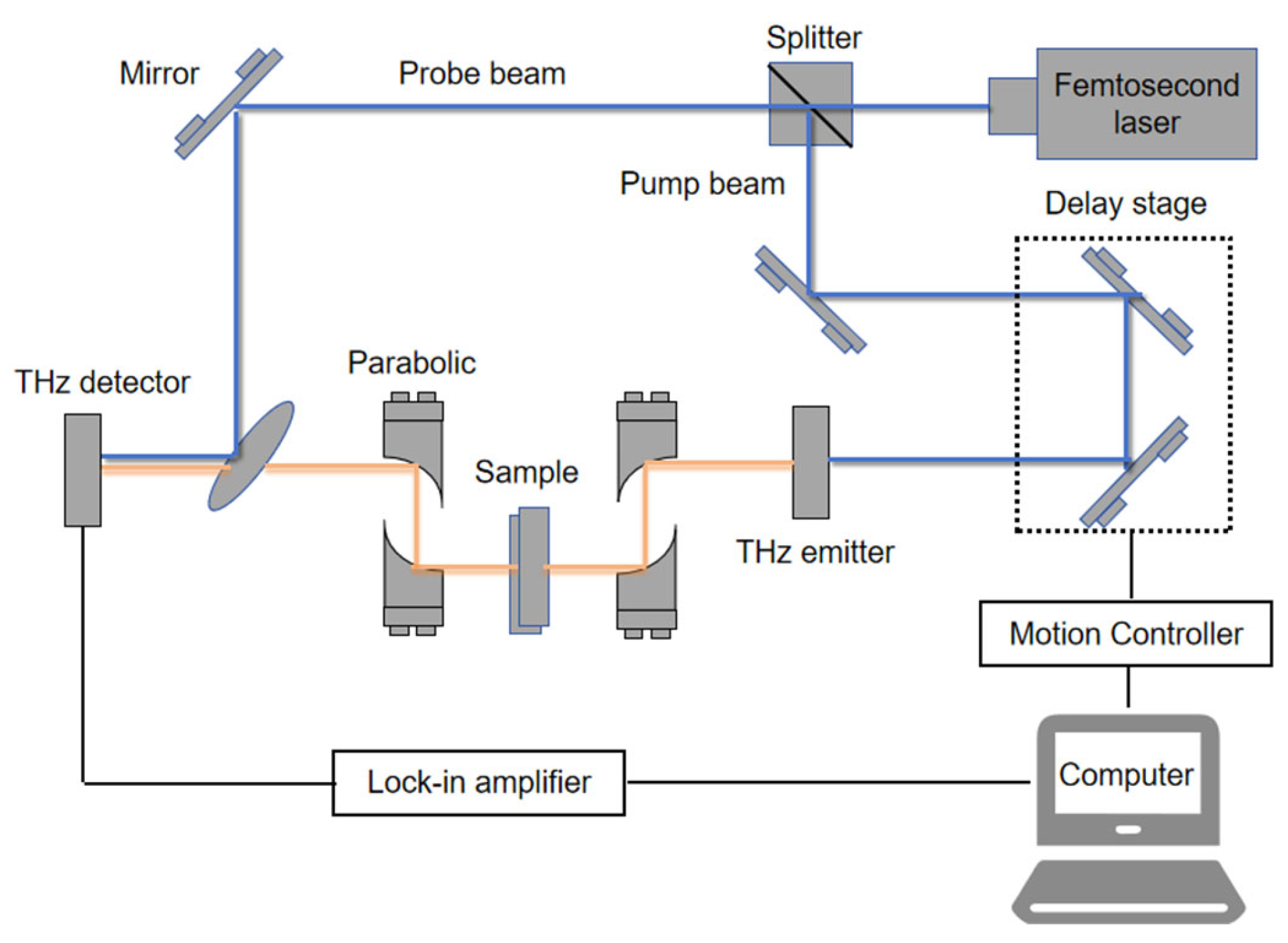
THz spectroscopy technology ingeniously combines the characteristics of microwaves and infrared rays and has unique advantages. On the one hand, its wavelength lies between that of microwaves and infrared rays, endowing terahertz waves with a certain penetrating ability and enabling them to deeply penetrate plant tissues. On the other hand, it can precisely obtain detailed information about the internal structure of plants by interacting with the substances inside these plants. This method of detecting the internal state of plants provides a brand-new perspective for the early and precise detection of plant diseases. It can replace traditional plant disease detection methods and provide strong technical support for disease prevention and control in agricultural production [72]. A typical terahertz spectrum experimental device is shown in Figure 3 [73].
Compared with traditional chemical detection methods, terahertz spectroscopy detection does not require any form of damage to plant samples. Traditional chemical testing often requires processing plant samples through methods such as cutting and grinding, which not only causes irreversible damage to the plants, but may also introduce errors during sample processing [74]. THz spectroscopy, on the other hand, directly detects plants to maintain their original state to the greatest extent. For instance, Gong et al. utilized terahertz spectroscopy technology to achieve the rapid identification of wheat mold conditions without the need for any complex processing of the wheat [75].
Meanwhile, terahertz spectroscopy is extremely sensitive to the water content within plant tissues. It can precisely detect changes in water content through indicators such as the intensity of characteristic absorption peaks, absorption coefficients, and refractive indices. Under favorable experimental conditions (such as a stable temperature and humidity environment and appropriate terahertz frequency band selection), for plant tissues with a relatively high degree of homogeneity such as leaves and fruits, terahertz technology can achieve an absolute error of ±0.5% to ±2% in water content. For instance, Adnan et al. combined terahertz spectroscopy technology with machine algorithms to accurately estimate the moisture content of leaves [76]. With this feature, this technology can effectively capture the early abnormal phenomena of plant physiological states, thereby achieving the early warning of plant diseases.
In addition, terahertz spectroscopy can simultaneously analyze multiple chemical components within plant tissues, such as cellulose, starch, and protein, thereby enabling a comprehensive observation of the impact of diseases on the composition and content of internal chemical components in plants. This provides a systematic and comprehensive research direction and technical support for the in-depth exploration of the occurrence mechanism, development process, and assessment of the severity of plant diseases. For example, Gu et al. used terahertz spectroscopy to simultaneously analyze ginsenoside R1, ginsenoside Rb1, ginsenoside Rg1, and other major bioactive components in Tianqi to identify its origin [77].
In practical applications, terahertz spectroscopy can be combined with other detection techniques, such as laser-induced breakdown spectroscopy (LIBS), to enhance the accuracy and efficiency of detection. For example, Li et al. combined terahertz spectroscopy with laser-induced breakdown spectroscopy to establish a classification model for camellia anthracnose, realizing the rapid, efficient, and high-precision detection of disease degree [78].
THz spectroscopy, as a detection technology that has emerged in recent years, has shown great application potential within the realm of plant disease detection due to its unique physical properties and detection advantages. With the continuous improvement of terahertz spectroscopy technology, such as the enhancement of spectral resolution and the miniaturization and portability of detection equipment, as well as the optimization of data processing algorithms, this technology will be increasingly widely and deeply applied in practical use for food safety and agricultural production. Table 1 lists the typical applications of spectral technology as a non-destructive detection method for plant diseases in recent times.

3. The Use of Imaging Technology in Plant Disease Detection

Imaging technology refers to the process and technique of converting real-world scenes, objects, or phenomena into visible images. It encompasses various forms such as optical imaging, medical imaging, thermal imaging, and spectral imaging and is widely applied in medical care, scientific research, and other fields [102,103]. By capturing image information through different principles and methods and analyzing this information in combination with relevant algorithms, it has become an important tool for human observation, diagnosis, and monitoring.

3.1. Hyperspectral Imaging

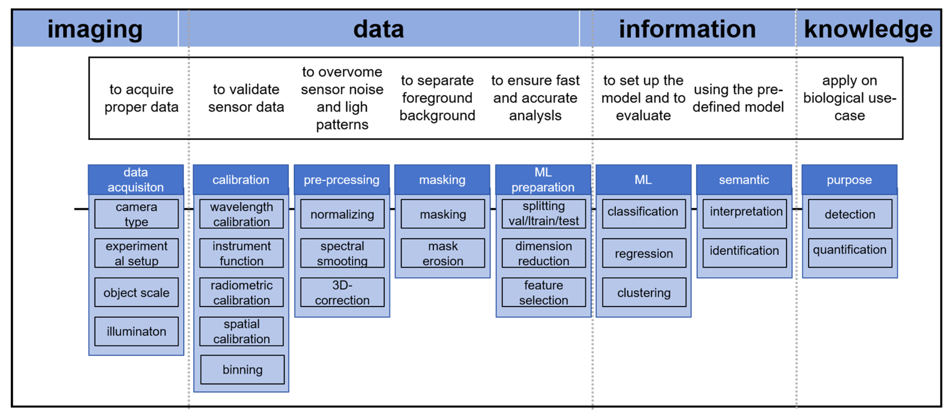
Hyperspectral imaging refers to the process of capturing the reflection or emission characteristics of an object within a continuous spectral band under natural light or laboratory lighting (such as xenon lamps, lasers, etc.) to obtain images rich in spectral information. The wavelength range of common hyperspectral devices is from 400 to 2500 nm. This technology is widely used in environmental monitoring and can be used to monitor water pollution, atmospheric composition, and vegetation coverage [104,105]. In geological exploration, hyperspectral imaging technology helps to identify mineral composition and geological structure and improve the exploration efficiency of mineral resources [106]. In the field of chemistry, hyperspectral imaging technology is also used to observe specific chemical compositions in samples [107]. In agriculture, it can be used to monitor crop growth [108,109] and assess crop diseases, pests, and their heavy metal content [110,111]. The general workflow of hyperspectral imaging technology is shown in Figure 4 [112].
Hyperspectral imaging technology is highly sensitive and can precisely capture the physiological changes occurring in plants at the initial stage of plant disease, thereby achieving the early identification of plant diseases. These changes cannot be observed in the conventional visible light environment and have certain concealment characteristics. This technology, through the analysis of spectral information in specific bands, enables the efficient and accurate identification of early plant disease symptoms, providing pivotal technical support for pre-diagnostic procedures in plant pathology. For example, Chen et al. obtained high-spectrum images of rice seedlings within the range of 414–1017 nm and used three models (VGG, CNN, and SVM) for training to develop an effective early monitoring system for rice bakanae disease, with an accuracy rate as high as 95.5% [113]. In addition, Liu et al. achieved the early identification of pine wilt disease by integrating UAV remote sensing with hyperspectral reconstruction [114].
Furthermore, different types of plant diseases present unique characteristic information in specific spectral bands. Hyperspectral imaging technology can clearly capture and effectively distinguish this information, thereby achieving the diagnosis of different plant diseases. For example, Bing et al. identified tea white star disease and anthrax using different spectral information [115].
Meanwhile, hyperspectral imaging technology can quantify the degree of impact of plant diseases on plant tissues. When plants are attacked by diseases, both their internal biochemical processes and external morphological structures will undergo corresponding changes, and these changes will be reflected in specific spectral characteristics. Through a series of complex and precise processing methods such as multivariate statistical analysis of spectral data and the establishment of mathematical models, hyperspectral imaging can present the changes in plant tissues caused by diseases as quantitative indicators, such as the variation range of spectral reflectance and the degree of deviation of characteristic bands. For example, Mei et al. used hyperspectral imaging technology and combined random forest (RF), extreme gradient lift, and other algorithms to build corresponding models to estimate the disease index of wheat stripe rust [116]. This quantitative result provides key data support for researchers to further explore the mechanism of disease occurrence and development in the laboratory environment, and also helps agricultural workers to quickly and intuitively assess the severity of disease in actual field operation scenarios, thus providing a solid basis for the formulation of reasonable scientific disease prevention and control strategies. Moreover, Thomas et al. utilized hyperspectral imaging technology to construct a corresponding model and conducted training, thereby successfully predicting the severity of Dothistroma needle blight in radiata pine. This provided crucial support for the prevention and control of the disease [117].
Hyperspectral imaging technology can also be used to study plant resistance to specific diseases and provide a scientific basis for disease resistance breeding. For instance, Zerdoner et al. used spectral information collected by hyperspectral equipment and combined it with the disease severity of rose petals infected by Botrytis cinerea for analysis, achieving the detection of the severity of Botrytis cinerea infection and applying the research results to plant breeding [118]. Leucker et al. revealed the influence of the quantitative trait loci of sugar beets on the spot resistance of Cercospora leaf through hyperspectral imaging technology, providing tools for improving resistance breeding [119].

3.2. Digital Imaging

Digital imaging technology operates on the principle of optical imaging, serving as a method to transform analog image signals into digital signals. In the digital imaging process, each point (pixel) of the image is assigned a specific value, and the entire image is a matrix composed of these values. Through digital processing technology, operations such as enhancement, compression, and recognition can be performed on the image [120,121]. In the field of medicine, it is used to diagnose diseases [122]. In materials science, it is used to analyze the microstructure and properties of materials [123]. It is used in agriculture for crop disease monitoring [124,125].
Since different diseases can cause different color changes in leaves, digital cameras can be used to capture the color changes of plant leaves and thereby preliminarily determine the type of disease. Meanwhile, disease can also cause changes in the shape and texture of leaves, and these changes can be identified through image processing technology. For example, Khirade et al. detected plant diseases through the color changes of leaves, combined color and texture to obtain image features, and then used the corresponding algorithms for calculation to detect plant disease [126].
As deep learning advances, digital imaging technology has gradually been combined with network models. By training a model with numerous images of plant diseases, the automatic recognition and classification of plant diseases can be achieved. For example, Zhao et al. utilized the improved convolutional neural network. After extensive training, they successfully achieved the automatic recognition and diagnosis of tomato leaf diseases [127].
In terms of detection speed, traditional chemical methods for preprocessing and modeling have short processing times and are suitable for small batch samples, but their efficiency decreases as the sample quantity increases. On the other hand, deep learning training takes a long time but has a fast detection speed and is suitable for large-scale real-time detection. In terms of noise stability, traditional chemical methods are sensitive to noise and require pre-processing to eliminate interference, while deep learning methods can automatically resist noise through a multi-layer structure. In terms of data labeling, traditional chemical methods require less data and are suitable for small sample scenarios, while deep learning methods rely on large-scale data, but transfer learning and data augmentation techniques such as flipping, cropping, etc., can reduce these requirements.
Furthermore, to further highlight the characteristics of a disease, image enhancement algorithms (such as histogram equalization, etc.) can be adopted to optimize the image quality. Meanwhile, image segmentation techniques (such as threshold segmentation, semantic segmentation, etc.) can be utilized to separate the disease area from the background, facilitating subsequent feature extraction. For example, Moupojou et al., after marking the healthy and diseased leaves in an image, cropped out the image of a single leaf from the original image to facilitate subsequent training [128].
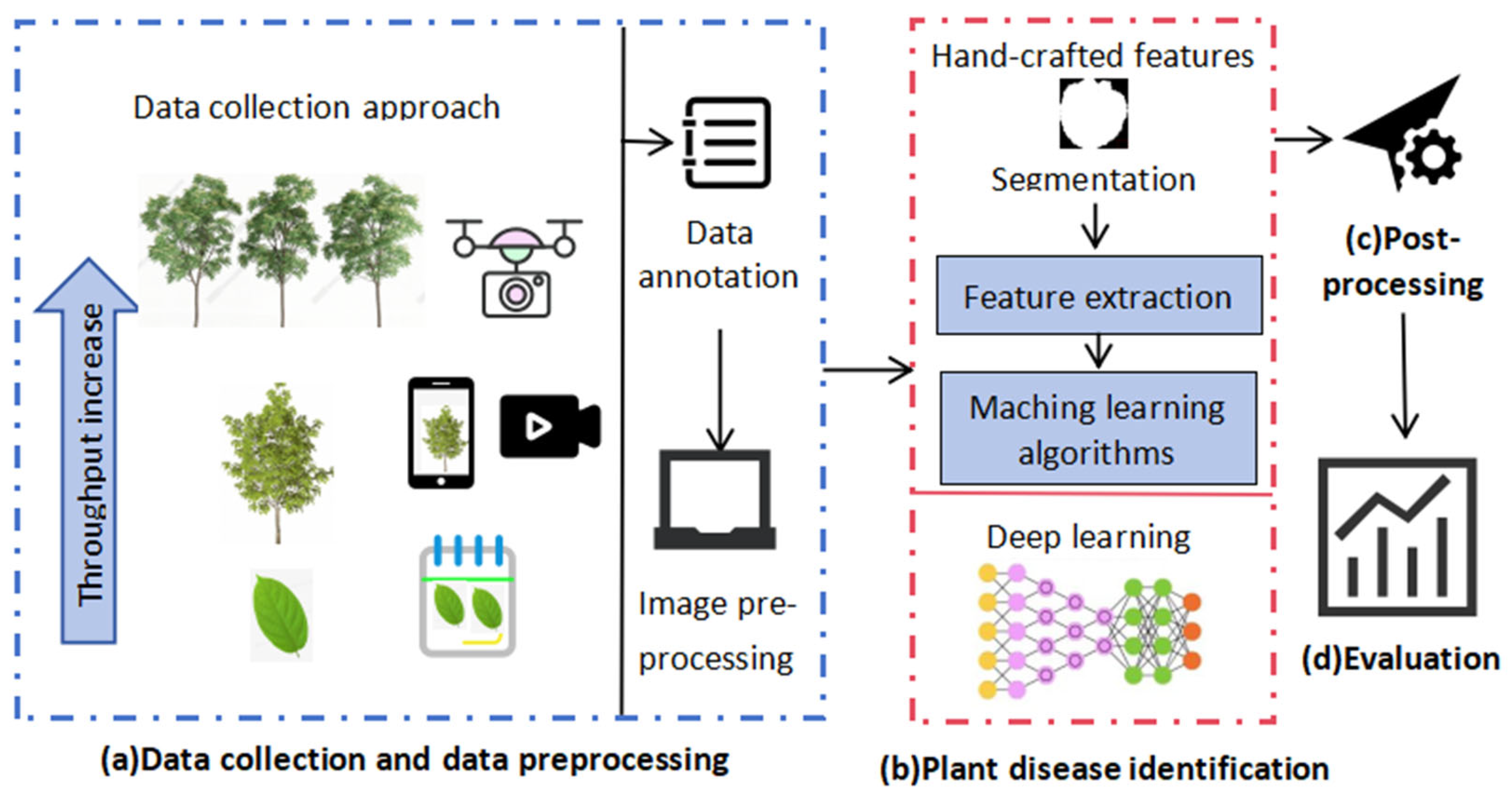
Digital imaging technology can also be combined with UAV-based and satellite remote sensing technologies to obtain large-scale crop images and monitor and warn of diseases on a macroscopic scale. When digital image technology is combined with drones, it is possible to take photos of hundreds of acres of farmland at once and conduct disease detection. When combined with satellite remote sensing, the detectable area can reach thousands or even tens of thousands of square kilometers. For instance, Zilberman et al. used drones equipped with high-resolution cameras to obtain color photos of banana plantations and used the color and shape changes as standards for identifying and classifying diseases, thereby achieving the detection of banana diseases [129]. The general flow of digital imaging technology for detecting plant diseases is shown in Figure 5 [130].
Digital imaging technology offers numerous advantages in plant disease detection. This technology can efficiently collect data and achieve the real-time detection of plant diseases. At the same time, it also has an excellent generalization performance, which can ensure the consistency of results when conducting plant disease detection across varying time points and conditions, providing solid and reliable technical support for plant protection work.

3.3. Thermal Imaging

Thermal imaging, also known as infrared imaging, works on the principle that all objects emit infrared energy due to their own temperature. Thermal imagers can detect this infrared energy and convert it into electrical signals. After signal processing and image reconstruction, the thermal distribution image of an object is ultimately presented [131]. Thermal imaging technology has a wide range of application scenarios and can be used for disease detection and breast cancer screening in the medical field [132]. In the field of materials, it can be used to evaluate the thermal conductivity of materials [133]. In agriculture, it can be used to detect crop health and assess crop moisture status [134]. A common thermal imaging experimental device is shown in Figure 6 [135].
The application of thermal imaging technology in plant disease detection is mainly reflected in the following aspects.
When plants are attacked by diseases, they usually show changes in temperature. Thermal imaging technology can capture such temperature changes, detect the existence of diseases before visible symptoms appear in plants, and achieve the early detection of plant diseases. Yang et al., for example, based on the principle that plant lesions would destroy the transpiration function and affect the relationship between the water content and temperature of plants, directly measured the leaf temperature of tea trees by using infrared thermal imaging technology, thereby indirectly obtaining the disease situation of the tea trees [136].
At the same time, thermal imaging technology has the characteristic of non-contact monitoring, allowing for monitoring without touching plants. This not only reduces human interference with plants, but also avoids possible cross-infection. For example, Feng et al. employed an unmanned aerial vehicle (UAV), integrated with a high-resolution thermal imaging sensor, to systematically capture multispectral thermal images of wheat fields. The UAV-based imaging system was operated at a fixed altitude to ensure uniform image acquisition, enabling the non-contact, large-scale monitoring of wheat canopy conditions [137]. Singh et al. used handheld cameras to photograph wheat plants in order to obtain temperature information that could reflect the condition of wheat stripe rust [138].
Thermal imaging can also cover large areas of plants, quickly scanning and identifying areas with abnormal temperatures. For instance, Sara et al. utilized drones integrated with thermal infrared and RGB cameras to monitor wheat under natural field conditions and detect Fusarium wilt (FHB), demonstrating the potential of drones equipped with thermal imaging technology in large-scale wheat disease detection [139]. Meanwhile, after performing an analysis of plant thermal imaging, it is possible to achieve a quantitative assessment of the severity of disease and visual monitoring of the spread range, which are conducive to formulating more effective prevention and control strategies. For instance, Singh et al. obtained visible light and thermal images of different resistant wheat varieties at the critical growth stage through field experiments and constructed a prediction model using a machine learning model for evaluation, achieving a reasonable prediction of the severity of wheat yellow rust disease and providing an effective method for assessing the severity of disease [140].
Thermal imaging technology has great potential and advantages. It can achieve the rapid and non-contact detection of plant diseases, providing real-time and accurate disease information for food safety and agricultural production. Meanwhile, thermal imaging technology can be coupled with other technologies (such as visible light imaging, deep learning, etc.) to further enhance the accuracy and reliability of disease detection. As technology continues to advance and mature, thermal imaging technology is anticipated to assume a more significant role in food safety and agricultural production. Table 2 summarizes the application cases of imaging technology as a non-invasive detection method for plant diseases over the past few years.

4. The Future Development Direction of Non-Destructive Detection Technology in Plant Disease

4.1. Limitations of Existing Non-Destructive Detection Technology

Although significant progress has been made in the relevant research on plant disease detection with NDDTs at present, there are still some problems.
Regarding spectral technology, although near-infrared spectroscopy technology has many advantages, spectral information is easily affected by environmental factors such as light intensity, temperature, and humidity, which may lead to fluctuations in spectral data and affect the accuracy of detection. Furthermore, the spectral characteristics of different plant varieties and the same plant at different growth stages vary, making it challenging to establish a universal detection model. However, the signal intensity of Raman spectroscopy is relatively weak, and its detection sensitivity is restricted by the sample concentration and scattering background. The detection effect on low-concentration disease samples is not good. Moreover, enhanced Raman spectroscopy technology has strict requirements for experimental conditions. The current problems faced by terahertz spectroscopy technology are the high cost of equipment and the fact that terahertz waves are easily affected by moisture during propagation, which leads to a decline in detection performance in high-humidity environments. Meanwhile, the spectral line analysis of terahertz spectra is rather difficult, and it is necessary to further develop precise data analysis methods to accurately identify disease-related information.
There are also deficiencies in imaging technology. Hyperspectral imaging equipment is expensive, and data acquisition is affected by conditions such as light intensity and weather conditions. Moreover, the amount of hyperspectral data is huge, and it has high requirements for storage, transmission and real-time processing, which limits its rapid application in actual production. Digital imaging technology is confronted with the problem of scarce high-quality labeled data, especially the lack of field images with multiple diseases and multiple growth stages, and model performance is poor, with insufficient multi-disease detection capabilities. Moreover, when factors such as light intensity change, additional preprocessing of the data is required and the environmental adaptability is poor. Thermal imaging technology is highly sensitive to the environment and is easily disturbed by external factors. Meanwhile, physiological activities such as transpiration in healthy plants can cause temperature fluctuations, which may be confused with disease signals. Secondly, single thermal imaging information is insufficient and usually needs to be combined with hyperspectral imaging, RGB images or sensor data, and other technologies to conduct a more comprehensive and detailed detection of plant diseases.
Furthermore, in terms of equipment size and complexity, digital imaging and thermal imaging technologies are the most convenient. Portable devices utilizing Raman spectroscopy and near-infrared spectroscopy technologies have also been developed and used for field testing, but they have a poor adaptability to environmental factors such as light and temperature and need to be used in relatively stable environments; terahertz spectroscopy technology and hyperspectral imaging technology usually have problems such as a large equipment size, complex structure, high environmental requirements, and poor mobility, making them difficult to operate outside the laboratory environment and unable to be flexibly deployed. At the same time, the operation of these types of equipment is very complex, making their wide use by the general public difficult. Comparisons of several technologies in terms of cost, convenience, penetration depth, and environmental sensitivity are presented in Table 3.

4.2. Future Development Direction

In view of the limitations of existing technologies, non-destructive testing technology can be developed for plant disease detection in the following directions in the future.
Firstly, the integration of spectral technology and imaging technology can be strengthened, combining the component analysis ability of spectral technology with the spatial feature capture ability of imaging technology. For instance, by combining the molecular fingerprint information of terahertz spectroscopy with the spatial distribution information of hyperspectral imaging, the multi-dimensional feature analysis of plant diseases can be achieved, thereby enhancing the accuracy of early detection. Secondly, deep learning algorithms, such as convolutional neural networks (CNNs) and Transformer models (such as Vision Transformer and Swin Transformer), can be introduced to optimize the processes of spectral data analysis and image recognition. For example, CNNs can be used to automatically extract disease features from hyperspectral images. Finally, it can also be combined with other technologies. For example, in the process of using imaging technology to conduct the real-time diagnosis of plant disease areas, by integrating multi-sensor data, a “detection–decision–execution” closed-loop control system can be constructed to achieve precise pesticide spraying, reducing waste and environmental pollution. The disease characteristics detected by spectral or imaging technology can be transmitted through blockchain technology to build a diagnostic network for disease prevention and control. In addition, quantum cascade lasers (QCLs) can be combined with spectral technology. By utilizing the characteristics of the terahertz waves (0.1–10 THz) generated by its interaction with substances, the limitations of traditional spectral technology in molecular vibration detection can be compensated, enabling the early and deep detection of plant diseases.
Secondly, portable detection devices can be developed to enhance the integration and miniaturization of equipment, such as handheld Raman spectrometers and hyperspectral imaging modules carried by small unmanned aerial vehicles, to reduce the volume and weight of the equipment and facilitate rapid field detection. For example, the handheld Raman devices that have emerged in existing studies can detect tomato viruses on-site and can be further optimized in the future. New types of photoelectric materials can also be adopted, which can reduce costs while enhancing the sensitivity and stability of equipment.
Continuous innovation is also needed in data processing and analysis methods. More efficient feature extraction methods can be developed to enable the automatic identification of key disease features in spectra or images. For example, in hyperspectral data, disease-sensitive bands can be focused on through the attention mechanism to reduce the interference of redundant information. Secondly, to address the issue of the lack of labeled data for rare crops or emerging diseases, transfer learning (such as the pre-trained Residual Network model) can be utilized to quickly transfer and adapt the disease detection models of other crops. Combined with fine-tuning with a small number of samples, the generalization ability of models can be enhanced. At the same time, an edge computing framework can also be developed to directly conduct data preprocessing and initial disease screening at the sensor end. Only suspicious data is transmitted to the cloud for in-depth analysis, reducing data transmission pressure and delay and achieving real-time disease response.
By overcoming the limitations of existing technologies and continuously promoting technological integration and equipment research and development, as well as innovation in data processing methods, non-destructive testing technology will have broader application prospects in the domain of plant disease detection, providing strong support for ensuring food safety and the sustainable development of agricultural production.

5. Conclusions

Non-destructive testing technology, with its unique advantages, plays a crucial role in related research on plant disease detection. By promptly and accurately identifying plant diseases, it is possible to effectively prevent the accumulation of pathogens in crops and prevent harmful substances in agricultural products from entering the market. This ensures food safety at the source. At the same time, through disease detection, infected plants can be quickly identified, allowing for targeted treatment to prevent a reduction in crop yield and ensure the sustainability and economic benefits of agricultural production.
Although spectral technology and imaging technology have different principles, they both demonstrate a unique performance in the detection of plant diseases. Near-infrared spectroscopy technology is simple to operate and has a fast analysis speed. It can effectively identify various diseases by detecting changes in plant components. Raman spectroscopy technology has a high sensitivity and convenient detection, and can accurately identify the types of pathogenic bacteria. Terahertz spectroscopy technology, with its penetrating power and high sensitivity, has obvious advantages in early disease detection and component analysis. Hyperspectral imaging technology can capture early changes in plants and achieve the early diagnosis of diseases. Digital imaging technology combined with deep learning models can automatically identify diseases, and its data acquisition is efficient. Thermal imaging technology can achieve early disease detection by monitoring temperature changes and has the advantages of non-contact monitoring and large-area scanning.
However, these technologies also have certain limitations. Spectral technology is greatly influenced by environmental factors, with a low detection sensitivity, high equipment cost, and difficult spectral line analysis. Imaging technology has problems such as complex data processing and an insufficient early disease detection ability: the early signs of plant diseases are not obvious, and imaging techniques struggle to capture the characteristics of these diseases. They also have an insufficient model generalization ability: the background of the training dataset is relatively simple, while the actual farmland environment has a complex background. Moreover, in order to meet the requirements of real-time detection, models need to be made more lightweight, which will result in a decline in the feature extraction capability.
To promote the further development of non-destructive testing technology, future research on plant disease detection should focus on aspects such as technology integration, equipment development, and innovation in data processing and analysis methods. By strengthening the integration of spectral technology and imaging technology and combining them with emerging technologies such as artificial intelligence, portable, low-cost, and highly sensitive detection equipment can be developed. At the same time, advanced data processing methods can be applied to identify potential disease characteristics and construct universal disease detection models. Thus, non-destructive testing technology can be further enhanced, enabling it to be more widely and deeply applied in fields such as food safety and agricultural production.

Author Contributions

Conceptualization, Y.W.; methodology, Z.W. and Y.J.; validation, J.S. and C.D.; formal analysis, Y.W.; investigation, Y.W.; resources, J.S. and C.D.; data curation, J.S.; writing original draft preparation, Y.W.; writing review and editing, Y.W.; supervision, J.S.; project administration, J.S. All authors have read and agreed to the published version of the manuscript.

Funding

This work was sponsored by Jiangsu Province and Education Ministry Co-Sponsored Synergistic Innovation Center of Modern Agricultural Equipment (XTCX2016). This article has been translated into English and has been polished for grammar and formatting by the Doubao AI tool (v9.7.0).

Institutional Review Board Statement

Not applicable.

Informed Consent Statement

Not applicable.

Data Availability Statement

The original contributions presented in this study are included in the article. Further inquiries can be directed to the corresponding authors.

Conflicts of Interest

The authors declare that they have no known competing financial interests or personal relationships that could have appeared to influence the work reported in this paper.

References

  1. Wan, L.; Li, H.; Li, C.; Wang, A.; Yang, Y.; Wang, P. Hyperspectral sensing of plant diseases: Principle and methods. Agronomy 2022, 12, 1451. [Google Scholar] [CrossRef]
  2. Zuo, X.; Chu, J.; Shen, J.; Sun, J. Multi-granularity feature aggregation with self-attention and spatial reasoning for fine-grained crop disease classification. Agriculture 2022, 12, 1499. [Google Scholar] [CrossRef]
  3. Shi, Y.; Yang, Q.Y.; Zhao, Q.H.; Dhanasekaran, S.; Ahima, J.; Zhang, X.Y.; Zhou, S.Q.; Droby, S.; Zhang, H.Y. Aureobasidium pullulans S-2 reduced the disease incidence of tomato by influencing the postharvest microbiome during storage. Postharvest Biol. Technol. 2022, 185, 111809. [Google Scholar] [CrossRef]
  4. Andriani, A.; Meyliana, A.; Susanto, W.E. Certainty factors in expert system to diagnose disease of chili plants. In Proceedings of the 6th International Conference on Cyber and IT Service Management (CITSM), Parapat, Indonesia, 7–9 August 2018. [Google Scholar] [CrossRef]
  5. Dewanto, S.; Lukas, J. Expert system for diagnosis pest and disease in fruit plants. In Proceedings of the International Conference on Advances Science and Contemporary Engineering (ICASCE), Jakarta, Indonesia, 23–24 October 2013. [Google Scholar] [CrossRef]
  6. Schaad, N.W.; Frederick, R.D. Real-time PCR and its application for rapid plant disease diagnostics. Can. J. Plant Pathol. 2002, 24, 250–258. [Google Scholar] [CrossRef]
  7. Li, M.; Hong, X.; Qiu, X.C.; Yang, C.Q.; Mao, Y.H.; Li, Y.; Liu, Z.J.; Du, D.L. Ultrasensitive monitoring strategy of PCR-like levels for zearalenone contamination based DNA barcode. J. Sci. Food Agric. 2021, 101, 4490–4497. [Google Scholar] [CrossRef]
  8. Brady, C.R.; Noll, L.W.; Saleh, A.A.; Little, C.R. Disease severity and microsclerotium properties of the sorghum sooty stripe pathogen, Ramulispora sorghi. Plant Dis. 2011, 95, 853–859. [Google Scholar] [CrossRef]
  9. Cagliari, D.; Dias, N.P.; Galdeano, D.M.; Dos Santos, E.A.; Smagghe, G.; Zotti, M.J. Management of pest insects and plant diseases by non-transformative RNAi. Front. Plant Sci. 2019, 10, 1319. [Google Scholar] [CrossRef]
  10. Kuo, Y.W.; Falk, B.W. RNA interference approaches for plant disease control. Biotechniques 2020, 69, 469–477. [Google Scholar] [CrossRef]
  11. Yu, P.; Ong, K.; Ueckert, J.; Gu, M.M. Biochar and Trichoderma reduce containerized poinsettia root rot caused by Pythium aphanidermatum. HortScience 2023, 58, 846–854. [Google Scholar] [CrossRef]
  12. Shi, J.; Hu, X.; Zou, X.; Zhao, J.; Zhang, W.; Holmes, M.; Huang, X.; Zhu, Y.; Li, Z.; Shen, T.; et al. A rapid and nondestructive method to determine the distribution map of protein, carbohydrate and sialic acid on edible bird’s nest by hyper-spectral imaging and chemometrics. Food Chem. 2017, 229, 235–241. [Google Scholar] [CrossRef]
  13. Huang, X.Y.; Lv, R.Q.; Wang, S.; Aheto, J.H.; Dai, C.X. Integration of computer vision and colorimetric sensor array for nondestructive detection of mango quality. J. Food Process Eng. 2018, 41, e12873. [Google Scholar] [CrossRef]
  14. Wu, X.; Liang, X.; Wang, Y.; Wu, B.; Sun, J. Non-destructive techniques for the analysis and evaluation of meat quality and safety: A review. Foods 2022, 11, 3713. [Google Scholar] [CrossRef] [PubMed]
  15. Li, H.H.; Sun, X.; Pan, W.X.; Kutsanedzie, F.; Zhao, J.W.; Chen, Q.S. Feasibility study on nondestructively sensing meat’s freshness using light scattering imaging technique. Meat Sci. 2016, 119, 102–109. [Google Scholar] [CrossRef] [PubMed]
  16. Chun, S.W.; Song, D.J.; Lee, K.H.; Kim, M.J.; Kim, M.S.; Kim, K.S.; Ae, C.M. Deep learning algorithm development for early detection of botrytis cinerea infected strawberry fruit using hyperspectral fluorescence imaging. Postharvest Biol. Technol. 2024, 214, 112918. [Google Scholar] [CrossRef]
  17. Weng, S.; Hu, X.; Wang, J.; Tang, L.; Li, P.; Zheng, S.; Zheng, L.; Huang, L.; Xin, Z. Advanced application of Raman spectroscopy and surface-enhanced Raman spectroscopy in plant disease diagnostics: A review. J. Agric. Food Chem. 2021, 69, 2950–2964. [Google Scholar] [CrossRef]
  18. Zhang, N.; Yang, G.; Pan, Y.; Yang, X.; Chen, L.; Zhao, C. A review of advanced technologies and development for hyperspectral-based plant disease detection in the past three decades. Remote Sens. 2020, 12, 3188. [Google Scholar] [CrossRef]
  19. Horigane, T.; Yamada, S.; Kimura-Suda, H.; Nishio, E.; Kawahara, S.; Mckeon, T. The use of spectroscopic analysis in component profiling of cereals. Comp. Biochem. Phys. A 2005, 141, S281. [Google Scholar]
  20. Qi, H.; Luo, J.; Wu, X.; Zhang, C. Application of nondestructive techniques for peach (Prunus persica) quality inspection: A review. J. Food Sci. 2024, 89, 6863–6887. [Google Scholar] [CrossRef]
  21. Wu, X.; Wang, Y.; Wu, B.; Sun, J. Classification of fritillaria using a portable near-infrared spectrometer and fuzzy generalized singular value decomposition. Ind. Crops Prod. 2024, 218, 119032. [Google Scholar] [CrossRef]
  22. Li, Q.; Wu, X.; Zheng, J.; Wu, B.; Jian, H.; Sun, C.; Tang, Y. Determination of pork meat storage time using near-infrared spectroscopy combined with fuzzy clustering algorithms. Foods 2022, 11, 2101. [Google Scholar] [CrossRef]
  23. Zhang, J.; Wu, X.; He, C.; Wu, B.; Zhang, S.; Sun, J. Near-infrared spectroscopy combined with fuzzy improved direct linear discriminant analysis for nondestructive discrimination of Chrysanthemum tea varieties. Foods 2024, 13, 1439. [Google Scholar] [CrossRef] [PubMed]
  24. Guo, Z.M.; Huang, W.Q.; Peng, Y.K.; Chen, Q.S.; Ouyang, Q.; Zhao, J.W. Color compensation and comparison of shortwave near infrared and long wave near infrared spectroscopy for determination of soluble solids content of ‘Fuji’ apple. Postharvest Biol. Technol. 2016, 115, 81–90. [Google Scholar] [CrossRef]
  25. Li, Y.; Pan, T.; Li, H.; Chen, S. Non-invasive quality analysis of thawed tuna using near infrared spectroscopy with baseline correction. J. Food Process Eng. 2020, 43, e13445. [Google Scholar] [CrossRef]
  26. Wu, X.; Zhou, H.; Wu, B.; Fu, H. Determination of apple varieties by near infrared reflectance spectroscopy coupled with improved possibilistic Gath–Geva clustering algorithm. J. Food Process. Preserv. 2020, 44, e14561. [Google Scholar] [CrossRef]
  27. Xu, Q.; Wu, X.; Wu, B.; Zhou, H. Detection of apple varieties by near-infrared reflectance spectroscopy coupled with SPSO-PFCM. J. Food Process Eng. 2022, 45, e13993. [Google Scholar] [CrossRef]
  28. Arslan, M.; Zou, X.B.; Tahir, H.E.; Hu, X.T.; Rakha, A.; Zareef, M.; Seweh, E.A.; Basheer, S. Nir spectroscopy coupled chemometric algorithms for rapid antioxidants activity assessment of Chinese dates (Zizyphus jujuba Mill.). Int. J. Food Eng. 2019, 15, 20180148. [Google Scholar] [CrossRef]
  29. Tahir, H.E.; Zou, X.B.; Shi, J.Y.; Mariod, A.A.; Wiliam, T. Rapid determination of antioxidant compounds and antioxidant activity of Sudanese karkade (Hibiscus sabdariffa L.) using near infrared spectroscopy. Food Anal. Methods 2016, 9, 1228–1236. [Google Scholar] [CrossRef]
  30. Sankaran, S.; Mishra, A.; Maja, J.M.; Ehsani, R. Visible-near infrared spectroscopy for detection of huanglongbing in citrus orchards. Comput. Electron. Agric. 2011, 77, 127–134. [Google Scholar] [CrossRef]
  31. Lucas Domingos Da Silva, A.; Alves Filho, E.G.; Silva, L.M.A.; Carlos Huertas Tavares, O.; Gervasio Pereira, M.; De Campos, T.; Manoel Da Silva, L. Near infrared spectroscopy to rapid assess the rubber tree clone and the influence of maturation and disease at the leaves. Microchem. J. 2021, 168, 106478. [Google Scholar] [CrossRef]
  32. Barthel, D.; Dordevic, N.; Fischnaller, S.; Kerschbamer, C.; Messner, M.; Eisenstecken, D.; Robatscher, P.; Janik, K. Detection of apple proliferation disease in Malus × domestica by near infrared reflectance analysis of leaves. Spectrochim. Acta Part. A 2021, 263, 120178. [Google Scholar] [CrossRef]
  33. Zhang, Z.; Gu, H.; Xie, K.; Jiang, H.; Xie, Q.; Sa, J. Pretreatment and combined method based on near infrared spectroscopy. Laser Optoelectron. Prog. 2021, 58, 1617001. [Google Scholar] [CrossRef]
  34. Fearer, C.J.; Conrad, A.O.; Marra, R.E.; Georskey, C.; Villari, C.; Slot, J.; Bonello, P. A combined approach for early in-field detection of beech leaf disease using near-infrared spectroscopy and machine learning. Front. For. Glob. Change 2022, 5, 934545. [Google Scholar] [CrossRef]
  35. Ghanei Ghooshkhaneh, N.; Golzarian, M.R.; Mollazade, K. VIS-NIR spectroscopy for detection of citrus core rot caused by Alternaria alternata. Food Control 2023, 144, 109320. [Google Scholar] [CrossRef]
  36. Sirakov, I.; Velichkova, K.; Dinev, T.; Slavcheva-Sirakova, D.; Valkova, E.; Yorgov, D.; Veleva, P.; Atanasov, V.; Atanassova, S. Detection of fungal diseases in lettuce by VIR-NIR spectroscopy in aquaponics. Microorganisms 2023, 11, 2348. [Google Scholar] [CrossRef] [PubMed]
  37. Zhou, X.; Sun, J.; Tian, Y.; Wu, X.H.; Dai, C.X.; Li, B. Spectral classification of lettuce cadmium stress based on information fusion and VISSA-GOA-SVM algorithm. J. Food Process Eng. 2019, 42, e13085. [Google Scholar] [CrossRef]
  38. Shen, L.F.; Jia, S.Q.; Guo, T.T.; Wu, W.J.; Yan, Y.L.; An, D. Study of feature extraction methods for maize’s near infrared spectra in biomimetic pattern recognition. Spectrosc. Spectr. Anal. 2012, 32, 939–943. [Google Scholar] [CrossRef]
  39. Seehanam, P.; Sonthiya, K.; Maniwara, P.; Theanjumpol, P.; Ruangwong, O.; Nakano, K.; Ohashi, S.; Kramchote, S.; Suwor, P. Ability of near infrared spectroscopy to detect anthracnose disease early in mango after harvest. Hortic. Environ. Biotechnol. 2024, 65, 581–591. [Google Scholar] [CrossRef]
  40. Fernandez-Cabanas, V.M.; Borrero, C.; Cozzolino, D.; Aviles, M. Feasibility of near infrared spectroscopy for estimating suppressiveness of carnation (Dianthus cariophyllus L.) fusarium wilt in different plant growth media. Spectrochim. Acta Part A 2022, 280, 121528. [Google Scholar] [CrossRef]
  41. Shi, J.Y.; Wang, Y.Y.; Li, Z.H.; Huang, X.W.; Shen, T.T.; Zou, X.B. Characterization of invisible symptoms caused by early phosphorus deficiency in cucumber plants using near-infrared hyperspectral imaging technology. Spectrochim. Acta A 2022, 267, 120540. [Google Scholar] [CrossRef]
  42. Li, M.D.; Chen, Q.W.; Huang, P.L.; Zhou, J.; Gong, W.K. A review of classification algorithms for data mining. In Proceedings of the 2nd International Conference on Mechanical, Electronic and Engineering Technology (MEET), Xi’an, China, 19–20 January 2019. [Google Scholar]
  43. Ong, P.; Jian, J.; Li, X.; Zou, C.; Yin, J.; Ma, G. New approach for sugarcane disease recognition through visible and near-infrared spectroscopy and a modified wavelength selection method using machine learning models. Spectrochim. Acta Part A 2023, 302, 123037. [Google Scholar] [CrossRef]
  44. Ying, L.; Kun, M.; Xinyu, Z.; Qifu, Y.; Jiaquan, W.; Shuangyan, Y. Identification of disease type of tobacco leaves based on near infrared spectroscopy and convolutional neural network. J. Braz. Chem. Soc. 2024, 35, e20230181. [Google Scholar] [CrossRef]
  45. Zahir, S.; Omar, A.F.; Jamlos, M.F.; Azmi, M.a.M.; Muncan, J. A review of visible and near-infrared (VIS-NIR) spectroscopy application in plant stress detection. Sens. Actuators A Phys. 2022, 338, 113468. [Google Scholar] [CrossRef]
  46. Mohd Hilmi Tan, M.I.S.; Jamlos, M.F.; Omar, A.F.; Dzaharudin, F.; Chalermwisutkul, S.; Akkaraekthalin, P. Ganoderma boninense disease detection by near-infrared spectroscopy classification: A review. Sensors 2021, 21, 3052. [Google Scholar] [CrossRef]
  47. Xiong, Y.; Mccarthy, C.; Humpal, J.; Percy, C. Near-infrared spectroscopy and deep neural networks for early common root rot detection in wheat from multi-season trials. Agron. J. 2024, 116, 2370–2390. [Google Scholar] [CrossRef]
  48. Hu, T.; Sun, Q.; Fernandes, F.a.N. Research progress on detection of apple watercore based on visible and near-infrared spectroscopy. J. Food Process. Preserv. 2025, 2025, 4394346. [Google Scholar] [CrossRef]
  49. Jiang, H.; Wang, Z.; Deng, J.; Ding, Z.; Chen, Q. Quantitative detection of heavy metal cd in vegetable oils: A nondestructive method based on Raman spectroscopy combined with chemometrics. J. Food Sci. 2024, 89, 8054–8065. [Google Scholar] [CrossRef]
  50. Zhang, X.; Bian, F.; Wang, Y.; Hu, L.; Yang, N.; Mao, H. A method for capture and detection of crop airborne disease spores based on microfluidic chips and micro Raman spectroscopy. Foods 2022, 11, 3462. [Google Scholar] [CrossRef]
  51. Xue, Y.; Jiang, H. Monitoring of chlorpyrifos residues in corn oil based on Raman spectral deep-learning model. Foods 2023, 12, 2402. [Google Scholar] [CrossRef]
  52. Jiang, H.; He, Y.; Xu, W.; Chen, Q. Quantitative detection of acid value during edible oil storage by Raman spectroscopy: Comparison of the optimization effects of boss and VCPA algorithms on the characteristic Raman spectra of edible oils. Food Anal. Methods 2021, 14, 1826–1835. [Google Scholar] [CrossRef]
  53. Guo, Z.M.; Wang, M.M.; Wu, J.Z.; Tao, F.F.; Chen, Q.S.; Wang, Q.Y.; Ouyang, Q.; Shi, J.Y.; Zou, X.B. Quantitative assessment of zearalenone in maize using multivariate algorithms coupled to Raman spectroscopy. Food Chem. 2019, 286, 282–288. [Google Scholar] [CrossRef]
  54. Huang, Y.; Wang, H.; Huang, H.; Tan, Z.; Hou, C.; Zhuang, J.; Tang, Y. Raman spectroscopy and its application in fruit quality detection. Agriculture 2025, 15, 195. [Google Scholar] [CrossRef]
  55. Wang, J.J.; Chen, Q.S.; Belwal, T.; Lin, X.Y.; Luo, Z.S. Insights into chemometric algorithms for quality attributes and hazards detection in foodstuffs using Raman/surface enhanced Raman spectroscopy. Compr. Rev. Food Sci. Food Saf. 2021, 20, 2476–2507. [Google Scholar] [CrossRef]
  56. Duan, N.; Chang, B.Y.; Zhang, H.; Wang, Z.P.; Wu, S.J. Salmonella typhimurium detection using a surface-enhanced Raman scattering-based aptasensor. Int. J. Food Microbiol. 2016, 218, 38–43. [Google Scholar] [CrossRef]
  57. Li, H.H.; Chen, Q.S.; Ouyang, Q.; Zhao, J.W. Fabricating a novel Raman spectroscopy-based aptasensor for rapidly sensing Salmonella typhimurium. Food Anal. Methods 2017, 10, 3032–3041. [Google Scholar] [CrossRef]
  58. Andersen, M.E.; Andersen, U.; Wiking, L.; Rasmussen, J.T.; Corredig, M.; Gregersen, S.B. The exploration of milk fat crystallization in milk fat globules by confocal Raman microscopy. Food Struct. 2024, 40, 100372. [Google Scholar] [CrossRef]
  59. Mosca, S.; Lin, Q.Q.; Stokes, R.; Bharucha, T.; Gangadharan, B.; Clarke, R.; Fernandez, L.G.; Deats, M.; Walsby-Tickle, J.; Arman, B.Y.; et al. Innovative method for rapid detection of falsified COVID-19 vaccines through unopened vials using handheld Spatially Offset Raman Spectroscopy (SORS). Vaccine 2023, 41, 6960–6968. [Google Scholar] [CrossRef]
  60. Liu, Z.F.; Huang, M.; Zhu, Q.B.; Zhao, X.; Yan, S.Q. Spatially offset Raman spectroscopy analysis technology and application in food subsurface detection. Spectrosc. Spectr. Anal. 2024, 44, 1201–1208. [Google Scholar]
  61. Tahir, H.E.; Zou, X.B.; Li, Z.H.; Shi, J.Y.; Xiaodong, Z.D.; Sheng, W.; Mariod, A.A. Rapid prediction of phenolic compounds and antioxidant activity of Sudanese honey using Raman and Fourier transform infrared (FT-IR) spectroscopy. Food Chem. 2017, 226, 202–211. [Google Scholar] [CrossRef] [PubMed]
  62. Mandrile, L.; Rotunno, S.; Miozzi, L.; Vaira, A.M.; Giovannozzi, A.M.; Rossi, A.M.; Noris, E. Nondestructive Raman spectroscopy as a tool for early detection and discrimination of the infection of tomato plants by two economically important viruses. Anal. Chem. 2019, 91, 9025–9031. [Google Scholar] [CrossRef] [PubMed]
  63. Sanchez, L.; Pant, S.; Irey, M.; Mandadi, K.; Kurouski, D. Detection and identification of canker and blight on orange trees using a hand-held Raman spectrometer. J. Raman Spectrosc. 2019, 50, 1875–1880. [Google Scholar] [CrossRef]
  64. Farber, C.; Bennett, J.S.; Dou, T.; Abugalyon, Y.; Humpal, D.; Sanchez, L.; Toomey, K.; Kolomiets, M.; Kurouski, D. Raman-based diagnostics of stalk rot disease of maize caused by Colletotrichum graminicola. Front. Plant Sci. 2021, 12, 722898. [Google Scholar] [CrossRef] [PubMed]
  65. Zhu, C.; Jiang, H.; Chen, Q. High precisive prediction of aflatoxin b(1) in pressing peanut oil using Raman spectra combined with multivariate data analysis. Foods 2022, 11, 1565. [Google Scholar] [CrossRef] [PubMed]
  66. Huang, C.C. Applications of Raman spectroscopy in herbal medicine. Appl. Spectrosc. Rev. 2016, 51, 1–11. [Google Scholar] [CrossRef]
  67. Zhu, S.S.; Zhong, Q.S.; Yan, D.H.; Yan, Z.J.; Chen, S.; Yao, Y.D.; Zhou, C. Rapid and nondestructive identification of the geographical origin of ophiopogonis radix by Raman spectroscopy and multivariate statistical analysis. Anal. Lett. 2024, 57, 3149–3163. [Google Scholar] [CrossRef]
  68. Saletnik, A.; Saletnik, B.; Zagula, G.; Puchalski, C. Raman spectroscopy for plant disease detection in next-generation agriculture. Sustainability 2024, 16, 5474. [Google Scholar] [CrossRef]
  69. Zhang, X.; Duan, Z.; Mao, H.; Gao, H.; Zuo, Z. A lettuce moisture detection method based on terahertz time-domain spectroscopy. Ciênc. Rural 2022, 52, e20210002. [Google Scholar] [CrossRef]
  70. Tong, Y.; Wang, S.; Han, K.; Song, X.; Zhang, W.; Ye, Y.; Ren, X. Development of a novel metal grating and its applications of terahertz spectroscopic detection of CuSO4 in fruit. Food Anal. Methods 2021, 14, 1590–1599. [Google Scholar] [CrossRef]
  71. Lu, Y.; Asante, E.A.; Duan, H.; Hu, Y. Quantitative assessment of cold injury in tea plants by terahertz spectroscopy method. Agronomy 2023, 13, 1376. [Google Scholar] [CrossRef]
  72. Gao, Y.; Peng, Z.; Yang, H.; Zhang, X.; Zhao, Y.; Hou, Z.; Su, B.; Li, K.; Zhang, C. Terahertz spectroscopy study of oridonin and ponicidin in the anticancer Chinese herbal medicine Rabdosia rubescens. Front. Plant Sci. 2024, 15, 1460123. [Google Scholar] [CrossRef]
  73. Fu, X.J.; Liu, Y.J.; Chen, Q.; Fu, Y.; Cui, T.J. Applications of terahertz spectroscopy in the detection and recognition of substances. Front. Phys. 2022, 10, 869537. [Google Scholar] [CrossRef]
  74. Zhu, C.X.; Liu, D.; Li, Y.Y.; Chen, T.; You, T.Y. Label-free ratiometric homogeneous electrochemical aptasensor based on hybridization chain reaction for facile and rapid detection of aflatoxin b1 in cereal crops. Food Chem. 2022, 373, 131443. [Google Scholar] [CrossRef] [PubMed]
  75. Gong, Y.-H.; Yang, T.-J.; Liang, Y.-T.; Ge, H.-Y.; Shen, E.-B. Comparative assessments between conventional and promising technologies for wheat aging or mold detection. Cereal Res. Commun. 2021, 49, 511–519. [Google Scholar] [CrossRef]
  76. Zahid, A.; Abbas, H.T.; Ren, A.; Zoha, A.; Heidari, H.; Shah, S.A.; Imran, M.A.; Alomainy, A.; Abbasi, Q.H. Machine learning driven non-invasive approach of water content estimation in living plant leaves using terahertz waves. Plant Methods 2019, 15, 138. [Google Scholar] [CrossRef]
  77. Gu, H.; Wang, S.; Hu, S.; Wu, X.; Li, Q.; Zhang, R.; Zhang, J.; Zhang, W.; Peng, Y. Identification of Panax notoginseng origin using terahertz precision spectroscopy and neural network algorithm. Talanta 2024, 274, 125968. [Google Scholar] [CrossRef]
  78. Bin, L.; Qiu, W.; Chao-Hui, Z.; Zhao-Yang, H.; Hai, Y.; Jun, L.; Yan-De, L. Research on anthracnose grade of Camellia oleifera based on the combined LIBS and THz technology. Plant Methods 2022, 18, 52. [Google Scholar] [CrossRef]
  79. Ong, P.; Jian, J.; Li, X.; Zou, C.; Yin, J.; Ma, G. Sugarcane disease recognition through visible and near-infrared spectroscopy using deep learning assisted continuous wavelet transform-based spectrogram. Spectrochim. Acta Part A 2025, 324, 125001. [Google Scholar] [CrossRef]
  80. Liu, Z.; Le, D.; Zhang, T.; Lai, Q.; Zhang, J.; Li, B.; Song, Y.; Chen, N. Detection of apple moldy core disease by fusing vibration and VIS/NIR spectroscopy data with dual-input MLP-transformer. J. Food Eng. 2024, 382, 112219. [Google Scholar] [CrossRef]
  81. Jiang, X.G.; Ge, K.; Li, B.; Ouyang, A.G.; Liu, Y.D.; Jiang, N.; Liu, H.F. Non-destructive detection of apple fungal infection based on VIS/NIR transmission spectroscopy. J. Food Compos. Anal. 2024, 133, 106469. [Google Scholar] [CrossRef]
  82. Jiang, X.G.; Ge, K.; Liu, Z.; Chen, N.; Ouyang, A.G.; Liu, Y.D.; Huang, Y.Y.; Li, J.H.; Hu, M.M. Non-destructive online detection of early moldy core apples based on VIS/NIR transmission spectroscopy. Chem. Biol. Technol. Agric. 2024, 11, 63. [Google Scholar] [CrossRef]
  83. Zhao, X.; Liu, Y.; Huang, Z.; Li, G.; Zhang, Z.; He, X.; Du, H.; Wang, M.; Li, Z. Early diagnosis of Cladosporium fulvum in greenhouse tomato plants based on visible/near-infrared (VIS/NIR) and near-infrared (NIR) data fusion. Sci. Rep. 2024, 14, 20176. [Google Scholar] [CrossRef] [PubMed]
  84. Li, C.; Xiang, D.; Yang, S.; Wang, X.; Li, C. Fusarium wilt of banana latency and onset detection based on visible/near infrared spectral technology. Agronomy 2024, 14, 2994. [Google Scholar] [CrossRef]
  85. Jin, X.; Gu, S.; Rao, Y.; Xiong, J.; Zhang, H.; Zhang, X.; Liu, L. An innovative fusion feature method of spectrum and visual image for diagnosing ‘Akizuki’ pear cork spot disorder. J. Food Compos. Anal. 2024, 127, 105963. [Google Scholar] [CrossRef]
  86. Tan, H.Z.; Liu, Y.; Tang, H.; Fan, W.; Jiang, L.W.; Li, P. Accurate discrimination of mold-damaged Citri Reticulatae Pericarpium using partial least-squares discriminant analysis and selected wavelengths. Foods 2024, 13, 3856. [Google Scholar] [CrossRef]
  87. Sahabi, H.; Baradaran-Motie, J. Detection of mite infested saffron plants using aerial imaging and machine learning classifier. Span. J. Agric. Res. 2025, 22, 20452. [Google Scholar] [CrossRef]
  88. Kim, S.; Lee, S.; Chi, H.Y.; Kim, M.K.; Kim, J.S.; Lee, S.H.; Chung, H. Feasibility study for detection of turnip yellow mosaic virus (TYMV) infection of Chinese cabbage plants using Raman spectroscopy. Plant Pathol. J. 2013, 29, 105–109. [Google Scholar] [CrossRef] [PubMed]
  89. Farber, C.; Kurouski, D. Detection and identification of plant pathogens on maize kernels with a hand-held Raman spectrometer. Anal. Chem. 2018, 90, 3009–3012. [Google Scholar] [CrossRef] [PubMed]
  90. Higgins, S.; Joshi, R.; Juarez, I.; Bennett, J.S.; Holman, A.P.; Kolomiets, M.; Kurouski, D. Non-invasive identification of combined salinity stress and stalk rot disease caused by Colletotrichum graminicola in maize using Raman spectroscopy. Sci. Rep. 2023, 13, 7661. [Google Scholar] [CrossRef] [PubMed]
  91. Yue, X.; Tan, Y.; Fan, W.; Song, S.; Ji, H.; Li, B. Raman spectroscopic analysis of paddy rice infected by three pests and diseases common in northeast Asia. J. Phys. Conf. Ser. 2019, 1324, 012050. [Google Scholar] [CrossRef]
  92. Lin, Y.J.; Lin, H.K.; Lin, Y.H. Construction of raman spectroscopic fingerprints for the detection of fusarium wilt of banana in Taiwan. PLoS ONE 2020, 15, e0230330. [Google Scholar] [CrossRef]
  93. Kim, S.; Hong, S.H.; Kim, J.H.; Oh, M.K.; Eom, T.J.; Park, Y.H.; Shin, G.H.; Yim, S.Y. Early on-site detection of strawberry anthracnose using portable Raman spectroscopy. Spectrochim. Acta Part. A 2023, 303, 123150. [Google Scholar] [CrossRef]
  94. Vallejo-Perez, M.R.; Sosa-Herrera, J.A.; Navarro-Contreras, H.R.; Alvarez-Preciado, L.G.; Rodriguez-Vazquez, A.G.; Lara-Avila, J.P. Raman spectroscopy and machine-learning for early detection of bacterial canker of tomato: The asymptomatic disease condition. Plants 2021, 10, 1542. [Google Scholar] [CrossRef]
  95. Orecchio, C.; Sacco Botto, C.; Alladio, E.; D’errico, C.; Vincenti, M.; Noris, E. Non-invasive and early detection of tomato spotted wilt virus infection in tomato plants using a hand-held Raman spectrometer and machine learning modelling. Plant Stress 2025, 15, 100732. [Google Scholar] [CrossRef]
  96. Ji, X.; Xue, J.; Shi, J.; Wang, W.; Zhang, X.; Wang, Z.; Lu, W.; Liu, J.; Fu, Y.V.; Xu, N.; et al. Noninvasive raman spectroscopy for the detection of rice bacterial leaf blight and bacterial leaf streak. Talanta 2025, 282, 126962. [Google Scholar] [CrossRef] [PubMed]
  97. Di Girolamo, F.V.; Pagano, M.; Tredicucci, A.; Bitossi, M.; Paoletti, R.; Barzanti, G.P.; Benvenuti, C.; Roversi, P.F.; Toncelli, A. Detection of fungal infections in chestnuts: A terahertz imaging-based approach. Food Control 2021, 123, 107700. [Google Scholar] [CrossRef]
  98. Penkov, N.V.; Goltyaev, M.V.; Astashev, M.E.; Serov, D.A.; Moskovskiy, M.N.; Khort, D.O.; Gudkov, S.V. The application of terahertz time-domain spectroscopy to identification of potato late blight and fusariosis. Pathogens 2021, 10, 1336. [Google Scholar] [CrossRef]
  99. Zhang, X.; Wang, Y.; Zhou, Z.; Zhang, Y.; Wang, X. Detection method for tomato leaf mildew based on hyperspectral fusion terahertz technology. Foods 2023, 12, 535. [Google Scholar] [CrossRef]
  100. Zhou, Y.; Wang, X.; Chen, K.; Han, C.; Guan, H.; Wang, Y.; Zhao, Y. Feasibility and potential of terahertz spectral and imaging technology for Apple Valsa canker detection: A preliminary investigation. Spectrochim. Acta A Mol. Biomol. Spectrosc. 2025, 327, 125308. [Google Scholar] [CrossRef]
  101. Wang, X.; Wu, Q.Y.S.; Zhang, N.; Ngo, A.C.Y.; Tanadi, J.; Khoo, E.H.; Zhu, Q.; Ke, L. Non-invasive early monitoring plant health using terahertz spectroscopy. J. Mater. Sci. Mater. Electron. 2024, 35, 1346. [Google Scholar] [CrossRef]
  102. Wang, Y.; Mao, H.; Zhang, X.; Liu, Y.; Du, X. A rapid detection method for tomato gray mold spores in greenhouse based on microfluidic chip enrichment and lens-less diffraction image processing. Foods 2021, 10, 3011. [Google Scholar] [CrossRef]
  103. Xu, M.; Sun, J.; Zhou, X.; Tang, N.; Shen, J.; Wu, X. Research on nondestructive identification of grape varieties based on EEMD-DWT and hyperspectral image. J. Food Sci. 2021, 86, 2011–2023. [Google Scholar] [CrossRef]
  104. Li, W.Z.; Fazli, S.; Maharjan, S.; El-Askary, H. Deciphering water quality and algal dynamics in clear lake through hyperspectral analysis using emit data. In Proceedings of the IEEE International Geoscience and Remote Sensing Symposium (IGARSS), Athens, Greece, 7–12 July 2024. [Google Scholar] [CrossRef]
  105. Kim, G.; Baek, I.; Stocker, M.D.; Smith, J.E.; Van Tassell, A.L.; Qin, J.W.; Chan, D.E.; Pachepsky, Y.; Kim, M.S. Hyperspectral imaging from a multipurpose floating platform to estimate chlorophyll-a concentrations in irrigation pond water. Remote Sens. 2020, 12, 2070. [Google Scholar] [CrossRef]
  106. Peyghambari, S.; Zhang, Y. Hyperspectral remote sensing in lithological mapping, mineral exploration, and environmental geology: An updated review. J. Appl. Remote Sens. 2021, 15, 031501. [Google Scholar] [CrossRef]
  107. Xu, M.; Sun, J.; Cheng, J.; Yao, K.; Wu, X.; Zhou, X. Non-destructive prediction of total soluble solids and titratable acidity in Kyoho grape using hyperspectral imaging and deep learning algorithm. J. Food Sci. Technol. 2022, 58, 9–21. [Google Scholar] [CrossRef]
  108. Zhong, Y.; Sun, J.; Yao, K.; Cheng, J.; Du, X. Detection of rice (with husk) moisture content based on hyperspectral imaging technology combined with MSLPP–ESMA–SVR model. J. Food Saf. 2024, 44, e13112. [Google Scholar] [CrossRef]
  109. Dai, C.; Sun, J.; Huang, X.; Zhang, X.; Tian, X.; Wang, W.; Sun, J.; Luan, Y. Application of hyperspectral imaging as a nondestructive technology for identifying tomato maturity and quantitatively predicting lycopene content. Foods 2023, 12, 2957. [Google Scholar] [CrossRef]
  110. Zhang, Y.; Sun, J.; Li, J.; Wu, X.; Dai, C. Quantitative analysis of cadmium content in tomato leaves based on hyperspectral image and feature selection. Appl. Eng. Agric. 2018, 34, 789–798. [Google Scholar] [CrossRef]
  111. Zhou, X.; Sun, J.; Zhang, Y.; Tian, Y.; Yao, K.; Xu, M. Visualization of heavy metal cadmium in lettuce leaves based on wavelet support vector machine regression model and visible-near infrared hyperspectral imaging. J. Food Process Eng. 2021, 44, e13897. [Google Scholar] [CrossRef]
  112. Paulus, S.; Mahlein, A.K. Technical workflows for hyperspectral plant image assessment and processing on the greenhouse and laboratory scale. GigaScience 2020, 9, giaa090. [Google Scholar] [CrossRef]
  113. Chen, S.; Lu, X.; Fang, H.; Perumal Anand, B.; Li, R.; Feng, L.; Wang, M.; Liu, Y. Early surveillance of rice bakanae disease using deep learning and hyperspectral imaging. aBIOTECH 2024, 5, 281–297. [Google Scholar] [CrossRef]
  114. Liu, W.T.; Xie, Z.R.; Du, J.; Li, Y.H.; Long, Y.B.; Lan, Y.B.; Liu, T.Y.; Sun, S.; Zhao, J. Early detection of pine wilt disease based on UAV reconstructed hyperspectral image. Front. Plant Sci. 2024, 15, 1453761. [Google Scholar] [CrossRef]
  115. Lu, B.; Jun, S.; Ning, Y.; Xiaohong, W.; Xin, Z. Identification of tea white star disease and anthrax based on hyperspectral image information. J. Food Process Eng. 2020, 44, e13584. [Google Scholar] [CrossRef]
  116. Mei, G.; Li, R.; Mei, X.; Chen, R.; Fan, Y.; Cheng, J.; Feng, Z.; Tao, T.; Zhao, Q.; Zhao, P.; et al. A VSURF-CA based hyperspectral disease index estimation model of wheat stripe rust. Sci. Agric. Sin. 2024, 57, 484–499. [Google Scholar] [CrossRef]
  117. Watt, M.S.; Poblete, T.; De Silva, D.; Estarija, H.J.C.; Hartley, R.J.L.; Leonardo, E.M.C.; Massam, P.; Buddenbaum, H.; Zarco-Tejada, P.J. Prediction of the severity of Dothistroma needle blight in radiata pine using plant based traits and narrow band indices derived from UAV hyperspectral imagery. Agric. For. Meteorol. 2023, 330, 109294. [Google Scholar] [CrossRef]
  118. Zerdoner, M.; Gabriëls, S.; Arens, P.; Visser, R.G.F.; Mishra, P. Detection of botrytis cinerea severity in rose petals using hyperspectral imaging for plant breeding applications. Comput. Electron. Agric. 2025, 233, 110210. [Google Scholar] [CrossRef]
  119. Leucker, M.; Wahabzada, M.; Kersting, K.; Peter, M.; Beyer, W.; Steiner, U.; Mahlein, A.K.; Oerke, E.C. Hyperspectral imaging reveals the effect of sugar beet quantitative trait loci on Cercospora leaf spot resistance. Funct. Plant Biol. 2017, 44, 1–9. [Google Scholar] [CrossRef]
  120. Chen, Y.; Huang, Y.; Zhang, Z.; Wang, Z.; Liu, B.; Liu, C.; Huang, C.; Dong, S.; Pu, X.; Wan, F. Plant image recognition with deep learning: A review. Comput. Electron. Agric. 2023, 212, 108072. [Google Scholar] [CrossRef]
  121. Batchuluun, G.; Nam, S.H.; Park, K.R. Deep learning-based plant-image classification using a small training dataset. Mathematics 2022, 10, 3091. [Google Scholar] [CrossRef]
  122. Leong, D.P.; Joseph, P.G.; Yusuf, S.; Mbiostat. Imaging asymptomatic individuals for coronary disease. JACC-Cardiovasc. Imaging 2017, 10, 318–320. [Google Scholar] [CrossRef]
  123. Xia, D.H.; Song, S.; Tao, L.; Qin, Z.; Wu, Z.; Gao, Z.; Wang, J.; Hu, W.; Behnamian, Y.; Luo, J.L. Review-material degradation assessed by digital image processing: Fundamentals, progresses, and challenges. J. Mater. Sci. Technol. 2020, 53, 146–162. [Google Scholar] [CrossRef]
  124. Hu, T.; Wang, W.; Gu, J.; Xia, Z.; Zhang, J.; Wang, B. Research on apple object detection and localization method based on improved YOLOX and RGB-D images. Agronomy 2023, 13, 1816. [Google Scholar] [CrossRef]
  125. Kior, A.; Yudina, L.; Zolin, Y.; Sukhov, V.; Sukhova, E. RGB imaging as a tool for remote sensing of characteristics of terrestrial plants: A review. Plants 2024, 13, 1262. [Google Scholar] [CrossRef]
  126. Khirade, S.D.; Patil, A.B. Plant disease detection using image processing. In Proceedings of the 2015 International Conference on Computing Communication Control and Automation, Pune, India, 26–27 February 2015; pp. 768–771. [Google Scholar] [CrossRef]
  127. Zhao, S.; Peng, Y.; Liu, J.; Wu, S. Tomato leaf disease diagnosis based on improved convolution neural network by attention module. Agriculture 2021, 11, 651. [Google Scholar] [CrossRef]
  128. Moupojou, E.; Tagne, A.; Retraint, F.; Tadonkemwa, A.; Wilfried, D.; Tapamo, H.; Nkenlifack, M. Fieldplant: A dataset of field plant images for plant disease detection and classification with deep learning. IEEE Access 2023, 11, 35398–35410. [Google Scholar] [CrossRef]
  129. Zilberman, A.; Ben Asher, J.; Kopeika, N.S.; Reshef, Y. Application of remote sensing for detecting plant disease using color and morphological features. In Proceedings of the Conference on Remote Sensing for Agriculture, Ecosystems, and Hydrology XXI held at SPIE Remote Sensing, Strasbourg, France, 9–11 September 2019. [Google Scholar] [CrossRef]
  130. Dang, M.; Wang, H.X.; Li, Y.F.; Nguyen, T.H.; Tightiz, L.; Xuan-Mung, N.; Nguyen, T.N. Computer vision for plant disease recognition: A comprehensive review. Bot. Rev. 2024, 90, 251–311. [Google Scholar] [CrossRef]
  131. Wilson, A.N.; Gupta, K.A.; Koduru, B.H.; Kumar, A.; Jha, A.; Cenkeramaddi, L.R. Recent advances in thermal imaging and its applications using machine learning: A review. IEEE Sens. J. 2023, 23, 3395–3407. [Google Scholar] [CrossRef]
  132. Brzezinski, R.Y.; Levin-Kotler, L.; Rabin, N.; Ovadia-Blechman, Z.; Zimmer, Y.; Sternfeld, A.; Finchelman, J.M.; Unis, R.; Lewis, N.; Tepper-Shaihov, O.; et al. Automated thermal imaging for the detection of fatty liver disease. Sci. Rep. 2020, 10, 15532. [Google Scholar] [CrossRef]
  133. Li, Y.H.; Sun, X.G.; Lian, J.H.; Liu, S. Research on heat conduction performance of carbon-fibre fabric based on infrared thermal imaging technology. In Proceedings of the International Symposium on Knowledge Acquisition and Modeling, Wuhan, China, 21–22 December 2008. [Google Scholar] [CrossRef]
  134. Batchuluun, G.; Nam, S.H.; Park, K.R. Deep learning-based plant classification and crop disease classification by thermal camera. J. King Saud Univ.-Comput. Inf. Sci. 2022, 34, 10474–10486. [Google Scholar] [CrossRef]
  135. Pathmanaban, P.; Gnanavel, B.K.; Anandan, S.S. Guava fruit (Psidium guajava) damage and disease detection using deep convolutional neural networks and thermal imaging. Imaging Sci. J. 2023, 70, 102–116. [Google Scholar] [CrossRef]
  136. Yang, N.; Yuan, M.; Wang, P.; Zhang, R.; Sun, J.; Mao, H. Tea diseases detection based on fast infrared thermal image processing technology. J. Sci. Food Agric. 2019, 99, 3459–3466. [Google Scholar] [CrossRef]
  137. Feng, Z.; Song, L.; Zhang, S.; Jing, Y.; Duan, J.; He, L.; Yin, F.; Feng, W. Wheat powdery mildew monitoring based on information fusion of multi-spectral and thermal infrared images acquired with an unmanned aerial vehicle. Sci. Agric. Sin. 2022, 55, 890–906. [Google Scholar] [CrossRef]
  138. Singh, R.N.; Krishnan, P.; Singh, V.K.; Banerjee, K. Application of thermal and visible imaging to estimate stripe rust disease severity in wheat using supervised image classification methods. Ecol. Inf. 2022, 71, 101774. [Google Scholar] [CrossRef]
  139. Francesconi, S.; Harfouche, A.; Maesano, M.; Balestra, G.M. UAV-based thermal, RGB imaging and gene expression analysis allowed detection of fusarium head blight and gave new insights into the physiological responses to the disease in durum wheat. Front. Plant Sci. 2021, 12, 628575. [Google Scholar] [CrossRef]
  140. Singh, R.N.; Krishnan, P.; Singh, V.K.; Das, B. Estimation of yellow rust severity in wheat using visible and thermal imaging coupled with machine learning models. Geocarto Int. 2023, 38, 2160831. [Google Scholar] [CrossRef]
  141. Xie, C.Q.; Shao, Y.N.; Li, X.L.; He, Y. Detection of early blight and late blight diseases on tomato leaves using hyperspectral imaging. Sci. Rep. 2015, 5, 16564. [Google Scholar] [CrossRef] [PubMed]
  142. Yeh, Y.-H.; Chung, W.-C.; Liao, J.-Y.; Chung, C.-L.; Kuo, Y.-F.; Lin, T.-T. Strawberry foliar anthracnose assessment by hyperspectral imaging. Comput. Electron. Agric. 2016, 122, 1–9. [Google Scholar] [CrossRef]
  143. Moghadam, P.; Ward, D.; Goan, E.; Jayawardena, S.; Sikka, P.; Hernandez, E. Plant disease detection using hyperspectral imaging. In Proceedings of the International Conference on Digital Image Computing-Techniques and Applications (DICTA), Sydney, Australia, 29 November–1 December 2017. [Google Scholar] [CrossRef]
  144. Morel, J.; Jay, S.; Feret, J.B.; Bakache, A.; Bendoula, R.; Carreel, F.; Gorretta, N. Exploring the potential of PROCOSINE and close-range hyperspectral imaging to study the effects of fungal diseases on leaf physiology. Sci. Rep. 2018, 8, 15933. [Google Scholar] [CrossRef]
  145. Förster, A.; Behley, J.; Behmann, J.; Roscher, R. Hyperspectral plant disease forecasting using generative adversarial networks. In Proceedings of the IEEE International Geoscience and Remote Sensing Symposium (IGARSS), Yokohama, Japan, 2 August 2019. [Google Scholar] [CrossRef]
  146. Jiang, D.Y.; Chang, Q.R.; Zhang, Z.J.; Liu, Y.F.; Zhang, Y.; Zheng, Z.K. Monitoring the degree of mosaic disease in apple leaves using hyperspectral images. Remote Sens. 2023, 15, 2504. [Google Scholar] [CrossRef]
  147. Guo, A.; Huang, W.; Dong, Y.; Ye, H.; Ma, H.; Liu, B.; Wu, W.; Ren, Y.; Ruan, C.; Geng, Y. Wheat yellow rust detection using UAV-based hyperspectral technology. Remote Sens. 2021, 13, 123. [Google Scholar] [CrossRef]
  148. Xie, Y.; Plett, D.; Evans, M.; Garrard, T.; Butt, M.; Clarke, K.; Liu, H. Hyperspectral imaging detects biological stress of wheat for early diagnosis of crown rot disease. Comput. Electron. Agric. 2024, 217, 108571. [Google Scholar] [CrossRef]
  149. Chen, S.T.; Ouyang, Y.C.; Shih, M.S.; Liu, T.S.; Chang, C. Fusarium wilt detection in phalaenopsis through integrated hyperspectral imaging and deep learning techniques. In Proceedings of the IEEE International Geoscience and Remote Sensing Symposium (IGARSS), Athens, Greece, 7–12 July 2024. [Google Scholar] [CrossRef]
  150. Raza, S.E.; Prince, G.; Clarkson, J.P.; Rajpoot, N.M. Automatic detection of diseased tomato plants using thermal and stereo visible light images. PLoS ONE 2015, 10, e0123262. [Google Scholar] [CrossRef]
  151. Ma, J.; Du, K.; Zheng, F.; Zhang, L.; Gong, Z.; Sun, Z. A recognition method for cucumber diseases using leaf symptom images based on deep convolutional neural network. Comput. Electron. Agric. 2018, 154, 18–24. [Google Scholar] [CrossRef]
  152. Yu, Y. Research on Precise Real-Time Detection of Corn Leaf Blight Based on Improved Convolutional Neural Network; Tongfang Knowledge Network (Beijing) Technology Co., Ltd.: Beijing, China, 2021. [Google Scholar] [CrossRef]
  153. Dong, X.Y.; Wang, Q.; Huang, Q.D.; Ge, Q.L.; Zhao, K.J.; Wu, X.C.; Wu, X.; Lei, L.; Hao, G.F. PDDD-PreTrain: A series of commonly used pre-trained models support image-based plant disease diagnosis. Plant Phenomics 2023, 5, 0054. [Google Scholar] [CrossRef] [PubMed]
  154. Pawar, R.; Jadhav, A. Pomogranite disease detection and classification. In Proceedings of the IEEE International Conference on Power, Control, Signals and Instrumentation Engineering (IEEE ICPCSI), Chennai, India, 21–22 September 2017. [Google Scholar] [CrossRef]
  155. Cohen, B.; Edan, Y.; Levi, A.; Alchanatis, V. Early detection of grapevine (Vitis vinifera) downy mildew (Peronospora) and diurnal variations using thermal imaging. Sensors 2022, 22, 3585. [Google Scholar] [CrossRef] [PubMed]
  156. Bhakta, I.; Phadikar, S.; Majumder, K.; Mukherjee, H.; Sau, A. A novel plant disease prediction model based on thermal images using modified deep convolutional neural network. Precis. Agric. 2022, 24, 23–39. [Google Scholar] [CrossRef]
  157. Ha, S.T.T.; Kim, Y.T.; In, B.C. Early detection of botrytis cinerea infection in cut roses using thermal imaging. Plants 2023, 12, 4087. [Google Scholar] [CrossRef]
  158. Farokhzad, S.; Modaress Motlagh, A.; Ahmadi Moghaddam, P.; Jalali Honarmand, S.; Kheiralipour, K. A machine learning system to identify progress level of dry rot disease in potato tuber based on digital thermal image processing. Sci. Rep. 2024, 14, 1995. [Google Scholar] [CrossRef]
  159. Liu, Y.; Liu, G.; Sun, H.; An, L.; Zhao, R.; Liu, M.; Tang, W.; Li, M.; Yan, X.; Ma, Y.; et al. Exploring multi-features in UAV based optical and thermal infrared images to estimate disease severity of wheat powdery mildew. Comput. Electron. Agric. 2024, 225, 109285. [Google Scholar] [CrossRef]
  160. Tuğrul, K.M. Early detection of sugar beet Cercospora leaf spot disease using machine learning-assisted thermal image processing method. Sugar Tech 2025, 27, 54–964. [Google Scholar] [CrossRef]
  161. Correa Da Silva, P.E.; Almeida, J. An edge computing-based solution for real-time leaf disease classification using thermal imaging. IEEE Geosci. Remote Sens. Lett. 2025, 22, 7000105. [Google Scholar] [CrossRef]
  162. Hashim, I.C.; Shariff, A.R.M.; Bejo, S.K.; Muharam, F.M.; Ahmad, K.; Hashim, H. Application of thermal imaging for plant disease detection. In Proceedings of the 10th Institution-of-Geospatial-and-Remote-Sensing-Malaysia (IGRSM) International Conference and Exhibition on Geospatial and Remote Sensing, Kuala Lumpur, Malaysia, 20–21 October 2020. [Google Scholar] [CrossRef]
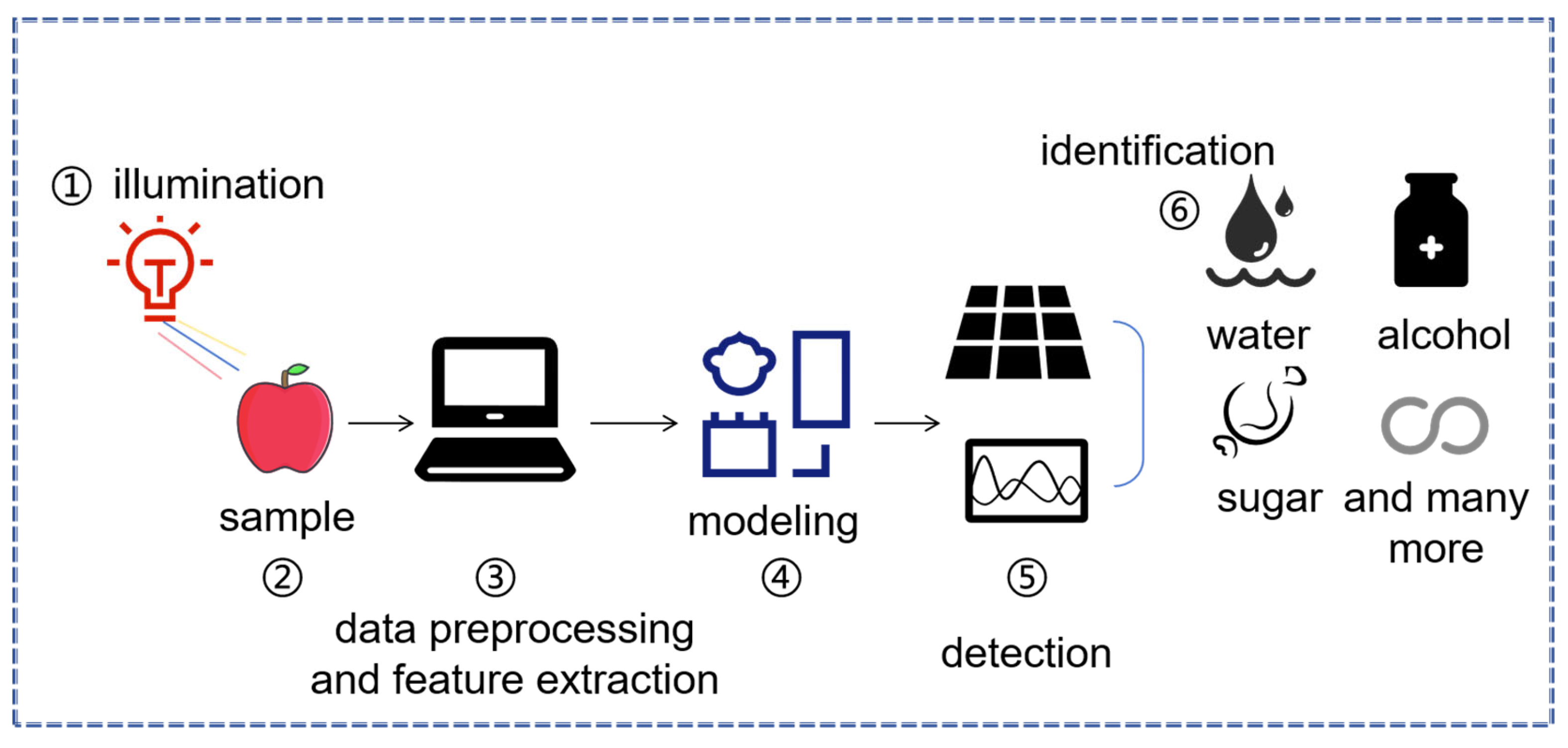
Figure 1. Steps for near-infrared spectroscopic measurement.
Agriculture 15 01670 g001
Figure 2. Structure diagram of Raman spectrometer.
Agriculture 15 01670 g002
Figure 3. Schematic of a typical THz experimental setup. The blue beam represents the pulse generated by the femtosecond laser, while the orange beam represents the terahertz pulse produced when the terahertz emitter is excited by the pulse generated by the femtosecond laser.
Agriculture 15 01670 g003
Figure 4. Hyperspectral imaging technology general workflow.
Agriculture 15 01670 g004
Figure 5. The plant disease identification system based on digital imaging mainly consists of four links, (a) data collection and data pre-processing; (b) plant disease recognition, in the deep learning section, different colors represent different neural network layers; (c) post-processing; and (d) evaluation.
Agriculture 15 01670 g005
Figure 6. A common thermal imaging experimental setup.
Agriculture 15 01670 g006
Table 1. Summaries focusing on diverse spectral techniques employed in plant disease detection.
TechniquesSamplesApplicationsAlgorithmsEquipmentReference
Near-infrared
spectroscopy
Sugar CaneSugarcane disease recognitionCNN, CWTUV2600[79]
AppleDetection of apple moldy core diseaseDMLPT, PLS-DA, SVM, ELMQE65pro[80]
AppleDetection of apple fungal infectionLDA, KNN, RFQE65pro[81]
AppleDetection of early moldy core applesSVM, ELM, KNNQE65pro[82]
TomatoDiagnosis of Cladosporium fulvum in greenhouse tomato plantsPCA, RBF, BP, SVMNIR system of Headwall Photonics Company, Bolton, MA, USA[83]
BananaDetection of the incubation period and onset period of banana wilt diseaseFDA, ELM, 1D-CNNUspectral-RIT-2.7.0[84]
‘Akizuki’ pearDiagnosing ‘Akizuki’ pear cork spot disorderSVM, RFNIR-S-G1[85]
Citri Reticulatae PericarpiumDiscrimination of mold-damaged Citri Reticulatae PericarpiumPLS-DA, MSCi-Spec Plus[86]
Saffron plantsDetection of mite-infested saffron plantsSVM, RBFHyspim, Sweden[87]
Raman spectroscopyChinese cabbageDetection of turnip yellow mosaic virus (TYMV) infectionPCA, LDADistributed Raman Microscope (Kaiser Optical Inc., Ann Arbor, MI, USA)[88]
Maize KernelsDetection and identification of plant pathogensOPLS-DAHandheld Rigaku Progeny ResQ Spectrometer[89]
MaizeIdentification of combined salinity stress and stalk rot diseaseSNV, PLS-DAHandheld Resolve Agilent Spectrometer[90]
Paddy riceAnalysis of paddy rice
infected by three pests and diseases
PLSDATriVista 555CRS Laser Raman Spectrometer[91]
BananaDetection of Fusarium wiltMDIP, IPDPPortable QE65 Pro Raman Spectrometer System[92]
StrawberryEarly on-site detection of strawberry anthracnosePCA, LDAPortable XPE85-NIR Spectrometer[93]
TomatoEarly detection
of bacterial canker of tomato
PCA, LDA, MLPHoriba XploRA ONETM Confocal Microscopy Spectrometer[94]
TomatoEarly detection of tomato spotted wilt virus infectionML, PLS-DAHandheld Bruker BRAVO Spectrometer[95]
RiceDetection of rice bacterial leaf blight and bacterial leaf streakCNN, SVM, RF, PCAPortable Raman spectrometer (produced by Ocean Optics of the United States, Largo, FL, USA)[96]
Terahertz
spectroscopy
ChestnutDetection of fungal infections in chestnutsBirnbaum-SaundersTHz camera (Tera-1024 32 × 32, Terasense, San Jose, CA, USA)[97]
PotatoIdentification of potato late blight and fusariosisRT-PCRTerahertz time-domain spectrometer (TPS Spectra 3000, Teraview, UK)[98]
TomatoDetection method for tomato leaf mildewPCA, BPNNTS7400 Terahertz Time Domain Spectroscopy Measurement System[99]
AppleApple Valsa canker detectionMSC, SGCCT-1800 Terahertz Time-Domain Imaging System[100]
Plant leafNon-invasive early monitoring of plant healthCNNTERA K15 Terahertz Time Domain Spectroscopy System[101]
Abbreviations: CNN, convolutional neural network; CWT, continuous wavelet transform; DMLPT, deep multi-layer perceptron; PLS-DA, partial least squares discriminant analysis; SVM, support vector machine; ELM, extreme learning machine; LDA, linear discriminant analysis; KNN, k-nearest neighbor; RF, random forest; PCA, principal component analysis; RBF, radial basis function; BP, back propagation; FDA, fisher discriminant analysis; 1D-CNN, one-dimensional convolutional neural network; MSC, multiplicative scatter correction; OPLS-DA, orthogonal projections to latent structures discriminant analysis; SNV, standard normal variate; MDIP, molecular detection of isolated pathogen; IPDP, in planta detection with PCR; MLP, multi-layer perceptron; ML, machine learning; BPNN, back-propagation neural network; SG, Savitzky–Golay; RT-PCR, reverse transcription-polymerase chain reaction.
Table 2. Summaries focusing on diverse imaging technology employed in plant disease detection.
TechniquesSamplesApplicationsAlgorithmsEquipmentReference
Hyperspectral imagingTomatoDetection of early blight and late blight diseasesELM, SPAImaging spectrometer (V10E-QE, Specim, Finland)[141]
Strawberry Strawberry foliar anthracnose assessmentSAM, SDA, PLSVNIR A series hyperspectral camera(Headwall HyperspecTM, Bolton, MS, USA)[142]
CapsicumPlant disease detectionSVM, RBFVNIR A series and SWIR M series hyperspectral cameras[143]
BananaThe effects of fungal diseases LDAHySpex VNIR-1600 Hyperspectral Camera[144]
Hordeum vulgarePlant disease forecastingGANHyper-spectral microscope[145]
Apple LeavesMonitoring the degree of mosaic diseaseSPA, CWT, PLSRSOC-710 Portable Hyperspectral Instrument (Surface Optics Corp, San Diego, CA, USA)[146]
WheatWheat yellow rust detectionPLSRHigh-spectrum imaging sensor (UHD 185)[147]
WheatEarly diagnosis of crown rot diseaseSVM, LDAFX10 camera and short-wave infrared camera[148]
PhalaenopsisFusarium wilt detection in Phalaenopsis2D-CNN, CBAM-EHyper-spectral sensor[149]
Digital imagingTomatoDetection of diseased tomato plantsSGM, SVMVisible light imaging camera (Canon Powershot S100)[150]
CucumberA recognition method for cucumber diseasesDCNN, SGDMNikon Coolpix S3100 Digital Camera[151]
CornDetection of corn leaf blightSSD, GIoUCamera[152]
Rice, Wheat, Tomato, Pepper, Cucumber, Squash, CornPlant disease diagnosisResNet50Camera, locator[153]
PomegranatePomegranate disease detection and classificationK-propagationCamera[154]
Thermal imagingGrapevineEarly detection of grapevine downy mildewSVMThermal imager (model FLIR SC655)[155]
Rice PlantsPlant disease predictionCNNFLIR C2 Camera[156]
RoseDetection of Botrytis cinerea infection in cut rosesLSDInfrared thermal imager (T530)[157]
PotatoIdentification of progress level of dry rot disease SVMInfrared thermal imager (model G120, NEC Avio, Tokyo, Japan)[158]
WheatEstimation of disease severity of wheat powdery mildewRFEAltum Camera (MicaSense USA, Inc., Raleigh, NC, USA)[159]
Sugar BeetEarly detection of sugar beet Cercospora leaf spot diseaseSVM, KNNHigh-resolution thermal imaging camera[160]
Persea americana, Malpighia emarginata, Myrciaria glaziovianaReal-time leaf disease classificationInceptionV3, MobileNetV1, VGG-16Infiray T3C Thermal Imaging Camera[161]
Cucumber, Sweet Potato, Wheat, Peanut, Oil PalmPlant disease detectionPCA, SVMPortable thermal imager[162]
Abbreviations: ELM, extreme learning machine; SPA, successive projections algorithm; SAM, spectral angle mapper; SDA, subspace discriminant analysis; PLS, partial least squares; GAN, generative adversarial network; CWT, continuous wavelet transform; PLSR, partial least squares regression; DW-ResNet, depth-wise residual network; SGM, superpixel graph matching; DCNN, deep convolutional neural network; SGDM, stochastic gradient descent with momentum; SSD, single shot multibox detector; GIoU, generalized intersection over union; DWConv, depth-wise convolution; IRB, inverted residual block; ResNet, residual network; LSD, local binary patterns-based spatial derivative; RFE, recursive feature elimination; PCA, principal component analysis.
Table 3. Comparison of different technologies.
TechniquesCostPortabilityDepth of PenetrationHumidity Sensitivity
Near-infrared
spectroscopy
Lower, relatively inexpensive equipment and low operation cost, suitable for large-scale applicationHigher, portable devices (such as handheld spectrometers) can be used in field sitesMedium, can obtain information on internal structure and composition of plant tissues, but with limited penetration depthHigher, spectral information is susceptible to humidity, which may cause data fluctuations and affect detection accuracy
Raman spectroscopyMedium, moderate cost for ordinary equipment, enhanced technologies (such as SERS) may be more expensiveHigher, handheld devices can be used for on-site detectionShallow, mainly detects molecular vibration information on the surface or shallow layers of samplesMedium, humidity may have some impact on detection, but the degree of influence is relatively small
Terahertz Spectroscopy Higher, expensive equipment limits its wide applicationLower, equipment is large in size and has poor portability, currently mainly used in laboratoriesDeeper, can penetrate many non-conductive materials and obtain deeper information on plant tissuesHigher, terahertz waves are susceptible to humidity during propagation and detection performance may decline in high-humidity environments
Hyperspectral imagingHigher, expensive equipment and high data processing and storage costsLower, equipment is usually large, although it can be combined with drones, overall portability is still limited, more used for laboratory fine analysisMedium, can simultaneously obtain image and spectral information, with moderate penetration depth for plant tissuesHigher, data collection is affected by environmental factors such as light and weather, humidity may also have some impact
Digital ImagingLower, equipment (such as ordinary cameras, cameras on drones) has low cost, easy to obtainHigher, equipment has good portability, can be collected on-site using smartphones, cameras, or dronesShallow, mainly obtains color, texture, etc., features of plant surfacesLower, humidity has a relatively small impact on digital imaging
Thermal ImagingMedium, moderate equipment cost, handheld devices and devices that can be mounted on dronesHigher, can be monitored on-site using handheld cameras or thermal imaging sensors mounted on dronesShallow, mainly detects temperature changes on the surface of plants to identify diseasesLower, humidity has a relatively small impact on thermal imaging
Disclaimer/Publisher’s Note: The statements, opinions and data contained in all publications are solely those of the individual author(s) and contributor(s) and not of MDPI and/or the editor(s). MDPI and/or the editor(s) disclaim responsibility for any injury to people or property resulting from any ideas, methods, instructions or products referred to in the content.