Article

Growth Kinetics Modeling and Evaluation of Antiphytopathogenic Activity of Newly Isolated Fungicolous Epicoccum nigrum Associated with Dryad’s Saddle (Polyporaceae)

1
Department of Microbiology and Biotechnology, University of Food Technologies, 26 Maritsa Blvd., 4002 Plovdiv, Bulgaria
2
Laboratory of Biologically Active Substances, Institute of Organic Chemistry with Centre of Phytochemistry, Bulgarian Academy of Sciences, 139 Ruski Blvd., 4000 Plovdiv, Bulgaria
*
Author to whom correspondence should be addressed.
Agriculture 2024, 14(12), 2179; https://doi.org/10.3390/agriculture14122179
Submission received: 30 October 2024 / Revised: 27 November 2024 / Accepted: 28 November 2024 / Published: 29 November 2024
(This article belongs to the Section Crop Protection, Diseases, Pests and Weeds)

Abstract

:
In the present study, an unknown fungal strain was isolated from the fruiting body of a local Dryad’s Saddle mushroom (Polyporaceae). The molecular identification of the isolate was performed by amplification of the ITS1-5.8S-ITS2 region and the strain was identified with 100.00% confidence as Epicoccum nigrum. The morphological characteristics, including the distinctive colony pigmentation, conidiophore structure, and conidial shape, were determined to ensure comprehensive characterization of the fungus. The modeling of the kinetics of the growth process was conducted with the applying the logistic curve model and the reverse autocatalytic growth model, and the concentrations of the compounds in the nutrient medium required for the E. nigrum development were established. Controlled submerged cultivation was carried out for cultural liquid obtaining, which was further used for the evaluation of the biological activities. The untreated cultural liquid demonstrated antimicrobial activity against Sclerotinia sclerotiorum where the minimal inhibitory concentration was 1.25 mg/mL. Antimicrobial activity was also detected toward Botrytis cinerea (2.5 mg/mL) and Aspergillus flavus (2.5 mg/mL). The direct utilization of crude cultural liquid for phytopathogenic control is a sustainable approach that will provide the opportunity for the development of an environmentally friendly manufacturing process.

1. Introduction

Fungicolous fungi, or fungi associated with other fungi, represent a very large, diverse, trophic and grossly underestimated group of organisms. Recent investigations revealed that fungicolous fungi were found across a wide range of lineages within the fungal kingdom, with the majority belonging to the Ascomycota phylum [1]. The interaction between these fungi and their host could be versatile and not always well defined. Currently, over 1550 fungal taxa have been identified as being in a relationship with other fungal [2], but most of them are described as mycoparasites or superparasites, often leading to the death of the fungal host [3]. Fungicolous fungi, as primary consumers, play an essential role in ecosystem functioning by accelerating nutrient cycling within food webs through their rapid and specialized life cycles. These fungi are integral to maintaining ecological balance as they influence host population dynamics and sizes across both aquatic and terrestrial ecosystems. By interacting with their hosts, fungicolous fungi can exert control over fungal communities, shaping biodiversity and ecosystem processes. A subset of fungicolous fungi operates as mycoparasites, targeting pathogenic fungi. This unique attribute makes them highly promising for use as biocontrol agents in agricultural and environmental management, helping to mitigate the spread of fungal diseases that affect crops and natural ecosystems. Conversely, fungicolous fungi can also act as harmful agents, causing significant diseases in cultivated edible and medicinal mushrooms. Such infections often lead to dramatic reductions in mushroom yield, quality, and economic value, presenting challenges to the mushroom cultivation industry. Beyond their practical applications and challenges, fungicolous fungi hold immense scientific value. They serve as model organisms for exploring the complexities of species interactions, offering insights into fungal evolution, genetic divergence, and the adaptive mechanisms that underpin fungicolous lifestyles. By studying these fungi, researchers can better understand the dynamic interplay between fungi and their environments, shedding light on broader ecological and evolutionary processes [3].
The members of the genus Epicoccum are ascomycete organisms, which are distributed worldwide and could be isolated from various substrata, but most commonly from colonized soil and host plants [4]. Although these species are primarily regarded as a saprotrophic fungus, they possess the potential to exhibit an endophytic lifestyle. Currently, the interactions between plants and their endo- and epiphytic microflora are of interest for the research teams around the world. Regarding the plant–endophyte interaction, it is well known that in some cases the endophyte is the one triggering the synthesis of a specific metabolite. On the other hand, endophytes are usually known to possess unique biological activities, which serve them in their interactions with the host organism and in some cases may lead to beneficial effects. In the search for metabolites with target biological activities, scientists increasingly turn their gaze to natural sources. As such, mushrooms are subjected to intense research in the last few decades. It is our hypothesis that some of the biological activities possessed by higher fungi are due to their interactions with fungicolous microorganisms, or only to the fungicolous fungus itself. In this case, the cultivation of the mushroom in order to obtain the target metabolite would not be necessary if the fungicolous fungus is the one possessing the desired activity.
Epicoccum species could be isolated from sugarcane or rice as the most common host organisms [5,6,7] but the available data in the literature regarding their possible interaction with mushrooms is scarce [8,9,10]. The knowledge about the fungicolous fungi associated with certain mushroom types could be crucial for their cultivation and metabolite production. Numerous biologically active secondary metabolites produced by Epicoccum species including polyketides, diketopiperazines, and pigments were characterized previously [11]. Biological activities such as antibacterial, antiparasitic, and immune suppression are related to polyketides, which makes them valuable from a pharmaceutical point of view. A broad range of activities is associated with the diketopiperazines. As a representative of genus Epicoccum, the species Epicoccum nigrum is also capable of synthesis of metabolites with biological activities of interest. Some E. nigrum strains produce polycyclic alkaloids, such as epicoccins, possessing antibacterial properties [9], and epicoccamides—reported to possess anticancer properties [10,12,13]. Actually, one of the most distinctive compounds produced by E. nigrum is the fluorescent pigment epicocconone, which has applications as a protein-binding dye and possesses broad antifungal activity [14]. In combination with the antifungal activity against the pathogenic strains, Fusarium verticillioides, Colletotrichum falcatum, Ceratocystis paradoxa, Xanthomomas albilineans, and E. nigrum P16 demonstrated capabilities of increasing the biomass of the root system of sugarcane [5].
There are several reports on the ability of E. nigrum strains to inhibit the growth of the Botrytis cinerea, one of the most abundant and important plant pathogens [15,16]. Some representatives demonstrate an ability to cause the inhibition of the conidial germination and growth of plenty of fungal phytopathogens, which is the main reason for several research papers on the possibility of its application as a biocontrol agent [16,17,18]. The introduction of such biocontrol agents, possessing antiphytopathogenic activities, in agriculture has several advantages. Firstly, phytopathogenic microorganisms could lead to significant crop damage and reduced yields. Preventing the proliferation of such pathogens with the employment of antiphytopathogenic agents will control or even prevent their growth, leading to stable and high crop yields. Secondly, traditional pesticides are effective but their overuse can cause environmental problems such as soil and water pollution, soil degradation, or even the development of pesticide-resistant organisms. The application of microorganisms with antiphytopathogenic activity is a sustainable alternative, which might lead to decreasing the need for synthetic pesticides thus minimizing the environmental impact. Also, these microorganisms could promote biodiversity and reduce the toxic load on ecosystems when combined with sustainable farming practices.
The fungal kingdom comprises millions of undiscovered species, which supposedly possess unique biological activities. In addition, the interaction between the mushrooms and fungicolous fungi, as well as the identification of these microorganisms, is of great interest to the scientific community. In the present study, we report on isolation, identification, mycelium growth kinetics, and the evaluation of the antiphytopathogenic activity of a newly isolated E. nigrum strain from the fruiting body of the Dryad’s Saddle mushroom.

2. Materials and Methods

2.1. Fungal Isolation, Cultivation, and Morphological Characterization

The fruiting body of the Dryad’s Saddle (Figure 1) was collected from a deciduous tree in May 2022 in a park near Maritza River, Plovdiv, Bulgaria (42.148732, 24.717380). The collection location is a public area, and the mushroom is not a protected species therefore no special permissions for the collection were required. After the collection, specimens with no visible damage were cut out from the fruiting body and cleaned with sterile distilled water. The surface of the samples was afterwards disinfected, following the protocol presented by Angelova et al. [19]. Briefly, the surface was disinfected by treatment with 70% ethanol, followed by a 2% NaClO solution and rinsing with sterile distilled water. The samples were further sliced to 2.5 by 2.5 mm pieces with a sterile scalpel and aseptically transferred onto a Rose Bengal Chloramphenicol Agar (RBCA) (HiMedia Laboratories GmbH, Modautal, Germany). The unknown fungal colonies were isolated by several transitions onto a fresh RBCA medium. The obtained unknown pure isolate was cultured in Petri dishes on Mushroom Complete Medium (MCM) with the following composition (g/L): glucose—20.0, KH2PO4—0.5, K2HPO4—1.0, MgSO4 × H2O—0.5, peptone—2.0, yeast extract—2.0, agar—2.0, and pH 5.5—6.0. The fully grown culture was stored at 4 °C and was further used for morphological characterization and molecular identification.
The macroscopic characteristics (the texture and size, color of the top of the colony and the bottom side of the plate) of the unknown fungal isolate were observed on a daily basis. Microscopic characterization was performed by observation of the culture under a biological microscope, Olympus CX33 (Olympus, Tokyo, Japan), after the mycelium was mounted in water or fixed with the scotch tape imprint method.

2.2. Molecular Identification by ITS1-5.8S-ITS2 rRNA Gene Sequence Analysis

The unknown fungal isolate was cultivated for 6 days on MCM agar plates. The fungal mycelium was scraped out with a sterile spatula (100–300 mg) and transferred to a 2 mL microtube. Total DNA was extracted using a modified CTAB method, according to Stefanova et al. [20]. The quality and concentration of the DNA extracts were assessed by determination of their absorbance at 260 nm and 280 nm (Shimadzu UV-VIS, Shimadzu Corporation, Kyoto, Japan).
The ITS-5.8S-ITS2 region was amplified by forward primer ITS 4 (5′-TCCTCCGCTTATTGATATGC-3′) and reverse primer ITS 5 (5′-GGAAGTAAAAGTGCTAACAAGG-3′) [21], obtained from Metabion (Martinsried, Germany). The PCR reaction mix contained 1 μL of DNA (50 ng), 0.5 μM of each primer, and 8 μL of Red-Taq DNA Polymerase Master Mix (Canvax Biotech, S.L., Valladolid, Spain) in a total volume of 20 μL. The amplification was carried out in a PCR 2720 Thermal Cycler (Applied Biosystems, Carlsbad, CA, USA) using the following program: initial denaturation at 95 °C for 10 min, followed by 35 cycles of 1 min at 95 °C, 1 min at 52 °C and 1 min at 72 °C, and final extension at 72 °C for 7 min. The PCR product was visualized in a 1% agarose gel stained with SafeView (NBS Biologicals, Huntingdon, UK) at 100 V for 50 min using VWR Mini Electrophoresis System (VWR, Darmstadt, Germany) and MiniBis Pro (DNR Bio-Imaging Systems, Jerusalem, Israel) for gel visualization. The PCR product was cut out from the gel and purified with Clean-Easy™ Agarose Purification Kit (Canvax Biotech, S.L., Valladolid, Spain).
Sequencing of the PCR product was performed by Microsynth Seqlab (Göttingen, Germany). The resulting sequence was analyzed using BLAST algorithm [22] and compared with the nucleotide sequences in the GenBank database [23]. The ITS1-5.8S-ITS2 rRNA gene sequence of the fungal isolate was deposited in the GenBank database and an accession number was assigned.

2.3. Phlylogenetic Analysis

The phylogenetic analysis was conducted using the closest matched ITS sequences from the GenBank database [23] and ITS sequences of six other species of genus Epicoccum. Didymella rosea (GenBank Accession No. NR_136125.1) was used as an outgroup taxa. CLC Genomics Workbench 20.0 [24] was used to align the sequences and build the phylogenetic tree by means of the Unweighted Pair Group Method using the Arithmetic Average (UPGMA) clustering algorithm [25]. The nucleotide distance was evaluated by Jukes–Cantor distance model and standard bootstrap analysis with 1000 replicates was performed.

2.4. Cultivation and Modeling of the Kinetics of the Process

The cultivation of the strain was performed in Petri dishes with the following nutrient media: MCM, glucose-peptone (GP), malt yeast extract (MYE), malt extract agar (MEA) and yeast extract malt extract agar (YMA), and pH 5.5–6.0 prior sterilization (Table 1). The dishes were inoculated with agar disks (d = 10 mm) of a fully grown culture, incubated at 25 °C, and the diameter of the growing colonies was measured daily. The resulting data were used for the modeling of the growth kinetics by applying the logistic curve model (Equation (1)) and the reverse autocatalytic growth model (Equation (2)) [26,27].
d D d τ = μ m a x 1 D D m D
d D d τ = k 1 S 0 D D k 2 D 2
where μmax—specific growth rate, d−1; D and Dm—current and maximal diameter of the mycelium, mm; k1—biomass yield rate constant, d−1; k2—biomass death rate constant, d−1; S0—initial substrate quantity in cell units described with the diameter of the mycelia, mm.
The applied models were solved by the Runge–Kutta methods of 4th order and the identification of their parameters was achieved by minimization of the difference in the square between the experimental and model data by applying Excel’s Solver function [28,29].

2.5. Submerged Cultivation of E. nigrum

The submerged cultivation of the pure culture was conducted in 500 mL Erlenmeyer’s flasks containing 100 mL MCM. The cultivation took place on a rotary shaker at 220 rpm and 25 °C for 5 days in the absence of light. At the end of the cultivation process, the biomass was separated from the medium by filtering on a Buhner’s funnel. The culture liquid was collected and used for the evaluation of the antiphytopathogenic activity of the E. nigrum strain after membrane sterilization through a sterile 0.20 µm membrane filter.

2.6. Antiphytopathogenic Activity

The minimal inhibitory concentration (MIC) of the culture liquid was determined against Penicillium chrysogenum, Fusarium verticillioides, Aspergillus flavus, Beauveria bassiana, Botrytis cinerea, and Sclerotinia sclerotiorum according to the CLSI method [30]. The fungal strains were provided from the collection of the Department of Microbiology and Biotechnology at the University of Food Technologies, Plovdiv, Bulgaria. The culture liquid was subjected to serial two-fold dilutions in RPMI-1640 broth (Merck KGaA, Darmstadt, Germany) in 96-well microtiter plates. Each well was inoculated with a microbial suspension at a concentration of 5 × 10⁵ CFU/mL. Two controls were used—a growth control (positive control) and a sample control for each dilution without inoculation (negative control). After mixing, the plates were incubated at 35 °C for 48 h, after which they were examined visually. The MIC was the lowest concentration that prevented any discernible growth (100% inhibition).

2.7. Statistical Analysis

All the experiments were conducted in triplicate and the values were expressed as mean values ± SD. The statistical significance was determined by the analysis of variance (ANOVA, Tukey’s test, and LSD test); the value of p < 0.05 indicated a statistical difference.

3. Results and Discussion

3.1. Isolation and Identification of Unknown Fungal Isolate

The fungus was isolated as a pure mycelial culture by several transitions onto fresh nutrient medium. The resulting culture was used for molecular analysis for achieving proper identification.
Molecular identification of the unknown fungal isolate was performed by amplification of the ITS1-5.8S-ITS2 region and the obtained PCR product was subjected to sequence analysis. The resulting sequence was analyzed using the BLAST algorithm and compared with the nucleotide sequences available at the GenBank database [23]. The strain was identified with a high level of confidence (100.00%) as Epicoccum nigrum. The ITS1-5.8S-ITS2 rRNA gene sequence of the newly isolated E. nigrum was deposited in GenBank and the accession number PQ438155 was assigned.

3.2. Phylogenetic Analysis

The genetic relationships between E. nigrum PQ438155, seventeen closely related E. nigrum strains, and six other species of genus Epicoccum were examined based on the partial sequences analysis of the ITS1-5.8S-ITS2 region (Figure 2). The phylogenetic tree showed that E. nigrum PQ438155 was clustered in the same clade with other E. nigrum strains. Some of the other Epicoccum species used in this study were grouped in a different clade (E. italicum PQ562358, E. ovisporum NR_158228, and Epicoccum isolate OK090879). E. proteae NR_158240 was also rather distantly related to the other E. nigrum strains. The presented results are in agreement with other authors, demonstrating the closely related relationship between some E. nigrum strains and E. layuense [6,31].

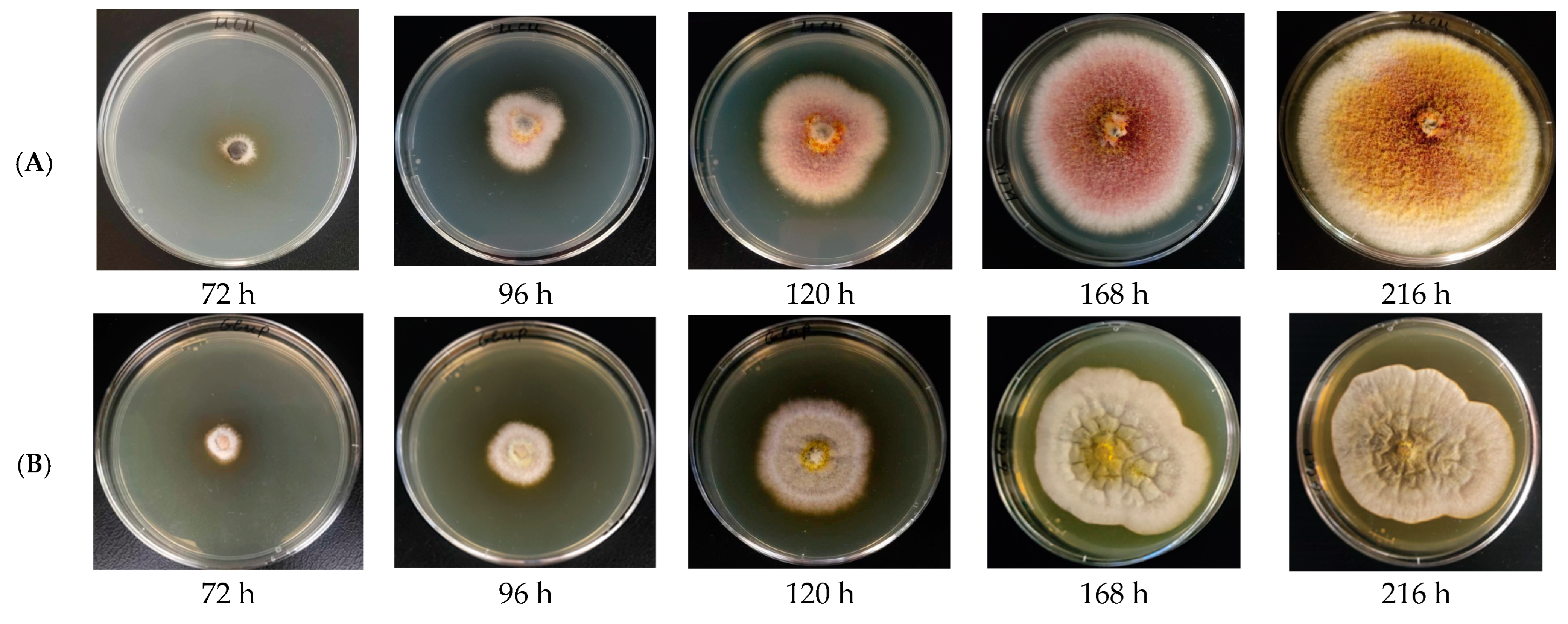
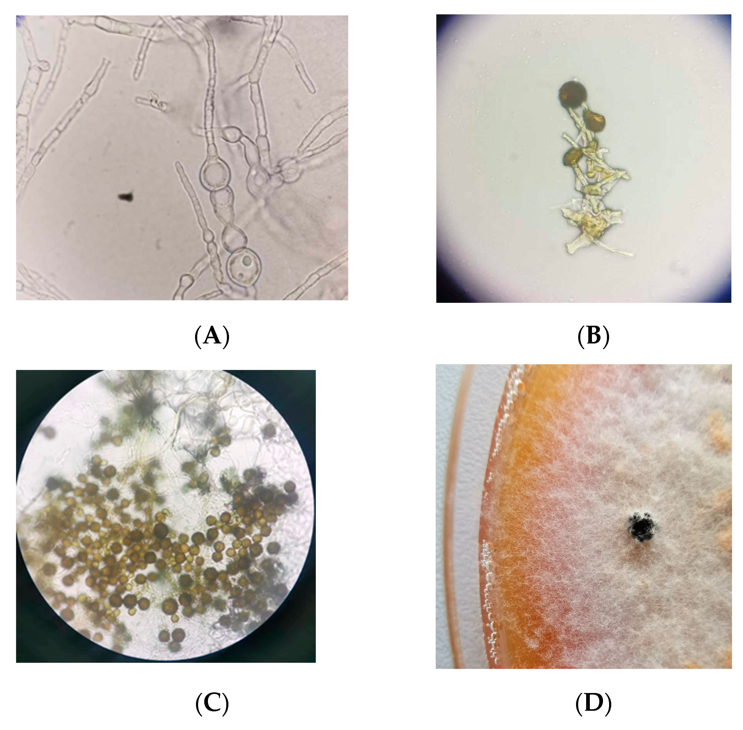
3.3. Morphological Characterization

The morphological characterization of E. nigrum is essential for accurate identification and comprehensive strain characterization. These characteristics encompass several distinct features like colony pigmentation, conidiophore structure, and conidial shape and size. The pigmentation and texture of the colony play a significant role in differentiating it from closely related species. The structure of the conidiophores is a key morphological trait where the shape, branching pattern, and size of the conidiophores serve as a vital criterion in distinguishing it and contributing to the verification of its molecular identification. Also, the conidial shape, size, and surface texture are crucial for precise identification. Together these morphological characteristics reinforce the accuracy of the molecular identification methods, ensuring reliable confirmation of the strain.
The morphology of the E. nigrum colonies was examined across different culture media. These colonies are slow growing, typically taking around 10 days to develop, and display a characteristic morphology. Initially, the colonies appear fluffy or wooly, becoming dense and compact in later stages. Over time, the colonies exhibit a distinctive color change, transitioning from white or pale to shades of pink, yellow, orange, red, or brown, due to the production of various pigments. This pigmentation is influenced by the composition of the culture medium (Figure 3A,B).
Notably, yellow to orange pigments begin to form after 72 h of cultivation on MCM. E. nigrum grows as an anamorph, primarily reproducing asexually through conidia. During cultivation at 25 °C, no exudates were observed, and only immature conidia were present (Figure 4A). The conidia are typically spherical to subspherical (globose to subglobose) and are borne on short, simple, or branched conidiophores, which aggregate to form sporodochia. Sporodochia were detected after 30 days of incubation at 4 °C (Figure 4D). The conidiophores are small and sometimes slightly curved, and due to their integration into the colony structure, they are often difficult to observe without magnification. Immature conidia are pale, while mature conidia are dark brown to black, heavily pigmented, and frequently possess rough walls with surface ornamentations or ridges. Conidia are produced singly or in short chains at the tips of the conidiophores (Figure 4B,C). The observed morphological features align with the previously described characteristics of E. nigrum strains and support the molecular identification of this species [15,32,33].

3.4. Modeling of the Kinetics of the Process

The growth dynamics of E. nigrum on the selected media were evaluated in a series of experiments. The main parameter used for the determination of the fungal growth was the diameter of the mycelium, which was measured periodically during the experiment. The obtained data are presented in Figure 5.
After observation of the growth curves, it is obvious that the strain forms mycelium with a maximal diameter of 75 mm. The maximal diameter was achieved for a different amount of time on the different media. When E. nigrum was cultivated on MEA, YMA, and GP media, the active growth of the culture continued until the 9th day of the cultivation when the maximal diameter was achieved. With this in mind, it can be concluded that if the goal is to obtain biomass, the cultivation could be carried out for up to 9–10 days. However, if the goal is the accumulation of secondary biologically active metabolites, then the cultivation should be carried out for up to 15 days. When the cultivation of E. nigrum was conducted on MCM and MYE, the mycelium diameter was constantly increasing intensively until the 9th day and afterwards slowly up until the 15th day when the maximal diameter was achieved.
The presented data clearly show that both models demonstrate high values for the correlation coefficient, ranging from 0.9970 to 0.9983. This proves the suitability of the models for the current analysis of the parameters and that they can be applied for modeling and prediction of the process. The data show that the specific growth rate values, calculated with the logistic curve model, are relatively high and similar for the different media, varying from 0.408 ± 0.018 d−1 to 0.495 ± 0.056 d−1. Due to the fact that the µmax values are analogs, the best medium for E. nigrum growth could not be determine, so the data were statistically processed by the method of least significant differences (LSD). This proved that in fact the strain will be able to grow well on all used media based on the µmax values. The statistically significant difference in the µmax values was established with the MYE and MEA media, giving advantage to the MEA. This is confirmed by the µmax values, which are 0.495 ± 0.056 d−1 and 0.408 ± 0.018 d−1 for MEA and MYE, respectively. The relationship between µmax and the maximal diameter is described with the coefficient of growth inhibition δ. For the different media, this coefficient has values between 0.0055 ± 0.0007 mm·d−1 and 0.0064 ± 0.0008 mm·d−1. Those values are significantly lower than 1, proving the absence of inhibition factors in the used media. This is also confirmed by the lower values of the k2 constant in the reverse autocatalytic growth model. The logistic curve model predicts a maximal diameter in the different media ranging from 75 ± 6 and 77 ± 1 mm, and those values are similar to the experimentally obtained data (74 ± 0.00 and 75 ± 0.00 mm).
The effect of the composition of the five nutrient media on the growth of E. nigrum was evaluated. Due to the fact that the increasing of the diameter is the main parameter characterizing the strain growth, the logistic curve and the reversed autocatalytic growth models were applied. In these models, it is possible for the diameter to be used instead of biomass concentration. The data obtained from the applied models is presented in Table 2.
Regarding the reverse autocatalytic growth model, the k1 constant also has similar values for all used media. The parameter K/1 + K, describing the substrate utilization for biomass gain, is of interest. The values for this parameter are close to 1, which indicates that the composition of all used media is suitable for E. nigrum growth. The similar values of the substrate utilization coefficient prevent the unequivocal determination of the most suitable medium and thus the LSD method was applied. The received data demonstrated no statistical difference between the MCM, GP, MYE, and MEA media, where the substrate will be utilized with similar speed by the strain. Regarding the K/1 − K parameter, there is statistical difference between all used media. The substrate is utilized to a greater extent in MEA compared to MCM. The same trend is observed with the comparison of MCM to YMA, where the K/1 + K values are, respectively, 0.9951 ± 0.0003 and 0.9989 ± 0.0011. Also, there are significant differences in the K/1 + K parameter regarding the GP compared with the MEA and YMA media, where the values for MEA and YMA are higher (0.9967 ± 0.0002 and 0.9989 ± 0.0011), meaning more complete substrate utilization compared to GP (0.9951 ± 0.0003). Significant difference in the parameter is identified in the comparison between MYE and YMA, and MEA to YMA. Here, the substrate is more completely utilized in MYE (0.9989 ± 0.0011), followed by MEA (0.9967 ± 0.0002) and finally MYE (0.9963 ± 0.0012).
The initial amount of the substrate in cell units, described as mycelium diameter, could be theoretically determined by the application of the reverse autocatalytic growth model. In other words, the exact concentration of the components of the medium required for E. nigrum growth or the nutrient supply of the media can be determined. The S0 values for the used media are 113 mm, 116 mm, 101 mm, 103 mm, and 83 mm, respectively, for MCM, GP, MYE, MEA, and YMA thus meaning that the nutrient supply in MCM and GP is best for E. nigrum growth.

3.5. Antiphytopathogenic Activity

The culture liquid obtained after the submerged cultivation of E. nigrum without any further processing was tested for antimicrobial activity against six common phytopathogenic fungi. Phytopathogenic fungi are responsible for the majority of diseases in agricultural and horticultural environments. These pathogens typically develop infection methods to invade plants, seeking entry points and forcibly extracting nutrients to support their growth and reproduction. They can bypass the plant’s immune defenses, which leads to disruptions in the plant’s health, balance, and functioning, potentially causing widespread damage. To combat the ongoing threat of phytopathogenic fungi in agriculture, various agrochemicals have been employed, though some pose toxicity risks to humans, requiring a waiting period between application and harvest. Additionally, these chemicals can have harmful effects on soil organisms, insects, and pollinators [34].
Inhibitory activity was detected against Penicillium chrysogenum, Aspegillus flavus, Botrytis cinerea, and Sclerotinia slerotorium, and the last fungus was the most susceptible to the effect of the culture liquid (Table 3). S. slerotorium is a phytophatogenic fungus that infects sunflower plants leading to mid-stalk rot, basal-stalk rot, rotting of tissues, and even falling of the seeds of the sunflower could occur. The phytophatogenic effect of E. nirgum extracts over S. slerotorium was reported previously [35,36,37] for various cultures where the spray application of spores was utilized. It was established that such application methods lead to losses in kiwi production [37]. It is important to note that the inhibition effect in our study was achieved only with cultural liquid, without any further extraction procedures with solvents or expensive chromatography methods. The other phytopathogen object to our study, B. cinerea, is at the top of the list of the most important fungal pathogens and it is able to cause diseases in over 200 different plant species, leading to huge economy loses. E. nigrum isolates have demonstrated the ability to inhibit the growth of B. cinerea in an in vitro antagonism test [15], meaning that these isolates possess antagonistic properties towards the pathogen. Keeping in mind these results and given the results in Table 3, the metabolites causing the inhibition of the growth of B. cinerea are likely to be extracellular.
Lee at al. (2020) [38] report the antimicrobial activity of an antifungal compound (Epipyrone A) produced by E. nigrum against B. cinerea, S. sclerotiorum, and several Aspergillus species. The reported antimicrobial activity was a result of the effect of the extracted metabolites on the test microorganisms. In our study, the antiphytopathogenic activity is due to the presence of certain metabolites in the culture liquid, which was used without any concentration of purification. The yellow mold, Aspergillus flavus, is a soil-dwelling saprobe, found globally, that infects a range of key agricultural crops. It frequently affects legumes, cereal grains, and tree nuts. This pathogen produces an aflatoxin, a secondary metabolite derived from polyketides, which is both carcinogenic and mutagenic. The species of genus Penicillium are one of the major groups of organisms causing spoilage in fruits and bread [39,40]. These fungi are known for their ability to produce mycotoxins in foods and feeds, for instance ochratoxin A and patulin [41]. Exposure to high levels of aflatoxin through inhalation or consumption of contaminated food or feed can lead to a condition known as aflatoxicosis [34]. Ochratoxin A could demonstrate direct or indirect genotoxic and nongenotoxic modes of action but in both cases, these lead to tumor formations [42], and the toxic action of patulin is related to the disruption of the immune system. It was established that the cultural liquid demonstrated antifungal activity against A. flavus, as well as against P. chrysogenum. There are no data in the available literature regarding the antifungal activity of the culture liquid obtained after the submerged cultivation of E. nigrum towards these strains and this is the first report of such an activity.
These results confirm that E. nigrum could be applied as a biocontrol agent against phytopathogenic fungi. It is important to be note that the cultural liquid was used without any treatments prior to the determination of MICs. The application of concentration or purification procedures could indeed enhance the concentration of the target metabolites in the sample but it will also increase the cost of the process. Thus, the usage of the cultural liquid without any processing will provide the opportunity for the development of an environmentally friendly manufacturing process with reduced production costs. Future experiments are needed for establishing the chemical nature of the metabolites providing the antiphytopathogenic activity of the culture liquid.

4. Conclusions

This study is the first to report the isolation of Epicoccum nigrum from the fruiting body of the Dryad’s Saddle mushroom. Logistic curve and reversed autocatalytic growth models were applied to assess the impact of five different nutrient media compositions on the growth of E. nigrum, allowing for the selection of the most suitable medium for fungal growth. For the first time, crude culture liquid, obtained after the submerged cultivation of the strain, was used to evaluate its antiphytopathogenic activity. Inhibitory effects were observed against Penicillium chrysogenum, Aspergillus flavus, Botrytis cinerea, and Sclerotinia sclerotiorum, with the latter being the most susceptible. These findings suggest that E. nigrum could be effectively used as a biocontrol agent against phytopathogenic fungi. Furthermore, the proposed approach of directly utilizing the crude culture liquid is more sustainable and helps reduce production costs.

Author Contributions

Conceptualization, G.A. and D.B.; methodology, M.B., R.B., D.B. and P.D.; software, P.S.; validation, R.B., M.B. and P.S.; formal analysis, Z.G. and R.B.; investigation, M.B. and D.T.; resources, D.B.; data curation, B.G.; writing—original draft preparation, R.B., M.B., P.S., G.A., D.B. and B.G.; writing—review and editing, G.A., D.B. and P.D.; visualization, M.B.; supervision, M.B.; project administration, R.B.; funding acquisition, R.B. All authors have read and agreed to the published version of the manuscript.

Funding

This research is supported by the Bulgarian Ministry of Education and Science under the National program “Young Scientists and Postdoctoral Students—2”, DCM #206/07.04.2022.

Institutional Review Board Statement

Not applicable.

Data Availability Statement

The original contributions presented in this study are included in the article. Further inquiries can be directed to the corresponding author.

Conflicts of Interest

The authors declare no conflicts of interest.

References

  1. Maurice, S.; Arnault, G.; Nordén, J.; Botnen, S.S.; Miettinen, O.; Kauserud, H. Fungal sporocarps house diverse and host-specific communities of fungicolous fungi. ISME J. 2021, 15, 1445–1457. [Google Scholar] [CrossRef] [PubMed]
  2. Sucharzewska, E.; Ejdys, E.; Kubiak, D. Fungicolous fungi on microscopic fungi parasitic to the vegetation of the urban environment. Acta Mycol. 2023, 58, 1–9. [Google Scholar] [CrossRef]
  3. Sun, J.-Z.; Liu, X.-Z.; McKenzie, E.H.C.; Jeewon, R.; Liu, J.-K.J.; Zhang, X.-L.; Zhao, Q.; Hyde, K.D. Fungicolous fungi: Terminology, diversity, distribution, evolution, and species checklist. Fungal Divers. 2019, 95, 337–430. [Google Scholar] [CrossRef]
  4. Fatima, N.; Ismail, T.; Muhammad, S.; Jadoon, M.; Ahmed, S.; Azhar, S.; Mumtaz, A. Epicoccum sp., An emerging source of unique bioactive metabolites. Acta Pol. Pharm. Drug Res. 2016, 73, 13–21. [Google Scholar]
  5. Fávaro, L.C.d.L.; Sebastianes, F.L.d.S.; Araújo, W.L. Epicoccum nigrum P16, a Sugarcane Endophyte, Produces Antifungal Compounds and Induces Root Growth. PLoS ONE 2012, 7, e36826. [Google Scholar] [CrossRef] [PubMed]
  6. Ali, S.A.-B.; Abdelmoaty, H.; Ramadan, H.; Salman, Y. The endophytic fungus Epicoccum nigrum: Isolation, molecular identification and study its antifungal activity against phytopathogenic fungus Fusarium solani. J. Microbiol. Biotechnol. Food Sci. 2024, 13, e10093. [Google Scholar] [CrossRef]
  7. Wen, J.; Okyere, S.K.; Wang, S.; Wang, J.; Xie, L.; Ran, Y.; Hu, Y. Endophytic Fungi: An Effective Alternative Source of Plant-Derived Bioactive Compounds for Pharmacological Studies. J. Fungi 2022, 8, 205. [Google Scholar] [CrossRef]
  8. Li, Q.; Chen, C.; Penttinen, P.; Xiong, C.; Zheng, L.; Huang, W. Microbial diversity associated with Tricholoma matsutake fruiting bodies. Microbiology 2016, 85, 531–539. [Google Scholar] [CrossRef]
  9. Zhang, Y.; Liu, S.; Che, Y.; Liu, X. Epicoccins A–D, epipolythiodioxopiperazines from a Cordyceps-colonizing isolate of Epicoccum nigrum. J. Nat. Prod. 2007, 70, 1522–1525. [Google Scholar] [CrossRef]
  10. Wangun, H.V.K.; Dahse, H.M.; Hertweck, C. Epicoccamides B–D, glycosylated tetramic acid derivatives from an Epicoccum sp. associated with the tree fungus Pholiota squarrosa. J. Nat. Prod. 2007, 70, 1800–1803. [Google Scholar] [CrossRef]
  11. Braga, R.M.; Padilla, G.; Araújo, W.L. The biotechnological potential of Epicoccum spp.: Diversity of secondary metabolites. Crit. Rev. Microbiol. 2018, 44, 759–778. [Google Scholar] [CrossRef] [PubMed]
  12. Wright, A.D.; Osterhage, C.; König, G.M. Epicoccamide, a novel secondary metabolite from a jellyfish-derived culture of Epicoccum purpurascens. Org. Biomol. Chem. 2003, 1, 507–510. [Google Scholar] [CrossRef] [PubMed]
  13. Yajima, A.; Kawajiri, A.; Mori, A.; Katsura, R.; Nakada, T. Total synthesis of epicoccamides A and D via olefin cross-metathesis. Tetrahedron Lett. 2014, 55, 4350–4354. [Google Scholar] [CrossRef]
  14. Randrianjatovo, I.; Girbal-Neuhauser, E.; Marcato-Romain, C.E. Epicocconone, a sensitive and specific fluorescent dye for in situ quantification of extracellular proteins within bacterial biofilms. Appl. Microbiol. Biotechnol. 2015, 99, 4835–4844. [Google Scholar] [CrossRef]
  15. Christova, P.; Slavov, S. Epicoccum nigrum—Isolation, characterization and potential for biological control of Botrytis cinerea. Bulg. J. Agric. Sci. 2021, 27, 693–698. [Google Scholar]
  16. Alcock, A.; Elmer, P.; Marsden, R.; Parry, F. Inhibition of Botrytis cinerea by epirodin: A secondary metabolite from New Zealand isolates of Epicoccum nigrum. J. Phytopathol. 2015, 163, 841–852. [Google Scholar] [CrossRef]
  17. Bhuiyan, S.A.; Ryley, M.J.; Galea, V.J.; Tay, D. Evaluation of potential biocontrol agents against Claviceps africana in vitro and in vivo. Plant Pathol. 2003, 52, 60–67. [Google Scholar] [CrossRef]
  18. Li, Y.; Xia, L.Q.Q.; Wang, Y.N.N.; Liu, X.Y.Y.; Zhang, C.H.H.; Hu, T.L.L.; Cao, K.Q.Q. The inhibitory effect of Epicoccum nigrum strain XF1 against Phytophthora infestans. Biol. Control 2013, 67, 462–468. [Google Scholar] [CrossRef]
  19. Angelova, G.; Stefanova, P.; Brazkova, M.; Krastanov, A. Molecular and morphological characterization of Xylaria karsticola (Ascomycota) isolated from the fruiting body of Macrolepiota procera (Basidiomycota) from Bulgaria. PLoS ONE 2023, 18, e0287679. [Google Scholar] [CrossRef]
  20. Stefanova, P.; Taseva, M.; Georgieva Tz Gotcheva, V.; Angelov, A. A modified CTAB method for DNA extraction from soybean and meat products. Biotechnol. Biotechnol. Equip. 2013, 27, 3803–3810. [Google Scholar] [CrossRef]
  21. Toju, H.; Tanabe, A.S.; Yamamoto, S.; Sato, H. High-coverage ITS primers for the DNA-based identification of ascomycetes and basidiomycetes in environmental samples. PLoS ONE 2012, 7, e40863. [Google Scholar] [CrossRef]
  22. Altschul, S.F.; Gish, W.; Miller, W.; Myers, E.W.; Lipman, D.J. Basic local alignment search tool. J. Mol. Biol. 1990, 215, 403–410. [Google Scholar] [CrossRef]
  23. Gene Bank Database. Available online: https://www.ncbi.nlm.nih.gov (accessed on 24 September 2024).
  24. QIAGEN Digital Insights. Available online: https://digitalinsights.qiagen.com (accessed on 25 September 2024).
  25. Saitou, N.; Nei, M. The neighbor-joining method: A new method for reconstructing phylogenetic trees. Mol. Biol. Evol. 1987, 4, 406–425. [Google Scholar] [CrossRef] [PubMed]
  26. Bouguettoucha, A.; Balannec, B.; Amrane, A. Unstructured Models for Lactic Acid Fermentation—A Review. Food Technol. Biotechnol. 2011, 49, 3–12. [Google Scholar]
  27. Ibarz, A.; Augusto, P.E. An autocatalytic kinetic model for describing microbial growth during fermentation. Bioprocess. Biosyst. Eng. 2015, 38, 199–205. [Google Scholar] [CrossRef] [PubMed]
  28. Choi, M.; Al-Zahrani, S.M.; Lee, S.Y. Kinetic model-based feed-forward controlled fed-batch fermentation of Lactobacillus rhamnosus for the production of lactic acid from Arabic date juice. Bioprocess. Biosyst. Eng. 2014, 37, 1007–1015. [Google Scholar] [CrossRef] [PubMed]
  29. Kemmer, G.; Keller, S. Nonlinear least-squares data fitting in Excel spreadsheets. Nat. Protoc. 2010, 5, 267–281. [Google Scholar] [CrossRef]
  30. CLSI. Method for Broth Dilution Antifungal Susceptibility Testing of Filamentous Fungi, Ap-Proved Standard, 2nd ed.; CLSI document М38-А2; Clinical and Laboratory Standards Institute: Wayne, PA, USA, 2008. [Google Scholar]
  31. Castro, P.; Parada, R.; Corrial, C.; Mendoza, L.; Cotoras, M. Endophytic Fungi Isolated from Baccharis linearis and Echinopsis chiloensis with Antifungal Activity against Botrytis cinerea. J. Fungi 2022, 8, 197. [Google Scholar] [CrossRef]
  32. Abdel-Hafez, S.; Nafady, N.; Abdel-Rahim, I.; Shaltour, A.; Daros, J.; Mohamed, M. Biosynthesis of Silver Nanoparticles Using the Compound Curvularin isolated form the Endophytic Fungus Epiccocum nigrum: Characterization and Antifungal activity. J. Pharm. Appl. Chem. 2016, 3, 135–146. [Google Scholar] [CrossRef]
  33. Guzmán-Moreno, J.; Ramírez-Santoyo, R.M.; Ortega-Sigala, J.J.; Ángeles-Chávez, C.; Rodríguez-González, V.; Rodríguez-Serrano, C.; Patrón-Soberano, A.; de Ávila-Araiza, G.I.; Luna-Álvarez, F.; Vidales-Rodríguez, L.E. Fungal biosynthesis of gold nanoparticles with sporicidal activity against bacterial endospores. Green Chem. Lett. Rev. 2024, 17, 2360489. [Google Scholar] [CrossRef]
  34. El-Baky, N.A.; Abdel Rahman, R.A.; Sharaf, M.M.; Al Fattah Amara, A.A. The Development of a Phytopathogenic Fungi Control Trial: Aspergillus flavus and Aspergillus niger Infection in Jojoba Tissue Culture as a Model. Sci. World J. 2021, 1, 6639850. [Google Scholar] [CrossRef] [PubMed]
  35. Pieckenstain, F.; Bazzalo, M.; Roberts, A.; Ugalde, R. Epicoccum purpurascens for biocontrol of Sclerotinia head hot of sunflower. Mycol. Res. 2001, 105, 77–84. [Google Scholar] [CrossRef]
  36. Fernando, W.G.D.; Nakkeeran, S.; Zhang, Y. Ecofriendly methods in combating Sclerotinia sclerotiorum (Lib.) de Bary. Recent. Res. Devel Environ. Biol. 2004, 1, 329–347. [Google Scholar]
  37. Elmer, P.A.G.; Hoyte, S.M.; Marsden, R.; Parry, F.; Reglinski, T. Epicoccum nigrum: A biological control agent for the control of Sclerotinia sclerotiorum in New Zealand kiwifruit (Actinidia deliciosa). In The XI th International Sclerotinia Workshop; Central Science Laboratory: York, UK, 2001; 41p. [Google Scholar]
  38. Lee, A.J.; Cadelis, M.M.; Kim, S.H.; Swift, S.; Copp, B.R.; Villas-Boas, S.G. Epipyrone A, a Broad-Spectrum Antifungal Compound Produced by Epicoccum nigrum ICMP 19927. Molecules 2020, 25, 5997. [Google Scholar] [CrossRef]
  39. Pitt, J.I. Penicillium and related genera. In Food Spoilage Microorganism; Elsevier: Amsterdam, The Netherlands, 2006; pp. 437–450. [Google Scholar] [CrossRef]
  40. Bao, Z.; Fan, M.; Hannachi, K.; Li, T.; Zhao, J.; Li, Y.; Qian, H.; Wang, L. Antifungal activity of star anise extract against Penicillium roqueforti and Aspergillus niger for bread shelf life. Food Res. Int. 2023, 172, 113225. [Google Scholar] [CrossRef] [PubMed]
  41. Perrone, G.; Susca, A. Penicillium Species and Their Associated Mycotoxins. Methods Mol. Biol. 2017, 1542, 107–119. [Google Scholar] [CrossRef]
  42. EFSA CONTAM Panel (EFSA Panel on Contaminants in the Food Chain); Schrenk, D.; Bodin, L.; Chipman, J.K.; delMazo, J.; Grasl-Kraupp, B.; Hogstrand, C.; Hoogenboom, L.; Leblanc, J.-C.; Nebbia, C.S.; et al. Scientific Opinion on the risk assessment of ochratoxin A in food. EFSA J. 2020, 18, 6113. [Google Scholar]
Figure 1. Dryad’s Saddle mushroom.
Agriculture 14 02179 g001
Figure 2. Phylogenetic relationship between Epicoccum nigrum PQ438155, closely related Epicoccum nigrum strains, and some other species of genus Epicoccum based on partial sequences analysis of ITS1-5.8S-ITS2 region.
Agriculture 14 02179 g002
Figure 3. Colonial morphology of E. nigrum on MCM (A) and YMA (B).
Agriculture 14 02179 g003
Figure 4. Micromorphology of E. nigrum on MCM after 30 days at 4 °C, septate branched hyphae and unmature conidia (100×) (A), conidiophores (40×) (B), mature conidia (40×) (C), and sporodochia (D).
Agriculture 14 02179 g004
Figure 5. Growth dynamics of E. nigrum on MCM (A), GP (B), MYE (C), MEA (D), and YMA (E) media. Data expressed as mean ± SD of replicates n = 4. Statistical significance of data determined by Tukey’s test at p < 0.05.
Agriculture 14 02179 g005
Table 1. Composition of nutrient media used for cultivation.
GPMYEMEAYMA
Glucose, g/L10.010.0-10.0
Peptone, g/L10.0-3.05.0
Yeast extract, g/L10.05.0-3.0
Malt extract, g/L15.03.030.03.0
Agar, g/L20.020.020.020.0
Table 2. The kinetic parameters of the models applied for the cultivation of E. nigrum of different synthetic media.
Logistic Curve ModelReversed Autocatalytic Growth Model
µmax, d−1δ, mm·d−1Dm, mmR2k1, d−1k2, d−1K/1 + KR2
MCM0.464 ± 0.0870.0062 ± 0.001376 ± 20.99830.0041 ± 0.00072.1 × 10−5 ± 4.8 × 10−60.9951 ± 0.00030.9983
GP0.475 ± 0.0520.0062 ± 0.000877 ± 10.99520.0044 ± 0.00072.9 × 10−5 ± 4.9 × 10−60.9950 ± 0.00040.9972
MYE0.408 ± 0.0180.0055 ± 0.000775 ± 50.99800.0040 ± 0.00021.5 × 10−5 ± 5.1 × 10−60.9963 ± 0.00120.9983
MEA0.495 ± 0.0560.0064 ± 0.000877 ± 10.99720.0048 ± 0.00051.6 × 10−5 ± 2.4 × 10−60.9967 ± 0.00020.9985
YMA0.442 ± 0.0320.0060 ± 0.000775 ± 60.99780.0051 ± 0.00025.6 × 10−5 ± 6.2 × 10−60.9989 ± 0.00110.9970
Table 3. Minimal inhibitory concentration (MIC) of Epicoccum nigrum culture liquid.
Test MicroorganismMIC, mg/mL Dry Weight
Penicillium chrysogenum2.5
Fusarium verticillioides-
Aspergillus flavus2.5
Beauveria bassiana-
Botrytis cinerea2.5
Sclerotinia sclerotiorum1.25
Disclaimer/Publisher’s Note: The statements, opinions and data contained in all publications are solely those of the individual author(s) and contributor(s) and not of MDPI and/or the editor(s). MDPI and/or the editor(s) disclaim responsibility for any injury to people or property resulting from any ideas, methods, instructions or products referred to in the content.

