Article

Effect of Different Levels of Calcium and Addition of Magnesium in the Diet on Garden Snails’ (Cornu aspersum) Condition, Production, and Nutritional Parameters

1
Division of Animal Nutrition, Institute of Animal Sciences, Warsaw University of Life Sciences, 166 Nowoursynowska St., 02-787 Warsaw, Poland
2
Institute of Wood Sciences and Furniture, Warsaw University of Life Sciences, 166 Nowoursynowska St., 02-787 Warsaw, Poland
3
Department of Animal Breeding, Institute of Animal Sciences, Warsaw University of Life Sciences, 166 Nowoursynowska St., 02-787 Warsaw, Poland
*
Author to whom correspondence should be addressed.
Agriculture 2023, 13(11), 2055; https://doi.org/10.3390/agriculture13112055
Submission received: 28 September 2023 / Revised: 23 October 2023 / Accepted: 25 October 2023 / Published: 26 October 2023

Abstract

:
Edible snails are an attractive protein source due to their high growth rate, cost-efficiency, and nutritional value. Calcium is crucial for snail growth, reproduction, and shell formation, while magnesium plays a role in enzyme function and muscle tone. This study aimed to optimise calcium and magnesium levels in Cornu aspersum diets to optimise the production and technological characteristics of the derived animal products. Snails were fed specific diets in controlled conditions with varying calcium and magnesium levels (44.3, 66.1, 88.7, 103.5 Ca g/kg feed and 3.3, 5.6, 7.2 Mg g/kg feed) for four months. Their growth, shell characteristics, and meat composition were evaluated. As calcium in the feed increased, carcass and shell weights were higher. Also, the crushing force of the shells was higher with increasing amount of calcium in the feed. In the group with 10.35% calcium and 0.72% magnesium, snail growth significantly slowed down after three months, with lower mortality. It is suggested that a shortened fattening cycle by 3–4 weeks compared to the magnesium-free diet is possible. However, based on meat, shell, mortality, and feed intake analysis, a 0.56% magnesium concentration in the feed seems to give better results, as magnesium content at 0.72% might be toxic to snails. Further investigation is to confirm the possibility of neutralising the negative effects of magnesium in the diet through increasing calcium and phosphorus intake.

1. Introduction

New animal protein sources are being investigated to meet the growing human population’s demands. Edible snails are an interesting source because they are easy to maintain, have a fast fattening cycle, are cost-effective, and are relatively environmentally friendly in production [1,2,3]. Snails are also valued for their low-calorie meat and caviar. In many European coastal countries (Italy, Spain, France, Greece), land snails are an integral part of the national cuisine, and the demand for their production is constantly high [4,5]. Snail meat is a good source of protein (59.53–67.42% of dry matter—DM), exogenous amino acids and unsaturated fatty acids. However, snail meat is relatively low in fat (4.24–7.30% DM). It is also a good source of vitamins and minerals [6].
One of the biggest problems in edible snail farming is achieving adequate mechanical strength in the shells. More shell brittleness hampers pre-processing—preventing mechanical cleaning and sorting of shells as well as long-distance transport, causing financial losses, as shells that are damaged, even if they only result in minor losses, are unsuitable for further use. Shell quality also affects the value of the snails for export, as a garden snail (Cornu aspersum) is traditionally served in the shell [7].
The shell makes up about a third of a snail’s body weight. The function of the shell is to protect the snail’s internal organs, prevent water loss, provide shelter from the cold, and protect from predators and microorganisms [8,9]. The outer part of the shell—the periostracum—can be divided into three layers: the outer conchiolin layer, made up of organic matter, which gives the shell its colour; the crystalline middle layer, made up mainly of calcium carbonate (about 95–99% of the total shell composition); and the inner layer, known as the pearl layer [10,11]. The shell ends with an opening called a peristomium. In adult specimens, the edge of the opening forms a characteristic upturned lip [12]. The quality of the shell is closely linked to the formation of its internal structure. This process involves the formation of an organic matrix of amino acids and glycoproteins in which calcium carbonate crystallises in the form of aragonite. The level of bioavailable calcium (Ca) forms in the feed influences the correct crystallisation and formation of aragonite layers under farm conditions. The mature shell exhibits species-specific microstructural variations in the arrangement of crystals and organic elements. The degree of mineral saturation of successive layers is highest in the inner layers and decreases outwards through the layers [9,13].
The shell also stores Ca, which the snails use to produce eggshells during the laying period. Excessive drainage of minerals from the shell leads to animal death or cannibalism (feeding on the shells of other living or dead individuals) [14].
Ca influences the adequate mineralisation of the skeleton in vertebrate organisms. It also affects muscle contraction, enzyme activation, cell differentiation, immune response, programmed cell death, and neuronal activity [15]. It is an essential element for the proper growth and development of animals [16,17].
In snails, Ca is crucial for shell formation [18,19,20] and during egg-laying by adults [7,21,22]. Ca deficiency can retard snail growth and render their shells more susceptible to damage. Conversely, excess Ca can reduce their body weight and cause shell thinning, increasing their vulnerability [7,23]. The Ca content in their diet influences the snails’ nutritional preferences [24], and its availability can be constrained by shell growth [25,26]. Snails acquire Ca ions through dietary consumption, water filtration, or passive diffusion across their bodies. Once absorbed, these ions may be temporarily stored in the mantle’s connective tissues or the calcifying mantle epithelium [27]. As previously noted, soluble Ca and bicarbonate ions circulate in the hemolymph, and Ca can be stored or transported as amorphous granules [28]. These granules are a Ca reserve readily available for release, especially during urgent situations like shell repair [29]. In minor quantities, Ca is deposited in the digestive gland, connective tissue cells, foot tissue, and surrounding major blood vessels [30,31].
Both Ca deficiency and its excess in the diet can lead to a decrease in snail body weight [7,32]. Adequate Ca intake enhances the reproductive outcomes of these animals [26,33,34], and a subsequent rise in their survival rate has been observed [7,35,36]. Depending on the snail species and the Ca source, 6.0 to 20.9% dry matter in the feed is advised [8,37,38].
Magnesium (Mg) is distinguished by its pronounced chemical activity [39,40], which influences various physiological functions, such as improving energy metabolism, protein synthesis, and replication prior to cell division [41] as well as cell proliferation and wound healing [42,43,44]. Magnesium supplementation has also been shown to have a beneficial effect on reproduction in animals [45,46]. Over 90% of intracellular Mg is associated with ribosomes, lysosomes, phospholipids, nucleotides, and mitochondria functions and structures. It aids in the structural stabilisation of proteins, nucleic acids, and membrane surface bonds [47,48]. Mg’s attributes related to membrane stabilisation have implications for generating reactive oxygen species and lipid peroxidation and releasing and modulating neurotransmitters [49]. This mineral orchestrates the function of approximately 300 enzymes pivotal in the metabolic transformation of carbohydrates, fats, proteins, nucleic acids, and redox reactions. It facilitates the synthesis of high-energy compounds, namely ATP and ADP, within cells and is involved in phosphorylation reactions, forming complexes with ATP (Mg2+ ATP) [43,50]. A myriad of enzymes, directly or indirectly, are influenced by fluctuations in Mg concentrations, encompassing those engaged in cell cycle regulation [51] and glucose utilisation [52].
Furthermore, Mg partially influences smooth muscle tone by counteracting Ca in potassium and Ca channels [53,54,55]. The metabolism of Mg is intricately intertwined with that of Ca and phosphorus [56,57,58]. The transport mechanisms of Mg are interconnected with other elements, predominantly via shared proteins that oversee their exchange and translocation across cellular membranes. These elements are Na [59,60,61], Cu, Fe, Mn [62,63,64], and K [53,65,66]. Particularly strong associations have been found between Mg and Na, K, and Ca [67,68,69]. Antagonistic interactions with Ca and zinc have also been demonstrated [70,71]. The interplay between Mg and phosphate absorption appears to be due to Mg–Ca interactions, mainly driven by renal phosphorus conservation [72].
Despite the absence of mucosal injury, Mg deficiency causes subclinical inflammation in the small intestine, leading to functional changes in local and distant organs and increasing susceptibility to oxidative stress [73]. Mg deficiency primarily results in elevated cellular Ca2+, which acts as a signal for priming cells to initiate an inflammatory response. The primary effects of Mg deficiency result from changes in extracellular Mg concentrations due to direct membrane effects and Mg–Ca interactions [74]. Mg deficiency also disrupts bone metabolism, impairing bone growth [75] and leading to bone loss [76].
Mg is absorbed primarily in the small intestine at an efficiency of approximately 60%, mostly via passive transport. In this site, potassium, Ca, and ammonia are its antagonists [77]. Mg is transported to various tissues, and its uptake occurs in response to physiological demand [78]. Analogous to Ca, the primary site of Mg retention is the kidneys [79]. In dietary Mg insufficiency, diminished plasma Mg concentrations can precipitate a swift release from the bone surface [80]. Consequently, it has been established that deficiencies in Mg can culminate in osteoporosis [81].
In snails, Mg constitutes a vital component of their shells [18,19,82]. Considering the literature addressing the impact of dietary Mg on fattening metrics or the quality of snail shells [83], this research endeavours to elucidate these associations.
Determining the optimal amount of Ca and Mg in snail feed can increase animal survival and improve reproductive performance. Farmers may also benefit from better feed utilisation by snails, increased mechanical shell strength, and accelerated animal maturation, reducing fattening time. These benefits may translate into direct economic benefits, allowing savings in the fattening period. Snails’ price depends on their maturity level, based on the appearance of their shell lip. Also, an increased final body weight of snails and animal survival rate result in higher earnings for the farmer. Snail meat’s enhanced quality and dietary benefits, including mineral enrichment, may be crucial to the end consumer.
The objective of this study was to determine the optimal Ca concentration and assess the influence of different levels of Mg addition in the diet for Cornu aspersum. That was done to optimise the production and technological characteristics of the derived animal products.

2. Materials and Methods

2.1. Animals and Experimental Design

In Experiments I and II, Cornu aspersum snails were housed at the Warsaw University of Life Sciences animal facility (52.15890, 21.04554). The animal facility was maintained under strict, controlled conditions: ambient temperature was consistently held at 22 °C ± 1 °C, with a relative humidity of 60% ± 10%. A 12:12 h light cycle was implemented. The room was provided with a mechanical-gravity ventilation system. The snails were housed in 10 L ventilated plastic containers with sterilised soil at the base to regulate humidity (refer to Figure 1). The mobile racks on which the containers were placed were repositioned twice a week. Additionally, a weekly rotation was carried out on the containers located on individual shelves.
A complete feed mixture, formulated for experiments at the Division of Animal Nutrition (detailed composition provided in Table 1), was administered ad libitum. This feed was presented in a flour form, with corn meal as its primary component. The composition of this compound feed was determined based on prior research and the standards set by the French INRA [84]. Per these standards, the protein content should range between 16–18% (and reach up to 24% [25]), while fat and fibre contents should not exceed 4% of the dry matter. Mayaki et al. [85] suggest that the optimal Ca content in snail feed should be 8%. However, other research recommends values ranging from 6% [37] to 17.5% [86] to achieve optimal production outcomes. Ca carbonate was selected as the Ca source primarily because of its prevalent use in snail production as fodder chalk and its distinction as the purest and most efficient form of Ca supplement in feed [7,87,88]. The premix included only vitamins and amino acids, devoid of any minerals.
Residual feed post-consumption was gathered to determine the FCR (Feed Conversion Ratio)—a metric representing the quantity of feed ingested in kg (DM) by the snails for every kg of body weight gain.
In both experiments, snails were procured commercially as hatchlings and introduced into the experimental containers when they were 2–3 days old. Upon their allocation to the containers, which was done randomly, both their body weight and shell width were ascertained using a scale and a caliper (AOS Absolute Digimatic Caliper, Mitutoyo, Japan). This instrument was consistently employed for all subsequent measurements. At the commencement of the experiment, each experimental group consisted of 120 snails, with 40 snails allocated to each experimental container within the group. The initial density was determined in alignment with the guidelines established for the commercial rearing of snails [12,89,90].
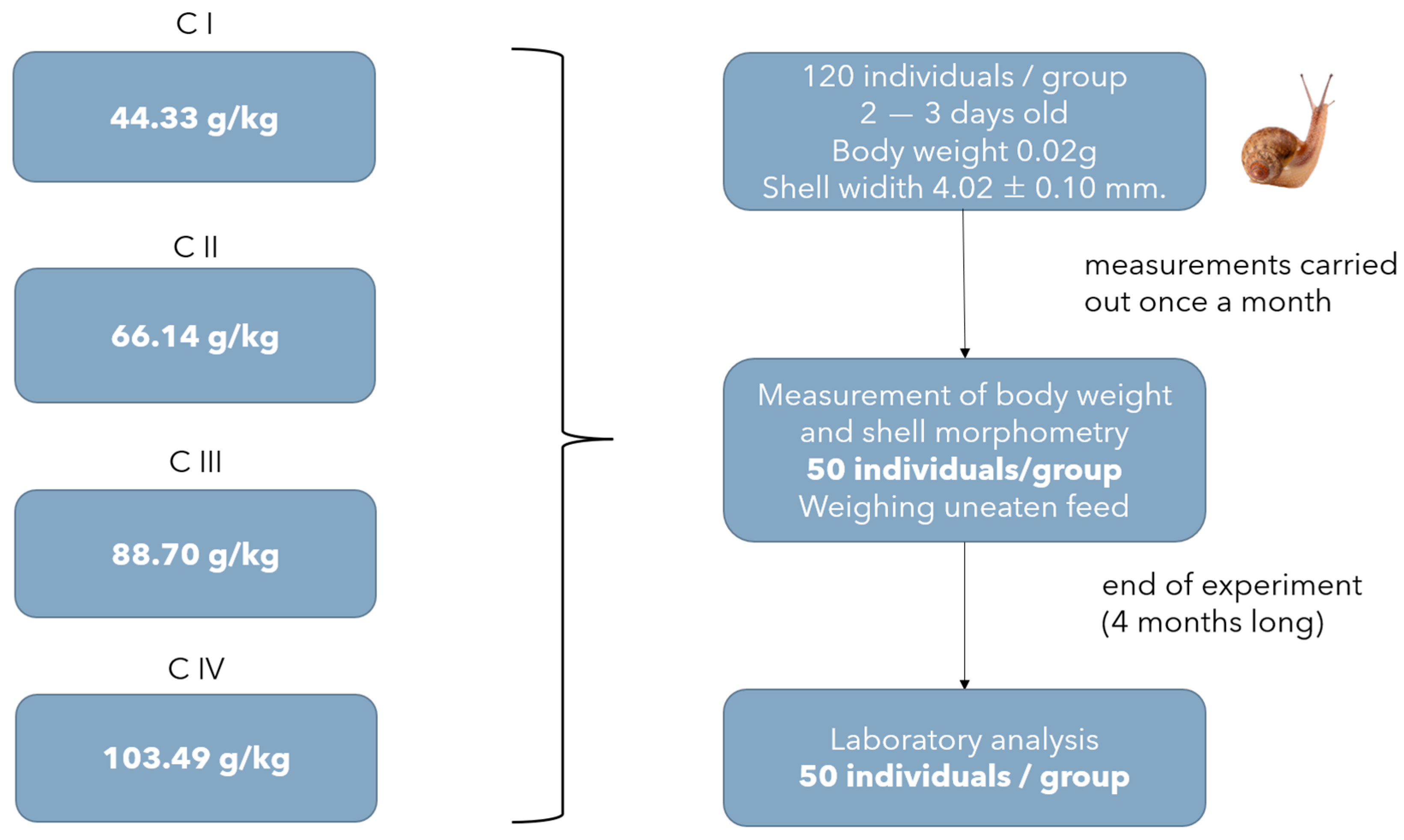
Experiment I was designed to explore the influence of varying Ca concentrations on snail production metrics, shell morphometry, and the nutritional attributes of the mollusc meat (refer to Figure 2). Each month, fifty individuals were randomly selected from each experimental group and their weight and shell width were measured, following the same procedure used at the start of the experiment. Mortality rates for animals within the group were determined, and feed consumption was calculated. Lastly, at the end of the experiment, fifty randomly selected individuals from each group were euthanised through freezing and utilised for further laboratory analyses pertaining to the quality of their carcasses and shells.
Given the extensive range of recommended dietary Ca levels cited in the literature, the decision was made to evaluate four distinct levels in this study. The C I group contained Ca levels of 44.22 g/kg feed, the C II group, 66.14 g/kg feed; the C III group, 88.70 g/kg feed; and the C IV group, 103.49 g/kg feed.
Experiment II, as depicted in Figure 3, aimed to assess the impact of Mg concentrations on snail production results, shell morphometry, and the nutritional attributes of the mollusc meat. The procedure in Experiment II mirrored that of Experiment I, except that forty-six individuals, chosen at random, underwent laboratory analysis instead of fifty. The diet from Experiment I, which yielded the most favourable results regarding snail fattening, was adopted as the benchmark for the control diet in this study. Mg oxide was chosen as the Mg supplement for the diet, given its status as the most prevalent mineral source with the highest available Mg concentration for feed [77]. Furthermore, Mg oxide typically ensures efficient absorption of Mg ions.
The control group (CG) had a Mg content of 2.45 g/kg feed (with no additional Mg). This level was determined based on prior research on poultry [91]. The M I group had a Mg content of 2.36 g/kg feed, the M II group was at 5.56 g/kg feed, and the M III group was set at 7.15 g/kg feed. The Mg concentrations in these rations were chosen based on studies in poultry, assuming that the nutritional requirements of laying hens may be similar to those of snails [92,93].

2.2. Experimental and Analytical Procedures

In the laboratory, carcasses were removed from shells. Carcass weight and the proportion of carcass to total body weight were calculated using data from fifty randomly chosen snails from Experiment I and forty-six from Experiment II. Then, further analyses of the chemical composition of snail meat, TBARS (thiobarbituric acid reactive substances) and mineral composition of the snail meat and shells were conducted in 6 replicates.
The chemical composition of the snail carcasses was determined according to AOAC (Association of Official Analytical Chemists) [94]: moisture and dry matter by drying at 105 °C to constant weight, crude ash by incineration at 550 °C for six h, crude protein (N × 6.25) using the micro-Kjeldahl technique (Kjeltec System 1026 Distilling Unit, Foss Tecator, Sweden), and crude fat after extraction with petroleum ether by the Soxhlet method.
Thiobarbituric acid reactive substances (TBARS) were expressed as equivalents of malondialdehyde (MDA), and MDA precursor—1.2.3.3-tetraethoxypropane (TEP) was used as a standard to plot a standard curve. The methodology presented by Uchiyam and Mihar [95] was used. The test tissues of snail carcasses were homogenised in 1% potassium chloride solution and then centrifuged at 2000 rpm for 15 min at 4 °C. Then, 1% phosphoric acid, 1% potassium chloride, 2% butyl hydroxyanisole (BHA), and 0.4% TBA were added to the resulting filtrate. After adding the reagents and mixing, the sample was capped and placed in a water bath at 100 °C for 60 min. After cooling, 4 mL of butanol was added to the sample and shaken for 2 min. The absorbance of compounds dissolved in the butanol phase was read at 532 nm. The results were read in nmol/mL from a standard curve against TEP.
Inductively coupled plasma optical emission spectrometry (ICP-OES) determined the mineral components of carcasses and snail shells. First, test samples were ground and mineralised in a chamber furnace at 450 °C for 12 h. Then, the acid digestion (hydrochloric acid 37%, AnalaR NORMAPUR®, VWR Chemicals, Radnor, PA, USA) occurred on a hotplate. The material was then filtered through blotting paper and diluted to 50 mL with distilled water. The total contents of Ca, Cu (copper), Fe (iron), Mg, Mn (manganese), P (phosphorus), K (potassium), Zn (zinc), Si (silicone), and Na (sodium) were determined with ICP-OEC equipment (Perkin Elmer Avio 200; Waltham, MA, USA).
During shell analysis, the share of matured individuals was determined by inspecting the presence of solid shell lip, indicating that the individual has reached maturity and ended the growth cycle [9]. Also, shell measurements were taken to establish the shell shape index—shell width-to-height ratio [96] and solidity index. This method is based on one used on poultry eggs [97], previously used in snail shell estimation by Ligaszewski et al. [98]. The formula follows [shell weight × (height width)−1] × 100.
An Instron 3382 testing device (Norwood, MA, USA) was employed to evaluate the crushing force exerted on the shells. This assessment aimed to discern the potential influence of varying Mg levels in the feed on shell robustness. The testing setup encompassed several components: a device designated for compression tests, plates marked for compression testing, and a computer system for data acquisition.
The device was calibrated to administer loads ranging from 0 to 100 kN (kilonewton). Static tests were conducted at a consistent speed of 5 mm per minute. The compressive strength was ascertained based on the orientation of the applied materials and the resistance they generated.

2.3. Statistical Analysis

The results were analysed using one-way analysis of variance (ANOVA), using Statistica 13.3 (TIBCO Software Inc., Palo Alto, CA, USA). A difference of p < 0.05 between means was significant. Further, the post-hoc LSD and repeated measures ANOVA tests were performed.
The used mathematical model followed Yij = μ + αi  +  eij, where Yij is the observation value, μ is the overall mean, αi is the effect of the experimental diet (calcium or magnesium level), and eij is random error.
Statistical analysis for the parameters of body weight, shell width, and mortality was performed with the PS IMAGO PRO 9.0 software, using ANOVA tests for repeated measures. The pairwise comparison method with Bonferroni correction was used for significant interaction effects. All study variables met the sphericity condition. A p < 0.05 was taken as statistically significant.

3. Results

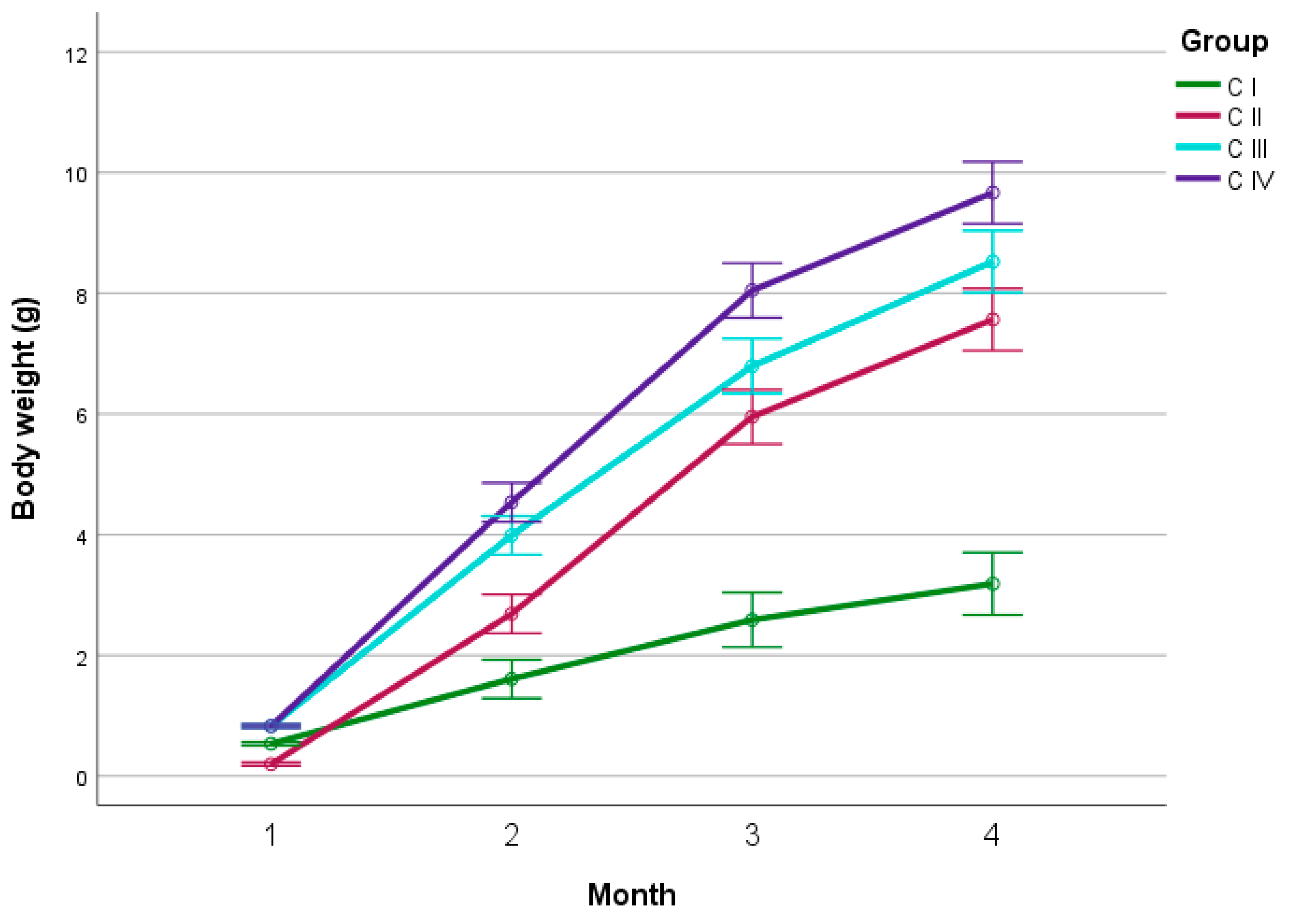
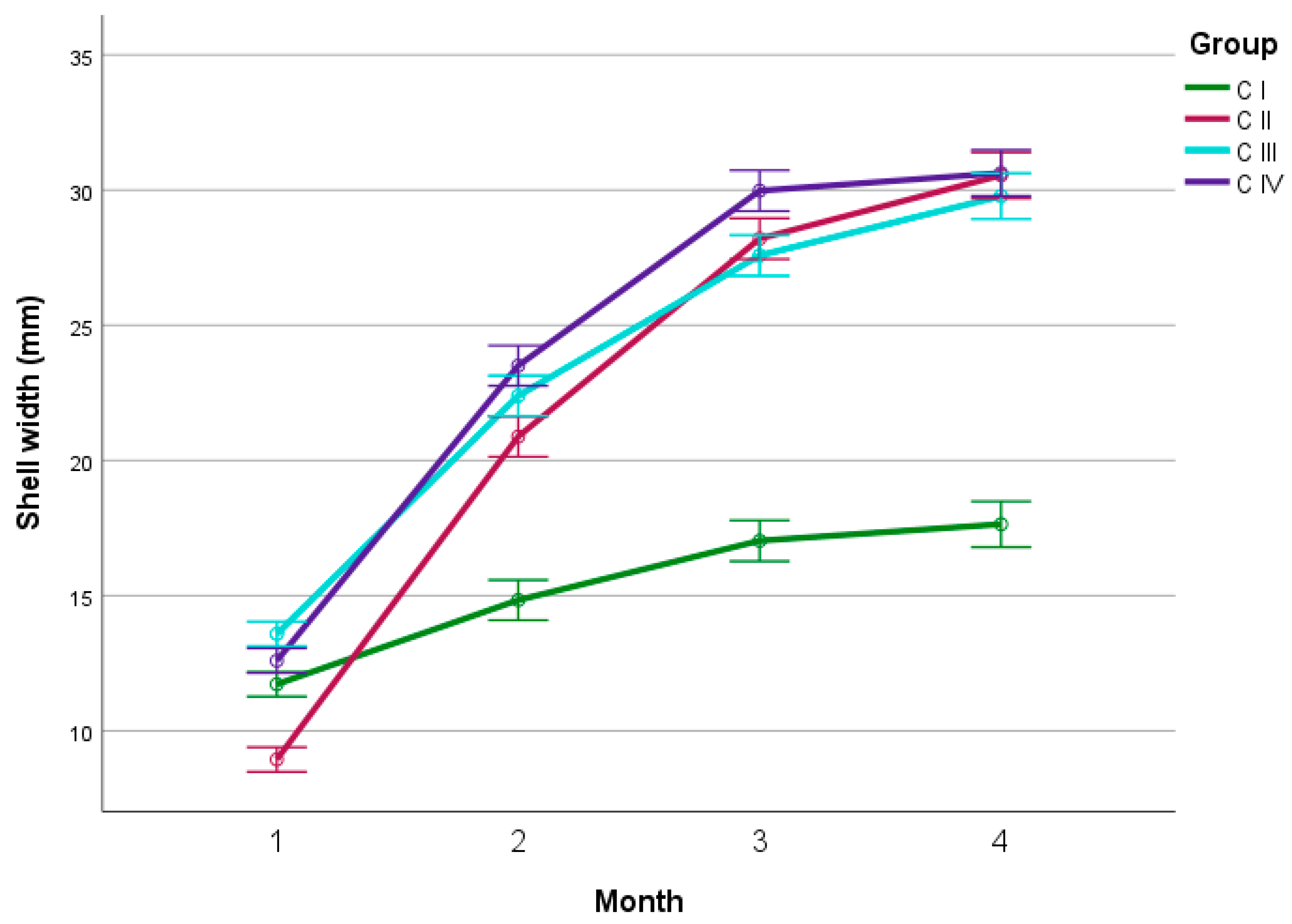
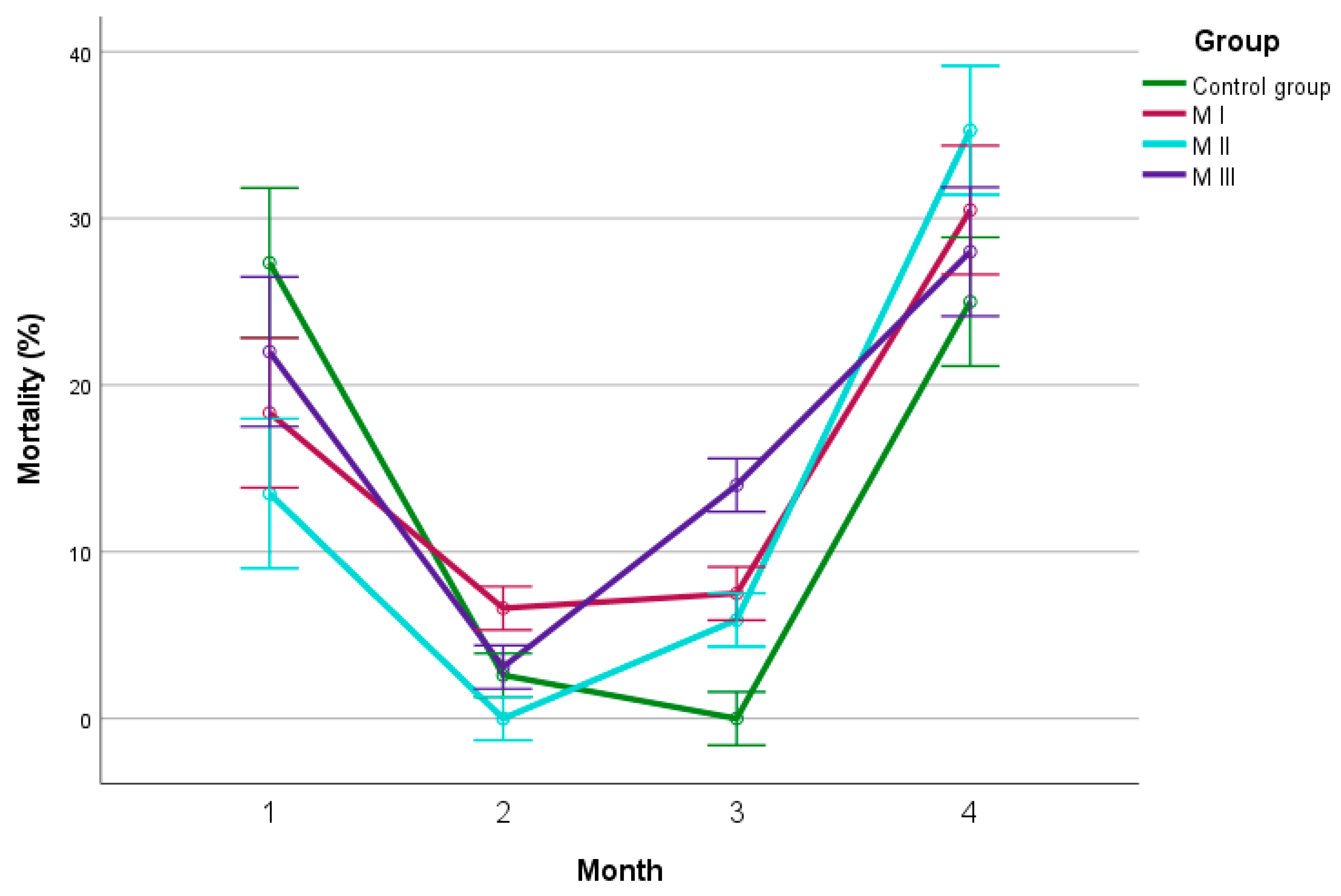
The development of snails is characterised by three stages that can be distinguished: slow growth of the body and rapid development of the internal organs in the first month, rapid increase in body weight in the second and third months, and stabilisation of body weight and a slowdown in growth after 13 weeks of age [12]. The results of the measurements carried out in each month of Experiment I and Experiment II are presented in Figure 4, Figure 5, Figure 6, Figure 7, Figure 8 and Figure 9.
There was no significant difference (p > 0.05) in the body weights of the snails between C III (0.85 g) and C IV (0.68 g) during the first month. Snails in group C II had the lowest weight gain, and those in group C III had the highest (0.18 g vs. 0.83 g). Throughout the second and third months, snails in group C I showed the lowest mean weight gains (1.08 g and 1.65 g), while animals in group C IV showed the greatest (3.87 g and 3.51 g). In the second month, the weights of snails in groups C III (3.99 g) and C IV (4.54 g) did not differ significantly (p > 0.05). Group C I showed the lowest body weight (1.61 g). During the third month, no significant differences were found (p > 0.05), only between C II (5.95 g) and C III (6.79 g). Group C I showed the lowest body weight (3.26 g), and group C IV showed the highest (8.05 g). In the final measurement, there was no significant difference (p > 0.05) in the body weights of snails between groups C II (7.56 g) and C III (8.52 g). At the end of the experiment, snails in the C I group had the lowest mean body weight (3.19 g) and those in C IV, the highest (9.67 g).
In the first month of the experiment, the snails belonging to group C III exhibited the largest shell width (13.58 mm). In comparison, those from C II had the smallest (8.94 mm). Significant shell width differences (p ≤ 0.01) were observed in all tested groups. However, during the second measurement, groups C III (22.37 mm) and C IV (23.47 mm) did not show significant differences in shell width (p > 0.05). Group C I showed the lowest shell width at 14.79 mm. At month 3, there were no significant differences (p > 0.05) in is-total between groups C II (28.17 mm) and C III (27.56 mm). Group C I had the lowest shell width throughout the Experiment I duration. In group C II, snails showed the highest shell width increases among all experimental groups in months 2–4 (11.95 mm, 7.28 mm and 2.37 mm, respectively). Finally, animals in group C I showed the smallest average shell width at 17.51 mm in the final month of the experiment, while snails in group C IV showed the largest average shell width at 30.63 mm. By the end of the experiment, there were no significant differences (p > 0.05) in shell width between snails in groups CII (30.54 mm) and CIII (29.77 mm), CII and CIV, and CIII and CIV. The most significant overall increase in shell width (p ≤ 0.01) over time was observed in CII (+21.60 mm), whereas the smallest was in CI (+5.92 mm).
During the first month, the mortality rate was the lowest (p ≤ 0.01) for group C III at 9.03%, while group C I registered the highest rate at 30%. For the first measurement, the number of deaths did not differ significantly (p > 0.05) between groups C I (30%) and C IV (27.17%), and between groups C II (10%) and C III (9.83%). For the second measurement, the number of deaths did not differ between groups C III (2.10%) and C IV (2.60%). For the third measurement, it did not differ between groups C II (1%) and C III (1.50%) and between groups C II and C IV (0%). By the fourth month of the experiment, the mortality rates for groups C I and C II were relatively low at 3.40% and 4.00%, respectively. However, group C III recorded a significantly higher mortality rate of 30.90%, while group C IV had a mortality rate of 25.00%. The number of deaths did not differ significantly (p > 0.05) between groups CI and CII.
Magnesium supplementation in the diet of snails in Experiment II had a significant effect (p ≤ 0.01) on their mean body weight. During the first measurement, there was no significant difference (p > 0.05) in mean body weight between the M II and M III groups. The control group had the highest body weight (0.83 g) whilst M I had the lowest (0.62 g). In the second month of measurements, only the CG (4.40 g) and M III (5.39 g), and M I (4.32 g) and M III groups had significantly different (p ≤ 0.05) body weights. Differences (p ≤ 0.05) in snail body weight were observed only between the M I (7.32 g) and M III (8.21 g) groups during the third measurement. No significant differences (p > 0.05) in the snails’ body weights were evident during the last month of measurements. At the end of the experiment, the snails had gained body weights of CG = 9.62 g; M I = 9.68 g; M II = 9.95 g; M III = 9.08 g. Animals in the M II group consistently showed the most significant differences in weight gain compared to the control group, specifically, in the first month (0.70 g vs. 0.40 g), in the third month (4.70 g vs. 3.71 g) and the fourth month (2.41 g vs. 1.49 g). Conversely, snails in the M III group showed the lowest weight gains in the mentioned months: 0.69 g, 2.82 g, and 0.87 g, respectively.
No significant difference was observed in shell width during the first month of the experiment (p > 0.05). Among all groups, M I had the smallest shell width (12.34 mm), while MIII had the largest (12.84 mm). However, shell width varied significantly (p ≤ 0.05) between CG and M II (25.41 mm), M I (22.30 mm) and M II, as well as between M I and M III (24.14 mm) during the second month of measurements. Similarly, during the third measurement round, significant differences were found (p ≤ 0.05) between the CG (29.98 mm) and M I (28.39 mm), CG and M II (28.72 mm), and CG and M III (28.68 mm) groups. Significant differences in shell width (p ≤ 0.01) were observed between groups CG (30.66 mm) and M III (29.03 mm), M I (30.79 mm) and MIII, and between M II (30.30 mm) and MIII by the end of the measurements. The largest shell width of 30.79 mm was observed in M I, while the smallest width of 29.06 mm was observed in M III. Regarding shell width gains, the M II group had significantly more substantial gains: 18.25% greater than the control group in the first month (13.04 mm vs. 11.03 mm). In contrast, in the third month, the M II group had significantly smaller gains: 47.77% less than the control group (3.31 mm vs. 6.36 mm). In the last month, the M I group had the highest increase in shell width—2.40 mm. Furthermore, the largest shell width change over time was found in MI (+18.45 mm) and the smallest in MIII (+16.21 mm).
During the initial measurement, mortality exhibited a substantial difference (p ≤ 0.01) exclusively between the CG (27.33%) and MII (13.50%) groups. In the second measurement, no significant differences (p > 0.05) were found while comparing CG (2.6%) and M II (0%), and CG and M III (2.9%). No significant differences in mortality were observed (p > 0.05) between the M I (7.6%) and M II (5.9%) groups after three months of measurements. The mortality rate of group M III was the highest at 13.6%. At the end of the study, mortality rates were noted as follows: CG = 25.00%; MI = 30.50%; MII = 35.28%; and MIII = 28.00%. Only the CG and MI groups showed significant differences (p ≤ 0.01).
During the examination of carcass and shell traits in Table 2, animals in group C IV in Experiment I had the highest mean carcass weight (8.27 g). The lowest body weight was noted in group C I at 2.89 g. The trend in shell mass was similar, with group C IV displaying the heaviest shells at 1.40 g and group C I at 0.29 g.
The share of carcass in total body weight was highest among snails in group C I at 91.24. Animals in group C IV showed the lowest share at 85.46%.
The shell shape index demonstrated minimal variation, with all groups exhibiting values around 1.44, signifying a similarity in shell shape among the experimental groups. The solidity index shows that animals in group C IV had the highest value of 21.15, while in group C I, the lowest score was recorded at 11.19 g/cm2.
Snails in group C IV displayed the highest crushing force at 54.83 N, and group C I had the lowest, at 12.44 N. The shells of the C I group showed unique characteristics, such as softness, flexibility and translucency. They exhibited a delayed fracture pattern characterised by stretching and planar flattening.
Thiobarbituric acid reactive substance (TBARS) results provide essential information in studies related to lipid oxidation processes in biological samples and indicate changes in health status and metabolism, or the effects of external factors on the studied organisms. The results showed that snails in group C III had the highest levels at 58.87 nmol/g lyophilizate in carcasses and snails in group C IV had the lowest, at 55.59 nmol/g.
Furthermore, group C IV had the highest percentage of mature individuals at 46%. In contrast, in groups C I, C II, and C III, the results were 10 times (C I) and 1.3 times (CII and C III) lesser, correspondingly.
The feed conversion ratio (FCR) in group C IV showed the highest value at 1.28 kg of feed (DM) per kg of snail body weight. Results of groups C I and C III showed comparable FCR values.
Regarding feed intake, individuals in group C II consumed the least amount of feed in the first month, with an average of 4.92 g per individual. Contrarily, group C IV had the highest feed intake, averaging 11.10 g per individual. The trend remained constant throughout the experiment, whereby Group C IV consistently displayed the greatest feed intake, and the largest discrepancy was noted in the final month. On the other hand, Group C I constantly demonstrated the lowest feed intake, with the minimum value of 2.06 g per specimen documented in the first month.
When carcass and shell characteristics were examined in Experiment II, the snails in group M II exhibited the highest mean carcass weight at 8.45 g compared with other groups. The lowest weight was recorded in group M II at 7.47 g.
Animals in group M III exhibited the heaviest shells, weighing 1.61 g, whereas the control group showed shells weighing 1.39 g. The mean proportion of the carcass of snails in total body weight was the highest in the control group, at 85.45%, while group M III had the lowest proportion, at 81.98%.
The control group displayed a shell shape index of snails of 1.43, the highest among the groups, while group M III recorded the lowest at 1.24. Group M I had the lowest solidity index of 20.26, whereas group M III had the highest solidity index of 23.58 g/cm2. The crushing force of the shells showed differences, but these were not statistically significant.
The TBARS levels in snail carcass tissues from group M III were the highest at 57.65 nmol/g lyophilisate, whereas those in snails from group M I were the lowest at 55.33 nmol/g.
The proportion of mature snails did not exhibit a distinct trend of maximum and minimum values (46–65%) between the groups but demonstrated fluctuation.
The FCR records revealed that group M III displayed the most efficient feed conversion, with the lowest FCR recorded at 0.94, while group M I reflected the highest FCR at 1.27. Also, group M III presented the highest feed intake at 11.09 g/individual, whereas group M I showed the lowest intake (8.34 g/individual).
The investigation into the percentage differences in proximate composition among the experimental groups in Experiment I and Experiment II demonstrates significant variations in the nutritional profiles of snail carcasses (Table 3).
The study found that snails in group C I exhibited the highest crude protein content in meat, at 60.26%. Group C III showed the lowest crude protein content of 53.38%.
The lipid content exhibited comparable changes across the experimental groups. Animals in group C I had the highest ether extract content, measuring 4.66%. Both groups C III and C IV exhibited the lowest ether extract content, measuring 1.49%.
Groups C I and C II exhibited the highest crude ash content values, registering at 14.07%. Snails in group C IV showed the lowest crude ash content among all experimental groups, measuring 9.20%.
The proximate composition analysis of the snail carcasses conducted in Experiment II illustrates substantial disparities in crude protein and ether extract content. Animals in the control group displayed the highest crude protein content at 59.97% in their carcasses. In contrast, the M III group showed the lowest content, a 13.16% reduction compared to the control group.
In contrast, the control group displayed the lowest ether extract content of 1.56%. In contrast, the M III group exhibited the highest ether extract content, with a significant increase of 83.33% compared to the control group. These results emphasise the substantial variations in these two components between the study groups.
Crude ash content was similar across all groups, ranging from 8.79% (group M III) to 9.22% (group M II).
Additional analyses were conducted concerning the mineral composition (in DM) of the snails’ meat and shells (in FM). The results are presented in Table 4.
In Experiment I, animals in group C III had the highest Ca content in meat (1.45%), with the lowest being in group C II (1.31%). In the shells, the highest Ca content was found in group C IV (38.96%), with the lowest being in group C II (36.16%). Snails in group C II recorded the highest concentration of Cu in meat (56.99 mg/kg), while group C IV presented the lowest (36.70 mg/kg). In the shells, group C I had the highest Cu content (6.12 mg/kg), and group C IV showed the lowest (1.88 mg/kg).
The Fe content in snail meat was highest in group C I (357.52 mg/kg) and lowest in group C IV (127.82 mg/kg). Regarding the shells, group C III had the highest Fe content (202.39 mg/kg), and group C IV had the lowest (135.82 mg/kg). While analysing potassium content, animals in group C II had the highest K content in meat (7718.49 mg/kg), while group C IV had the lowest (6220.12 mg/kg). There were no significant differences in K content in the shells among the groups.
Snails in group C II had the highest Mg content in meat at 3532.52 mg/kg, while the lowest was detected in group C IV at 2670.22 mg/kg. In the shells, group C I exhibited the highest Mg content at 508.48 mg/kg, and group C IV had the lowest at 320.12 mg/kg. Animals in group C I displayed the highest manganese concentration in meat (25.98 mg/kg) and shells (14.58 mg/kg). Group C IV had the lowest manganese content in both meat and shells, with measurements of 21.82 mg/kg and 9.76 mg/kg, respectively.
Animals in group C II showed the highest Na content in meat at 7934.27 mg/kg, whereas group C IV showed the lowest at 5299.06 mg/kg. In the shells, inversely, group C IV had the highest Na content at 1050.68 mg/kg, and group C II had the lowest at 806.95 mg/kg. The highest phosphorus content in snail meat was observed in group C II (11,616.93 mg/kg), whereas the lowest was in group C IV (9271.07 mg/kg). In the shells, group C I had the highest phosphorus content (896.51 mg/kg), whereas group C IV had the lowest (428.37 mg/kg).
The meat from group C I exhibited the highest silicon content (66.00 mg/kg), while the lowest was in group C II (36.40 mg/kg). In the shells, group C I demonstrated the greatest Si concentration (182.30 mg/kg), whereas group C IV presented the lowest (24.98 mg/kg). Group C I exhibited the highest zinc content in meat (105.27 mg/kg); conversely, group C IV had the lowest (57.26 mg/kg). In the shells, the Zn content was highest in group C IV (17.91 mg/kg) and lowest in group C I (6.40 mg/kg).
In Experiment II, animals in the control group had the highest Ca content in the meat at 1.40% and in shells (38.96%), while M I had the lowest content in meat at 1.16% and in the shell at 33.78%. Copper content displayed differences across the groups, with M I having the lowest at 48.69 mg/kg and the control group having the highest at 60.33 mg/kg in snail meat. Cu content increased across the groups in the shell, with the control group having the lowest (1.90 mg/kg) and M III having the highest (4.98 mg/kg).
The iron content in the snail meat exhibited a significant increase in M I (228.16 mg/kg) compared with the control group (127.82 mg/kg). Regarding the shell, the Fe content was the highest in group M II (200.70 mg/kg) and the lowest in group M III (131.80 mg/kg). There were slight, not statistically significant, differences in the potassium content of the snail meat. However, in the shell, the control group exhibited a high concentration of 1096.70 mg/kg, while M I showed the lowest concentration at 750.43 mg/kg.
Differences across the groups in the Mg content in the snail meat were noted, with M I recording the minimum value (2279.38 mg/kg) and the control group showing the maximum value (2684.13 mg/kg). However, the Mg content in the shell varied significantly, with the highest value recorded in M III (408.85 mg/kg) and the lowest in M I (271.84 mg/kg). The Mn content in the meat in group M III was the lowest (21.17 mg/kg); in group M II, the Mn content recorded was the highest (25.34 mg/kg). The M III group shells exhibited the lowest Mn content (8.42 mg/kg), in contrast to M II, which contained the highest Mn content (9.25 mg/kg).
The sodium levels in the meat were most elevated in the control group (5299.06 mg/kg) and least in M III (4497.35 mg/kg). Na levels varied in the shells, with M I exhibiting the lowest (738.78 mg/kg) and the control group the highest (1050.70 mg/kg). Phosphorus content in the meat was the lowest in group M I (7905.68 mg/kg) and the highest in the control group (9271.07 mg/kg). In shell analysis, the highest P content was found in the control group (428.40 mg/kg) and the lowest in the M I group (244.33 mg/kg).
The silicon content in the snails’ meat was highest in the M II group, measuring 119.27 mg/kg, while the lowest value was recorded in the control group at 66.00 mg/kg. Regarding the shell, Si content in the control group was the lowest (25.00 mg/kg), and M I had the highest (179.36 mg/kg) values. The lowest zinc content in the meat was detected in the M I group (49.63 mg/kg), with the highest value found in M II (53.86 mg/kg). Zn content in the shell varied among the groups, with the highest content in the control group (17.90 mg/kg) and the lowest in group C II (4.42 mg/kg).

4. Discussion

4.1. Growth Rates during Fattening

4.1.1. Experiment I

As anticipated, the body weights of the snails increased concerning the Ca concentration in the mixtures. As an indicator of shell development, the shell width parameter showed similar results across all groups from the second month until the fattening period’s end, except for the group receiving the lowest Ca supplement (C I). Based on the findings, it can be inferred that the C I group (44.3 g Ca/kg) lacked sufficient calcium for the adequate growth and development of snails. Individuals in this group displayed indications of arrested development at approximately 1 month of age compared to the other experimental groups. From day 30 onwards, the low Ca content was deficient and impeded proper shell development. This finding corroborates previous research linking low Ca levels in their diet to the development of dwarfism in these animals [99,100,101]. However, it is worth mentioning that in the first fattening stage, a level of 4.4% satisfies the animals’ Ca requirements. Although the shell widths in groups C II, C III, and C IV during the second and third months differ considerably, they eventually equalise by the end of the fattening period. That implies that shell growth is sustained regardless of the Ca level in the mixtures.
Previous studies on Archachatina marginata demonstrated that increasing the proportion of Ca in the feed by up to 20% (above the standard level in snail nutrition) increased body weight and shell width [32]. Also, Oluokun et al. [25] conducted studies that indicated body weight and shell width of snails were positively influenced by Ca levels, showing that animals fed with an 8% added Ca mixture feed noted an increase in shell width of approx. 30%, and an approx. 25% increase in body weight compared with animals fed with a feed composition containing 4% Ca.
The experimental results show that the C IV group had the highest total FCR results compared to other experimental groups, yet it was not significantly different. Also, group C IV showed minimal growth during the final month of the fattening period, which was attributed to the onset of reproduction. As a result, this group may have had an overestimated FCR compared to other groups, as the snails’ growth ceased despite their continuous feed consumption. The study results show that total feed intake was rising along with Ca levels in the feed, suggesting that feed containing higher doses of Ca is more palatable or encourages animals to eat in larger quantities, supported by previous research [24]. It was previously shown that increased Ca supply likely affects digestive processes and the absorption of other nutrients, which may affect metabolic efficiency and feed energy utilisation [7]. However, current results contrast with those of Oluokun et al. [25], who demonstrated that FCR improved with higher percentages of Ca in animal feed. Experimental groups, except C IV, exhibited FCRs comparable to those documented in the literature for Cornu aspersum [102].
The results show an association between Ca in the diet at 4.43% and 8.87% and higher mortality rates in the early fattening stages. A different trend is observed in the 6.61% Ca group, which can reduce mortality throughout the entire fattening period. It is assumed that low calcium levels in snails’ feed increase mortality in the first month of rearing due to deficiencies. In contrast, high levels contribute to high mortality in the final phase of fattening due to the exhaustion of animals during laying, which commences earlier due to the accelerated maturation process. Maintaining sufficient calcium levels in the snails’ feed is recommended to reduce mortality rates.
Studies by Tchakounte et al. [36] on Archachatina marginata showed reduced snail mortality with increasing Ca concentration (14–18%) in the feed, valid for animals in the middle fattening period in the current study. Notably, snails experience their highest mortality during the first month of development and when entering the breeding and egg-laying phase [21,36]. Notably, none of the experimental groups achieved a mortality rate of 40% during the first month of fattening, which has been reported in the literature as the standard [12]. The results were closer to the 30% obtained by Desbuquois (1997) [103]. Studies on Helix Aspersa Maxima reported mortality rates of 8–10% [102], and comparable results, below 12% mortality, were observed in all experimental groups in the current study in the second and third month of fattening.

4.1.2. Experiment II

It was shown that Mg at 0.72% promoted increased animal weight in the second and third months of rearing. Therefore, it appears justifiable to incorporate magnesium supplementation during the intermediate stage of snail feeding (second and third month). Adding Mg to the diet did not significantly affect the final body weight of the animals, yet it was observed that it accelerated the snails’ growth in time. Previous studies on farm animals and shrimps confirm the beneficial effect of Mg supplementation on animal weight gain [104,105,106]. Compared to the Berillis et al. [8] study (20% Ca in feed mixture), snails’ body weights were higher, and the widths of the shells were comparable to the current research.
In addition to compound feed, results of Experiment II showed that total FCR and feed consumption were lower in the groups with Mg at 3.3 g and 5.6 g/kg feed (approx. 1 kg DM/kg and approx. 8.5 g/individual), which indicates better feed utilization by animals and the possibility of savings for the farmer due to lower feed expenses. The high feed conversion rate observed in both the control and M III groups, coupled with the weight gain and shell width measurements in the last two months of fattening and the number of mature individuals in each group, suggests that the snails’ growth was nearly complete. The snails began to reproduce after the development and fattening phase ended. Consequently, the fattening period of the snails could be completed 2–3 weeks earlier, thereby positively impacting the economic factors of snail fattening. It was shown that higher Mg content had a beneficial effect on feed conversion in farm animals and shrimps, which is, to some extent, consistent with the study’s findings [104,105]. All experimental groups exhibited FCRs better than those documented in the literature for Cornu aspersum, with values up to 1.85 [9,12,107].
Groups with additional Mg in their diet exhibited a comparatively lower mortality rate in the initial month of fattening than CG. Mg at 0.56% in the diet increased mortality in the last month. Results below 14% of mortality were observed in all experimental groups in the current study in the second and third month of fattening. This study’s findings suggest that adding magnesium to the diet at levels of 3.3 g/kg and 5.6 g/kg in the first month of fattening and 5.6 g/kg in the second month resulted in favourable survival of the snails. However, during the following two months, the addition of magnesium to the diet showed no better results than its absence in the control group. All groups receiving Mg supplementation exhibited more significant mortality in the final month of fattening, potentially due to the snails laying eggs and maturing faster than the control group. This faster maturation is believed to be the primary reason for the higher mortality rate observed [108]. A study on shrimps indicated higher animal survival rates with increasing Mg supplementation. However, this association was not observed in the present snail study [105,106]. The mortality results were close to 20–30% obtained in previous studies on snails [109,110] in the first and fourth month of fattening. Earlier studies on Helix Aspersa Maxima reported mortality rates of up to 10% [111].

4.2. Carcass Characteristics

4.2.1. Experiment I

Increasing dietary Ca levels were associated with increased carcass weight from 2.89 g to 8.27 g. The increased calcium content in the diet is thought to have a positive effect on the development of the snails and their growth, not only in terms of total body weight, but also in terms of carcass weight. Calcium is speculated to enhance the rapidity and effectiveness of fattening these animals. At the same time, it was noted that under the influence of increased dietary Ca intake, the percentage of carcass weight in the total body weight of the snail was reduced from 91.24% to 85.46%. Previous studies corroborate the present findings [31]. It can be assumed that this effect is due to the maturation and saturation of the shell with minerals and increasing its weight [9,25,32]. Therefore, calcium supplementation has a positive impact not only on the weight of the snail’s body but also on the weight of its shell and overall weight.
The proximate composition analyses showed that introducing 4.43% Ca in the diet contributes to increased snail meat protein. In contrast, the presence of 8.87% Ca in the diet appears to have a negative effect on the protein content of the meat. As the amount of Ca in the feed increased, a decrease in the fat and ash content of the snail meat was observed (down to 1.48% and 9.20%, respectively). The findings indicate that an elevated calcium content in the diet correlates with a heightened proportion of carbohydrates in snail carcasses. Such carbohydrates mostly exist in the form of glycogen and galactose, which enable the animals to prepare for hibernation, among other functions [112,113]. Current findings show comparable crude protein and fat levels and higher crude ash content than previous snail studies [102,114]. The current research shows higher amounts of crude ash in snail meat and lower levels of crude protein than previous studies on Helix Aspersa Maxima [111,115].
Mineral analyses of the carcasses revealed that Ca at 4.43% in the diet influences higher iron content and that Ca at 6.61% contributes to the meat’s potassium, Mg, sodium, and phosphorus content while reducing the amount of silicon. In addition, a higher amount of Ca in feed was linked to higher silicon content and lower potassium, Mg, sodium, and phosphorus content. Ca interacts with these elements, frequently dislodging them from their binding and transportation positions on the exterior of cell membranes. Based on the study results, it can be assumed that Ca at a level above 8.87% starts to have negative influence on these elements’ absorption. Also, Ca at 10.35% increased snail carcasses’ copper. It is worth noting that copper is a component of haemocyanin, which is responsible for transporting oxygen in the snail’s haemolymph throughout the body [12,116]. This relationship may be attributed to the presence of larger and heavier carcasses, which require a greater supply of nutrients to a larger body surface area. Consequently, an increased amount of hemolymph is necessary for proper body function. An association was observed among vertebrates (rats, dogs, chickens) between increased Ca supply and Mg deficits [117,118,119]. In the current study, no such relationship was shown.
As a result of the experiments related to the mineral analysis of snail meat, significant differences were observed in the carcasses’ Ca, iron, phosphorus, Mg, sodium, potassium, manganese, and zinc contents. The values of these minerals were significantly different compared with the results of previous studies [120,121], where higher Ca, iron, and phosphorus contents were found in the carcasses studied. In contrast, a lower Ca content was recorded compared with the results of Çağıltay (2011) [115]. Moreover, copper and Mg contents were lower compared with the study of Gomot (1998) [120]. Compared with A. achatina, it was proven that the Cornu aspersum snails’ meat contained more Ca, iron, copper, potassium, and phosphorus and less zinc [122]. It was established that Cornu aspersum snails can accumulate copper in their foot tissues [123]. The snails in the current experiment exhibited decreased Ca, Mg, and phosphorus levels and elevated iron and zinc levels in their carcasses, except in the C IV group, compared with the Niemiec et al. [124] study. On the other hand, the potassium and manganese levels were higher than in previous studies [115,125]. Fagbuaro et al. [126] showed lower iron, sodium and potassium levels than in the current study. Thus, the available literature confirms variations in the mineral composition of meat.
The increased TBARS concentration in the C II and C III groups, compared with the C I Ca group, could indicate a possible increase in lipid oxidation processes or biological tissue reactivity within these groups. A decrease in TBARS values in the C IV group might signify a particular reduction in these oxidation processes. The results indicate that the effect of calcium in the diet at a level of 10.35% is beneficial from the point of view of lipid peroxidation in snail tissues.

4.2.2. Experiment II

Post-mortem analyses showed that Mg at 0.72% decreased the proportion of carcass weight in the body weight of the animals along with carcass weight, favouring an increase in shell weight. It can be assumed that Mg in the feed above this level begins to have a negative effect on the weight gain of snails, especially carcass weight. Compared with the Ligaszewski et al. [127] study, lower carcass weights were demonstrated, along with a higher share of the carcass in the total body weight of animals.
It was proven that Mg supplementation increases protein utilisation in foods due to increased protein absorption and synthesis [128]. Stronger protein digestion may be related to increased sodium–potassium triphosphatase activity in enterocytes and changes in membrane transport systems [129]. That could explain the weight gain observed during the second and third month of fattening of the snails. At 2–3 months, snails achieve complete development of their internal organs, marking the beginning of their fattening period. However, this did not result in an increase in final protein quantity in snail carcasses, as analyses of the proximate composition showed that adding Mg to the diet contributed to a linear decrease in protein and ash while increasing carcass fat. It is possible that increasing magnesium levels in the diet may result in increased deposition of elements in the shell (thus reducing the ash content of the meat) or reduce the digestibility and absorption of nutrients such as protein and minerals. This may be related to the possibility of diarrhea when taking high doses of magnesium in the diet [40]. A study on shrimps confirms Mg supplementation’s positive impact on animal carcasses’ fat content [105]. It was also shown that Cornu aspersum carcasses contained five times less crude fat compared with the research conducted by Niemiec et al. [124] on reared snails fed a diet without added Mg. The crude protein levels were comparable across the groups.
Experiment II meat mineral assays showed that Mg at 0.72% reduced the meat’s manganese content. Mg at 0.33% increased the iron and decreased the carcass’s Ca, copper, Mg, potassium and zinc content. Mg at 0.56% increased manganese and silicone in the meat. Adding Mg to the diet also resulted in a linear decrease in the sodium and potassium content of the snail meat. It is worth pointing out that sodium and potassium are the main positive ions responsible for maintaining animals’ physiological pH levels. Objectively, the ions’ presence affects muscle contractility and tension. Proper sodium concentrations in the body also aid the absorption of monosaccharides and amino acids [16]. Reducing the percentage of these constituents in carcasses may suggest the potential for several disorders in the organism. The current findings challenge preceding research demonstrating an inverse relationship between Ca concentrations and Mg and copper concentrations [31]. Reductions in the content of elements like K, Mg, Mn, Na, Fe, and P in the meat of snails fed with feed mix containing 0.72% Mg can indicate the antagonistic effects between these elements.
These results partially align with the study’s findings on Roman snails, which showed an association between Mg, Cu, and Zn in soft tissues [130]. The study by Beeby and Richmond (2015) displayed that Mg levels did not impact Mg deposition in Cantareus Aspersus snails’ bodies, contradicting the present study’s results [131]. However, the element concentrations used in the cited research were notably low (720–2263 µg/g). In contrast to studies on poultry [118,132], the present study demonstrates that Mg addition affects the mineral composition of meat by increasing iron levels and decreasing Ca and Mg levels. Similarly, studies on shrimps that received an elevated Mg dose (254.36 μg/g) in the feed yielded conflicting outcomes to the current findings. The heightened Mg dosage raised the copper, zinc, Ca, sodium, and potassium concentration in the organisms’ carcasses. The sole shared result was the rise in iron levels [105]. The study’s findings corroborate previously reported links between elevated Mg intake and reduced absorption of manganese [133,134], zinc [135,136], and Ca [137,138] into body tissues. Both Mg and zinc were proven to minimise oxidative stress and free radicals [139,140]. Like Mg, zinc is a component or an activator of nearly 300 enzymes and plays various biological roles in the body [127,128], like regulating hormonal functions [141,142,143] and the immune response [144,145]. Higher iron content in Mg groups can indicate increasing immunity because immune responses are, among others, aided by enzymes containing iron, which promote the improvement of non-specific immunity. [146,147]. Mg supplementation also enhances protein utilisation in food by increasing protein absorption and synthesis [128]. It was shown that excessive Mg results in increased phosphate resorption [148], which promotes the formation of Ca salts that may favour Fe retention in the body [149].
In Experiment II, it was observed that the Mg content of snail meat was higher compared with the results of Çağıltay (2011) [115] and Fagbuaro et al. [126] but lower than in the study by Özogul et al. [125]. In contrast, potassium and manganese contents were higher than in the last-mentioned study [125]. Snail meat’s zinc content was also lower than previous studies’ results [120,125,126]. Furthermore, it was noted that the Mg content was lower than in the results of Özogul et al. [125]. The levels of potassium and manganese in Experiment II were similar to the results of Çağıltay (2011) [115].
The current study showed that Mg content at 0.72% increased TBARS levels compared with other experimental groups, indicating an increased incidence of oxidative stress.
Significant reductions in the content of a number of elements in the meat of snails fed with feed mix containing 0.72% Mg can confirm antagonistic effects between these elements, as shown in previous studies [59,63,66]. Alongside a statistically significant increase in TBARS, these findings suggest that a high Mg level may be toxic to snails. Generally, the lack of this element is associated with oxidative stress, inflammation, cytokine production, and increased free radicals [150,151,152]. It was proven that the negative impact of Mg on the body can be neutralised by increasing the availability of Ca and phosphorus in the diet [153].

4.3. Characteristics of Shells

4.3.1. Experiment I

The study showed that increasing Ca levels positively affected shell weight, as it also positively affects shell width, and shells may have a higher mineral saturation. It was proven that increasing Ca content in feed has a positive effect on snails’ development and growth. Also, the results of Ireland and Marigomez (1992) confirm the beneficial impact of an increased Ca proportion in the feed on the weight of shells [31]. Yet, the results were lower than those of the Berillis et al. [8] study.
Mineral analyses of the shells showed that Ca at 4.43% increased the Mg and sodium in shells. For Ca at 6.61%, a reduction in the Ca and iron content of the shells was observed. Ca at 8.87% contributed to an increase in iron and silicon while decreasing sodium and zinc. In contrast, Ca at 10.35% increased the Ca content and reduced the amount of silicon in the shells. As the proportion of Ca in the diet increased, a decrease in copper, Mg, manganese, and phosphorus and an increase in zinc was observed in the snail shells. The mechanisms responsible for the deposition of individual minerals in snail shells are still poorly understood and require further research.
A solidity index increase in response to higher dietary Ca levels was observed. This change may have implications for shell structural resistance. As such, the study showed that snail shell crushing force also increased (from 12.44 N to 54.83 N). In addition, an association was observed between shell crushing force, solidity index, and shell weight (from 0.29 g to 1.40 g). The current shells’ crushing force was lower than in previous studies [84,98]. Also, in the present study, if differences in the shell shape index were statistically significant, they had little impact on crush resistance, and besides the Ca-deficient group, a higher Ca content enhanced the shell’s crush resistance, unlike the results of the previous research on Cornu aspersum [84,98]. Also, less Ca was present in the shells: 37–38%, compared to 41.5%.
These relationships may indicate significant interactions between dietary Ca and physical characteristics and shell strength and the potential influence of individual maturity on its strength [26,154,155]. Studies on marine gastropods have demonstrated that the low Ca content of the water causes a substantial decrease in shell hardness [156]. Previous studies noted that shells of snails fed with insufficient Ca exhibited discolouration, thin walls, and reduced resistance to crushing. That was particularly evident in the group with a Ca level of 4.43% [34].
In the current study, there is evidence of earlier maturation of animals under the influence of increased dietary Ca, like higher body weight, a higher percentage of matured individuals, and smaller weight and shell size gains in the last month of the experiment.

4.3.2. Experiment II

The study showed that the addition of Mg increased snail shell weight from 1.39 g to 1.61 g. Considering the absence of considerable variations in shell dimensions, it is likely that mineral saturation within the shell structure may cause an increase in their mass. The results are comparable to those of the Berillis et al. [8] experiment.
It was also observed that Mg at 0.72% promoted a higher solidity index of shells (23.58 g/cm2) and higher shell weight than other experimental groups. In addition, a reduction in the shell shape index was observed, meaning that the shells became flatter. Shell crush strength increased up to a level of 0.56% of the dietary Mg content (61.41 N) and then decreased significantly (to 49.73 N), which is even lower than that of the control group (57.29 N). The experimental findings have established that the augmentation of Mg to the extent of 0.56% has a beneficial effect on the strength of the shell, as well as on its hardness and density (strength index). The shell crushing force can also be linked to the Fe amount in shell composition. Interestingly, a study on commercially raised Cornu aspersum in outdoor plots demonstrated more than twice the shell-crushing force compared to the best-observed result in the current experiment [157]. On the other hand, in a study by Berillis et al. 2013 [8], the shell-crushing force was up to five times lower than in the current study. It is plausible that dissimilar measurement techniques account for the disparity.
Mineral analyses of the shells showed that Mg at 0.33% in the diet reduced the Mg, potassium, Na, and P while increasing the Si content of the shell. Mg at 0.56% increased iron in the shell while decreasing Zn. In contrast, Mg at 0.72% resulted in a decrease in iron in the shell. Adding Mg to the diet also contributes to increased Mg in the shell while reducing the levels of manganese. The antagonistic interaction between magnesium and other elements is also apparent in the elemental composition of shells, although it is not as evident as it is in the composition of meat.
Assuming a similarity between hen eggshells and snail shells, the relationship between increased Mg in the feed and increased Mg in the shell composition is confirmed. However, this is not the case for Ca—studies on poultry showed that a higher proportion of Mg in the feed reduced the amount of Ca in the eggshell. The Ca level remained statistically unchanged in snail shells [158,159]. A study on eggshells found that adding Mg to feed increased zinc and reduced manganese levels [160]. This study affirms the association between Mg addition and decreased manganese levels in the shell. Still, the zinc content was lower in the Mg-added group than in the control group. However, there was an observable trend of increasing zinc concentration with increasing Mg levels within the experimental groups. Substantial decreases in the elemental levels, including Fe, K, and Mn, highlight the antagonistic interactions between these minerals and Mg. It is worth mentioning that the deposition of Mg and copper within the chicken eggshells increases along with their respective concentrations in the feed [53,61,62].
It was noted that Mg addition helped accelerate the snails’ maturation process (from 46% to 65% mature individuals), potentially allowing for a shorter fattening period.

4.4. Impact of Ca and Mg

It is crucial to consider that snails excrete some nutrients absorbed from their feed, which ultimately affects the soil. According to Gomot et al. [83], consuming Ca and Mg-rich soil, which is a natural behaviour of snails, positively impacts their growth and enhances the resilience and hardness of their shells. Soils richer in Ca and Mg were also shown to reduce oxidative stress physiology parameters in snails Pila globose [161]. In addition, a previous study showed that these kinds of soils are preferred by a wide range of gastropod snail species [162].
When considering the effect of soil Ca on plants, it should be noted that Ca deficiency results in reduced plant growth, impairs root system growth, and causes leaf discolouration. Mg plays a role in the structure of chlorophyll, and thus in photosynthesis, and is also required for many enzymatic reactions in plant organisms [163]. It was proven that it is Mg deficiency that is related to the occurrence of plant diseases (17) rather than its excess (6 linked diseases), which may be linked to the impact of these ion elements on the pH of the soil and plant immune mechanisms [164]. However, the intended Mg level application is unlikely to impact soil pH or the availability of Ca to plants (Mg-inducted Ca deficiency), as it is comparably low in the feed compared with Ca levels [165,166]. Infiltration of Ca and Mg into groundwater is possible. Nevertheless, the augmented presence of these minerals in drinking water has been shown to have beneficial effects on human health [167,168].
Typically, to mitigate low pH, toxic ions, and other ionic imbalances and their harmful effects on plant roots, soil solution concentrations of Ca ranging from 1 to 5 mM are necessary. Ca and Mg requirements for plants vary with plant species, from 100 to 2000 µg/g [169]. A study on maize showed that ratios of 2:1 to 3:1 of Ca:Mg gave optimum yield of plants without symptoms of Mg deficiency. Ratios of up to 6:1 also gave satisfactory results [170]. It can be assumed that additional amounts of Ca in soil can be beneficial for plants as long as the Ca:Mg ratio is kept at the right level.

5. Conclusions

In the group featuring 10.35% Ca and 0.72% Mg, snail growth was observed to slow down after three months, accompanied by a decrease in mortality. However, based on the analysis of total animal mortality and feed intake, it seems that a feed containing 0.56% magnesium would be the safer option, as high Mg (0.72%) could potentially exhibit slight toxicity towards snails. Shortening the fattening period by 3–4 weeks could increase profitability for snail farmers.

Author Contributions

Conceptualisation, A.R.-G. and T.N.; methodology, T.N.; investigation, A.R.-G., K.Z., M.R., W.B. and K.R.; formal analysis: A.R.-G.; data curation, A.R.-G. and D.B.; resources, K.Z., K.R., M.R. and W.B.; writing—original draft preparation A.R.-G.; writing—review T.N.; visualisation, A.R.-G. and D.B.; supervision, T.N. All authors have read and agreed to the published version of the manuscript.

Funding

This research received no external funding.

Institutional Review Board Statement

Not applicable.

Data Availability Statement

Not applicable.

Acknowledgments

This manuscript is part of a PhD thesis.

Conflicts of Interest

The authors declare no conflict of interest.

References

  1. Zucaro, A.; Forte, A.; De Vico, G.; Fierro, A. Environmental Loading of Italian Semi-Intensive Snail Farming System Evaluated by Means of Life Cycle Assessment. J. Clean. Prod. 2016, 125, 56–67. [Google Scholar] [CrossRef]
  2. Hatziioannou, M.; Issari, A.; Neofitou, C.; Aifadi, S.; Matsiori, S. Economic Analysis and Production Techniques of Snail Farms in Southern Greece. World J. Agric. Res. 2014, 2, 276–279. [Google Scholar] [CrossRef]
  3. Ogunniyi, L.T. Economic Analysis of Snail Production IN Ibadan, Oyo Stat. Int. J. Agric. Econ. Rural. Dev. 2009, 2, 26–34. [Google Scholar]
  4. Zagata, L.; Sutherland, L.A. Deconstructing the ‘Young Farmer Problem in Europe’: Towards a Research Agenda. J. Rural Stud. 2015, 38, 39–51. [Google Scholar] [CrossRef]
  5. Elmslie, L. Snail Collection and Small-Scale Production in Africa and Europe. In Ecological Implications of Minilivestock: Potential of Insects, Rodents, Frogs, and Snails; Paoletti, M.G., Ed.; Science Publishers: New Hampshire, UK, 2005; pp. 93–121. ISBN 9781482294439. [Google Scholar]
  6. Rygało-Galewska, A.; Zglińska, K.; Niemiec, T. Edible Snail Production in Europe. Animals 2022, 12, 2732. [Google Scholar] [CrossRef]
  7. Ireland, M.P. The Effect of Dietary Calcium on Growth, Shell Thickness and Tissue Calcium Distribution in the Snail Achatina fulica. Comp. Biochem. Physiol. A Physiol. 1991, 98, 111–116. [Google Scholar] [CrossRef]
  8. Berillis, P.; Hatziioannou, M.; Panagiotopoulos, N.; Neofitou, C. Similar Shell Features between Rear and Wild Cornu aspersum Snails. World J. Agric. Res. 2013, 1, 1–4. [Google Scholar]
  9. Murphy, B. Breeding and Growing Snails Commercially in Australia; Rural Industries Research Development Corporation: Wagga Wagga, Australia, 2001; ISBN 0-642-58219-X.
  10. Lehmann, U.; Hillmer, G. Bezkręgowce Kopalne; Wydawnictwo Geologiczne: Warszawa, Poland, 1991. [Google Scholar]
  11. Zhang, C.; Zhang, R. Matrix Proteins in the Outer Shells of Molluscs. Mar. Biotechnol. 2006, 8, 572–586. [Google Scholar] [CrossRef]
  12. Sowiński, G.; Wąsowski, R. Chów Ślimaków. Pielęgnacja, Żywienie, Zarys Chorób z Profilaktyką Oraz Kulinaria; Wydawnictwo Uniwersytetu Warmińsko-Mazurskiego: Olsztyn, Poland, 2000; ISBN 83-88343-40-8. [Google Scholar]
  13. Ligaszewski, M. Opracowanie Metody Oceny Jakości Produkcji Ślimaków Jadalnych z Gatunku Helix Aspersa z Różnych Warunków Hodowlanych i Środowiskowych na Podstawie Analizy cech Jakościowych I Ilościowych ich Muszli; Instytut Zootechniki Pib: Balice, Poland, 2008. [Google Scholar]
  14. Fournié, J.; Chétail, M. Calcium Dynamics in Land Gastropods. Am. Zool. 1984, 24, 857–870. [Google Scholar] [CrossRef]
  15. Pu, F.; Chen, N.; Xue, S. Calcium Intake, Calcium Homeostasis and Health. Food Sci. Hum. Wellness 2016, 5, 8–16. [Google Scholar] [CrossRef]
  16. Radwińska, J.; Zarczyńska, K. Effects of Mineral Deficiency on the Health of Young Ruminants. J. Elem. 2014, 19, 915–928. [Google Scholar]
  17. Jaiswal, J.K. Calcium—How and Why? J. Biosci. 2001, 26, 357–363. [Google Scholar] [CrossRef] [PubMed]
  18. Jatto, O.E.; Asia, I.O.; Medjor, W.E. Proximate and Mineral Composition of Different Species of Snail Shell. Pac. J. Sci. Technol. 2010, 11, 416–419. [Google Scholar]
  19. Beeby, A.; Richmond, L. Magnesium and the Deposition of Lead in the Shell of Three Populations of the Garden Snail Cantareus aspersus. Environ. Pollut. 2011, 159, 1667–1672. [Google Scholar] [CrossRef] [PubMed]
  20. Hotopp, K.P. Land Snails and Soil Calcium in Central Appalachian Mountain Forest. Southeast. Nat. 2002, 1, 27–44. [Google Scholar] [CrossRef]
  21. Tompa, A.S.; Wilbur, K.M. Calcium Mobilisation during Reproduction in Snail Helix aspersa. Nature 1977, 270, 53–54. [Google Scholar] [CrossRef]
  22. Emelue, G.U.; Omonzogbe, E.A. Growth Performance of African Giant Land Snails (Archachatina marginata) Fed with Feed Formulated with Different Calcium Sources. Malays. J. Sustain. Agric. 2018, 2, 1–4. [Google Scholar] [CrossRef]
  23. Voelker, J. Der Chemische Einfluss von Kalzimkarbonat auf Wachstum, Entwicklung und Gehausebau von Achatina fulica Bowdich (Pulmonata). Mitteilungen Hambg. Zool. Mus. Inst. Univ. Hambg. 1959, 57, 37–78. [Google Scholar]
  24. Chevalier, L.; Desboquois, C.; Papineau, J.; Charrier, M. Influence of Inorganic Compounds on Food Selection by the Brown Garden Snail Cornu aspersum (Müller) (Gastropoda: Pulmonata). J. Moll. Stud. 2000, 66, 61–68. [Google Scholar] [CrossRef]
  25. Oluokun, J.A.; Omole, A.J.; Fapounda, O. Effect of Increasing the Level of Calcium Supplementation in the Diets of Growing Snail on Performance Characteristics. Res. J. Agric. Biol. Sci. 2005, 1, 76–79. [Google Scholar]
  26. Gouveia, A.R.; Pearce-Kelly, P.; Quicke, D.L.J.; Leather, S.R. Effects of Different Calcium Concentrations Supplemented on the Diet of Partula gibba on Their Morphometric Growth Parameters, Weight and Reproduction Success. Malacologia 2011, 54, 139–146. [Google Scholar] [CrossRef]
  27. Marin, F.; Roy, N.L.; Marie, B. The Formation and Mineralization of Mollusk Shell. Front. Biosci. 2012, 4, 1099–1125. [Google Scholar] [CrossRef] [PubMed]
  28. Roinel, N.; Morel, F.; Istin, M. Etude des Granules Calcifiés du Manteau des Lamellibranches à L’aide de la Microsonde Électronique. Calcif. Tissue Res. 1973, 11, 163–170. [Google Scholar] [CrossRef] [PubMed]
  29. Fleury, C.; Marin, F.; Marie, B.; Luquet, G.; Thomas, J.; Josse, C.; Serpentini, A.; Lebel, J.M. Shell Repair Process in the Green Ormer Haliotis tuberculata: A Histological and Microstructural Study. Tissue Cell 2008, 40, 207–218. [Google Scholar] [CrossRef] [PubMed]
  30. Simkiss, K.; Watkins, B. The Influence of Gut Microorganisms on Zinc Uptake in Helix aspersa. Environ. Pollut. 1990, 66, 263–271. [Google Scholar] [CrossRef]
  31. Ireland, M.P.; Marigomez, I. The Influence of Dietary Calcium on the Tissue Distribution of Cu, Zn, Mg & P and Histological Changes in the Digestive Gland Cells of the Snail Achatina fulica Bowdichm. J. Molluscan Stud. 1992, 58, 157–168. [Google Scholar] [CrossRef]
  32. Kouassi Kouadio, D.; Jean-Baptiste, A.; Mamadou, K. Growth Performance of Archachatina marginata Bred on the Substrate Amended with Industrial Calcium: Mikhart. Int. J. Sci. Res. 2016, 5, 582–586. [Google Scholar] [CrossRef]
  33. Aman, J.B.; Adou, C.F.D.; Karamoko, M.; Otchoumou, A. Effect of Source and Amendment Rate of Rearing Substrate on the Growth and Yield of Archachatina marginata. J. Res. Ecol. 2019, 7, 2546–2554. [Google Scholar]
  34. Crowell, H.H. Laboratory Study of Calcium Requirements of the Brown Garden Snail, Helix aspersa Müller. J. Molluscan Stud. 1973, 40, 491–503. [Google Scholar] [CrossRef]
  35. Wacker, A.; Baur, B. Effects of Protein and Calcium Concentrations of Artificial Diets on the Growth and Survival of the Land Snail Arianta arbustorum. Invertebr. Reprod. Dev. 2004, 46, 47–53. [Google Scholar] [CrossRef]
  36. Tchakounte, F.M.; Kana, J.R.; Azine, P.C.; Meffowoet, C.P.; Djuidje, V.P. Growth and Reproductive Performances of African Giant Snail (Archachatina marginata) as Affected by Dietary Calcium Levels. Sci. J. Vet. Adv. 2019, 8, 263–271. [Google Scholar]
  37. Otchoumou, A.; Dupont-Nivet, M.; Dosso, H. Effects of Diet Quality and Dietary Calcium on Reproductive Performance in Archachatina ventricosa (Gould 1850), Achatinidae, under Indoor Rearing Conditions. Invertebr. Reprod. Dev. 2012, 56, 14–20. [Google Scholar] [CrossRef]
  38. Lobo, H.L.; Santos, A.G. Níveis de Cálcio em Rações para Escargot (Cornu aspersum). Bachelor’s Thesis, Federal University of São João del-Rei, São João del-Rei, Brazil, 2017; p. 32. [Google Scholar]
  39. Drapała, T. Chemia Ogólna Nieorganiczna; Pwn: Warszawa, Poland, 1986. [Google Scholar]
  40. Karmanska, A.; Stanczak, A.; Karwowski, B. Magnez Aktualny Stan Wiedzy. Bromatol. Chem. Toksykol. 2015, 48, 677–689. [Google Scholar]
  41. Rubin, H. Central Role for Magnesium in Coordinate Control of Metabolism and Growth in Animal Cells. Proc. Natl. Acad. Sci. USA 1975, 72, 3551–3555. [Google Scholar] [CrossRef] [PubMed]
  42. Banai, S.; Haggroth, L.; Epstein, S.E.; Casscells, W. Influence of Extracellular Magnesium on Capillary Endothelial Cell Proliferation and Migration. Circ. Res. 1990, 67, 645–650. [Google Scholar] [CrossRef]
  43. Wolf, F.I.; Cittadini, A. Magnesium in Cell Proliferation and Differentiation. Front. Biosci. 1999, 4, 607–617. [Google Scholar] [CrossRef]
  44. Maier, J.A.M.; Bernardini, D.; Rayssiguier, Y.; Mazur, A. High Concentrations of Magnesium Modulate Vascular Endothelial Cell Behaviour In Vitro. Biochim. Biophys. Acta-Mol. Basis Dis. 2004, 1689, 6–12. [Google Scholar] [CrossRef]
  45. Chandra, A.K.; Sengupta, P.; Goswami, H.; Sarkar, M. Effects of Dietary Magnesium on Testicular Histology, Steroidogenesis, Spermatogenesis and Oxidative Stress Markers in Adult Rats. Indian J. Exp. Biol. 2013, 51, 37–47. [Google Scholar]
  46. Gaál, K.K.; Sáfár, O.; Gulyás, L.; Stadler, P. Magnesium in Animal Nutrition. J. Am. Coll. Nutr. 2004, 23, 754s–757s. [Google Scholar] [CrossRef]
  47. Cowan, J.A. Introduction to the Biological Chemistry of Magnesium Ion. In The Biological Chemistry of Magnesium; Cowan, J.A., Ed.; Vch Publishers: New York, NY, USA, 1995; pp. 1–23. ISBN 1560816279. [Google Scholar]
  48. Rubin, H. The Logic of the Membrane, Magnesium, Mitosis (MMM) Model for the Regulation of Animal Cell Proliferation. Arch. Biochem. Biophys. 2007, 458, 16–23. [Google Scholar] [CrossRef]
  49. Vink, R.; Cernak, I. Regulation of Intracellular Free Magnesium in Central Nervous System Injury. Front. Biosci. 2000, 5, A541. [Google Scholar] [CrossRef] [PubMed]
  50. Pasternak, K.; Kocot, J.; Horecka, A. The Biochemistry of Magnesium. J. Elementol. 2010, 15, 601–616. [Google Scholar] [CrossRef]
  51. Cefaratti, C.; Romani, A.; Scarpa, A. Differential Localization and Operation of Distinct Mg2+ Transporters in Apical and Basolateral Sides of Rat Liver Plasma Membrane. J. Biol. Chem. 2000, 275, 3772–3780. [Google Scholar] [CrossRef]
  52. Garfinkel, D.; Garfinkel, L. Magnesium and Regulation of Carbohydrate Metabolism at the Molecular Level. Magnesium 1988, 7, 249–261. [Google Scholar]
  53. Tammaro, P.; Smith, A.L.; Crowley, B.L.; Smirnov, S.V. Modulation of the Voltage-Dependent K+ Current By Intracellular Mg2+ in Rat Aortic Smooth Muscle Cells. Cardiovasc. Res. 2005, 65, 387–396. [Google Scholar] [CrossRef] [PubMed]
  54. Shi, J.; Krishnamoorthy, G.; Yang, Y.; Hu, L.; Chaturvedi, N.; Harilal, D.; Qin, J.; Cui, J. Mechanism of Magnesium Activation of Calcium-Activated Potassium Channels. Nature 2002, 418, 876–880. [Google Scholar] [CrossRef] [PubMed]
  55. Agus, Z.S.; Morad, M. Modulation of Cardiac Ion Channels by Magnesium. Annu. Rev. Physiol. 1991, 53, 299–307. [Google Scholar] [CrossRef]
  56. Mcdonald, P.; Edwards, R.A.; Greenhalgh, J.F.D.; Morgan, C.A.; Sinclair, L.A.; Wilkinson, R.G. Animal Nutrition, 7th ed.; Longman Scientific and Technical: Madison, WI, USA, 2011; ISBN 9781408204238. [Google Scholar]
  57. Horky, P.; Sochor, J.; Skladanka, J.; Klusonova, I.; Nevrkla, P. Effect of Selenium, Vitamins E and C on Antioxidant Potential and Quality of Boar Ejaculate. J. Anim. Feed Sci. 2016, 25, 29–36. [Google Scholar] [CrossRef]
  58. Freitag, J.J.; Martin, K.J.; Conrades, M.B.; Bellorin-Font, E.; Teitelbaum, S.; Klahr, S.; Slatopolsky, E. Evidence for Skeletal Resistance to Parathyroid Hormone in Magnesium Deficiency: Studies in Isolated Perfused Bone. J. Clin. Investig. 1979, 64, 1238–1244. [Google Scholar] [CrossRef]
  59. Schlingmann, K.P.; Gudermann, T. A Critical Role of TRPM Channel-Kinase for Human Magnesium Transport. J. Physiol. 2005, 566, 301–308. [Google Scholar] [CrossRef]
  60. Monteilh-Zoller, M.K.; Hermosura, M.C.; Nadler, M.J.S.; Scharenberg, A.M.; Penner, R.; Fleig, A. TRPM7 Provides an Ion Channel Mechanism for Cellular Entry of Trace Metal Ions. J. Gen. Physiol. 2003, 121, 49–60. [Google Scholar] [CrossRef] [PubMed]
  61. Okorodudu, A.O.; Yang, H.; Elghetany, M.T. Ionized Magnesium in the Homeostasis of Cells: Intracellular Threshold for Mg in Human Platelets. Clin. Chim. Acta 2001, 303, 147–154. [Google Scholar] [CrossRef] [PubMed]
  62. Féray, J.C.; Garay, R. A One-to-One Mg2+:Mn2+ Exchange in Rat Erythrocytes. J. Biol. Chem. 1987, 262, 5763–5768. [Google Scholar] [CrossRef] [PubMed]
  63. Goytain, A.; Quamme, G.A. Functional Characterization of the Human Solute Carrier, SLC41A2. Biochem. Biophys. Res. Commun. 2005, 330, 701–705. [Google Scholar] [CrossRef]
  64. Schmitz, C.; Perraud, A.-L.; Johnson, C.O.; Inabe, K.; Smith, M.K.; Penner, R.; Kurosaki, T.; Fleig, A.; Scharenberg, A.M. Regulation of Vertebrate Cellular Mg2+ Homeostasis by TRPM7. Cell 2003, 114, 191–200. [Google Scholar] [CrossRef]
  65. Matsuda, H. Magnesium Gating of the Inwardly Rectifying K+ Channel. Annu. Rev. Physiol. 1991, 53, 289–298. [Google Scholar] [CrossRef]
  66. Gröber, U.; Schmidt, J.; Kisters, K. Magnesium in Prevention and Therapy. Nutrients 2015, 7, 8199–8226. [Google Scholar] [CrossRef]
  67. Shils, M.E. Experimental Human Magnesium Depletion. Medicine 1969, 48, 61–85. [Google Scholar] [CrossRef]
  68. Whang, R.; Flink, E.B.; Dyckner, T.; Wester, P.O.; Aikawa, J.K.; Ryan, M.P. Magnesium Depletion as a Cause of Refractory Potassium Repletion. Arch. Intern. Med. 1985, 145, 1686–1689. [Google Scholar] [CrossRef]
  69. Chang, C.; Bloom, S. Interrelationship of Dietary Mg Intake and Electrolyte Homeostasis in Hamsters: I. Severe Mg Deficiency, Electrolyte Homeostasis, and Myocardial Necrosis. J. Am. Coll. Nutr. 1985, 4, 173–185. [Google Scholar] [CrossRef]
  70. Goytain, A.; Quamme, G.A. Functional Characterization of ACDP2 (Ancient Conserved Domain Protein), a Divalent Metal Transporter. Physiol. Genom. 2005, 22, 382–389. [Google Scholar] [CrossRef] [PubMed]
  71. Toba, Y.; Kajita, Y.; Masuyama, R.; Takada, Y.; Suzuki, K.; Aoe, S. Nutrient Metabolism Dietary Magnesium Supplementation Affects Bone Metabolism and Dynamic Strength of Bone in Ovariectomized Rats. J. Nutr. 2000, 130, 216–220. [Google Scholar] [CrossRef] [PubMed]
  72. Clark, I. Effects of Magnesium Ions on Calcium and Phosphorus Metabolism. Am. J. Physiol. 1968, 214, 348–356. [Google Scholar] [CrossRef] [PubMed]
  73. Scanlan, B.J.; Tuft, B.; Elfrey, J.E.; Smith, A.; Zhao, A.; Morimoto, M.; Chmielinska, J.J.; Tejero-Taldo, M.I.; Mak, I.T.; Weglicki, W.B.; et al. Intestinal Inflammation Caused by Magnesium Deficiency Alters Basal and Oxidative Stress-Induced Intestinal Function. Mol. Cell. Biochem. 2007, 306, 59–69. [Google Scholar] [CrossRef] [PubMed]
  74. Gunther, T.; Hollriegl, V.; Vormann, J.; Bubeck, J.; Classen, H.G. Increased Lipid Peroxidation in Rat Tissues by Magnesium Deficiency and Vitamin E Depletion. Magnes. Bull. 1994, 16, 38–43. [Google Scholar]
  75. Rude, R.K.; Gruber, H.E.; Wei, L.Y.; Frausto, A.; Mills, B.G. Magnesium Deficiency: Effect on Bone and Mineral Metabolism In the Mouse. Calcif. Tissue Int. 2003, 72, 32–41. [Google Scholar] [CrossRef]
  76. Rude, R.K.; Kirchen, M.E.; Gruber, H.E.; Stasky, A.A.; Meyer, M.H. Magnesium Deficiency Induces Bone Loss in the Rat. Miner. Electrolyte Metab. 1998, 24, 314–320. [Google Scholar] [CrossRef]
  77. Lipiński, K.; Stasiewicz, M.; Purwin, C.; Zuk-Gołaszewska, K. Effects of Magnesium on Pork Quality. J. Elem. 2011, 16, 325–337. [Google Scholar] [CrossRef]
  78. Vormann, J.; Günther, T.; Höllriegl, V.; Schumann, K. Pathobiochemical Effects of Graded Magnesium Deficiency in Rats. Z. Ernahrungswissenschaft 1998, 37 (Suppl. S1), 92–97. [Google Scholar]
  79. Quamme, G.A.; De Rouffignac, C. Epithelial Magnesium Transport and Regulation by the Kidney. Front. Biosci. 2000, 5, D694–D711. [Google Scholar] [CrossRef]
  80. Elin, R.J. Magnesium: The Fifth but Forgotten Electrolyte. Am. J. Clin. Pathol. 1994, 102, 616–622. [Google Scholar] [CrossRef] [PubMed]
  81. Weaver, V.M.; Welsh, J. 1,25-Dihydroxycholecalciferol Supplementation Prevents Hypocalcemia in Magnesium-Deficient Chicks. J. Nutr. 1993, 123, 764–771. [Google Scholar] [CrossRef]
  82. Yoshimura, T.; Tamenori, Y.; Kawahata, H.; Suzuki, A. Fluctuations of Sulfate, S-Bearing Amino Acids and Magnesium in a Giant Clam Shell. Biogeosciences 2014, 11, 3881–3886. [Google Scholar] [CrossRef]
  83. Gomot, A.; Gomot, L.; Boukraa, S.; Bruckert, S. Influence of Soil on the Growth of the Land Snail Helix aspersa. An Experimental Study of the Absorption Route for the Stimulating Factors. J. Molluscan Stud. 1989, 55, 1–7. [Google Scholar] [CrossRef]
  84. Ligaszewski, M.; Pol, P. Wybrane Zagadnienia z Dziedziny Helikultury; Zespół Wydawnictw i Poligrafii iz Pib: Kraków, Poland, 2019; ISBN 9788376073927. [Google Scholar]
  85. Mayaki, O.M.; Ozumba, A.U.; Aderele, A.A.; Daramola, A.O. Effect of Sources of Fibre on Performance of Growing Snail. Niger. Food J. 2013, 31, 28–32. [Google Scholar] [CrossRef]
  86. Sampelayo, M.R.S.; Fonolla, J.; Extremera, F.G. Factors Affecting the Food Intake, Growth and Protein Utilization in the Helix Aspersa Snail. Protein Content of the Diet and Animal Age. Lab. Anim. 1991, 25, 291–298. [Google Scholar] [CrossRef]
  87. Bonnet, J.-C.; Vrillon, J.-L.; Aupinel, P. L’escargot Helix aspersa; Inra: Paris, France, 1992; ISBN 9782759201976. [Google Scholar]
  88. García, A.; Perea, J.; Martín, R.; Acero, R.; Mayoral, A.; Peña, F.; Luque, M. Effect of Two Diets on the Growth of the Helix aspersa Müller During the Juvenile Stage. In Proceedings of the 56th Annual Meeting EAAP, Uppsala, Sweden, 5–8 June 2005; pp. 1–9. [Google Scholar]
  89. Cameron, R.A.D.; Carter, M.A. Intra- and Interspecific Effects of Population Density on Growth and Activity in Some Helicid Land Snails (Gastropoda: Pulmonata). J. Anim. Ecol. 1979, 48, 237. [Google Scholar] [CrossRef]
  90. Staikou, A.; Dimitriadou, L. Effect of Crowding on Growth and Mortality in the Edible Snail Helix lucorum (Gastropoda: Pulmonata) in Greece. Isr. J. Zool. 1989, 36, 1–9. [Google Scholar]
  91. Guo, Y.; Zhang, G.; Yuan, J.; Nie, W. Effects of Source and Level of Magnesium and Vitamin E on Prevention of Hepatic Peroxidation and Oxidative Deterioration of Broiler Meat. Anim. Feed Sci. Technol. 2003, 107, 143–150. [Google Scholar] [CrossRef]
  92. Lee, S.; Britton, W.M. Magnesium Toxicity: Effect on Phosporus Utilization by Broiler Chicks. Poult. Sci. 1980, 59, 1989–1994. [Google Scholar] [CrossRef]
  93. Hess, J.; Britton, W. Effects of Dietary Magnesium Excess in White Leghorn Hens. Poult Sci 1997, 76, 703–710. [Google Scholar] [CrossRef] [PubMed]
  94. AOAC. Official Methods of Analysis of the Association of Official Analytical Chemists; AOAC Intl.: Gaithersburg, MD, USA, 2005. [Google Scholar]
  95. Uchiyama, M.; Mihara, M. Determination of Malonaldehyde Precursor in Tissues by Thiobarbituric Acid Test. Anal. Biochem. 1978, 86, 271–278. [Google Scholar] [CrossRef]
  96. Chevallier, H. La Variabilité de L’escargot Petit-Gris Hélix aspersa Muller. Bull. Muséum Natl. D’histoibe Nat. 1977, 3, 425–442. [Google Scholar]
  97. Cooke, A.S. Shell Thinning in Avian Eggs by Environmental Pollutants. Environ. Pollut. 1973, 4, 85–152. [Google Scholar] [CrossRef]
  98. Ligaszewski, M.; Surówka, K.; Stekla, J. The Shell Features of Cornu aspersum (Synonym Helix aspersa) And Helix pomatia: Characteristics and Comparison. Am. Malacol. Bull. 2009, 27, 173–181. [Google Scholar] [CrossRef]
  99. Fournie, J.; Chetail, M. Evidence for a Mobilization of Calcium Reserves for Reproduction Requirements in Deroceras reticulatum (Syn: Agriolimax reticulatus) (Gastropoda: Pulmonata). Malacologia 1980, 22, 285–291. [Google Scholar]
  100. Cowie, R.H.; Cain, A.J. Laboratory Maintenance and Breeding of Land Snails, with an Example from Helix aspersa. J. Molluscan Stud. 1983, 49, 176–177. [Google Scholar] [CrossRef]
  101. Dan, N.A. Studies on the Growth and Ecology of Helix asposa Muller. Ph.D. Thesis, University of Manchester, Manchester, UK, 1978. [Google Scholar]
  102. Milinsk, M.C.; Padre, R.D.G.; Hayashi, C.; De Oliveira, C.C.; Visentainer, J.V.; De Souza, N.E.; Matsushita, M. Effects of Feed Protein and Lipid Contents on Fatty Acid Profile of Snail (Helix aspersa maxima) Meat. J. Food Compos. Anal. 2006, 19, 212–216. [Google Scholar] [CrossRef]
  103. Desbuquois, C. Influence of Egg Cannibalism on Growth, Survival and Feeding in Hatchlings of the Land Snail Helix aspersa Müller (Gastropoda, Pulmonata, Stylommatophora). Reprod. Nutr. Dev. 1997, 37, 191–202. [Google Scholar] [CrossRef]
  104. Suttle, N. Mineral Nutrition of Livestock; Suttle, N., Ed.; Cabi: Cambridge, UK, 2010; ISBN 9781845934736. [Google Scholar]
  105. Srinivasan, V.; Bhavan, P.S.; Rajkumar, G.; Satgurunathan, T.; Muralisankar, T. Dietary Supplementation of Magnesium Oxide (MgO) Nanoparticles for Better Survival and Growth of the Freshwater Prawn Macrobrachium rosenbergii Post-Larvae. Biol. Trace Elem. Res. 2017, 177, 196–208. [Google Scholar] [CrossRef]
  106. Jahan, I.; Reddy, A.K.; Sudhagar, S.A.; Harikrishna, V.; Singh, S.; Varghese, T.; Srivastava, P.P. The Effect of Fortification of Potassium and Magnesium in the Diet and Culture Water on Growth, Survival and Osmoregulation of Pacific White Shrimp, Litopenaeus vannamei Reared in Inland Ground Saline Water. Turk. J. Fish Aquat. Sci. 2018, 18, 1235–1243. [Google Scholar] [CrossRef] [PubMed]
  107. Wacker, A. Lipids in the Food of a Terrestrial Snail. Invertebr. Reprod. Dev. 2005, 47, 205–212. [Google Scholar] [CrossRef]
  108. Egonmwan, R.I. Effects of Dietary Calcium on Growth and Oviposition of the African Land Snail Limicolaria flammea (Pulmonata: Achatinidae). Rev. Biol. Trop. 2008, 56, 333–343. [Google Scholar] [CrossRef] [PubMed]
  109. Jess, S.; Marks, R.J. Population Density Effects on Growth in Culture of the Edible Snail Helix aspersa Var. Maxima. J. Molluscan Stud. 1995, 61, 313–323. [Google Scholar] [CrossRef]
  110. Daguzan, J.; Bonnet, J.C.; Perrin, Y.; Perrin, E.; Rouet, H. Contribution à L’élevage de L’escargot Petit-Gris: Helix aspersa Müller (Mollusque Gastéropode Pulmoné Stylommatophore). I.—Reproduction et Éclosion des Jeunes, en Bâtiment et en Conditions Thermohygrométriques Contrôlées. Ann. Zootech. 1981, 30, 249–272. [Google Scholar] [CrossRef]
  111. Milinsk, M. Influence of Diets Enriched with Different Vegetable Oils on the Fatty Acid Profiles of Snail Helix aspersa maxima. Food Chem. 2003, 82, 553–558. [Google Scholar] [CrossRef]
  112. Nicolai, A.; Filser, J.; Lenz, R.; Bertrand, C.; Charrier, M. Adjustment of Metabolite Composition in the Haemolymph to Seasonal Variations in the Land Snail Helix pomatia. J. Comp. Physiol. B 2011, 181, 457–466. [Google Scholar] [CrossRef]
  113. Dimitriadis, V.K.; Domouhtsidou, G.P. Carbohydrate Cytochemistry of the Intestine and Salivary Glands of the Snail Helix lucorum: Effect of Starvation and Hibernation. J. Molluscan Stud. 1995, 61, 215–224. [Google Scholar] [CrossRef]
  114. Baby, R.L.; Hasan, I.; Kabir, K.A.; Naser, M.N. Nutrient Analysis of Some Commercially Important Molluscs of Bangladesh. J. Sci. Res. 2010, 2, 390–396. [Google Scholar] [CrossRef]
  115. Çağıltay, F. Amino Acid, Fatty Acid, Vitamin and Mineral Contents of the Edible Garden Snail (Helix aspersa). J. Fishscicom. 2011, 5, 354–363. [Google Scholar] [CrossRef]
  116. Mikkelsen, F.F.; Weber, R.E. Oxygen Transport and Hemocyanin Function in the Pulmonate Land Snail, Helix pomatia: Physiological and Molecular Implications of Polyphasic Oxygen-Binding Curves. Physiol. Zool. 1992, 65, 1057–1073. [Google Scholar] [CrossRef]
  117. Tufts, E.V.; Greenberg, D.M. The Biochemistry of Magnesium Deficiency. J. Biol. Chem. 1938, 122, 693–714. [Google Scholar] [CrossRef]
  118. Bunce, G.E.; Chiemchaisri, Y.; Phillips, P.H. The Mineral Requirements of the Dog IV. Effect of Certain Dietary and Physiologic Factors upon the Magnesium Deficiency Syndrome. J. Nutr. 1962, 76, 23–29. [Google Scholar] [CrossRef]
  119. Nugara, D.; Edwards, H.M. Influence of Dietary Ca and P Levels on the Mg Requirement of the Chick. J. Nutr. 1963, 80, 181–184. [Google Scholar] [CrossRef]
  120. Gomot, A. Biochemical Composition of Helix Snails: Influence of Genetic and Physiological Factors. J. Molluscan Stud. 1998, 64, 173–181. [Google Scholar] [CrossRef]
  121. Çelik, M.Y.; Duman, M.B.; Sariipek, M.; Uzun Gören, G.; Kaya Öztürk, D.; Kocatepe, D.; Karayücel, S. Comparison of Fatty Acids and Some Mineral Matter Profiles of Wild and Farmed Snails, Cornu aspersum Müller, 1774. Molluscan Res. 2019, 39, 234–240. [Google Scholar] [CrossRef]
  122. Engmann, F.; Afoakwah, N.A.; Darko, P.O.; Sefah, W. Proximate and Mineral Composition of Snail (Achatina achatina) Meat; any Nutritional Justification for Acclaimed Health Benefits? J. Basic Appl. Sci. Res. 2013, 3, 8–15. [Google Scholar]
  123. Gomot, A.; Pihan, F. Comparison of the Bioaccumulation Capacities of Copper and Zinc in Two Snail Subspecies (Helix). Ecotoxicol. Environ. Saf. 1997, 38, 85–94. [Google Scholar] [CrossRef]
  124. Niemiec, T.; Łozicki, A.; Pietrasik, R.; Pawęta, S.; Rygało-Galewska, A.; Matusiewicz, M.; Zglińska, K. Impact of Ag Nanoparticles (AgNPs) and Multimicrobial Preparation (EM) on the Carcass, Mineral and Fatty Acid Composition of Cornu aspersum aspersum Snails. Animals 2021, 11, 1926. [Google Scholar] [CrossRef]
  125. Özogul, Y.; Özogul, F.; Olgunoglu, A.I. Fatty Acid Profile and Mineral Content of the Wild Snail (Helix pomatia) from the Region of the South of the Turkey. Eur. Food Res. Technol. 2005, 221, 547–549. [Google Scholar] [CrossRef]
  126. Fagbuaro, O.; Oso, J.A.; Edward, J.B.; Ogunleye, R.F. Nutritional Status of Four Species of Giant Land Snails in Nigeria. J. Zhejiang Univ. Sci. B 2006, 7, 686–689. [Google Scholar] [CrossRef] [PubMed]
  127. Ligaszewski, M.; Pol, P. Ocena Wpływu Różnych Systemów Chowu Ślimaka Szarego (Helix aspersa) na Wartość Odżywczą i Wydajność Jego Mięsa. Wiadomości Zootech. 2016, 54, 18–34. [Google Scholar]
  128. Rico, M.C.; Lerma, A.; Planells, E.; Aranda, P.; Gonzalez, J.L. Changes in the Nutritive Utilization of Protein Induced by Mg Deficiency in Rats. Int. J. Vitam. Nutr. Res. 1995, 65, 122–126. [Google Scholar]
  129. Bara, M.; Guiet-Bara, A.; Durlach, J. Regulation of Sodium and Potassium Pathways by Magnesium in Cell Membranes. Magnes. Res. 1993, 6, 167–177. [Google Scholar] [PubMed]
  130. Nica, D.V.; Bordean, D.M.; Hărmănescu, M.; Bura, M.; Gergen, I. Interactions among Heavy Metals (Cu, Cd, Zn, Pb) and Metallic Macroelements (K, Ca, Na, Mg) in Roman Snail (Helix pomatia) Soft Tissues. Acta Met.–MEEMB 2014, 11, 65. [Google Scholar]
  131. Beeby, A.; Richmond, L. Magnesium and the Regulation of Lead in Three Populations of the Garden Snail Cantareus aspersus. Environ. Pollut. 2010, 158, 2288–2293. [Google Scholar] [CrossRef]
  132. Liu, Y.X.; Guo, Y.M.; Wang, Z. Effect of Magnesium on Reactive Oxygen Species Production in the Thigh Muscles of Broiler Chickens. Br. Poult. Sci. 2007, 48, 84–89. [Google Scholar] [CrossRef]
  133. Woerpel, H.R.; Balloun, S.L. Effect of Iron and Magnesium on Manganese Metabolism. Poult. Sci. 1964, 43, 1134–1142. [Google Scholar] [CrossRef]
  134. Sanchez-Morito, N.; Planells, E.; Aranda, P.; Llopis, J. Magnesium-Manganese Interactions Caused by Magnesium Deficiency in Rats. J. Am. Coll. Nutr. 1999, 18, 475–480. [Google Scholar] [CrossRef]
  135. Lease, J.G.; Williams, W.P. The Effect of Added Magnesium on the Availability of Zinc with Some High-Protein Feedstuffs. Poult. Sci. 1967, 46, 242–248. [Google Scholar] [CrossRef]
  136. Planells, E.; Aranda, P.; Lerma, A.; Llopis, J. Changes in Bioavailability and Tissue Distribution of Zinc Caused by Magnesium Deficiency in Rats. Br. J. Nutr. 1994, 72, 315–323. [Google Scholar] [CrossRef]
  137. Planells, E.; Llopis, J.; Peran, F.; Aranda, P. Changes in Tissue Calcium and Phosphorus Content and Plasma Concentrations of Parathyroid Hormone and Calcitonin after Long-Term Magnesium Deficiency in Rats. J. Am. Coll. Nutr. 1995, 14, 292–298. [Google Scholar] [CrossRef] [PubMed]
  138. O’dell, B.L. Magnesium Requirement and Its Relation to other Dietary Constitunents. Fed. Proc. 1960, 19, 648–654. [Google Scholar] [PubMed]
  139. Afanas’ev, I.B.; Suslova, T.B.; Cheremisina, Z.P.; Abramova, N.E.; Korkina, L.G. Study of Antioxidant Properties of Metal Aspartates. Analyst 1995, 120, 859–862. [Google Scholar] [CrossRef] [PubMed]
  140. Sahin, K.; Onderci, M.; Sahin, N.; Gulcu, F.; Yildiz, N.; Avci, M.; Kucuk, O. Responses of Quail to Dietary Vitamin E and Zinc Picolinate at Different Environmental Temperatures. Anim. Feed Sci. Technol. 2006, 129, 39–48. [Google Scholar] [CrossRef]
  141. Al-Saad, K.M.; Al-Sadi, H.I.; Abdul-Majeed, M.O. Clinical, Hematological, Biochemical and Pathological Studies on Zinc Deficiency (Hypozincemia) in Sheep. Vet. Res. 2010, 3, 14–20. [Google Scholar]
  142. Miao, X.; Sun, W.; Fu, Y.; Miao, L.; Cai, L. Zinc Homeostasis in the Metabolic Syndrome and Diabetes. Front. Med. 2013, 7, 31–52. [Google Scholar] [CrossRef]
  143. Alves, C.X.; Vale, S.H.L.; Dantas, M.M.G.; Maia, A.A.; Franca, M.C.; Marchini, J.S.; Leite, L.D.; Brandao-Neto, J. Positive Effects of Zinc Supplementation on Growth, Gh, Igf1, and Igfbp3 in Eutrophic Children. J. Pediatr. Endocrinol. Metab. 2012, 25, 881–887. [Google Scholar] [CrossRef]
  144. Kincaid, R.L. Assessment of Trace Mineral Status of Ruminants: A Review. J. Anim. Sci. 2000, 77, 1. [Google Scholar] [CrossRef]
  145. El-Far, A.H. Biochemical Alterations in Zinc Deficient Sheep Associated by Hyperlactatemia. Am. J. Anim. Vet. Sci. 2013, 8, 112–116. [Google Scholar] [CrossRef]
  146. Walter, T.; Olivares, M.; Pizarro, F.; Muñoz, C. Iron, Anemia, and Infection. Nutr. Rev. 1997, 55, 111–124. [Google Scholar] [CrossRef]
  147. Benito, P.; Miller, D. Iron Absorption and Bioavailability: An Updated Review. Nutr. Res. 1998, 18, 581–603. [Google Scholar] [CrossRef]
  148. Planells, E.; Aranda, P.; Peran, F.; Llopis, J. Changes in Calcium and Phosphorus Absorption and Retention during Long-Term Magnesium Deficiency in Rats. Nutr. Res. 1993, 13, 691–699. [Google Scholar] [CrossRef]
  149. Sanchez-Morito, N.; Planells, E.; Aranda, P.; Llopis, J. Influence of Magnesium Deficiency on the Bioavailability and Tissue Distribution of Iron in the Rat. J. Nutr. Biochem. 2000, 11, 103–108. [Google Scholar] [CrossRef] [PubMed]
  150. Yang, Y.; Wu, Z.; Chen, Y.; Qiao, J.; Gao, M.; Yuan, J.; Nie, W.; Guo, Y. Magnesium Deficiency Enhances Hydrogen Peroxide Production and Oxidative Damage in Chick Embryo Hepatocyte In Vitro. Biometals 2006, 19, 71–81. [Google Scholar]
  151. Bussière, F.I.; Zimowska, W.; Gueux, E.; Rayssiguier, Y.; Mazur, A. Stress Protein Expression cDNA Array Study Supports Activation of Neutrophils During Acute Magnesium Deficiency in Rats. Magnes. Res. 2002, 15, 37–42. [Google Scholar] [PubMed]
  152. Weglicki, W.B.; Dickens, B.F.; Wagner, T.L.; Chmielinska, J.J.; Phillips, T.M. Immunoregulation by Neuropeptides in Magnesium Deficiency: Ex Vivo Effect of Enhanced Substance P Production on Circulating T Lymphocytes from Magnesium-Deficient Mice. Magnes. Res. 1996, 9, 3–11. [Google Scholar]
  153. Haag, J.R.; Palmer, L.S. The Effect of Variations in the Proportions of Calcium, Magnesium, and Phosphorus Contained in the Diet. J. Biol. Chem. 1928, 76, 367–389. [Google Scholar] [CrossRef]
  154. Ligaszewski, M. Kształtowanie się Wskaźników Wartości Użytkowej Muszli Ślimaków Jadalnych z Rodzaju Helix w Zróżnicowanych Warunkach Hodowlanych i Środowiskowych. Rocz. Nauk. Zootech. 2005, 19, 1–104. [Google Scholar]
  155. Ligaszewski, M.; Pol, P. Edible Snails Breeding in Poland. Zagadnienia Doradz. Rol. 2021, 1, 67–85. [Google Scholar]
  156. Glass, N.H.; Darby, P.C. The Effect of Calcium and Ph on Florida Apple Snail, Pomacea paludosa (Gastropoda: Ampullariidae), Shell Growth and Crush Weight. Aquat. Ecol. 2009, 43, 1085–1093. [Google Scholar] [CrossRef]
  157. Łozicki, A.; Niemiec, T.; Pietrasik, R.; Pawęta, S.; Rygało-Galewska, A.; Zglińska, K. The Effect of Ag Nanoparticles and Multimicrobial Preparation as Factors Stabilizing the Microbiological Homeostasis of Feed Tables for Cornu aspersum (Müller) Snails on Snail Growth and Quality Parameters of Carcasses and Shells. Animals 2020, 10, 2260. [Google Scholar] [CrossRef] [PubMed]
  158. Atteh, J.O.; Leeson, S. Influence of Increasing Dietary Calcium and Magnesium Levels on Performance, Mineral Metabolism, and Egg Mineral Content of Laying Hens. Poult. Sci. 1983, 62, 1261–1268. [Google Scholar] [CrossRef] [PubMed]
  159. Shastak, Y.; Rodehutscord, M. A Review of the Role of Magnesium in Poultry Nutrition. World Poult. Sci. J. 2019, 71, 125–137. [Google Scholar] [CrossRef]
  160. Holder, D.P.; Huntley, D.M. Influence of Added Manganese, Magnesium, Zinc, and Calcium Level on Egg Shell Quality. Poult. Sci. 1978, 57, 1629–1634. [Google Scholar] [CrossRef]
  161. Pati, S.G.; Panda, F.; Samanta, L.; Paital, B. Spatio-Temporal Changes in Oxidative Stress Physiology Parameters in Apple Snail Pila globosa as a Function of Soil Mg, Ca, Organic Carbon and Aquatic Physico-Chemical Factors. Environ. Geochem. Health 2023, 45, 2591–2610. [Google Scholar] [CrossRef]
  162. Ondina, P.; Mato, S.; Hermida, J.; Outeiro, A. Importance of Soil Exchangeable Cations and Aluminium Content on Land Snail Distribution. Appl. Soil Ecol. 1998, 9, 229–232. [Google Scholar] [CrossRef]
  163. Schwartzkopf, C. Potassium, Calcium, Magnesium—How They Relate to Plant Growth. Usga Green Sect. Rec. 1972, 10, 1–2. [Google Scholar]
  164. Huber, D.M.; Jones, J.B. The Role of Magnesium in Plant Disease. Plant Soil 2013, 368, 73–85. [Google Scholar]
  165. Kobayashi, H.; Masaoka, Y.; Sato, S. Effects of Excess Magnesium on the Growth and Mineral Content of Rice and Echinochloa. Plant Prod. Sci. 2005, 8, 38–43. [Google Scholar] [CrossRef]
  166. Franklin, W.T.; Olsen, J.S.; Soltanpour, P.N. Effects of Excessive Magnesium in Irrigation Waters on Wheat and Corn Growth. Commun. Soil Sci. Plant Anal. 1991, 22, 49–61. [Google Scholar] [CrossRef]
  167. Razowska-Jaworek, L. Calcium and Magnesium in Groundwater Occurrence and Significance for Human Health, 1st ed.; Razowska-Jaworek, L., Ed.; CRC Press Taylor & Francis Group: Boca Raton, FL, USA, 2019. [Google Scholar]
  168. Rapant, S.; Cvečková, V.; Fajčíková, K.; Sedláková, D.; Stehlíková, B. Impact of Calcium and Magnesium in Groundwater and Drinking Water on the Health of Inhabitants of the Slovak Republic. Int. J. Environ. Res. Public Health 2017, 14, 278. [Google Scholar] [CrossRef]
  169. Clark, R.B. Physiological Aspects of Calcium, Magnesium, and Molybdenum Deficiencies in Plants. Soil Acidity Liming 2015, 12, 99–170. [Google Scholar] [CrossRef]
  170. Osemwota, I.O.; Omueti, J.A.I.; Ogboghodo, A.I. Effect of Calcium/Magnesium Ratio in Soil on Magnesium Availability, Yield, and Yield Components of Maize. Commun. Soil Sci. Plant Anal. 2007, 38, 2849–2860. [Google Scholar] [CrossRef]
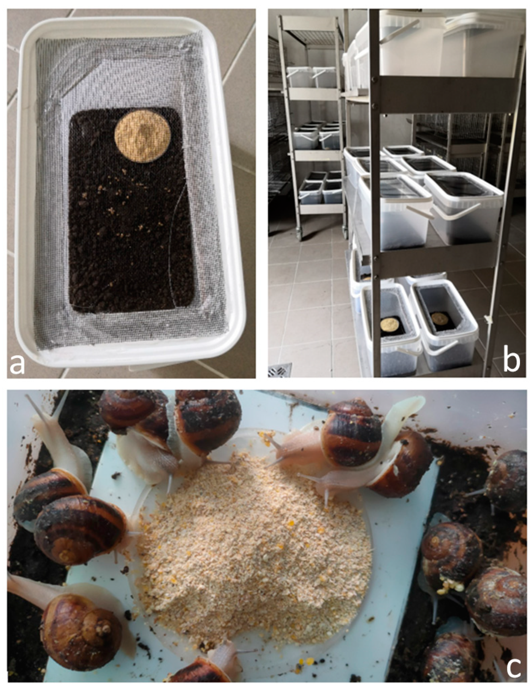
Figure 1. (a) Experimental container; (b) Setting of containers in the animal house; (c) Adult snails foraging on experimental feed.
Agriculture 13 02055 g001
Figure 2. Scheme illustrating the design of Experiment I: categorisation of the groups based on Ca content, details of the snails’ initial body parameters, the methodology for measuring the snails, and the end of the research.
Agriculture 13 02055 g002
Figure 3. Scheme illustrating the design of Experiment II: categorisation of the groups based on Mg content, details of the snails’ initial body parameters, the methodology for measuring the snails, and the end of the research.
Agriculture 13 02055 g003
Figure 4. Results of monthly body weight (g) measurements of snails in Experiment I. Data are given as mean ± SD (n = 50). C I, C II, C III, C IV—diet with Ca dose respectively: 44.3, 66.1, 88.7, 103.5 g/kg feed.
Agriculture 13 02055 g004
Figure 5. Results of monthly shell width (mm) measurements of snails in Experiment I. Data are given as mean ± SD (n = 50). C I, C II, C III, C IV—diet with Ca dose respectively: 44.3, 66.1, 88.7, 103.5 g/kg feed.
Agriculture 13 02055 g005
Figure 6. Results of monthly mortality (%) records of snails in Experiment I. Data are given as mean ± SD (n = 3). C I, C II, C III, C IV—diet with Ca dose respectively: 44.3, 66.1, 88.7, 103.5 g/kg feed.
Agriculture 13 02055 g006
Figure 7. Results of monthly body weight measurements (g) of snails in Experiment I. Data are given as mean ± SD (n = 50). No-Mg control diet; M I, M II, M III—diet with Mg dose respectively: 3.3 g/kg, 5.6, 7.2 g/kg feed.
Agriculture 13 02055 g007
Figure 8. Results of monthly shell width (mm) measurements of snails in Experiment I. Data are given as mean ± SD (n = 50). No-Mg control diet; M I, M II, M III—diet with Mg dose respectively: 3.3 g/kg, 5.6, 7.2 g/kg feed.
Agriculture 13 02055 g008
Figure 9. Results of monthly mortality (%) records of snails in Experiment I. Data are given as mean ± SD (n = 3). No-Mg control diet; M I, M II, M III—diet with Mg dose respectively: 3.3 g/kg, 5.6, 7.2 g/kg feed.
Agriculture 13 02055 g009
Table 1. Ingredients and determined chemical composition of snail diets in Experiments I and II.
Ingredients (%)Experiment IExperiment II
C IC IIC IIIC IVCONM IM IIM III
Corn meal53.050.045.043.343.843.543.343.1
Post-extraction soybean meal 46018.019.021.021.021.021.021.021.0
Wheat bran10.08.06.05.05.05.05.05.0
Fodder yeast4.04.04.04.04.04.04.04.0
Rapeseed oil2.02.02.52.52.02.02.02.0
Monocalcium phosphate2.02.02.02.02.02.02.02.0
NaCl0.20.20.20.20.20.20.20.2
Vitamin premix2.02.02.02.02.02.02.02.0
Ca carbonate8.913.217.320.020.020.020.020.0
Mg oxide (g)-----0.83.14.7
Composition (% DM)
Crude protein 18.22 ± 0.5318.86 ± 0.7618.72 ± 0.4418.30 ± 0.6818.40 ± 0.5818.74 ± 0.6319.03 ± 0.7118.82 ± 0.42
Ether extracts 2.93 ± 0.093.11 ± 0.123.32 ± 0.113.31 ± 0.073.01 ± 0.103.33 ± 0.133.15 ± 0.103.12 ± 0.08
Crude fibre 3.62 ± 0.094.05 ± 0.093.47 ± 0.093.30 ± 0.093.33 ± 0.093.62 ± 0.093.59 ± 0.093.81 ± 0.09
Crude ash 21.15 ± 0.6223.48 ± 0.7325.48 ± 0.9327.30 ± 1.0327.24 ± 0.7926.30 ± 0.8127.00 ± 0.5327.60 ± 0.86
Nitrogen-free extract54.08 ± 2.0950.50 ± 1.8349.01 ± 1.1247.78 ± 1.0648.02 ± 1.6648.01 ± 0.6747.23 ± 1.3446.65 ± 1.12
Ca 4.43 ± 0.136.61 ± 0.218.87 ± 0.3210.35 ± 0.3910.35 ± 0.2210.35 ± 0.3110.35 ± 0.4010.35 ± 0.25
Mg 0.26 ± 0.010.25 ± 0.010.26 ± 0.010.24 ± 0.010.25 ± 0.010.33 ± 0.010.56 ± 0.020.72 ± 0.02
Calculated BE (MJ)13.21 ± 0.5212.79 ± 0.4412.59 ± 0.4912.32 ± 0.3712.26 ± 0.4212.43 ± 0.5012.28 ± 0.3812.14 ± 0.45
Table 2. Mean (±SD) results of carcass and shell characteristics and feed intake at the end of Experiment I and Experiment II.
Experiment I
IndicesnExperimental GroupsSEMp-Value
C IC IIC IIIC IV
Carcass weight (g)502.89 ± 1.95 a6.87 ± 1.40 b7.41 ± 1.39 b8.27 ± 1.68 c0.23<0.0001
Shell weight (g)500.29 ± 0.22 a0.70 ± 0.34 b1.12 ± 0.21 c1.40 ± 0.24 d0.03<0.0001
Share of the carcass in total body weight (%)5091.24 ± 0.93 c90.78 ± 1.12 c86.87 ± 2.32 b85.46 ± 2.20 a0.24<0.0001
Shell shape index501.44 ± 0.361.39 ± 0.081.44 ± 0.071.43 ± 0.120.030.5572
Solidity index (g/cm2) × 1005011.19 ± 2.43 a10.25 ± 2.10 a18.08 ± 2.85 b21.15 ± 3.55 c0.39<0.0001
Crushing force of the shells (N)1512.44 ± 6.68 a17.47 ± 8.01 a32.85 ± 13.46 b54.83 ± 17.39 c3.38<0.0001
TBARS (nmol/g lyophilisate)656.94 ± 0.65 ab58.87 ± 1.47 c58.02 ± 1.84 bc55.59 ± 0.38 a0.45<0.0001
Mature individuals (%)504 ± 0.02 a20 ± 0.04 b20 ± 0.04 b46 ± 0.05 c0.06<0.0001
Total FCR (kg DM/kg)30.86 ± 0.32 a1.10 ± 0.14 ab0.94 ± 0.10 a1.28 ± 0.05 b0.090.0009
Total feed intake (g/individual)311.09 ± 0.318.34 ± 0.038.75 ± 0.5110.64 ± 0.250.920.1647
Experiment II
IndicesnControl GroupExperimental GroupsSEMp-Value
M IM IIM III
Carcass weight (g)468.23 ± 1.30 b8.18 ± 1.51 b8.45 ± 1.53 b7.47 ± 1.65 a0.220.0134
Shell weight (g)461.39 ± 0.23 a1.50 ± 0.35 ab1.49 ± 0.28 a1.61 ± 0.27 b0.040.0047
Share of the carcass in total body weight (%)4685.45 ± 2.12 c84.52 ± 2.07 b84.92 ± 1.87 bc81.98 ± 3.04 a0.30<0.0001
Shell shape index461.43 ± 0.10 c1.29 ± 0.08 b1.27 ± 0.08 ab1.24 ± 0.11 a0.02<0.0001
Solidity index (g/cm2) × 1004621.17 ± 3.13 a20.26 ± 3.52 a20.51 ± 3.03 a23.58 ± 3.84 b0.51<0.0001
Crushing force of the shells (N)1557.29 ± 17.3159.38 ± 23.4861.41 ± 22.6149.73 ± 10.735.770.5123
TBARS (nmol/g lyophilisate)655.59 ± 0.35 a55.33 ± 0.32 a55.90 ± 1.36 a57.65 ± 1.96 b0.390.0246
Mature individuals (%)4646.00 ± 0.4163.00 ± 0.4965.00 ± 0.4859.00 ± 0.520.070.2287
Total FCR (kg DM/kg)31.27 ± 0.06 b1.10 ± 0.25 ab0.94 ± 0.17 a1.61 ± 0.12 c0.090.0049
Total feed intake (g/individual)311.09 ± 0.298.34 ± 0.238.75 ± 0.8810.64 ± 3.040.910.1643
C I, C II, C III, C IV—diet with Ca dose respectively: 44.3, 66.1, 88.7, 103.5 g/kg feed; No-Mg control diet; M I, M II, M III—diet with Mg dose respectively: 3.3 g/kg, 5.6, 7.2 g/kg feed. The means indicated with different superscripts (a, b, c, d) are significantly different (p < 0.05).
Table 3. Mean (±SD; n = 6) proximate composition of snail carcasses (% of DM) in Experiment I and Experiment II.
Experiment I
ItemExperimental GroupsSEMp-Value
C IC IIC IIIC IV
Crude protein60.26 ± 1.33 c58.06 ± 1.56 b53.38 ± 1.79 a59.97 ± 1.96 bc0.69<0.0001
Ether extracts4.66 ± 0.62 c3.14 ± 2.41 b1.49 ± 0.26 a1.48 ± 0.24 a0.510.0008
Crude ash14.07 ± 0.72 c14.30 ± 1.38 c11.09 ± 1.26 b9.20 ± 0.62 a0.43<0.0001
Experiment II
ItemControl GroupExperimental GroupsSEMp-Value
M IM IIM III
Crude protein59.97 ± 1.92 b52.16 ± 1.72 a52.48 ± 1.00 a52.14 ± 0.76 a0.56<0.0001
Ether extracts1.56 ± 0.28 a1.68 ± 0.72 a2.29 ± 1.51 ab2.86 ± 0.25 b0.330.0442
Crude ash9.20 ± 0.589.22 ± 0.249.01 ± 0.398.79 ± 0.970.250.6056
C I, C II, C III, C IV—diet with Ca dose respectively: 44.3, 66.1, 88.7, 103.5 g/kg feed; No-Mg control diet; M I, M II, M III—diet with Mg dose respectively: 3.3, 5.6, 7.2 g/kg feed. The means indicated with different superscripts (a, b, c) are significantly different (p < 0.05).
Table 4. The mean (±SD; n = 6) mineral content of snail carcasses (in DM) and shells (in FM) in Experiment I and Experiment II.
Experiment I
Item Experimental GroupsSEMp-Value
C IC IIC IIIC IV
Ca (%)Meat1.36 ± 0.231.31 ± 0.091.45 ± 0.051.40 ± 0.120.070.5847
Shell37.99 ± 0.96 bc36.16 ± 0.97 a37.05 ± 1.15 ab38.96 ± 0.22 c0.450.0048
Cu (mg/kg)Meat36.70 ± 1.19 a56.99 ±4.04 c45.33 ±1.58 b60.33 ± 3.90 c1.49<0.0001
Shell6.12 ± 2.60 b3.47 ± 0.47 a3.51 ± 0.72 a1.88 ± 0.02 a0.680.0072
Fe (mg/kg)Meat357.52 ± 31.75 d312.01 ± 28.83 c218.19 ± 30.51 b127.82 ± 9.70 a13.38<0.0001
Shell196.76 ± 35.77 bc129.26 ± 50.56 a202.39 ±23.17 c135.82 ± 26.76 ab17.410.0181
K (mg/kg)Meat7097.36 ± 430.28 b7718.49 ± 275.80 c6960.40 ± 529.03 b6220.12 ± 214.21 a191.530.0012
Shell806.19 ± 184.37791.41 ±289.89726.69 ± 126.451096.67 ± 48.9092.060.0624
Mg (mg/kg)Meat2948.14 ± 425.59 a3532.52 ± 122.51 b2670.22 ± 308.84 a2684.13 ± 186.15 a142.780.0026
Shell508.48 ± 32.46 b322.13 ± 9.54 a356.97 ± 26.00 a320.12 ±5.89 a10.74<0.0001
Mn (mg/kg)Meat25.98 ± 2.9023.31 ± 2.6524.57 ± 2.0821.82 ± 0.941.140.1148
Shell14.58 ± 0.68 c10.98 ± 0.78 b9.99 ± 0.56 a9.76 ± 0.20 a0.30<0.0001
Na (mg/kg)Meat5915.44 ± 886.60 a7934.27 ± 175.55 b5467.21 ± 783.89 a5299.06 ± 298.35 a308.26<0.0001
Shell1198.09 ± 169.30 c806.95 ± 242.83 ab752.42 ± 117.97 a1050.68 ± 27.87 bc79.870.0057
P (mg/kg)Meat11,109.90 ± 1289.60 b11,616.93 ± 265.68 b10,415.35 ± 880.25 ab9271.07 ± 415.49 a409.350.0089
Shell896.51 ± 55.47 c558.19 ± 14.21 b532.45 ± 47.52 b428.37 ± 16.78 a18.92<0.0001
Si (mg/kg)Meat57.49 ± 15.46 bc36.40 ± 9.59 a45.59 ± 14.02 ab66.00 ± 10.35 c6.300.0293
Shell182.30 ± 45.08 c93.11 ± 27.66 b190.42 ± 36.81 c24.98 ± 3.69 a16.13<0.0001
Zn (mg/kg)Meat105.27 ± 11.43 b104.58 ± 8.85 b95.25 ± 7.30 b57.26 ± 3.06 a4.12<0.0001
Shell6.40 ± 1.04 a6.28 ± 2.25 a5.26 ± 1.16 a17.91 ± 1.75 b0.77<0.0001
Experiment II
Item Control GroupExperimental GroupsSEMp-Value
M IM IIM III
Ca (%)Meat1.38 ± 0.11 b1.16 ± 0.05 a1.29 ± 0.09 ab1.26 ± 0.23 ab0.050.0274
Shell38.96 ± 0.2133.78 ± 6.4838.70 ± 2.3937.64 ± 1.081.480.0977
Cu (mg/kg)Meat60.33 ± 3.88 b48.69 ± 4.43 a57.20 ± 2.94 b48.86 ± 2.72 a1.780.0013
Shell1.90 ± 0.023.06 ± 1.533.84 ±1.454.98 ± 2.660.820.1048
Fe (mg/kg)Meat127.82 ± 9.72 a228.16 ± 8.39 c223.68 ± 18.68 c174.40 ± 7.05 b5.93<0.0001
Shell135.80 ± 26.76 a184.22 ± 2.28 b200.70 ± 25.37 b131.80 ± 10.40 a8.78<0.0001
K (mg/kg)Meat6220.12 ± 214.236102.19 ± 233.645829.50 ± 443.645759.94 ± 228.28147.780.1424
Shell1096.70 ± 27.92 b750.43 ± 90.35 a882.32 ± 138.52 a827.05 ± 140.74 a53.640.0043
Mg (mg/kg)Meat2684.13 ± 186.13 c2279.38 ± 44.22 a2500.01 ± 154.35 bc2381.87 ± 92.34 ab65.650.0055
Shell320.10 ± 5.78 b271.84 ± 2.85 a367.77 ± 42.97 c408.85 ± 16.86 d11.62<0.0001
Mn (mg/kg)Meat21.82 ± 0,92 a22.77 ± 1,10 a25.34 ± 1.80 b21.17 ± 1.30 a0.660.0043
Shell9.80 ± 0.22 b9.18 ± 0.47 ab9.25 ± 0.58 b8.42 ± 0.46 a0.210.0058
Na (mg/kg)Meat5299.06 ± 298,35 c4899.43 ± 142,00 b4644.27 ± 262,32 ab4497.35 ± 112,38 a109.150.0013
Shell1050.70 ± 26.98 c738.78 ± 32.79 a849.92 ± 101.69 ab941.13 ± 180.83 bc52.600.0082
P (mg/kg)Meat9271.07 ± 415.42 b7905.68 ± 332.83 a8164.64 ± 330.69 a7958.51 ± 327.05 a176.73<0.0001
Shell428.40 ± 16.79 b244.33 ± 24.08 a259.35 ± 39.55 a296.59 ± 38.89 a15.10<0.0001
Si (mg/kg)Meat66.00 ± 10.32 a102.26 ± 19.74 b119.27 ± 10.67 b104.25 ± 3.47 b6.24<0.0001
Shell25.00 ± 3.66 a179.36 ± 24.08 b178.05 ± 39.55 b149.24 ± 38.89 b29.650.0094
Zn (mg/kg)Meat57.26 ± 3.09 c49.63 ± 1.24 a53.86 ± 1.85 b53.35 ± 1.71 b1.040.0022
Shell17.90 ± 1.72 c4.52 ± 0.96 ab4.42 ± 0.62 a6.63 ± 1.74 b0.62<0.0001
C I, C II, C III, C IV—diet with Ca dose respectively: 44.3, 66.1, 88.7, 103.5 g/kg feed; No-Mg control diet; M I, M II, M III—diet with Mg dose respectively: 3.3, 5.6, 7.2 g/kg feed. The means indicated with different superscripts (a, b, c, d) are significantly different (p < 0.05).
Disclaimer/Publisher’s Note: The statements, opinions and data contained in all publications are solely those of the individual author(s) and contributor(s) and not of MDPI and/or the editor(s). MDPI and/or the editor(s) disclaim responsibility for any injury to people or property resulting from any ideas, methods, instructions or products referred to in the content.