Article

Vaginal and Uterine Microbiomes during Puerperium in Dairy Cows

1
Department for Animal Hygiene and Animal Health, Institute of Animal Breeding and Husbandry, Christian-Albrechts-University, Olshausenstr. 40, 24098 Kiel, Germany
2
Institute for Animal Hygiene, Animal Welfare and Farm Animal Behaviour, University of Veterinary Medicine Hannover, Foundation, Bischofsholer Damm 15, 30173 Hannover, Germany
*
Author to whom correspondence should be addressed.
Agriculture 2022, 12(3), 405; https://doi.org/10.3390/agriculture12030405
Submission received: 31 January 2022 / Revised: 1 March 2022 / Accepted: 7 March 2022 / Published: 14 March 2022
(This article belongs to the Special Issue Animal Hygiene on Farms - Realising Animal Health Prevention)

Abstract

:
Knowledge of how vaginal and uterine microbiomes are composed is essential to prevent uterine diseases and to understand their pathogenesis. For this purpose, 50 dairy cows were involved in a prospective trial. Cows were clinically examined and vaginal and uterine swabs were taken (day 2, 4, 6 and 14 p.p.), followed up by clinical examination on day 21 ± 1 and 41 ± 1. All samples (n = 364) were analyzed with cultural bacteriological methods and bacteria were identified by MALDI-TOF MS. In animals with a pathological puerperium, bacterial diversity was reduced and the genera Fusobacteria spp., Bacteroides spp. and Helcococcus spp. occurred significantly more frequently. By contrast, other well-known uterine pathogens such as Escherichia spp. and Trueperella pyogenes did not show significant differences between healthy and diseased cattle. Bacterial occurrences in the vagina and the uterus were sometimes correlated. Cows that suffered from puerperal disorders showed a significantly lower incidence of coagulase- negative staphylococci even before symptoms occurred. This may point towards a protective role of the vaginal microbiome. In addition, we discuss several shortcomings in calving hygiene that might have contributed to the high percentage of puerperal disorders on the study farm (58.7%).

1. Introduction

Puerperal infections of the reproductive tract in dairy cows can lead to fertility problems and, due to their frequency, still represent a relevant economic problem [1,2,3,4]. The reduction in dairy cow welfare due to inflammation of the reproductive tract should also not be underestimated [5]. Pre- and postpartum hygiene has a non-negligible influence on the development of puerperal disorders and the recovery of sick animals [6,7]. In healthy cows, the female genitalia form segregated compartments, since the vagina and uterus are separated by the cervix, which hinders bacteria from ascending [8]. During parturition, this segregation is lost. Natural biological barriers are broken and thus bacteria ascend into the vagina and uterus. This is an inevitable consequence, but it seems that some cows face more difficulties to control this bacterial contamination than others [9,10]. Hence, there is a risk that bacterial contamination will develop into bacterial infections because the natural defense mechanisms are overwhelmed [10]. The puerperium thus represents a critical phase in the cow’s life cycle [4]. Sheldon and Dobson [11] state that in the first two weeks after calving, a wide variety of bacteria can be detected in about 90% of cows.
According to the authors, this bacterial contamination is not specific, but includes a large number of different bacteria. Sheldon et al. [12] attempted to categorise the bacteria isolated from the postpartum uterus based on their potential pathogenicity and attributed high pathogenicity to Trueperella pyogenes (T. pyogenes), Prevotella spp., Escherichia coli (E. coli) and Fusobacterium spp. Williams et al. [13] came to a similar conclusion; according to their data, T. pyogenes, E. coli, Prevotella melaninogenicus and Fusobacterium necrophorum were associated with endometrial lesions, whereas the presence of coagulase-negative staphylococci and alpha-haemolytic streptococci appeared to exert a protective effect against postpartum illness. The small number of studies on microbial colonisation of the postpartum vagina using culture-based methods is striking, given the frequent therapeutic intervention with antibiotics. In view of the postpartum rapid closure of the cervix, it is essential to know whether the vaginal and the uterine microbiomes are correlated, especially by comparing uterine and vaginal microbiomes in the same animal. Prospective studies allow for the depiction of changes in the microbiome before clinical disease develops, which is important for any preventive approach.
Therefore, our study aimed to prospectively investigate the microbial composition in the uterus and vagina during the first 14 days after parturition, and to retrospectively compare these data between cattle which did or did not develop postpartum (p.p.) disease during the clinical study period of 41 ± 1 days. We hypothesize that (i) the microbial composition differs between healthy cattle and clinically diseased cattle, (ii) these differences are present already before the onset of clinical signs, (iii) differences include presence of pathogenic microorganisms and absence of protective microorganisms, and (iv) vaginal and uterine microbiomes are correlated.

2. Materials and Methods

The research project was duly notified and approved by the Ministry of Environment and Agriculture in Schleswig-Holstein (V 242- 46504/2019). Sampling procedures and experimental manipulations were authorized by the farm owner, who was aware of the procedure.

2.1. Study Farm and Management

The study was conducted between September 2019 and July 2020 on a conventional dairy farm in Schleswig-Holstein. The herd comprised 330 dairy cows of the Holstein Friesian breed. The animals were kept in a free stall with high boxes. The dry cows were housed in a separate stall and shortly before calving, the animals were transferred to individual straw boxes. After calving, the animals remained in a group for the entire puerperium until they were re-housed in the group with bulls for re-stocking. The cattle were thus mated by means of natural insemination. Systematic puerperal control did not exist. Animals that were found to have abnormal vaginal discharge or postpartum retention were treated with antibiotics and their core body temperature was checked by the farmer. All treatments were individually recorded. In general, animals without fever were treated locally, three times at an interval of two days with antibiotic uterine sticks (6000 mg tetracycline hydrochloride) and those with fever were additionally treated twice at an interval of 24 h with systemic antibiotics (7500 mg benzylpenicillin procaine).

2.2. Study Population and Design

A total of 50 animals were examined and sampled. Heifers were excluded from the study. Four animals aborted prematurely, so that the examination results of 46 animals were included in the evaluation. The preliminary report contained information about the dam, such as identification number, breed, date of birth, calving date, time of calving, total number of calvings, previous diseases of the reproductive tract, pre-treatments and postpartum discharge. Further information was obtained on the course of parturition. To that purpose, we inquired whether it was a spontaneous birth, heavy birth, stillbirth, or twin birth, whether malformations of the calf occurred, whether a fetotomy or cesarean section had to be performed, and whether obstetric assistance was provided and by which personnel. Thereafter, in the longitudinal cohort study conducted here, measurement of rectal body temperature, a gynecologic examination, and collection of swab samples from the vagina and uterus were performed on days 2, 4, 6, and 14 p.p. Clinical follow-up was performed on days 22 ± 1 and 41 ± 1, respectively.

2.3. Clinical and Gynecological Examination

Before each examination, identity was checked by ear tag number and the animals were fixed in the feeding fence. This was done with as little manipulation as possible by performing the examinations at feeding times. After measuring rectal body temperature with a digital thermometer (Microlife® VT 1831, Microlife AG, Widnau, Switzerland; measurement accuracy: ±0.1 °C), the tail was tied to the side and the external genitalia were washed thoroughly with iodine-containing soap (Iodosept®PVP, Vetoquinol GmbH, Ismaning, Germany) and dried with a paper towel. The subsequent vaginoscopy was performed with a tube speculum, which had previously been made slippery with lubricant gel (Gleitcreme Bengen®, WDT, Garbsen, Germany), and with the aid of a head lamp. The degree of moisture and color of the mucosa of the vagina and portio vaginalis, the shape and degree of opening of the portio vaginalis, and the presence of vaginal discharge and its character were assessed. Vaginal discharge was classified according to a point scheme (Table 1). Based on the clinical and gynecologic findings, animals were scored as healthy or as having a pathologic puerperium (postpartum retention, metritis, or clinical endometritis) after each examination.

2.4. Antimicrobial Treatment

Antimicrobial treatment was prescribed by the treating vet of the farm, independent of study involvement. Thus, antimicrobial treatments were not governed by the authors, but were recorded retrospectively.

2.5. Bacteriological Samples

Intravaginal and intrauterine samples were taken on day 2, 4, 6 and 14 p.p. After thorough washing with iodine soap and drying of the pubic area with paper towels, collection of the vaginal swab was performed before vaginoscopy to avoid potential bacterial contamination. Sterile cotton headed swabs with polypropylene backing were used for this (cotton stick, Boettger, Bodenmais, Germany). The labia were spread with the gloved hand, the swab inserted and rotated on the vaginal roof for over 10 seconds. After the vaginoscopy, the swab samples were taken from the uterus. A sterile double-protected swab was used, also with a cotton head and polypropylene carrier (uterine culture swab Minitube, Tiefenbach, Germany). With the gloved hand, the cervix was grasped over the rectum and carefully pulled cranially to allow the cervical folds to pass and to prevent the swab from getting stuck; with the other hand, the uterine culture swab was inserted into the vagina and advanced through the cervical canal into the uterus. Inside the uterine body, the swab was extruded from the protective covers and rotated for 10 s on the endometrium of the uterine roof. While still in the uterus, the swab was withdrawn into the inner plastic cover and these two were then withdrawn into the outer cover before the entire uterine culture swab was withdrawn from the genital tract. The use of the double-protected swab thus enabled the collection of a contamination-free sample from the uterus. After collection, the vaginal and uterine samples were transferred into 15 mL sterile Greiner tubes (Greiner Bio-One tubes, Sarstedt, Nümbrecht, Germany) with 3 mL Amies medium (Amies Transport Medium, Thermo Scientific TM, Schwerte, Germany) and transported refrigerated to the laboratory within 12 h.

2.6. Bacteriological Cultures

Samples were thoroughly vortexed under the safety workbench, swabs were discarded and dilution series (10−1 to 10−3) were set up. 0.1 mL of the original sample and dilutions were spat out on Columbia agar with 7% sheep blood, Gassner agar, chocolate agar with vitox, MRS agar and Schaedler agar (all from Thermo ScientificTM, Schwerte, Germany). The original samples were cryopreserved at −80 °C. Culture conditions were as follows: Columbia agar with 7% sheep blood and Gassner agar aerobic for 24 h at 37°C, MRS agar and Schaedler agar anaerobic for 48 h at 37 °C and chocolate agar with vitox microaerophilic for 48 h at 37 °C. After incubation, all morphologically distinguishable germs were described in terms of their size, shape, surface area, marginal zone, density, colour and haemolysis form. For simplification, phenotypically identical and repeatedly occurring germs could be assigned to a morphological type. Each morphologically distinguishable germ was subcultured to obtain pure cultures. Each pure culture was transferred into a cryotube with nutrient broth (nutrient broth, OxoidTM, Wesel, Germany) and glycerol, and the pure cultures obtained from the MRS agar were transferred into MRS nutrient (MRS nutrient, OxoidTM, Wesel, Germany) with glycerol. All pure cultures were cryopreserved at −80 °C.

2.7. Identification (MALDI-TOF MS)

The isolates were identified by MALDI-TOF MS. For this purpose, the cryopreserved pure cultures were subcultured on the corresponding agar plates and incubated under the respective incubation conditions. For mass spectrometry, care was taken to ensure that the cultures were fresh so as to ensure that the ribosomal proteins of the bacteria did not degrade. The pure cultures were transferred to the target (MSP 96- well plate, Bruker, Billerica, MA, USA) in duplicate using autoclaved toothpicks. Then, 1 µL of 70% formic acid was pipetted onto each target position to ensure better accessibility of the bacterial ribosomal proteins by cell lysis. Finally, 1 µL of the matrix solution (alpha-ciano-4- hydroxy-cinnamic acid, HCCA) was added to each target position. If the measurement of the spectra was unsuccessful, the formic acid extraction protocol was followed: using a flamed and cooled inoculation loop, material from the pure culture was transferred to an Eppendorf reaction vessel with 300 µL distilled water and thoroughly vortexed. Then, 900 µL ethanol was added and thoroughly mixed again.
This was followed by centrifugation at 11,000 rpm for four minutes, and the supernatant was then shaken off. To remove the remaining ethanol, this was carefully pipetted off without damaging the pellet and the Eppendorf reaction vessel was left open for three minutes. Depending on the size of the pellet, 5 to 40 µL of 70% formic acid was added and the pellet resuspended. Then, the same amount of acetonitrile was added and mixed carefully. The resulting mixture was then centrifuged at 11,000 rpm for four minutes. A total of 1 µL of the supernatant containing the ribosomal proteins for analysis was pipetted as a thin coating onto the target positions. After drying at room temperature, 1 µL of the matrix solution (alpha-ciano-4-hydroxy-cinnamic acid, HCCA) was added to the target positions. For quality control, an extract of Escherichia coli DH5 alpha (Bacterial Test Standard, Bruker, Billerica, MA, USA) was applied to one analysis position, with the help of which calibration was carried out. The measurement was carried out with Autoflex III smartbeam (Bruker, Billerica, MA, USA). The measured mass spectra were compared with the reference database so that isolates could be identified down to species level. Validity was indicated by an evaluation number. Values can range from 0 to 3, whereby the range from 0.000 to 1.699 means an unreliable identification and thus remained without result. The range between 1700 and 1999 made identification at the genus level probable, values between 2000 and 2299 made genus identification certain and species identification probable, and values between 2300 and 3000 made species identification very likely. All identifications with a score of at least 1700 were included in the evaluation.

2.8. Statistical Analysis

The statistical analysis and creation of corresponding graphs were carried out with GraphPad Prism (version 9.3.1; GraphPad Software, San Diego, CA, USA). An unpaired t-test was used to compare the body temperatures between healthy and diseased animals. Fisher’s exact test was used to compare whether bacterial genera were more common in the vagina than in the uterus, or in cows that remained healthy compared to diseased animals. In addition, Spearman’s rank test was used to test whether there was a correlation between the presence of potentially pathogenic bacteria in the vagina and the presence of the same bacteria in the uterus. The chi-squared test was applied to test whether animals that later became diseased or were already diseased had a positive detection of potentially pathogenic or potentially protective bacteria significantly more often/ less often on day 2 p.p. than the cows that remained healthy. Furthermore, the chi-squared test was used to test for further significant differences in microbiome dynamics at the genus level between healthy and diseased animals.

3. Results

3.1. Clinical Results

A total of 50 animals were examined, of which the findings of 46 animals were included in the evaluation, as four cattle left the farm prematurely. The average age of the multiparous cows at calving was 3.83 years. The median number of calvings was 2.0, including the calvings within the study term (min: 2, max: 6). During the clinical gynaecological examination, a total of 364 swabs were taken from the vagina and uterus.
Based on the findings of the clinical gynaecological examination, 19 cows (41.3%) were diagnosed as having remained healthy within 41 ± 1 days p.p. and 27 cows (58.7%) showed a pathological puerperium. Of the 27 diseased animals, 12 suffered from postpartum retention. In two cows, the fetal membranes were shed between 12 and 24 h and in 10 animals, postpartum retention persisted for more than 24 h. Eight (17.4%) animals developed puerperal metritis, of which one cow showed typical signs of toxaemia in addition to fever. A total of 24 (52.2%) of the cows showed clinical uterine inflammation and 18 (39.1%) animals showed clinical endometritis. A single cow was found to have urovagina. A total of 15.8% of the healthy and 11.1% of the diseased animals had pre-reported diseases of the reproductive tract. Four animals required obstetric care due to a feto-maternal mismatch. Other causes for obstetric assistance were a twin birth (healthy cow), a position-posture anomaly, a stillbirth and a uterine torsion A total of four twin births occurred, whereby only one of the twin-mothers remained healthy during puerperium. The incidences and results of the preliminary reports are summarised in Table 2. The Venn diagram (Figure 1) shows the correlations of the inflammatory uterine diseases: one cow had singular puerperal metritis, but this cow left the farm before day 14 p.p. Two animals developed only clinical metritis, but one cow also left the farm before day 14 p.p. There was no cow that showed a singular clinical endometritis. Five cows showed all forms of uterine inflammation, two cows showed puerperal metritis and clinical endometritis, again with early departure, and 13 animals developed clinical metritis and endometritis.

3.1.1. Antibiotic Treatment

Of the diseased cows, 37% were not treated with antibiotics, 29.6% were treated intrauterinally with a tetracycline (6000 mg tetracycline hydrochloride), 11.1% were additionally treated with a beta-lactam antibiotic (7500 mg benzylpenicillin procaine), and 11.1% of the animals received a single beta-lactam antibiotic. Tetracycline was applied intrauterinally three times at intervals of two days and benzylpenicillin-procaine was administered intramuscularly twice at intervals of 24 h. Animals that additionally developed mastitis were also treated with another beta-lactam antibiotic (5000 mg penethamate hydroiodide; i.m.) daily for three days. One of the animals that did not develop puerperal disorders of the reproductive tract was treated with antibiotics due to a different diagnosis (Table 3). Of the 18 animals treated with antibiotics, treatment started on the day of calving for three animals, on day 1 p.p. for six animals, on day 2 p.p. for four animals and on day 3, 4 and 7 p.p. for one cow each. Two cows were treated with antibiotics only after the sampling period, but within the study period. These cows appear within the diseased and antibiotic treated cows in Figure 2.

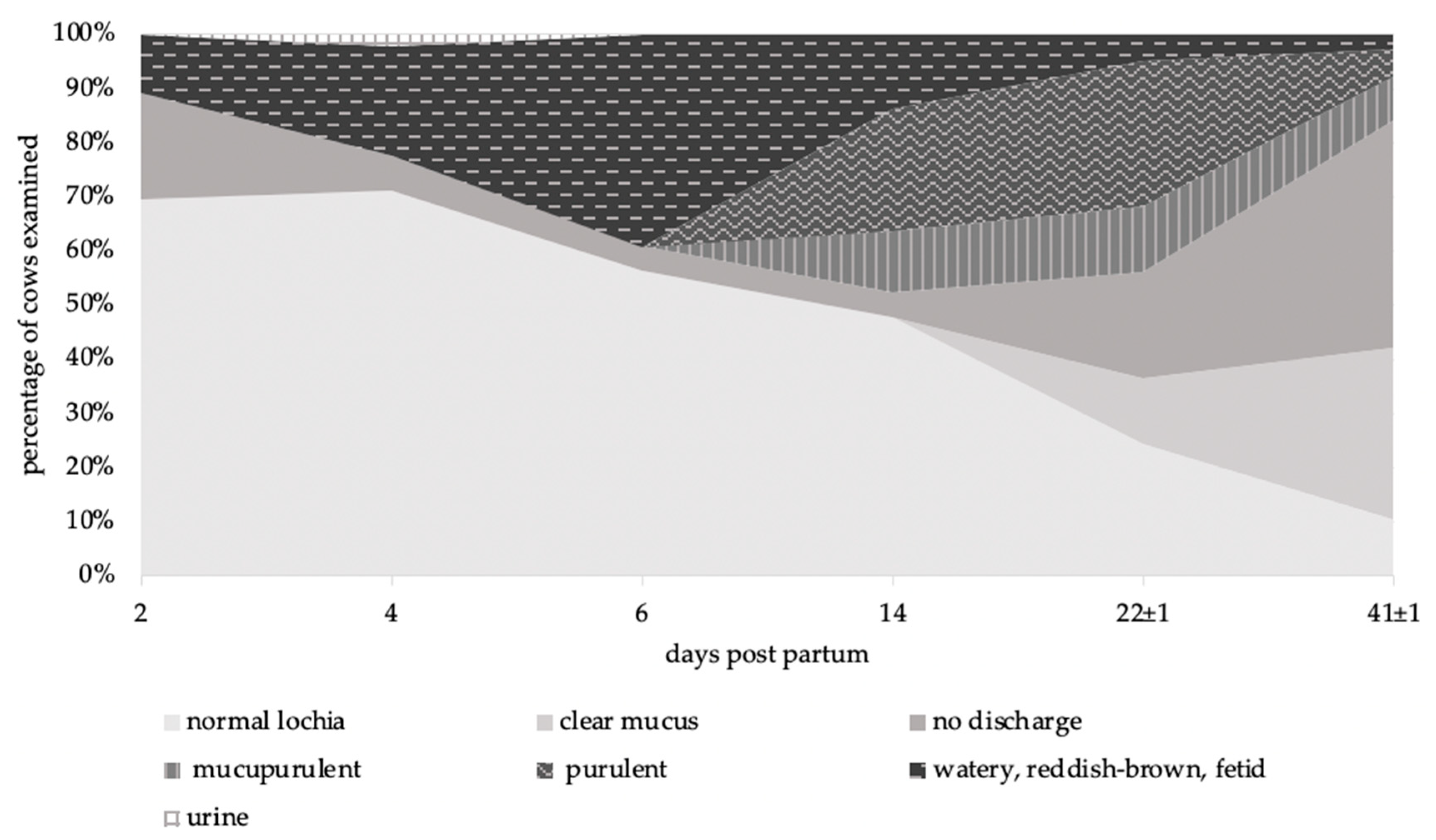
3.1.2. Vaginal Discharge

Vaginal discharge was characterised via vaginoscopy according to an assessment scheme (Table 1) at six time points during the puerperium. Pathological discharge was present for 1 to 39 days p.p. (mean: 18) in treated cows and for 0 to 27 days p.p. (mean: 11) in the untreated ones, taking into account the examination intervals.
On day 6 p.p., 39.1% of the animals examined had a watery, reddish-brown and fetid vaginal discharge; this proportion decreased to 13.6% on day 14 p.p., at which time purulent (22.7%) and mucupurulent (11.4%) vaginal discharge first appeared. On day 22 p.p., 43.9% of the cows still showed a pathological vaginal discharge, 36.6% of the cows had clinically unremarkable vaginal discharge and no vaginal discharge could be detected in 19.5% of the cows. At the end of puerperium, the proportion of cows with a pathological vaginal discharge decreased from a peak at day 14 p.p. (47.7%) to 15.8% and the proportion of cows with clinically inconspicuous vaginal discharge increased to 42.1%, with 42.1% showing no vaginal discharge (Figure 3). It was found that the vaginal discharge in some cows took on different characters over the study period and that different forms merged into one another.

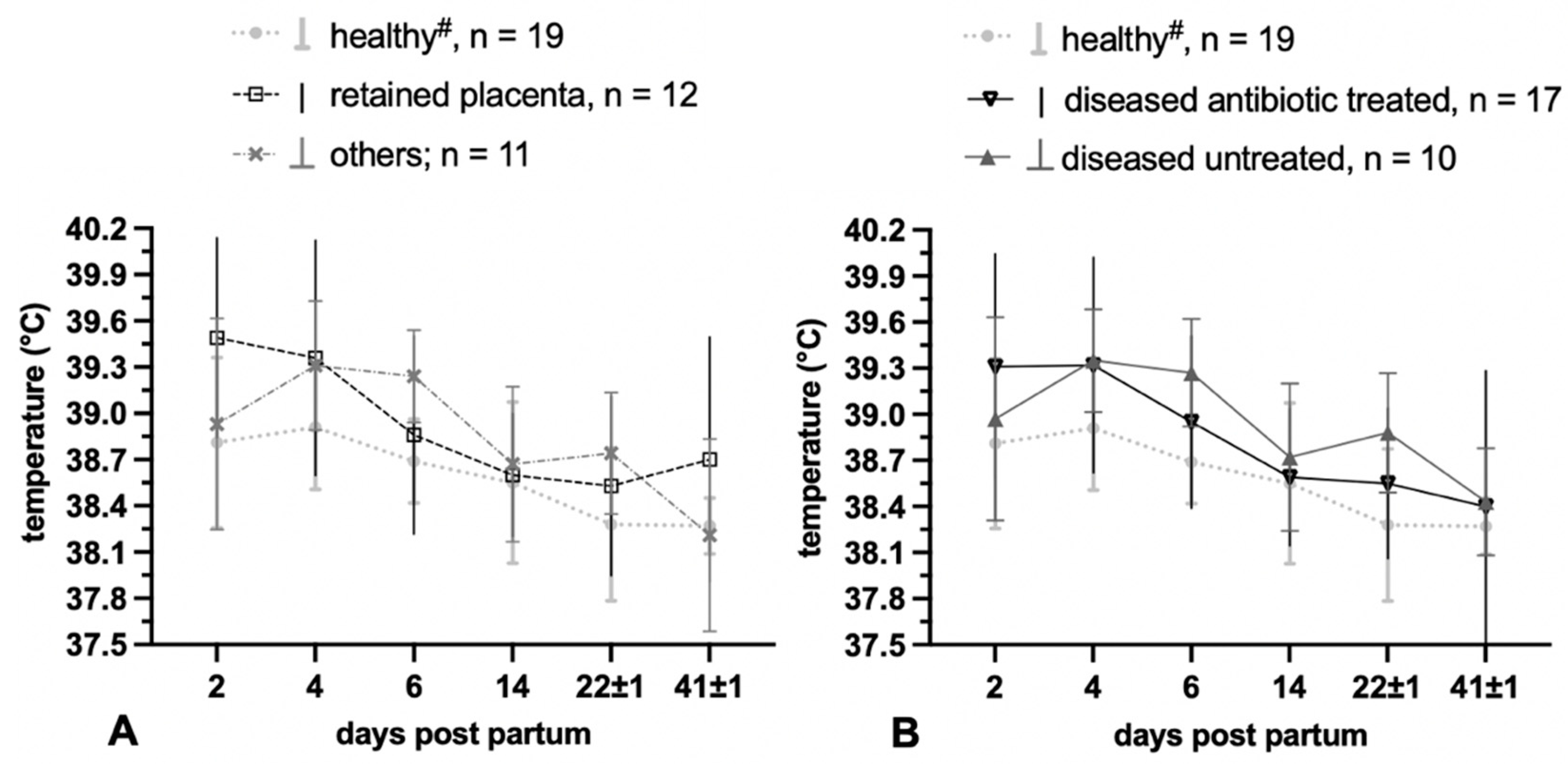
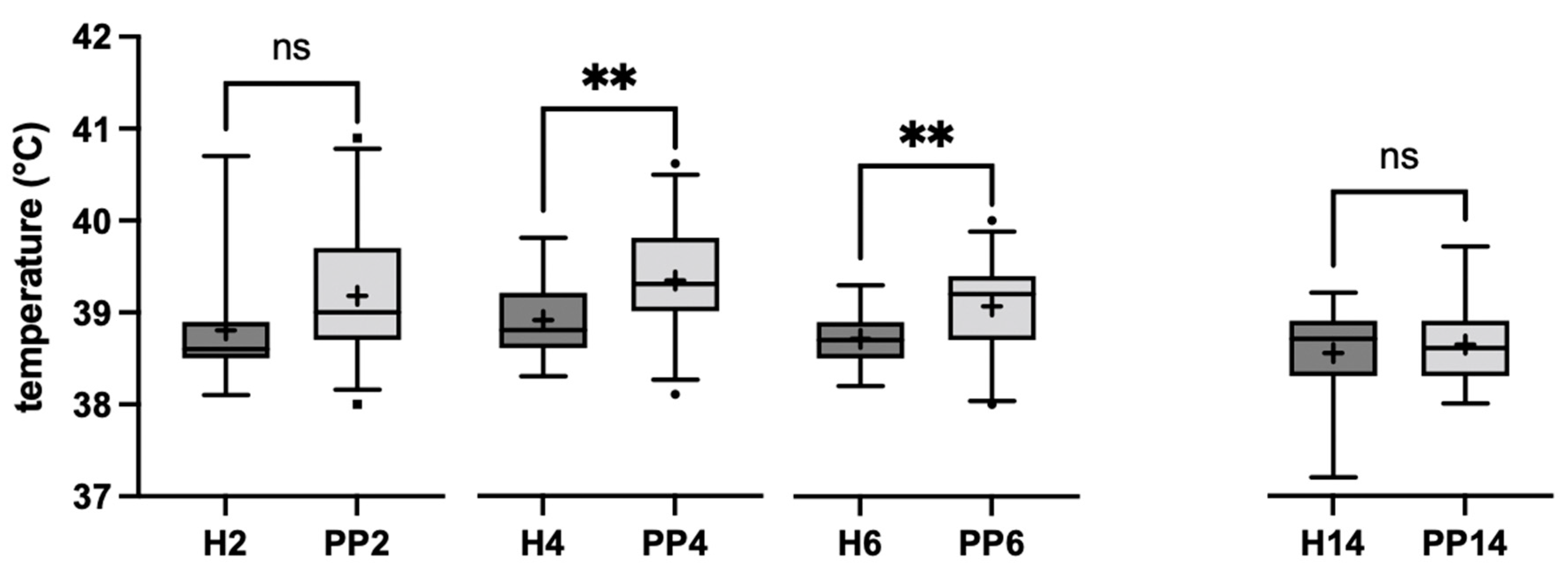
3.1.3. Temperature Measurement

With regard to rectal body temperature, the animals that remained healthy showed an average body temperature of 38.6 °C over the entire puerperium. Compared to them, the animals with puerperal disorders of the reproductive tract showed a lower body temperature at each time point (Figure 2). However, one animal in the group of healthy cows showed fever due to a diagnosis other than a disorder of the reproductive tract. This cow was responsible for the maximum temperature values in the group of healthy cows on day two after calving, with a maximum body temperature of 40.7 °C on day two p.p. Nevertheless, statistical analysis showed significant differences in rectal body temperatures between diseased and healthy animals on day 4 (p = 0.008) and day 6 (p = 0.008) p.p. The body temperature of cows with a pathological puerperium was significantly higher than the rectal temperature of the healthy ones (Figure 4). Comparing the trend of the rectal body temperature of diseased animals (n = 17) that received antibiotic treatment or remained untreated (n = 10), the antibiotic-treated animals initially showed a higher mean body temperature on day 2 p.p. On day 4 p.p., the mean values converged. In the further course, the temperature of the untreated animals was higher until the temperatures at the end of the puerperium (day 41 ± 1 p.p.) converged to the same mean value (38.4 °C). Overall, all cows showed a higher body temperature at the beginning of puerperium (day 2 to 4), which decreased continuously from the fourth day p.p. until the 14th day p.p. At two weeks p.p., the cows showed the least divergence in body temperature during the puerperium. From day 22 ± 1 to day 41 ± 1, only the animals with postpartum retention showed an increase in body temperature (Figure 2A).

3.2. Microbiological Results

Detailed microbial results are available from 22 animals, of which ten developed clinical disease. During clinical gynaecological examination, a total of 364 swab samples were taken from the vagina and uterus. In total, 985 isolates were cultured from the reproductive tract, belonging to five phyla, 56 genera and 129 bacterial species. When considering the postpartum reproductive tract over the entire sample period, the phylum Firmicutes was the most frequently detected with 40.4%, followed by Proteobacteria (24.2%), Actinobacteria (19.0%), Fusobacteria (10.1%) and Bacteroidetes (6.2%). Among the genera represented, Streptococci were the most frequently detected (20.2% of isolates), followed by T. pyogenes (11.8%), Escherichia spp. (10.8%) and Fusobacterium spp. (10.1%). The following genera were also detected with a prevalence >3%: Staphylococcus spp. (6.0%), Bacillus spp. (3.4%) and Actinobacillus spp. (3.1%). The genera Staphylococcus spp. and Streptococcus spp. showed the greatest species diversity with 12 different species each. The genera Corynebacterium spp. (n = 10), Bacillus spp. (n = 9), Lactobacillus spp. (n = 7) and Enterococcus spp. (n = 6) were also represented by a considerable number of different species.

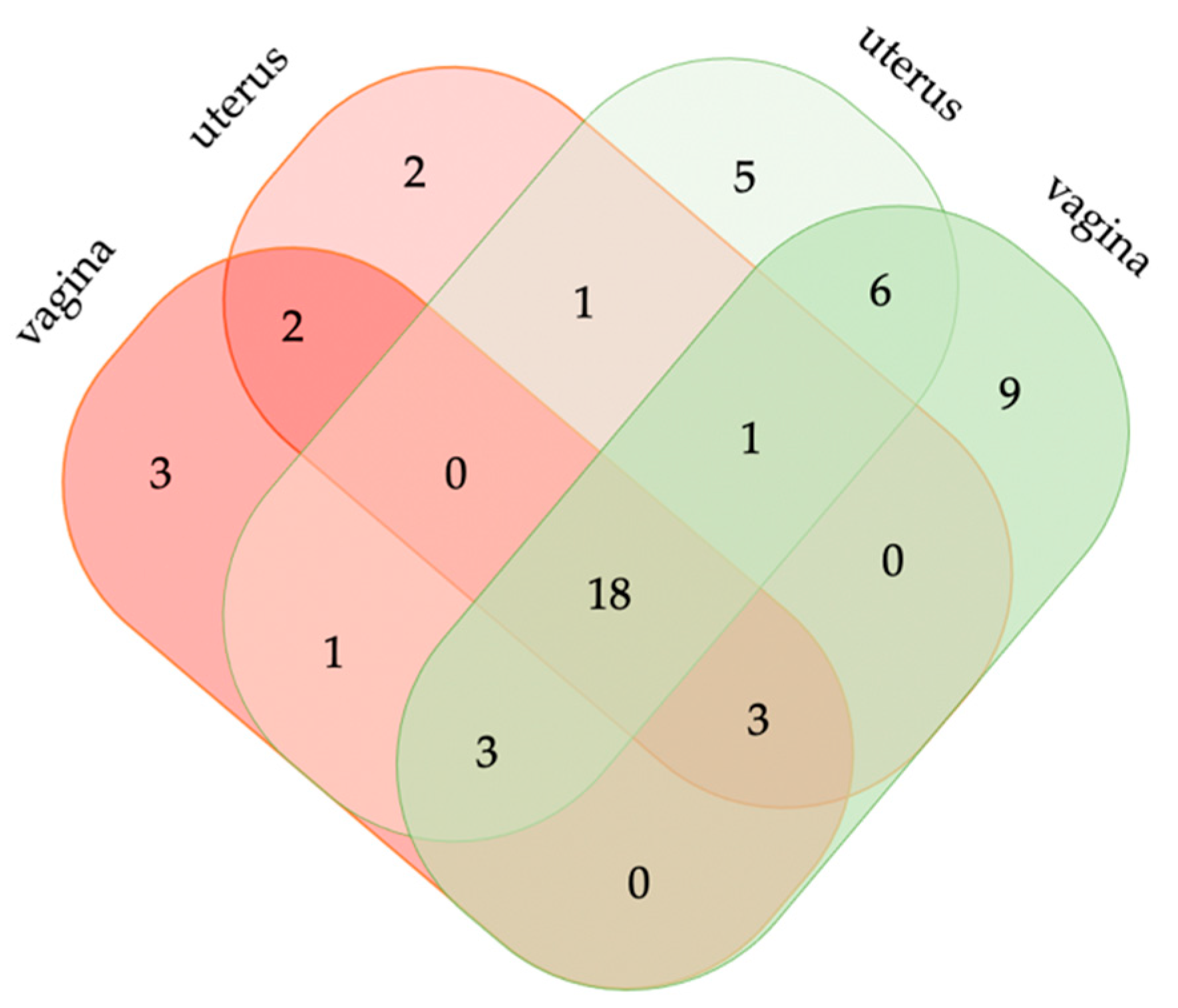
3.2.1. Postpartum Vaginal and Uterine Bacterial Diversity

In total, 561 bacteria from 46 genera were isolated from the postpartum vagina. The isolates from the postpartum uterus sum up to a total number of 409 and represent 42 genera. Table 4 summarises the core bacterial community of the postpartum vagina and uterus as revealed by cultural methods. Bacteria of the genus Streptococcus spp. were most frequently isolated in the vagina and uterus. The genus Streptococcus was represented in the vagina by 11 different species and in the uterus by eight different species, with Streptococcus pluranimalium (S. pluranimalium; vagina n = 36; uterus n = 10) and Streptococcus uberis (S. uberis; vagina n = 32; uterus n = 35) dominating. The second and third most common genera were Trueperella spp. and Escherichia spp., again in the vagina as well as in the uterus. The genus Trueperella was represented exclusively by the species T. pyogenes. From the genus Escherichia, E. coli and Escherichia fergusonii (E. fergusonii) were isolated. Fisher’s exact test showed that the genera Bacillus spp. and Corynebacterium spp. were significantly more frequent in the vagina than in the uterus. The genus Clostridium spp. was significantly more frequent in the uterus than in the vagina (Table 4). In addition, significant correlations between the presence of potentially pathogenic bacteria in the vagina and the presence of the same genera in the uterus were demonstrated using Spearman’s rank test: Bacteroides spp. (ρ = 0.81; p = < 0.0001), Proteus spp. (ρ = 0.73; p = 0.0001), Fusobacterium spp. (ρ = 0.65; p = 0.001), T. pyogenes (ρ = 0.61; p = 0.002), Helcococcus spp. (ρ = 0.61; p = 0.002), Prevotella spp. (ρ = 0.54; p = 0.009), Escherichia spp. (ρ = 0.50; p = 0.01).

3.2.2. Vaginal Bacterial Diversity in Healthy and Diseased Animals

In Table 5, the microbial community of the vagina and the uterus of healthy and diseased cows is compared at genus level. From the postpartum vagina of 12 cows that remained healthy, 295 bacteria belonging to 40 genera could be isolated. The bacterial community is dominated by the genus Streptococcus, the genus being represented by six species, with the three most common species being S. pluranimalium (n = 28), S. uberis (n = 14) and Streptococcus suis (S. suis; n = 11). Other frequently isolated genera were Staphylococcus (six species in total), with the three most common species Staphylococcus chromogenes (n = 19), Staphylococcus sciuri (n = 5) and Staphylococcus haemolyticus (n = 3); the genus Trueperella, represented by the species T. pyogenes (n = 28) and the genus Escherichia also represented by only one species: E. coli (n = 25). The genus Acinetobacter reached a prevalence above 2% only in the vaginal swab samples from cows that remained healthy, the two most common species of this genus were Acinetobacter johnsonii (n = 4) and Acinetobacter lwoffii (n = 3).
In total, from the vagina of ten cows with puerperal disorders, 266 bacteria were isolated, belonging to 30 genera. The most common bacteria from the vagina of cows with puerperal disorders also belonged to the genus Streptococcus, represented by 10 species, with S. uberis (n = 18) being the most common species isolated, followed by S. pluranimalium (n = 8) and Streptococcus lutetiensis (S. lutetiensis; n = 4). In contrast to the healthy animals, the genera Trueperella with the species T. pyogenes (n = 35), Fusobacterium spp. (n = 32) and Escherichia with the species E. coli (n = 27) and E. fergusonii (n = 2) follow. Several genera show up with a prevalence above 2% only in the vagina of the diseased cows, i.e., the genera Atopobium with the species Atopobium minutum (n = 9), Enterococcus with the most common species Enterococcus faecalis (n = 6) and Porphyromonas with the species Porphyromonas levii (n = 6).

3.2.3. Uterine Bacterial Diversity in Healthy and Diseased Animals

From the uterus of cows that remained healthy, 190 bacteria belonging to 35 genera could be isolated. Like in the vagina, Streptococcus spp. was the most frequently isolated genus. It was represented by five species, of which the three most common species were shared with the vagina (S. uberis (n = 15), S. pluranimalium (n = 9) and S. suis (n = 5)). In addition, frequently isolated bacteria belonged to the genera Trueperella and Escherichia, with only one species each (T. pyogenes, n = 24 and E. coli, n = 21). Only in the healthy uterus, the genus Peptoniphilus was found with the species Peptoniphilus indolicus (n = 6) with a prevalence above 2%.
A total of 220 bacteria from 27 genera were detected from the uterus of cattle with puerperal disorders of the reproductive tract. In particular, bacteria of the genus Streptococcus where shared between the uteri of healthy and diseased animals. The genus was composed of seven species, wherein S. uberis (n = 20) was the most common species, followed by S. lutetiensis (n = 8) and Streptococcus dysgalactiae (n = 4). The species E. fergusonii (n= 4) could additionally be isolated from the uterus of diseased animals, as from the vaginal samples. Several species were found in the uterus of diseased animals exclusively. This was true for the genera Clostridium with four species—most frequently Clostridium perfringens (n = 4)—and the genus Proteus (in total n = 5), with the main species Proteus mirabilis (n = 3).
The Venn diagram (Figure 5) illustrates the number of genera that were shared between the uterus and/or vagina in a healthy and/or diseased state at any of the samplings (day 2, 4, 6 and 14 p.p.) within a 14-day period. Only 18 genera were found in all kinds of specimen and health states. Vice versa, seven genera were found in diseased cattle only: three in the vagina, two in the uterus and two in both localizations.

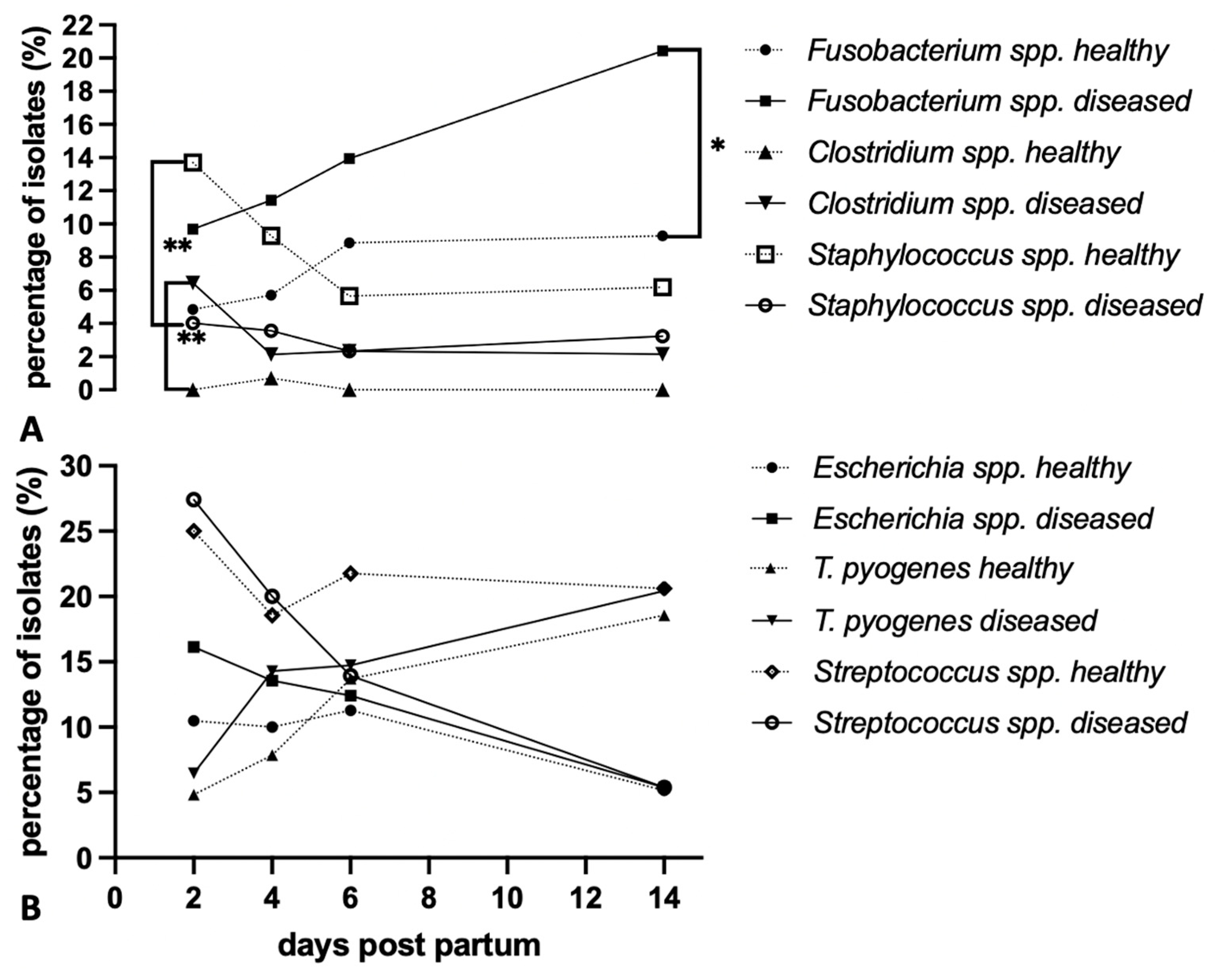
3.2.4. Microbiota Community Structure and Dynamics in Healthy and Diseased Cattle

Four genera were significantly more abundant in the vagina and uterus of healthy animals, compared to animals with puerperal disorders. Vice versa, six genera showed a significantly higher occurrence in animals with puerperal disorders, compared to the healthy animals; see Table 5.
The genus Streptococcus spp. occurred quite frequently in the first two weeks after calving in the healthy animals, without significant decrease. In comparison, the Streptococcus genus in the diseased cows showed a steady, significant decline over the sampling period (Chi-squared test: day 2 vs. day 6 p.p. p = 0.008, day 2 vs. day 14 p.p., p = 0.0001). At the beginning of the puerperium, Escherichia spp. occurred more frequently in the diseased animals, compared to healthy animals, but the percentages of the isolates converged more and more during the course until they were finally almost identical on day 14 p.p. Significant differences between the healthy and diseased animals could not be found at any sampling time. Nevertheless, there was a significant decrease in the genus Escherichia spp. from day 2 to day 14 p.p. only in the cows with the pathological puerperium (Chi-squared test: p = 0.01). The species T. pyogenes increased in parallel in healthy and diseased cows in the first two weeks p.p. Only at day four p.p., a difference became apparent, with a higher occurrence in the diseased animals (Chi-squared test: day 2 vs. day 4 p.p. of the diseased animals, p = 0.04); see Figure 6B.
Staphylococcus spp. occurred significantly more often in the healthy animals than in the diseased animals at each sampling time, especially close to calving (Chi-squared test: day 2 p.p. of healthy animals vs. day 2 p.p. of diseased cows, p = 0.007; Figure 6A). Clostridium spp. occurred significantly more often especially on day two p.p., in diseased cows (Chi-squared test day 2 p.p. of healthy animals vs. day 2 p.p. of diseased cows, p = 0.004; Figure 6A). On day 14 p.p., the genus Fusobacteria spp. occurred significantly more often in the diseased animals than in the cows that remained healthy (Chi-squared test: day 14 vs. day 14 p.p. between healthy and diseased cows, p = 0.03; Figure 6A).

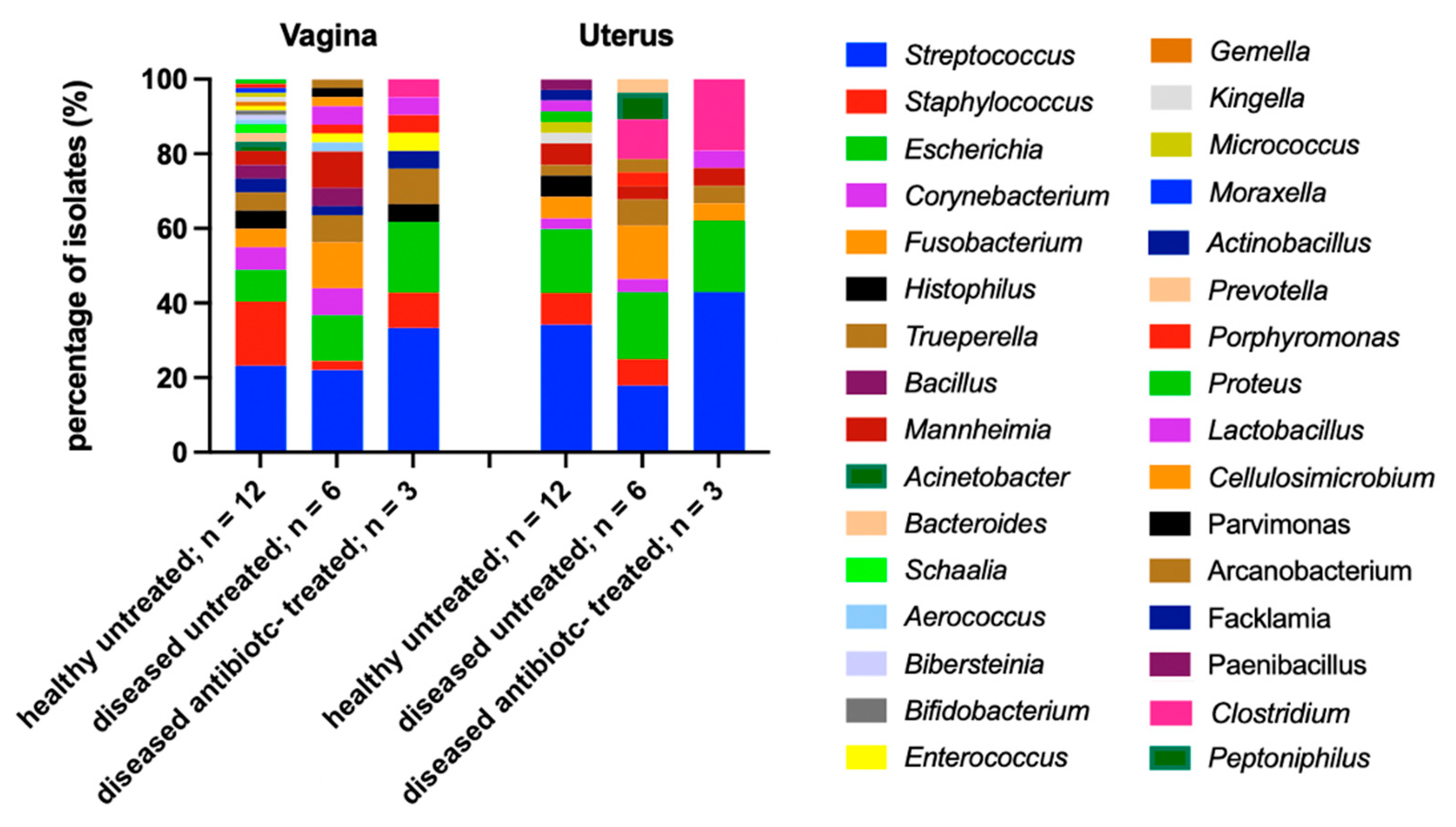
3.2.5. Vaginal and Uterine Microbiome of Antibiotic-Treated and Untreated Cows

Comparing the influence of antibiotic use in the vagina and uterus depending on health state and antibiotic treatment, an absence of the genus Staphylococcus spp. can be observed in the uteri of the diseased, antibiotic-treated cows at day 2 p.p. The presence of the genus Clostridium spp. in the diseased animals and even an increased occurrence in the cows under antibiotic treatment should be emphasized (Figure 7). The genus Escherichia spp. still accounts for 19.0% and 18.2% of isolates, respectively, in animals under antibiosis in both the vagina and uterus. Thus, there is even a higher occurrence of the genus Escherichia spp. in the vagina (19.0% compared to 12.2%) and an almost equal occurrence in the uterus (17.9% compared to 18.2%) in treated cows, compared to the sick, untreated animals. Bacterial diversity was greatest in the untreated healthy animals with a total of 26 genera, compared to 19 genera in the diseased, untreated animals and 12 genera in the diseased, antibiotic-treated cows; however, different numbers of cows in each group (12 vs. 6 vs. 3) may have contributed to that observation.

4. Discussion

This study found an incidence of 27 cases in 46 observed dairy cows (58.7%) for pathological puerperium. Of the 27 diseased animals, almost half showed postpartum retention, giving an incidence of 26.1%. The incidence of puerperal metritis amounted to 17.4%, for clinical metritis, 52.2%, and for clinical endometritis, 39.1%. The incidence of these clinical pictures in the literature vary greatly, wherein incidence is differently defined—sometimes by cases per calving cows in one year [14], in other studies—as in ours—as incidence within the study period (here: 11 months). The incidence of postpartum retention of the fetal membranes ranges from 3% to 39% [15]. It is difficult to define an acceptable percentage, but a value below 10% is envisaged [16]. Pohl et al. [17] give an incidence of 7.2% to 38.1% for puerperal metritis, which is also called acute puerperal metritis in the literature. For clinical metritis, incidences of 25% to 40% within the first two weeks after calving and 20% for clinical endometritis are reported [18]. These varying figures are partly due to the fact that the clinical pictures can be defined differently and the incidence rates vary greatly depending on the farm [15,19]. In addition, disease patterns can exist in parallel and merge into each other in the absence of controls for the infection. However, the values show that there is a need for action on the farm to reduce these high numbers of illnesses. When we attempted to find reasons for the comparatively high incidence of puerperal disorders in the farm, we could not escape the conclusion that calving hygiene was suboptimal and impaired the health state of cows p.p. Calving areas were neither cleaned nor disinfected before cows were moved there. Birth assistance was given without gloves, and ropes were only cleaned with warm water. No systematic puerperal monitoring—apart from the study conditions—was established on the farm, such as control of afterbirth expulsion and measurements of body temperature.
Animals with metritis or endometritis do not show fever. However, since puerperal metritis is defined by the occurrence of fever, it is obvious from the case numbers that the whole group of diseased animals must have a higher mean body temperature than those which stayed healthy. Most cows with a retained placenta reacted with fever within the first two days after calving, which underlines the importance of controlling body temperature, complete shedding of fetal membranes, or—at best—both. Cattle with other puerperal disorders developed fever late in the course of disease. Within 14 days after calving, fever was cured with or without antibiotic treatment. However, cows that received antibiotics recovered more quickly from fever than cows that did not receive antibiotic treatment. Similar observations were made by Benzaquen et al. [20]; after antibiotic treatment of cows with puerperal metritis, fever decreased to a similar level as in the healthy untreated cows. In a study by Chenault et al. [21], the antibiotic-treated animals also showed a faster decrease in body temperature compared to a control group receiving a 0.9% NaCl solution.
Vaginoscopy is a simple and quick examination procedure, in addition to the measurement of body temperature, that helps to better distinguish healthy from diseased cows [22,23,24]. Pathological vaginal discharge was present on average longer in diseased antibiotic-treated cows than in diseased untreated animals. This seemingly counterintuitive observation could be due to the fact that the group of treated animals included cows that showed a severe course of puerperal disorders. However, Jeremejeva et al. [25] also concluded that the administration of antibiotics to metritic animals had no effect on the development of vaginal discharge compared to an untreated control group. Based on the results, we concluded that antibiotic administration should be better based on body temperature and not only on vaginal discharge. The period in which pathological discharge was present varied greatly from individual to individual and in some cows even persisted from the first to the last time of examination. While the inconspicuous lochial discharge existed for 14 to 18 days p.p. [26], pathological discharge can last up to 30 days p.p. according to Youngquist and Shore [27], or, according to our observations, even up to about 40 days p.p.
As mentioned earlier, bacterial colonisation of the postpartum vagina and uterus is an inevitable fact [9,10]. Accordingly, we found close correlation between the presence of potential pathogens in the vagina and the presence of the same genera in the uterus. This was true for Bacteroides spp., Fusobacteria spp. and Helcococcus spp., for example. These bacteria were also detected significantly more often in diseased cattle, compared to healthy ones. Fusobacterium necrophorum is a recognised uterine pathogen according to many authors [13,28,29]. Karstrup et al. [30] also point to tissue invasiveness of Fusobacterium necrophorum and Porphyromonas levii. However, Porphyromonas levii showed only an insignificantly increased incidence in the diseased animals in our study. Bacteroides spp. and Helcococcus spp. are also genera known in the literature as uterine pathogens [9,31]. Furthermore, correlations were found for the occurrence of the genera Proteus spp., Prevotella spp., T. pyogenes and Escherichia spp. in the vagina and uterus, but no significant differences were found between healthy and diseased cows for these genera, although they are discussed as opportunistic metritis pathogens [13,32]. Thus, ascendency and mixing of the vaginal and uterine microbiome did not seem to result in puerperal disorder here. Likewise, Cunha et al. [31] also question the pathogenic significance of these bacteria.
The genera Staphylococcus spp., Actinobacillus spp., Acinetobacter spp., and Histophilus spp. were isolated significantly more frequently from the cows that remained healthy, so they could qualify as potentially protective. In our study, we found a significantly lower incidence of the genus Staphylococcus spp. in the diseased cows on day 2 p.p., i.e., before clinical symptoms developed in most of the cattle. Huszenicza et al. [33] also reported that coagulase-negative Staphylococci were more prevalent in animals that did not show uterine infections. Williams et al. [13] demonstrated that coagulase-negative staphylococci reduced the risk of abnormal vaginal discharge. The genus Actinobacillus spp. [34] was isolated from the postpartum uterus of dairy cows, but has not been associated with positive or negative effects on uterine health, only with epididymitis in rams [35]. Acinetobacter spp. is described by Sheldon et al. [12] as a potential pathogen of uterine infections. However, Acinetobacter spp. was isolated from the uterus of heifers and pregnant cows [36] and Wang et al. [37] described the genus as commensal. Histophilus somni is found, among other places, on the mucous membranes of the genital tract [38]. Contrary to our findings, other authors have associated Histophilus somni with diseases of the genital tract [39,40,41].
The results of this study reveal a common postpartum bovine bacterial core community. The vaginal bacterial community proved to be more diverse compared to the uterine one, as was the microbiota of the cows that remained healthy compared to the diseased ones. Animals with puerperal disorders had a remarkably lower microbial diversity at genus level. However, a considerable number of genera was shared between all departments (Figure 5), including several pathogens well-known for causing uterine disease (Bacteroides spp., Fusobacteria spp., Helcococcus spp.). Other bacteria occurred only in diseased animals, but have not been reported to be involved in puerperal disease up to now. Interestingly, the diseased cows lacked more bacteria than they acquired: while diseased cattle were positive for seven genera for which healthy cows were negative, they lacked twenty genera that were found in healthy cows. Future research is needed to clarify whether these are occasional or significant findings, due to the moderate number of animals involved in this study. Antimicrobial treatment further reduced bacterial diversity, as described for a broad variety of body sites under antibiotic treatment [42,43], sometimes also resulting in dysbiosis. Dysbiosis may implicate the absence of potentially protective genera, such as Staphylococcus spp. in our study, and the increased occurrence of potentially pathogenic genera, as seen with Clostridium spp. in the diseased and treated animals at the site of inflammation in the uterus. Thus, antibiosis may even have a negative impact on the course of the disease.

5. Conclusions

The puerperium sets the course for a healthy start into lactation and a successful conception. We conclude that measurement of body temperature, at best combined with vaginoscopy, is suitable to monitor the health of dairy cows during this decisive period. Without the establishment of strict hygiene management during the period of puerperium, which includes close monitoring of body temperature and control of afterbirth expulsion and lochia, the incidence of puerperal disorders can reach unacceptable levels, which may result in the high use of antibiotics. In particular, the frequent use of tetracyclines should be critically reconsidered. Tetracyclines strongly bind calcium ions [44]—at a time of high calcium requirement—and are excreted as antimicrobially active molecules mainly through milk. Our study reconfirmed that bacterial colonisation of the reproductive tract is a normal finding. However, during the period of broken anatomical barriers, it is important to limit potentially pathogenic bacteria and, at best, promote potentially protective bacteria.
Our findings also confirmed the presence of known uterine pathogens such as Bacteroides spp. and Fusobacteria spp., as well as more recently discussed uterine pathogens such as Helcococcus spp. However, our results question the role of the bacteria Escherichia spp. and T. pyogenes as triggers of uterine inflammation. Future studies could focus on the role of the microbiota of healthy animals, as a physiological, diverse microbiome is essential for the prevention of dysbiosis or infectious disease. This may bring the genus Staphylococcus spp. to the forefront of future investigations for protective properties, such as the production of bacteriocins (here: staphylococcin). Thus, our findings depict the complexity of the bovine bacterial community of the vagina and uterus and provide the basis for further investigations aiming to promote reproductive tract health in cattle.

Author Contributions

Conceptualization, H.K., N.K. and C.S.H.; methodology, H.K. and C.S.H.; validation, H.K. and C.S.H.; formal analysis, H.K. and C.S.H.; investigation, H.K.; resources, H.K. and C.S.H.; data curation, H.K.; writing—original draft preparation, H.K. and C.S.H.; writing—review and editing, H.K., N.K. and C.S.H.; visualization, H.K and C.S.H.; supervision, C.S.H. and N.K.; project administration, C.S.H. All authors have read and agreed to the published version of the manuscript.

Funding

This research received no external funding.

Institutional Review Board Statement

The animal study protocol was approved by the Institutional Review Board (or Ethics Committee) of the Ministry of Environment and Agriculture in Schleswig-Holstein (V 242- 46504/2019).

Informed Consent Statement

Not applicable.

Data Availability Statement

The data presented in this study is available on reasonable request from the corresponding author. Data are not publicly available due to privacy reason for the farm involved.

Acknowledgments

The authors are grateful for the support of the farmer. We also thank the technical assistants Evelyn Laß and Meike Friedrichsen (Institute of Animal Breeding and Husbandry, Kiel, Germany), who provided support in the laboratory. We sincerely thank U. Schotte for providing the MALDI- TOF biotyper and Maike Siewers (Central Institute of the Bundeswehr Medical Service Kiel, Kronshagen, Germany) for providing the MALDI-TOF instrument and technical support.

Conflicts of Interest

The authors declare no conflict of interest.

References

  1. Gilbert, R.O. Management of Reproductive Disease in Dairy Cows. Vet. Clin. N. Am. Food Anim. Pract. 2016, 32, 387–410. [Google Scholar] [CrossRef] [PubMed]
  2. Overton, M. Economics of postpartum uterine health. In Proceedings of the Dairy Cattle Reproduction Council Convention, Omaha, NE, USA, 7–8 November 2008; pp. 39–43. [Google Scholar]
  3. Wagener, K.; Prunner, I.; Pothmann, H.; Drillich, M.; Ehling-Schulz, M. Diversity and health status specific fluctuations of intrauterine microbial communities in postpartum dairy cows. Vet. Microbiol. 2015, 175, 286–293. [Google Scholar] [CrossRef] [PubMed]
  4. Neubrand, L.; Wagener, K.; Drillich, M. Bovine uterine diseases: Aspects of microbiology, molecular biology, and immunology. Tierarztl. Praxis. Ausg. G Grosstiere/Nutztiere 2020, 48, 253–261. [Google Scholar] [CrossRef]
  5. Stojkov, J.; von Keyserlingk, M.A.; Marchant-Forde, J.N.; Weary, D.M. Assessment of visceral pain associated with metritis in dairy cows. J. Dairy Sci. 2015, 98, 5352–5361. [Google Scholar] [CrossRef] [PubMed]
  6. Palmer, C. Postpartum Uterine Infection. In Bovine Reproduction; Hopper, R.M., Ed.; Wiley-Blackwell: Hoboken, NJ, USA, 2015; p. 440. [Google Scholar]
  7. Frazer, G.S. A rational basis for therapy in the sick postpartum cow. Vet. Clin. N. Am. Food Anim. Pract. 2005, 21, 523–568. [Google Scholar] [CrossRef] [PubMed]
  8. Dadarwal, D.; Palmer, C.; Griebel, P. Mucosal immunity of the postpartum bovine genital tract. Theriogenology 2017, 104, 62–71. [Google Scholar] [CrossRef] [PubMed]
  9. Miranda-CasoLuengo, R.; Lu, J.; Williams, E.J.; Miranda-CasoLuengo, A.A.; Carrington, S.D.; Evans, A.C.O.; Meijer, W.G. Delayed differentiation of vaginal and uterine microbiomes in dairy cows developing postpartum endometritis. PLoS ONE 2019, 14, e0200974. [Google Scholar] [CrossRef] [Green Version]
  10. Foldi, J.; Kulcsar, M.; Pecsi, A.; Huyghe, B.; de Sa, C.; Lohuis, J.A.; Cox, P.; Huszenicza, G. Bacterial complications of postpartum uterine involution in cattle. Anim. Reprod. Sci. 2006, 96, 265–281. [Google Scholar] [CrossRef]
  11. Sheldon, I.M.; Dobson, H. Postpartum uterine health in cattle. Anim. Reprod. Sci. 2004, 82–83, 295–306. [Google Scholar] [CrossRef]
  12. Sheldon, I.M.; Noakes, D.E.; Rycroft, A.N.; Pfeiffer, D.U.; Dobson, H. Influence of uterine bacterial contamination after parturition on ovarian dominant follicle selection and follicle growth and function in cattle. Reproduction 2002, 123, 837–845. [Google Scholar] [CrossRef]
  13. Williams, E.J.; Fischer, D.P.; Pfeiffer, D.U.; England GC, W.; Noakes, D.E.; Dobson, H.; Sheldon, I.M. Clinical evaluation of postpartum vaginal mucus reflects uterine bacterial infection and the immune response in cattle. Theriogenology 2005, 63, 102–117. [Google Scholar] [CrossRef] [PubMed]
  14. Andriamanga, S.; Steffan, J.; Thibier, M. Metritis in dairy herds: An epidemiological approach with special reference to ovarian cyclicity. Ann. Rech. Vet. 1984, 15, 503–508. [Google Scholar] [PubMed]
  15. Kimura, K.; Goff, J.P.; Kehrli, M.E., Jr.; Reinhardt, T.A. Decreased Neutrophil Function as a Cause of Retained Placenta in Dairy Cattle. J. Dairy Sci. 2002, 85, 544–550. [Google Scholar] [CrossRef]
  16. Peter, A.T. Retained Fetal Membranes. In Bovine Reproduction, 2nd ed.; Hopper, R.M., Ed.; John Wiley & Sons: Hoboken, NJ, USA, 2021. [Google Scholar]
  17. Pohl, A.; Bertulat, S.; Borchardt, S.; Burfeind, O.; Heuwieser, W. Randomized, controlled clinical trial on the efficacy of nonsteroidal antiinflammatory drugs for the treatment of acute puerperal metritis in dairy cows. J. Dairy Sci. 2016, 99, 8241–8249. [Google Scholar] [CrossRef] [Green Version]
  18. Sheldon, I.M.; Williams, E.J.; Miller, A.N.; Nash, D.M.; Herath, S. Uterine diseases in cattle after parturition. Vet. J. 2008, 176, 115–121. [Google Scholar] [CrossRef]
  19. Sheldon, I.M.; Cronin, J.; Goetze, L.; Donofrio, G.; Schuberth, H.J. Defining postpartum uterine disease and the mechanisms of infection and immunity in the female reproductive tract in cattle. Biol. Reprod. 2009, 81, 1025–1032. [Google Scholar] [CrossRef]
  20. Benzaquen, M.E.; Risco, C.A.; Archbald, L.F.; Melendez, P.; Thatcher, M.J.; Thatcher, W.W. Rectal temperature, calving-related factors, and the incidence of puerperal metritis in postpartum dairy cows. J. Dairy Sci. 2007, 90, 2804–2814. [Google Scholar] [CrossRef] [Green Version]
  21. Chenault, J.R.; McAllister, J.F.; Chester, S.T.; Dame, K.J.; Kausche, F.M.; Robb, E.J. Efficacy of ceftiofur hydrochloride sterile suspension administered parenterally for the treatment of acute postpartum metritis in dairy cows. J. Am. Vet. Med. Assoc. JAVMA 2004, 224, 1634–1639. [Google Scholar] [CrossRef]
  22. Leutert, C.; von Krueger, X.; Plontzke, J.; Heuwieser, W. Evaluation of vaginoscopy for the diagnosis of clinical endometritis in dairy cows. J. Dairy Sci. 2012, 95, 206–212. [Google Scholar] [CrossRef] [Green Version]
  23. Sheldon, I.M.; Rycroft, A.N.; Zhou, C. Associationbetweenpostpartumpyrexia and uterinebacterial infectionin dairy cattle. Vet. Rec. 2004, 154, 289–293. [Google Scholar] [CrossRef]
  24. LeBlanc, S.J.; Duffield, T.F.; Leslie, K.E.; Bateman, K.G.; Keefe, G.P.; Walton, J.S.; Johnson, W.H. Defining and Diagnosing Postpartum Clinical Endometritis and its Impact on Reproductive Performance in Dairy Cows. J. Dairy Sci. 2002, 85, 2223–2236. [Google Scholar] [CrossRef]
  25. Jeremejeva, J.; Orro, T.; Waldmann, A.; Kask, K. Treatment of dairy cows with PGF2alpha or NSAID, in combination with antibiotics, in cases of postpartum uterine inflammation. Acta Vet. Scand. 2012, 54, 45. [Google Scholar] [CrossRef] [Green Version]
  26. Senger, P.L. The puerperium and Lactation. In Pathways to Pregnancy and Parturition, 2nd ed.; Current Conceptions: Redmond, OR, USA, 2003; p. 329. [Google Scholar]
  27. Youngquist, R.S.; Shore, M.D. Postpartum uterine infection. In Current Therapy of Large Animal; Youngquist, R.S., Ed.; Saunders Elsevier: St. Louis, MO, USA, 1997; pp. 335–340. [Google Scholar]
  28. Sheldon, I.M.; Lewis, G.S.; LeBlanc, S.; Gilbert, R.O. Defining postpartum uterine disease in cattle. Theriogenology 2006, 65, 1516–1530. [Google Scholar] [CrossRef] [PubMed]
  29. Bicalho, M.L.; Machado, V.S.; Oikonomou, G.; Gilbert, R.O.; Bicalho, R.C. Association between virulence factors of Escherichia coli, Fusobacterium necrophorum, and Arcanobacterium pyogenes and uterine diseases of dairy cows. Vet. Microbiol. 2012, 157, 125–131. [Google Scholar] [CrossRef]
  30. Karstrup, C.C.; Agerholm, J.S.; Jensen, T.K.; Swaro, L.R.V.; Klitgaard, K.; Rasmussen, E.L.; Krogh, K.M.; Pedersen, H.G. Presence and localization of bacteria in the bovine endometrium postpartum using fluorescence in situ hybridization. Theriogenology 2017, 92, 167–175. [Google Scholar] [CrossRef] [PubMed]
  31. Cunha, F.; Jeon, S.J.; Daetz, R.; Vieira-Neto, A.; Laporta, J.; Jeong, K.C.; Barbet, A.F.; Risco, C.A.; Galvão, K.N. Quantifying known and emerging uterine pathogens, and evaluating their association with metritis and fever in dairy cows. Theriogenology 2018, 114, 25–33. [Google Scholar] [CrossRef] [PubMed]
  32. Madoz, L.V.; Giuliodori, M.J.; Migliorisi, A.L.; Jaureguiberry, M.; de la Sota, R.L. Endometrial cytology, biopsy, and bacteriology for the diagnosis of subclinical endometritis in grazing dairy cows. J. Dairy Sci. 2014, 97, 195–201. [Google Scholar] [CrossRef] [PubMed] [Green Version]
  33. Huszenicza, G.; Fodor, M.; Gacs, M.; Kulcsar, M.; Dohmen, M.J.V.; Vamos, M.; Porkolab, L.; Kegl, T.; Bartyik, J.; Lohuis, J.A.C.M.; et al. Uterine bacteriology, resumption of cyclic ovarian activity and fertility in postpartum cows kept in large-scale dairy herds. Reprod. Domest. Anim. 1999, 34, 237–245. [Google Scholar] [CrossRef]
  34. Moore, S.G.; Ericsson, A.C.; Behura, S.K.; Lamberson, W.R.; Evans, T.J.; McCabe, M.S.; Poock, S.E.; Lucy, M.C. Concurrent and long-term associations between the endometrial microbiota and endometrial transcriptome in postpartum dairy cows. BMC Genom. 2019, 20, 405. [Google Scholar] [CrossRef]
  35. Al-Katib, W.A.; Dennis, S.M. Epididymal and testicular lesions in rams following experimental infection with Actinobacillus seminis. N. Z. Vet. J. 2007, 55, 125–129. [Google Scholar] [CrossRef]
  36. Moore, S.G.; Ericsson, A.C.; Poock, S.E.; Melendez, P.; Lucy, M.C. Hot topic: 16S rRNA gene sequencing reveals the microbiome of the virgin and pregnant bovine uterus. J. Dairy Sci. 2017, 100, 4953–4960. [Google Scholar] [CrossRef] [PubMed] [Green Version]
  37. Wang, M.L.; Liu, M.C.; Xu, J.; An, L.G.; Wang, J.F.; Zhu, Y.H. Uterine Microbiota of Dairy Cows With Clinical and Subclinical Endometritis. Front. Microbiol. 2018, 9, 2691. [Google Scholar] [CrossRef] [PubMed]
  38. Jánosi, K.; Stipkovits, L.; Glávits, R.; Molnár, T.; Makrai, L.; Gyuranecz, M.; Varga, J.; Fodor, L. Aerosol infection of calves with Histophilus somni. Acta Vet. Hung. 2009, 57, 347–356. [Google Scholar] [CrossRef]
  39. Miller, R.B.; Barnum, D.A.; McEntee, K.E. Haemophilus somnus in the reproductive tracts of slaughtered cows: Location and frequency of isolations and lesions. Vet. Pathol. 1983, 20, 515–521. [Google Scholar] [CrossRef] [PubMed]
  40. Gonzalez Moreno, C.; Fontana, C.; Cocconcelli, P.S.; Callegari, M.L.; Otero, M.C. Vaginal microbial communities from synchronized heifers and cows with reproductive disorders. J. Appl. Microbiol. 2016, 121, 1232–1241. [Google Scholar] [CrossRef] [PubMed] [Green Version]
  41. van der Burgt, G.; Clark, W.; Knight, R.; Colles, K. Cattle fertility problems and Histophilus somni. Vet. Rec. 2007, 160, 600. [Google Scholar] [CrossRef] [PubMed]
  42. Francino, M.P. Antibiotics and the Human Gut Microbiome: Dysbioses and Accumulation of Resistances. Front. Microbiol. 2016, 6, 1543. [Google Scholar] [CrossRef] [Green Version]
  43. Karpinets, T.V.; Solley, T.N.; Mikkelson, M.D.; Dorta-Estremera, S.; Nookala, S.S.; Medrano, A.Y.D.; Petrosino, P.F.; Mezzari, M.P.; Zhang, J.; Futreal, P.A.; et al. Effect of Antibiotics on Gut and Vaginal Microbiomes Associated with Cervical Cancer Development in Mice. Cancer Prev. Res. 2020, 13, 997–1006. [Google Scholar] [CrossRef]
  44. Neuvonen, P.J. Interactions with the Absorption of Tetracyclines. Drugs 1976, 11, 45–54. [Google Scholar] [CrossRef]
Figure 1. Relationships of inflammatory diseases of the uterus of dairy cows over the period of puerperium (n = 25). The numbers reflect the number of cows that suffered from one of the diseases or a combination of them; #: one cow left the farm prematurely in each case.
Agriculture 12 00405 g001
Figure 2. Mean values and standard deviation of rectal body temperature over the period of puerperium in cattle that remained healthy (grey dotted); (A) additionally specific disease patterns; (B) additionally diseased cows treated with antibiotic treatment and without treatment. Others: cows showing a pathological puerperium but no singular postpartum retention of the fetal membranes or all forms of puerperal disorders; # one healthy cow showed fever due to a different diagnosis.
Agriculture 12 00405 g002
Figure 3. Development of vaginal discharge over the period of puerperium (n = 46).
Agriculture 12 00405 g003
Figure 4. Rectal body temperature of healthy cattle (H #, n = 19) and cows with a pathological puerperium (PP, n = 27) on day 2, 4, 6 and 14 after calving. Unpaired t-test: ns = not significant; ** p < 0.01. Box = from the 25th to 75th percentile; midline = median value; plus = mean value; whiskers = 5–95 percentile; # = one healthy cow showed fever due to a different diagnosis.
Agriculture 12 00405 g004
Figure 5. Venn diagram showing the number of shared genera isolated over the sampling period in the vagina and uterus of animals that remained healthy (green) and diseased (red).
Agriculture 12 00405 g005
Figure 6. (A) Percentage of isolates of selected genera that were significantly more abundant in healthy animals (n = 12; Staphylococcus spp.) or in diseased animals (n = 10; Fusobacteria spp., Clostridium spp.) over the entire sampling period (day 2, 4, 6, 14 p.p.); Chi-squared test comparison between healthy and diseased cows per sampling time point and genus: * p < 0.05, ** p < 0.01. (B) Percentage of isolates of selected genera that showed no significant differences between healthy and diseased animals over the combined sampling period but were among the most frequently isolated genera.
Agriculture 12 00405 g006
Figure 7. The vaginal and uterine microbiome of cows that remained healthy and received no treatment, compared to diseased cows with or without antibiotic treatment.
Agriculture 12 00405 g007
Table 1. The assessment scheme of the character of vaginal discharge.
DescriptionScore
Mucus Character
Clear mucus0
Normal lochia (reddish- brown, yellowish, non-smelling, mucous discharge)1
Mucupurulent (discharge containing ≤ 50% white or off- white mucupurulent material)2
Purulent (discharge containing > 50% purulent material, usually white, yellow or occasionally sanguineous)3
Watery, reddish-brown, fetid4
No discharge5
Urine6
Table 2. Incidences and results of preliminary reports of 46 dairy cows over the period of puerperium (until day 42 p.p.).
TotalPrevious IllnessesObstetricsBirth of TwinsStillbirth
%n%n %n%n%n
Diagnosis
Healthy41.31915.83 # 15.835.31
Pathological puerperium58.72711.13 ## 18.55 ###11.133.71
thereof
retained fetal membranes26.1128.31 16.7225.038.31
puerperal metritis17.48 12.5112.5112.51
grade 115.27 14.3114.31
grade 22.21 100.01
clinical metritis52.22412.53 20.8512.534.21
clinical endometritis39.11811.12 22.2416.73
urovagina 2.21
# one cow with retained fetal membranes and two cases of metritis; ## one cow with metritis and two cases of retained fetal membranes; ### Birth assistance by farmer (once by the veterinarian); first column: % of all 46 animals; second and following columns: % of 19 animals (healthy) or the number of the respective disease pattern of the 27 animals with a pathological puerperium; bold: group of cows that remained healthy and group of cows that became diseased.
Table 3. Treatments with antibiotics over the study period. TC was applied locally in the uterus (6000 mg three times at two-day intervals) BP (7500 mg twice at 24-hour intervals) and P (5000 mg daily for three days) were injected into the muscle.
No TreatmentTCTC + BPBPBP + PTC + BP + PP
% (n)% (n)% (n)% (n)% (n)% (n)% (n)
Diagnosis
Healthy94.7 (18) 5.3 (1)
Pathological puerperium37.0 (10)29.6 (8)11.1 (3)11.1 (3) #3.7 (1)3.7 (1)3.7 (1)
thereof
retained fetal membranes16.7 (2)58.3 (7)16.7 (2) 8.3 (1)
puerperal metritis
grade 142.9 (3)28.6 (2)14.3 (1) 14.3 (1)
grade 2 100.0 (1)
clinical metritis33.3 (8)33.3 (8)12.5 (3)12.5 (3) #4.2 (1)4.2 (1)
clinical endometritis33.3 (6)33.3 (6)11.1 (2)16.7 (3) #5.6 (1)
urovagina 100 (1)
Percentages are given linewise; # two cows were treated with antibiotics only after sampling; TC = Tetracyclinhydrochlorid; BP = Benzylpenicillin-Procain; P = Penethamathydroiodid; bold: group of cows that remained healthy and group of cows that became diseased.
Table 4. Microbial core community of the vagina and uterus at genus level of 22 cows within 14 days after calving.
Bacterial FindingsLocalization
p.p. Vaginap.p. Uterus
% (n)% (n)(ρ)
Streptococcus20.3 (114)18.6 (76)n.s
Trueperella11.2 (63)13.5 (55)0.61 *
Escherichia9.6 (54)12.7 (52)0.50 *
Fusobacterium9.1 (51)11.7 (48)0.65 **
Staphylococcus7.3 (41)4.4 (18)n.s.
Bacillus4.5 (25) *1.7 (7)n.s.
Corynebacterium3.9 (22) **1.0 (4)n.s.
Actinobacillus2.7 (15)3.7 (15)n.s.
Mannheimia2.7 (15)2.9 (12)n.s.
Bacteroides2.7 (15)2.0 (8)0.81 ***
Histophilus2.5 (14)2.0 (8)n.s.
Atopobium2.1 (12)1.2 (5)n.s.
Enterococcus2.0 (11)1.0 (4)n.s.
Acinetobacter1.8 (10)0.2 (1)n.s.
Helcococcus1.8 (10)2.2 (9)0.61 *
Porphyromonas1.8 (10)1.5 (6)n.s.
Prevotella1.8 (10)2.4 (10)0.54 **
Lactobacillus1.1 (6)1.0 (4)n.s.
Peptoniphilus1.1 (6)2.4 (10)n.s.
Proteus1.1 (6)1.5 (6)0.73 ***
Aerococcus0.7 (4)0.7 (3)n.s.
Clostridium0.7 (4)3.2 (13) **n.s.
Micrococcus0.7 (4)0.7 (3)n.s.
Bibersteinia0.7 (4)0.5 (2)n.s.
Glutamicibacter0.5 (3)0.0 (0)n.s.
Peptostreptococcus0.5 (3)0.7 (3)n.s.
Schaalia0.5 (3)1.0 (4)n.s.
Shewanella0.5 (3)1.0 (4)n.s.
Others4.1(23)4.6 (19)n.d.
Total100 (561)100 (409)
Fisher’s-exact-test/Spearman rank test: * p < 0.05; ** p < 0.01, *** p < 0.001. n.s.: not significant. n.d.: not determined.
Table 5. Microbial community of the vagina and uterus at genus level of 22 cows within 14 days of calving, cows that remained healthy and cows with puerperal disorders compared. Genera are ordered by frequency.
Bacterial Findings Localization and Health Status
Healthy VaginaHealthy UterusDiseased VaginaDiseased Uterus
% (n)% (n)% (n)(n)
Streptococcus23.4 (69)18.4 (35)16.9 (45)18.2 (40)
Staphylococcus ***10.5 (31)6.3 (12)3.8 (10)2.7 (6)
Trueperella9.5 (28)12.6 (24)13.2 (35)14.1 (31)
Escherichia8.5 (25)11.1 (21)11.0 (29)14.1 (31)
Fusobacterium **6.4 (19)7.9 (15)12.0 (32)15.0 (33)
Bacillus5.1 (15)1.6 (3)3.8 (10)1.8 (4)
Actinobacillus **3.4 (10)6.8 (13)1.9 (5)0.9 (2)
Corynebacterium3.4 (10)1.6 (3)4.5 (12)0.0 (0)
Histophilus *3.4 (10)4.2 (8)1.5 (4)0.9 (2)
Acinetobacter **3.1 (9)0.5 (1)0.4 (1)0.0 (0)
Mannheimia2.7 (8)3.7 (7)2.6 (7)2.3 (5)
Prevotella2.0 (6)3.7 (7)1.5 (4)1.4 (3)
Peptoniphilus1.7 (5)3.2 (6)0.4 (1)1.8 (4)
Bacteroides *1.4 (4)1.1 (2)4.1 (11)2.7 (6)
Porphyromonas1.4 (4)0.0 (0)2.3 (6)2.7 (6)
Atopobium *1.0 (3)0.5 (1)3.4 (9)1.8 (4)
Enterococcus *0.7 (2)0.5 (1)3.4 (9)1.4 (3)
Proteus0.7 (2)0.5 (1)1.5 (4)2.3 (5)
Helcococcus *0.3 (1)1.6 (3)3.4 (9)2.7 (6)
Clostridium ***0.3 (1)0.0 (0)1.1 (3)5.9 (13)
Others11.2 (33)14.2 (27)7.5 (20)7.3 (16)
Total100 (295)100 (190)100 (266)100 (220)
Prevalences < 2% are printed in grey colour. Genera which have a prevalence <2% in all specimen are combined and labeled “others”; Fisher’s-exact-test: * p < 0.05; ** p < 0.01; *** p < 0.001. In the Fisher’s exact-test, isolates of the vagina and uterus were added and healthy and diseased cows were compared.
Publisher’s Note: MDPI stays neutral with regard to jurisdictional claims in published maps and institutional affiliations.

