Article

Influence of Effective Microorganisms and Clinoptilolite on Gut Barrier Function, Intestinal Health and Performance of Broiler Chickens during Induced Eimeria tenella Infection

1
Department of Epizootiology and Clinic of Infectious Diseases, Faculty of Veterinary Medicine, University of Life Sciences in Lublin, Głęboka 30, 20-612 Lublin, Poland
2
Sub-Department of Preventive Veterinary and Avian Diseases, Faculty of Veterinary Medicine, Institute of Biological Bases of Animal Diseases, University of Life Sciences in Lublin, 20-950 Lublin, Poland
3
Department of Animal Breeding and Product Quality Assessment, Poznań University of Life Sciences, Wołynska 33, 60-637 Poznań, Poland
4
Department of Botany, Mycology and Ecology, Maria Curie-Skłodowska University, Akademicka 19, 20-033 Lublin, Poland
*
Author to whom correspondence should be addressed.
Agriculture 2022, 12(12), 2176; https://doi.org/10.3390/agriculture12122176
Submission received: 3 November 2022 / Revised: 12 December 2022 / Accepted: 15 December 2022 / Published: 19 December 2022

Abstract

:
The prohibition of certain coccidiostats in poultry has created a need to seek an alternative to control Eimeria infection. The aim of this study was to evaluate the effects of effective microorganisms (EM) in a multi-strain probiotic (Bokashi®), with clinoptilolite as a feed supplement on the mRNA expression of tight junction proteins and redox enzymes in the caecal tissue of chickens infected with E. tenella. The integrity of the intestinal barrier was tested by determining the concentration of fluorescein isothiocyanate dextran (FITC-d) in the chicken’s serum. A total of 600 1-day-old Ross 308 male chickens received diets with a 0.5% or 0.8% concentration of the probiotic together with clinoptilolite. The experiment used 5 treatment groups, and a control group, each with 5 replicates with 20 birds. The results indicate that the use of the 8 kg/t of feed multi-strain probiotic together with clinoptilolite in the diet of poultry caused a significant reduction in the number of E. tenella oocysts in the faeces and caecum and significantly improved the growth rate of chicken broilers infected with E. tenella. In addition, the probiotic and clinoptilolite enhanced antioxidant processes in the caecal mucosa and reduced oxidative stress induced by E. tenella infection.

1. Introduction

One of the most important health and economic problems in intensive poultry production is coccidiosis, caused by infection with Eimeria tenella [1,2]. This protozoon is found in the caecal mucosa, where it damages epithelial cells and leads to the development of bloody diarrhoea and malabsorption, resulting in a slower growth rate, poorer feed conversion, and ultimately increased mortality [3,4]. At the subcellular level, E. tenella damages tight junctions (TJ) in the intestinal barrier, which are an important element regulating mucosal permeability and the integrity of the intestinal epithelium [5,6,7,8]. Impairment or loss of intercellular junctions caused by this protozoon, as a consequence of changes in TJ structure, adversely affects the selective permeability of the intestines, responsible for passive transport of small water-soluble molecules, and leads to reduced nutrient absorption and utilisation [8,9]. Disturbances of the integrity of the epithelium of the intestinal barrier during E. tenella infection also lead to the impairment of GALT (gut-associated lymphoid tissue) activity and stimulate the release of pro-inflammatory cytokines and the expression of numerous proteins taking part in the immune response to infection, which is conducive to the development of intestinal inflammation [10,11]. Moreover, E. tenella affects the composition of the caecal microbiome of birds, decreasing the number of saprophytic bacteria and increasing the number of conditionally pathogenic bacteria, which leads to dysbacteriosis and disturbances of the fermentation of the digesta in the caecum, creating conditions favourable to the development of inflammation [4,12].
E. tenella infection and the spread of coccidiosis in poultry flocks are currently prevented by conventional methods, mainly chemoprophylaxis and immunoprophylaxis [13]. The emergence of drug-resistant E. tenella strains in the breeding environment and the ban on the use of certain coccidiostats, as well as the occurrence of subclinical post-vaccination Eimeria infections, have created the need for new, alternative strategies to control these infections in poultry [14]. Modern feeding strategies based on the use of essential oils, pro-, pre- and synbiotics, or various plant extracts make it possible to prevent or mitigate the negative effects of E. tenella infection in broilers by stimulating intestinal epithelial cells, modulating the intestinal microbiome, and stimulating GALT mechanisms [15,16,17].
Effective microorganisms (EM) and clinoptilolite (zeolite) are increasingly used in poultry production to stabilise the intestinal microbiome, improve digestion and nutrient absorption, and increase the immunity of birds. Effective microorganisms (EM) are widely used in livestock farming, e.g., as feed additives that regulate intestinal function by stabilising and maintaining the microbial balance between pathogenic and saprophytic microbes [18]. EM also takes part in the intestinal digestion of proteins, carbohydrates, and fats, stimulates the synthesis of biologically active compounds, including enzymes and vitamins, and is involved in detoxification processes [19,20]. The beneficial effects of EM in poultry production are manifested as increased daily weight gains, improved feed absorption, increased production performance in birds, and decreased mortality rates [21,22]. Aluminosilicates, which include clinoptilolite, are hydrated volcanic rock soils used to ensure good sanitary conditions and as feed additives to improve growth performance and meat quality [23]. Natural clinoptilolite is a hydrated aluminosilicate. Hydrated aluminosilicates have ion-exchange and adsorption properties, and when added to litter and feed, they adsorb ammonia and mycotoxins, thereby improving the quality and biosecurity of poultry production [24,25]. Numerous studies have shown that clinoptilolite used as a dietary supplement for cattle, pigs, and poultry increases the digestibility of feed nutrients, which improves productivity and reduces susceptibility to disease. Clinoptilolite has also been shown to influence the morphology of intestinal cells in broiler chickens [26] and to modify the composition of the gastrointestinal microbiome [27,28,29]. Moreover, clinoptilolite improves digestion and absorption in rats, lambs, pigs, and laying hens [28,30,31,32] and increases weight gains and feed conversion [31], thereby reducing production costs. The mechanisms of action of probiotics containing effective microorganisms and clinoptilolite on birds are complex and not yet fully understood, especially with regard to infection with Eimeria spp. Previously published research indicates that probiotic bacteria favourably influence the functions of the intestinal barrier by maintaining paracellular permeability, increasing the production of mucus coating the enterocytes, stimulating the immune system, and modulating the composition of the intestinal microbiome [33]. In vitro studies have shown that Enterococcus faecium decreases the permeability of the intestinal epithelium by increasing the expression of transmembrane proteins that form tight junctions (TJ) [34]. Similar observations have been made by Chang et al. [35], who demonstrated that a multi-strain probiotic including Lactobacillus acidophilus LAP5, Lactobacillus fermentum P2, Pediococcus acidilactici LS, and Lactobacillus casei L21 used as a feed additive in poultry diets increased the mRNA expression of OCLDN (occludin), ZO1 (zonulin), and MUC (mucin). The increased concentrations of these proteins inhibited the multiplication and harmful effects of pathogenic Salmonella bacteria by increasing mucus production, which prevents bacteria from adhering to enterocytes and ensures the integrity of epithelial cells and tight junctions (TJ). A similar effect is achieved in poultry through dietary supplementation with clinoptilolite, which has a strong capacity to adsorb intestinal bacteria, toxic substances, and other harmful compounds [28,29]. In addition, clinoptilolite causes morphological abnormalities in sporulated oocysts, which collapse and disintegrate. These processes reduce the secretion of sporulated oocysts to the environment and thus their infectious potential [36].
Little is known of the effect of formulations combining effective microorganisms (EM) and clinoptilolite on the intestinal barrier of birds in the case of simultaneous infection with E. tenella. So, we made the hypothesis that a high-quality feed supplement based on a mixture of effective microorganisms (EM) and clinoptilolite could be used to combat E. tenella infection. The aim of this study was to evaluate the mRNA expression of OCLDN (occludin), CLDN1 (claudin 1), CLDN2 (claudin 2), ZO1 (zonula occludens 1), ZO2 (zonula occludens 2), JAM2 (junctional adhesion molecule 2), and MUC2 (intestinal mucin 2) in the caecal tissue of chickens infected with E. tenella and supplemented with feed additives containing effective microorganisms (EM) and clinoptilolite. In addition, the mRNA expression of redox enzymes SOD (superoxide dismutase 1), CAT (catalase), and HMOX1 (haem oxygenase 1) was assessed, as well as growth performance and health parameters as indicators of the profitability of production.

2. Materials and Methods

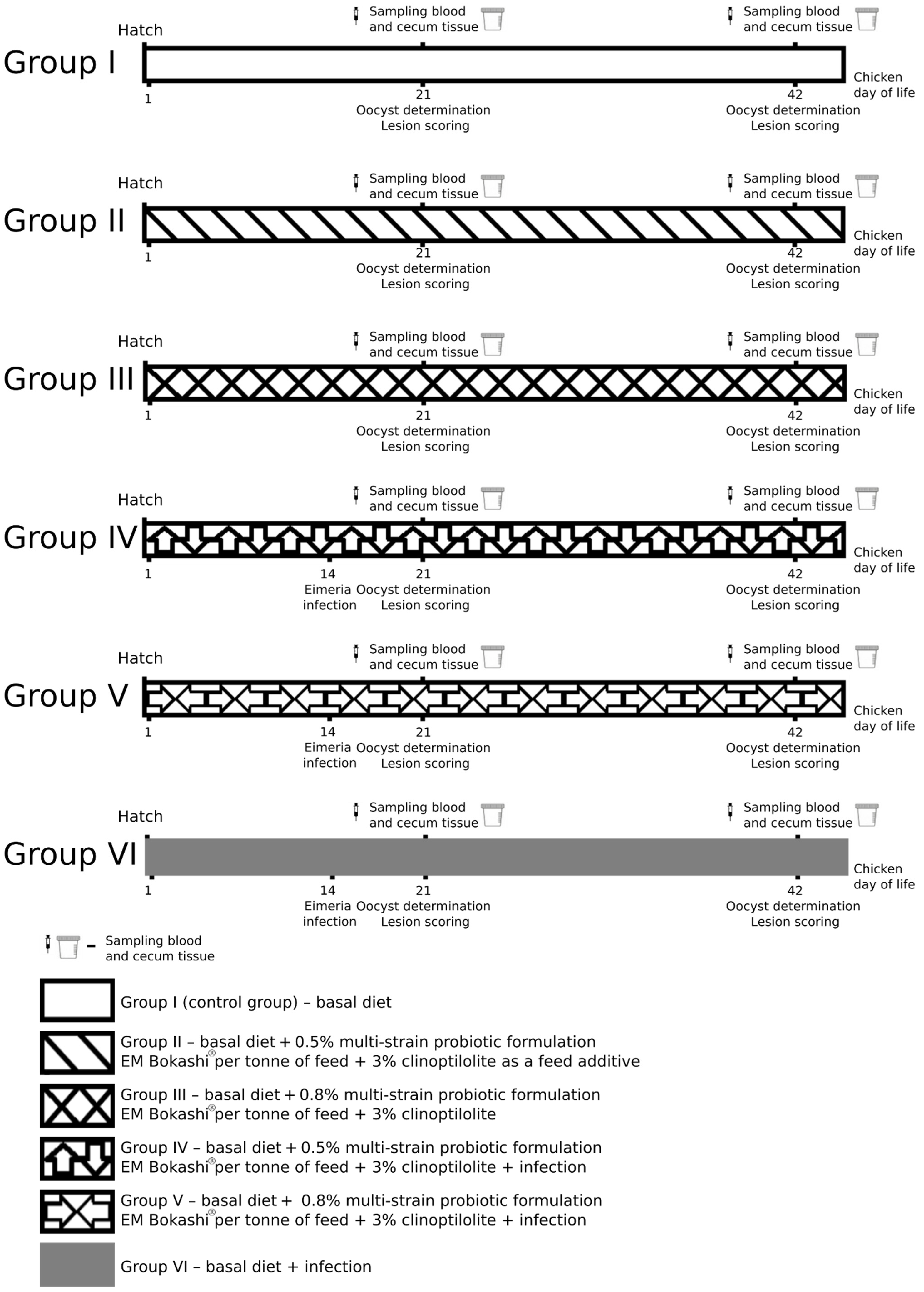
2.1. Experimental Animals

The experiment was conducted at the Experimental Station of the Poznan University of Life Sciences, Gorzyń 4, Międzychód commune. Consent for all research procedures was obtained from the Local Ethics Committee for Animal Testing at the University of Life Sciences in Lublin, Poland (approval no. 11/2021 of 1 March 2021).
A total of 600 1-day-old Ross 308 male chickens were used in the experiment. There were 6 treatments, each of which had 5 replicates with 20 birds per replicate pen. The treatments were as follows: a basal diet (control group—group I); basal diet + 0.5% multi-strain probiotic formulation EM Bokashi® per tonne of feed + 3% clinoptilolite (group II); basal diet + 0.8% multi-strain probiotic formulation EM Bokashi® per tonne of feed + 3% clinoptilolite (group III); basal diet + 0.5% multi-strain probiotic formulation EM Bokashi® per tonne of feed + 3% clinoptilolite + E. tenella infection (group IV); basal diet + 0.8% multi-strain probiotic formulation EM Bokashi® per tonne of feed + 3% clinoptilolite and E. tenella infection (group V); and basal diet + E. tenella infection (group VI). The additives were introduced into the experimental diets in place of wheat. The experimental design and composition of the basal diet are shown in Table 1 and Table 2 and in Figure 1. The basal diet was formulated to meet dietary recommendations for Ross 308 broiler chickens [37].
The chickens had unlimited access to feed and water. They received compound feeds appropriate for each rearing period: starter, S (days 1–21); grower, G (days 22–35); and finisher, F (days 36–42). The starter feed was provided to the chickens in crumble form, and the grower and finisher feeds were pelleted. No coccidiostats or antibiotics were used in the experiment.
The rearing period was 42 days. The experimental birds were housed in pens on wood shavings in a room with controlled temperature and humidity. Before the birds were placed in the pens, the wood shavings were tested for the presence of Eimeria spp. oocysts, according to Hauck and Pacheco [38], and no Eimeria oocysts were found. The pens were equipped with feeding lines and nipple drinkers. The lighting regime was adjusted to the age and diurnal rhythm of the birds. The light intensity was 30–40 lux up to day 7 and 20–30 lux thereafter. Three days before the chickens were placed in the cages, the floor was heated to 29 °C and the air to 33 °C. The temperature was maintained at 31–33 °C up to day 7 and then gradually reduced by 2 °C a week to a final temperature of 22–23 °C. The relative humidity throughout the experiment was 60% +/− 10%. The concentrations of gases were <10 ppm for ammonia and <3000 ppm for carbon dioxide.
The multi-strain probiotic formulation EM Bokashi° (batch number 45/01/2020) used in the experiment is manufactured by the commercial company Greenland Technologia EM, Janowiec, Poland, and contains a mixture of microorganisms (Table 3). The manufacturer tested the viability of probiotic bacterial cells and their content per gram of product. The company laboratory operates in compliance with all criteria for food safety and production quality, and the manufacturer has obtained a veterinary approval number for the product (α PL 0614002p, Quality Certificate, Supplementary Materials). Before the start of the experiment, the microbiological purity of the probiotic preparation was tested in the National Reference Laboratory of the Department of Hygiene of Animal Feedingstuffs, National Veterinary Research Institute in Puławy (Certificate of Analysis—Test Report no. P/20/59139, Supplementary Materials). To prevent loss of viability of the microbial strains in the product, the feed for the control and experimental groups was prepared once a week throughout the experiment.
In groups II–V, clinoptilolite (Andalusia sp. z o.o., Warsaw, Poland) was added to the feed in the amount of 3%. The preparation contained at least 87% clinoptilolite as the active substance, with the following composition: 67.07% SiO2, 12.4% Al2O3, 2.09% CaO, 2.8% K2O, 0.9% Fe2O3, 0.72% MgO, 2.05% Na2O, 0.19% TiO2, 0.04% MnO, and 0.014% P2O5.

2.2. Parasites and Inoculum Preparation

A local field isolate of Eimeria tenella recovered from a case of caecal coccidiosis in a poultry flock (a flock of 150,000 broiler chickens), whose carcasses were submitted for postmortem examination at the Department of Avian Diseases, Faculty of Veterinary Medicine, University of Life Sciences in Lublin, was used for the challenge of chickens in groups IV, V, and VI. Oocysts were replicated, isolated, and sporulated using standard procedures described by Raether et al. [39]. Only sporulated oocysts that had been stored for no longer than 4 weeks after their acquisition were used to infect the chickens. The oocysts were confirmed to belong to the species E. tenella by PCR using species-specific forward primer 5′- AATTTAGTCCATCGCAACCCTTG -3′ and reverse primer 5′- CGAGCGCTCTGCATACGACA -3′ as described by Lee et al. [40]. All chickens from each replicate in groups IV, V, and VI were infected at 14 days of age with 1.7 × 10⁴ E. tenella sporulated oocysts per bird by oral inoculation into the crop [41].

2.3. Clinical Signs and Growth Performance in Chickens

The birds were under clinical observation throughout the experiment, with special attention paid to their activity, appetite, respiratory symptoms, and the occurrence of digestive disorders manifesting as diarrhoea. The health status of the birds was evaluated by determining clinical parameters, anatomopathological changes in dead birds, and the mortality rate (Table 4). During the experiment, the birds were weighed before feeding on days 0, 7, 14, 21 (end of starter period), 28, 35 (end of grower period), and 42 (end of finisher period). In addition, feed intake (FI) and feed conversion ratio (FCR) were recorded on a per-pen basis on days 0, 7, 14, 21, 28, 35, and 42. Finally, adjusted average daily gain (ADG), feed intake (FI), and feed conversion ratio (FCR) were calculated for each period (days 0–21, 22–35, and 36–42) and also for the cumulative experimental period (days 0–42). The feed conversion ratio (FCR) for all experimental groups during the 42-day period was calculated as mean feed consumption/mean weight. Chicken mortality was recorded daily during morning and afternoon inspections and used for chicken-day calculations. Feed intake and FCR were corrected for mortality accordingly (Table 5).

2.4. Faecal Collection and Counting of Oocysts
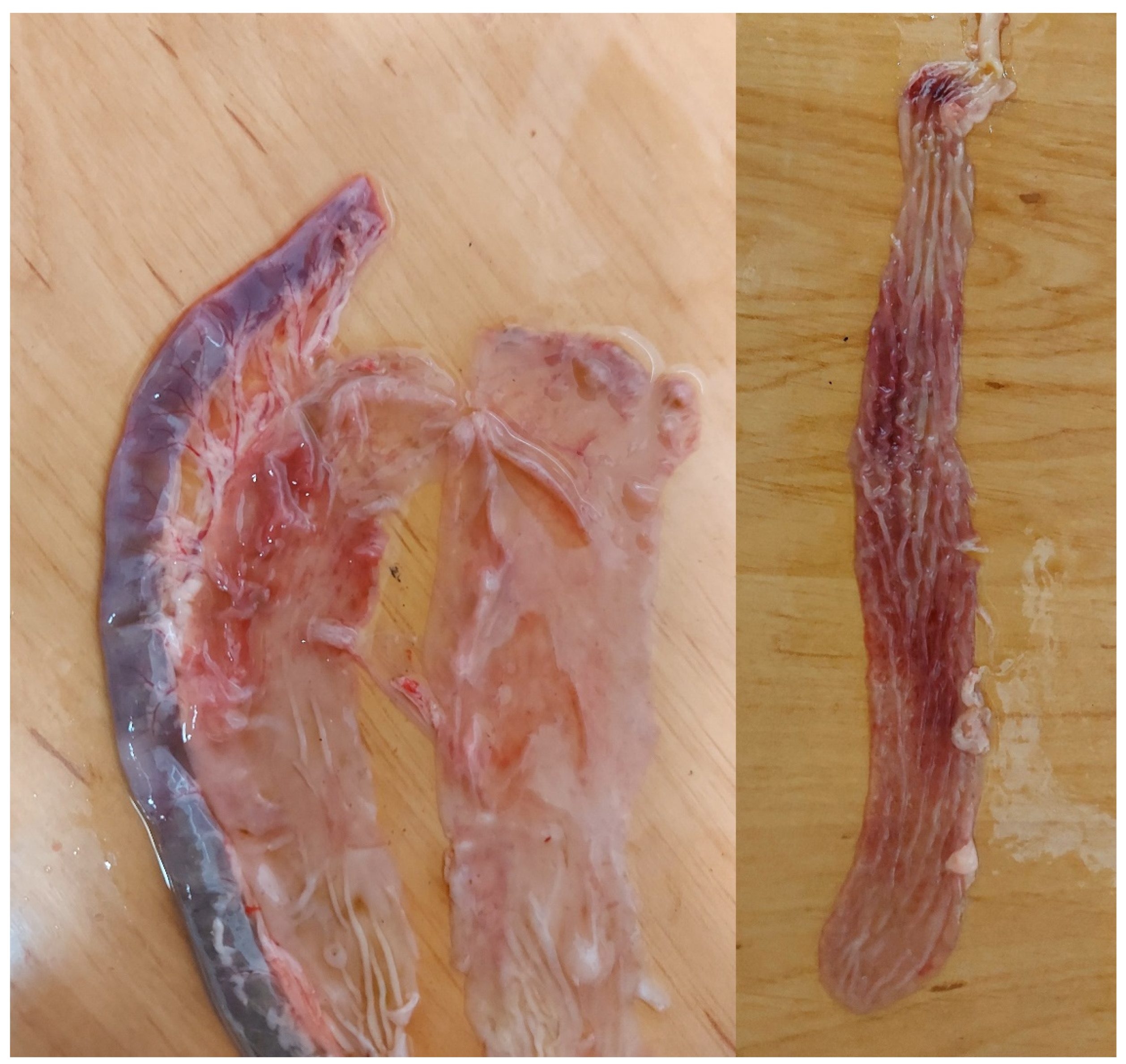
The numbers of oocysts per gram (OPG) of faeces were determined in samples collected from each pen and from each group on days 21 and 42 of the experiment. For each pen, fresh excreta samples were collected from every corner of the pen and from the centre of the pen and placed in separate airtight plastic bags. The number of oocysts per gram of faeces was counted using a modified McMaster technique as described by Hodgson [42]. Briefly, a 10% (w/v) faeces suspension in a salt solution (151 g NaCl mixed into 1 L of water) was prepared. After thorough shaking to obtain a homogenous mixture, 1 mL of the suspension was mixed with 9 mL of a salt solution (131 g of NaCl mixed into 1 L of water). Then, the suspension was pipetted into a McMaster chamber, which has two chambers with two identical 10 mm × 10 mm × 1.5 mm grids (0.15 mL). The total number of oocysts under both grids was recorded. The oocyst counts (oocysts per gram) were calculated by multiplying the total number of oocysts in the two chambers by 100. The caecum contents (2 g) were collected aseptically post-mortem from all birds on days 21 and 42 of the experiment. The oocyst counts in the caecum contents were determined as described above.

2.5. Lesion Scoring

On days 21 and 42, two birds per replicate (10 birds/treatment) were randomly selected for scoring of coccidian intestinal lesions. The 0–4 lesion scoring system of Johnson and Reid [43] was used. The areas scored were the duodenum, jejunum, ileum, and caecum. Based on the severity of the lesions, a score of 0 (no lesions), 1 (mild lesions), 2 (moderate lesions), 3 (severe lesions), or 4 (extremely severe lesions) was recorded for each chicken. Dead birds were scored as 4 [43]. Based on the anatomopathological changes observed in the internal organs of the dead birds, the cause of their deaths was established as coccidiosis.

2.6. Gastrointestinal Permeability

Fluorescein isothiocyanate dextran (FITC-d; MW 4000; Sigma-Aldrich, Poznań, Poland) was administered to evaluate gastrointestinal permeability. On days 21 and 42 of the experiment, 10 chicks from each group (two chickens selected randomly from each replicate) were gavaged with 1 mL of FITC-d solution (2.2 mg/mL) according to the method described by Kuttappan et al. [44]. At 2 h post-inoculation, peripheral blood was collected from the wing vein. Blood samples were stored in a dark container for an additional 2 h at room temperature before centrifugation at 1000× g for 15 min. A 100 μL volume of serum was transferred from the blood sampling tubes to a dark 96-well microplate. Five levels of FITC-d standard solution were formulated with the stock solution (2.2 mg/mL) and pooled serum from 10 additional unchallenged chickens raised in the same house. The FITC-d levels in the serum samples and standard solution were measured at an excitation wavelength of 485 nm and an emission wavelength of 528 nm using a microplate reader (Spectramax M5, Molecular Devices, San Jose, CA, USA). Blood processing and preparation of the standard solution were performed in a dark environment to protect the FITC-d from light exposure.

2.7. Intestinal Sample Collection

On day 21, 10 chicks from each group (two chickens selected randomly from each replicate) were sacrificed for tissue sampling. The remaining chicks continued to receive experimental diets up to day 42 of the experiment. On day 42, 10 birds (two chickens selected randomly from each replicate) from all groups were sacrificed for tissue sampling. The tissue was sampled from approximately the same central part of the caecum and flushed with cold PBS. Slices were stored at −80 °C until gene expression analysis. The intestinal samples were not pooled.

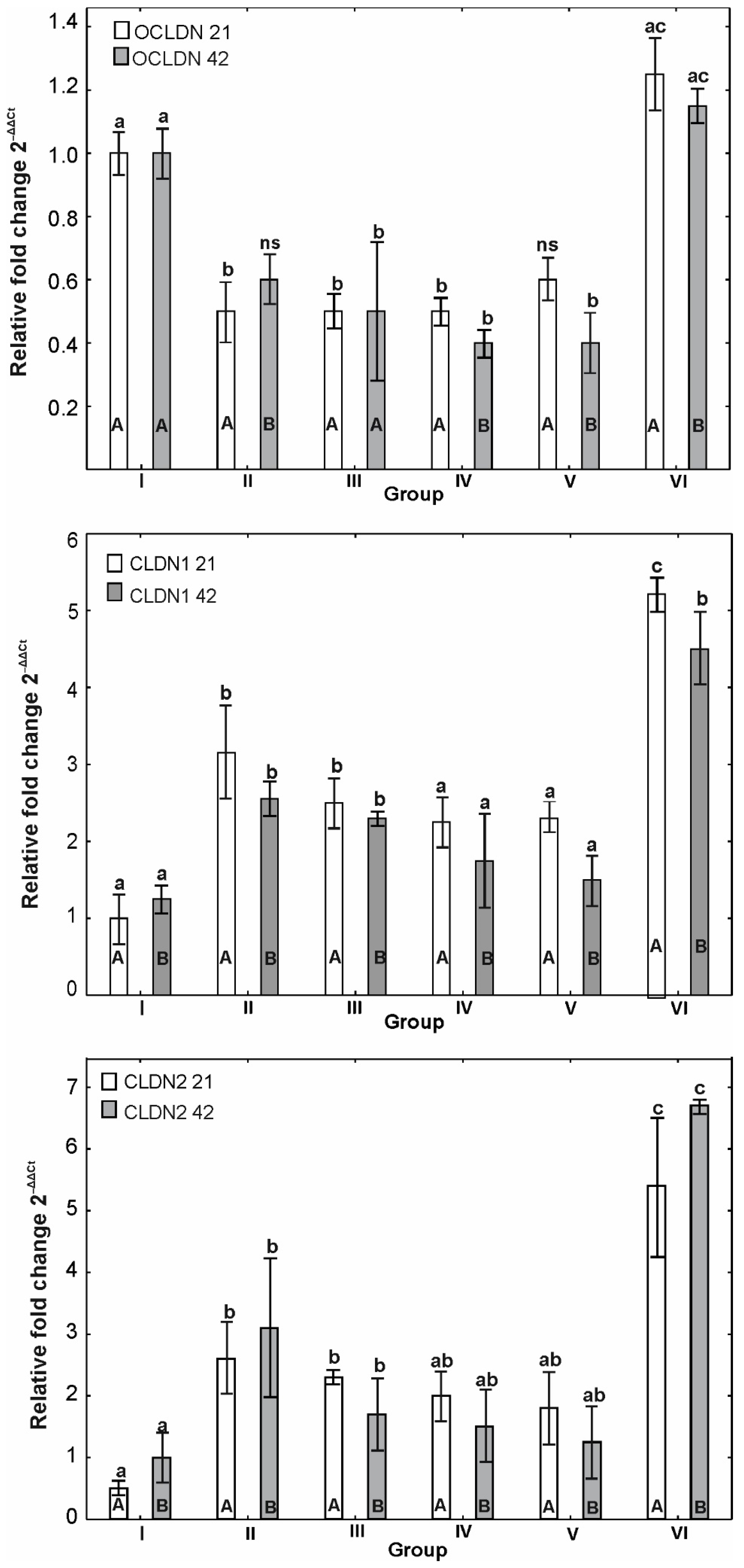
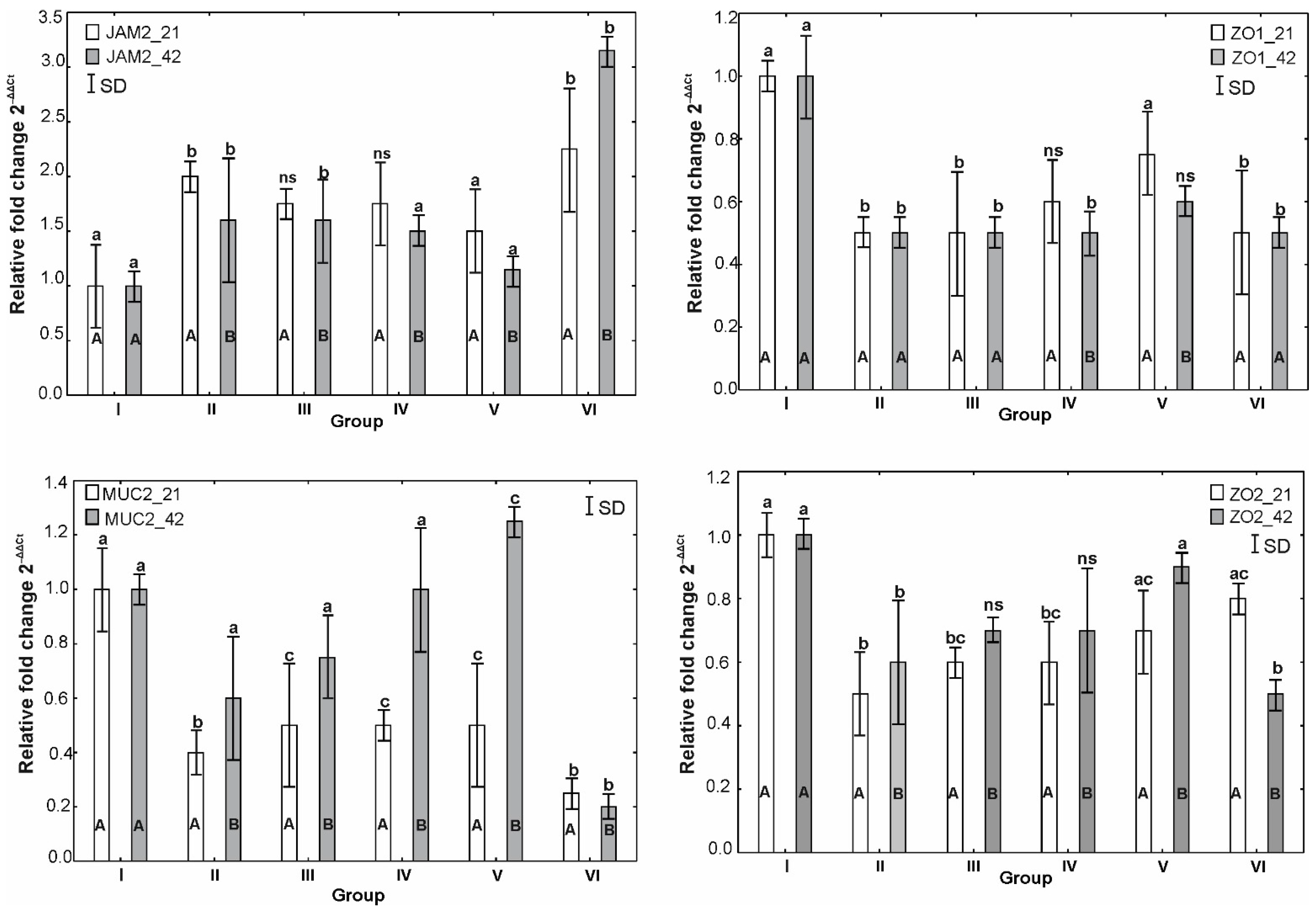
2.8. Quantitative Real-Time PCR for Tight Junction Proteins, Mucin and Antioxidant Gene Expression

Total RNA was isolated from homogenised samples of the caecum of each bird using the RNeasy Mini Kit (QIAGEN, Crawley, United Kingdom) following the manufacturer’s instructions. Purified RNA was eluted in 50 μL RNase-free water and stored at −80 °C until use. RNA was quantified using an ND-1000 spectrophotometer at 260 nm/280 nm (NanoDrop Technologies, Silverside, Wilmington, NC, USA). cDNA for quantitative reverse transcription-PCR (qRT-PCR) was synthesised from 1 μg of purified RNA using 50 ng of random hexamers and the SuperScript II first-strand cDNA synthesis kit according to the manufacturer’s instructions (Invitrogen, Chorzów, Poland). The mRNA expression of tight junction proteins, mucin, and antioxidants was determined by qRT-PCR using the Applied BioSystems 7500 real-time PCR system (Applied Biosystems, Warrington, United Kingdom) as previously described by Lammers et al. [45]. The PCR conditions were denaturation at 95 °C for 10 min followed by amplification at 60 °C for 1 min for 40 cycles. The primer sequences used for qRT-PCR are listed in Table 6. Each sample was subjected to qRT-PCR in triplicate, and the mean values were used for analysis. Glyceraldehyde 3-phosphate dehydrogenase (GAPDH) was used as the reference gene for gene expression. The threshold cycle (CT) values for the genes of interest were normalised to the average CT value of the housekeeping genes, and the relative expression of each replicate was calculated as 2−ΔΔCt (Applied Biosystems user Bulletin #2; AI prism 7700 detection system, 2001). The CT values of each gene were normalised against reference genes.

2.9. Statistical Analysis

Statistical analysis of the results for LS (lesion scoring), OPG (oocysts per gram), and protein expression for the six experimental groups on the two testing days (days 21 and 42) was performed using Statistica 13.2 PL software (StatSoft, Krakow, Poland). Due to the qualitative nature of the lesion score (LS) data (0–4), we used a non-parametric Kruskal–Wallis one-way ANOVA on ranks with multiple comparisons and the Mann–Whitney U test to show statistically significant differences between groups on the two testing days. The significance of differences in the numbers of oocysts (OPG) and expression of proteins, due to the lack of normal distribution of the data, was analysed by a non-parametric Kruskal–Wallis one-way ANOVA on ranks with multiple comparisons. The results were presented in graphic form using the mean and standard deviation (SD), with the same letters indicating the absence of statistically significant differences for p < 0.05.
The statistical evaluation of performance results was carried out using SAS® v. 9.4 statistics software (SAS, 2011). All data were presented as mean values with pooled standard error of the mean (SE). A two-way ANOVA was used to determine the effect of the experimental factors. For all characteristics, the significance of differences between group mean values was verified by Tukey’s test.

3. Results

3.1. Clinical Signs and Growth Performance in Chickens

During the experiment, the highest percentage of mortality rate, 11%, was observed in the group of birds receiving the basal diet and infected with E. tenella (group VI). Symptoms of diarrhoea with mucus and blood lasting 4 to 6 days were observed in birds from all groups infected with E. tenella. Intestinal hyperaemia, isolated pinpoint petechiae in the small intestinal mucosa, and catarrhal enteritis were observed among the anatomopathological changes in dead birds in groups infected with E. tenella and simultaneously supplemented with probiotic and clinoptilolite (groups IV and V). Additionally, haemorrhagic enteritis was observed in the group receiving the basal diet and infected with E. tenella. Detailed data are presented in Table 4 and Figure 2.
The effect of experimental factors on broiler chicken performance is presented in Table 5. During the first 10 days of the experiment, the infection did not affect the performance of broiler chickens (p > 0.05). However, the use of the multi-strain probiotic formulation and clinoptilolite statistically significantly increased BWG and FI (p < 0.05). Irrespective of the dose of the probiotic, broilers fed supplemented diets had increased FI and poorer FCR (p < 0.05). In the final periods of the experiment (days 25–42), infection adversely affected FCR, but the higher dose of the multi-strain probiotic formulation and clinoptilolite statistically significantly decreased the value of this parameter. At the same time, the supplement increased FI but also significantly increased BWG (p < 0.05). No interactions between the experimental factors were confirmed during the finisher period (p > 0.05). Analysis of the entire experimental period (days 0–42) revealed an interaction between the use of the supplement and infection with BWG (p < 0.05). This study showed that the multi-strain probiotic formulation and clinoptilolite were more effective in infected birds. In the case of other parameters (FI and FCR), the supplement increased FI (p < 0.05) but did not affect FCR (p > 0.05).

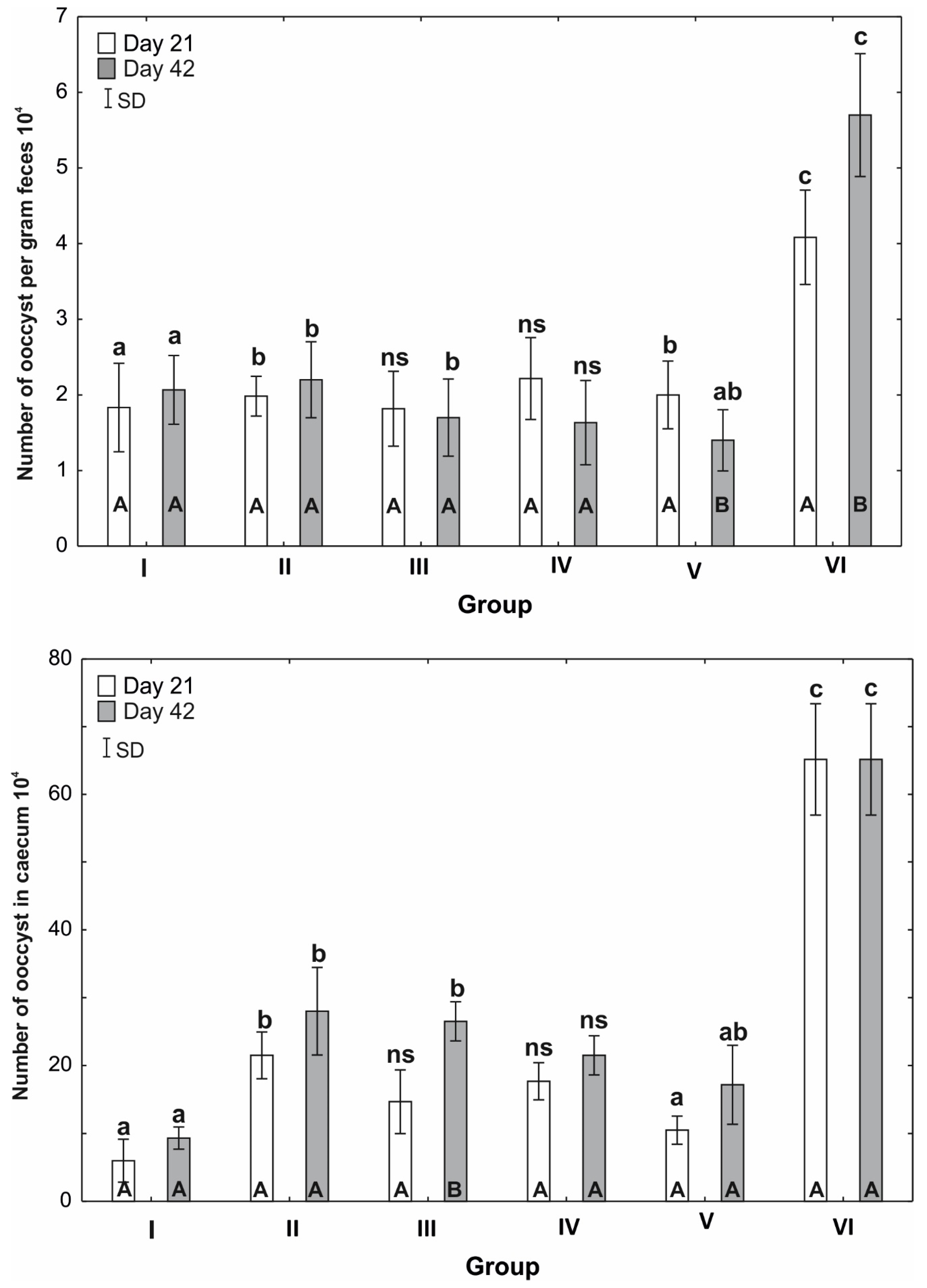
3.2. Numbers of Oocysts per Gram of Faeces

Analysis of the mean number of oocysts in the faeces showed that the testing day was statistically significant only for group V (0.8% probiotic + clinoptilolite, infected with E. tenella) and group VI (infected with E. tenella). The number of oocysts in the faeces of birds in group V was significantly higher (p ≤ 0.05) on the 21st day of this study and decreased significantly (p ≤ 0.05) on day 42. In group VI, the number of oocysts in both periods was the highest among all groups and significantly increased (p ≤ 0.05) on day 42. The number of oocysts in group I (control) was significantly lower (p ≤ 0.05) than in groups II and V. The mean number of oocysts per gram of faeces in the control group was slightly lower than in group II and slightly higher than in groups III and V. In group VI (infected with E. tenella), there was a pronounced increase in the number of oocysts compared to the other groups of chickens (Figure 3).

3.3. Numbers of oocysts in the Caecum

A detailed analysis of the mean number of oocysts in the caecum showed that the measurement day was a significant factor only for group III (0.8% Bokashi® formulation + 3 clinoptilolite), in which the number of oocysts on day 21 of this study was statistically significantly lower (p ≤ 0.05) than on day 42. The number of oocysts in the caecum was the lowest in groups I (control) and V and was significantly different from that observed in groups II and VI. Similarly, on day 42 of this study, the control group and group V had the lowest number of oocysts, which significantly differed from the number of oocysts recorded in groups II and III. In group VI (infected with E. tenella), the number of oocysts tripled to over 60 (Figure 3).

3.4. Scoring of Coccidial Intestinal Lesions

On day 21 of this study, the lesion score (LS) for the caecum and rectum in group V was statistically significantly lower (p ≤ 0.05) than in group VI (with the highest LS value, p = 0.0042). On day 42, differences in LS values were also shown only for the caecum and rectum; in groups III and V, the LS was statistically significantly lower (p ≤ 0.05) than in group VI (infected with E. tenella).
The results of the Kruskal–Wallis ANOVA for individual intestinal sections on day 21 of this study showed that the mean LS for the duodenum and jejunum was statistically significantly lower (p ≤ 0.05) than for the caecum and rectum in groups I and VI. On day 42 of this study, the LS was lower for the duodenum than for the caecum and rectum in groups II, III, and VI. There were significant differences in lesion scores between the upper and middle intestines and the lower intestine in groups I–V. In groups I, III, IV, and V, the LS did not differ significantly for the duodenum, jejunum, and ileum, while clear differences were found for the caecum and rectum. In group II, statistically significant differences (p ≤ 0.05) were found between the ileum and the duodenum and jejunum and between the caecum and rectum and the other sections of the intestines (Table 7).

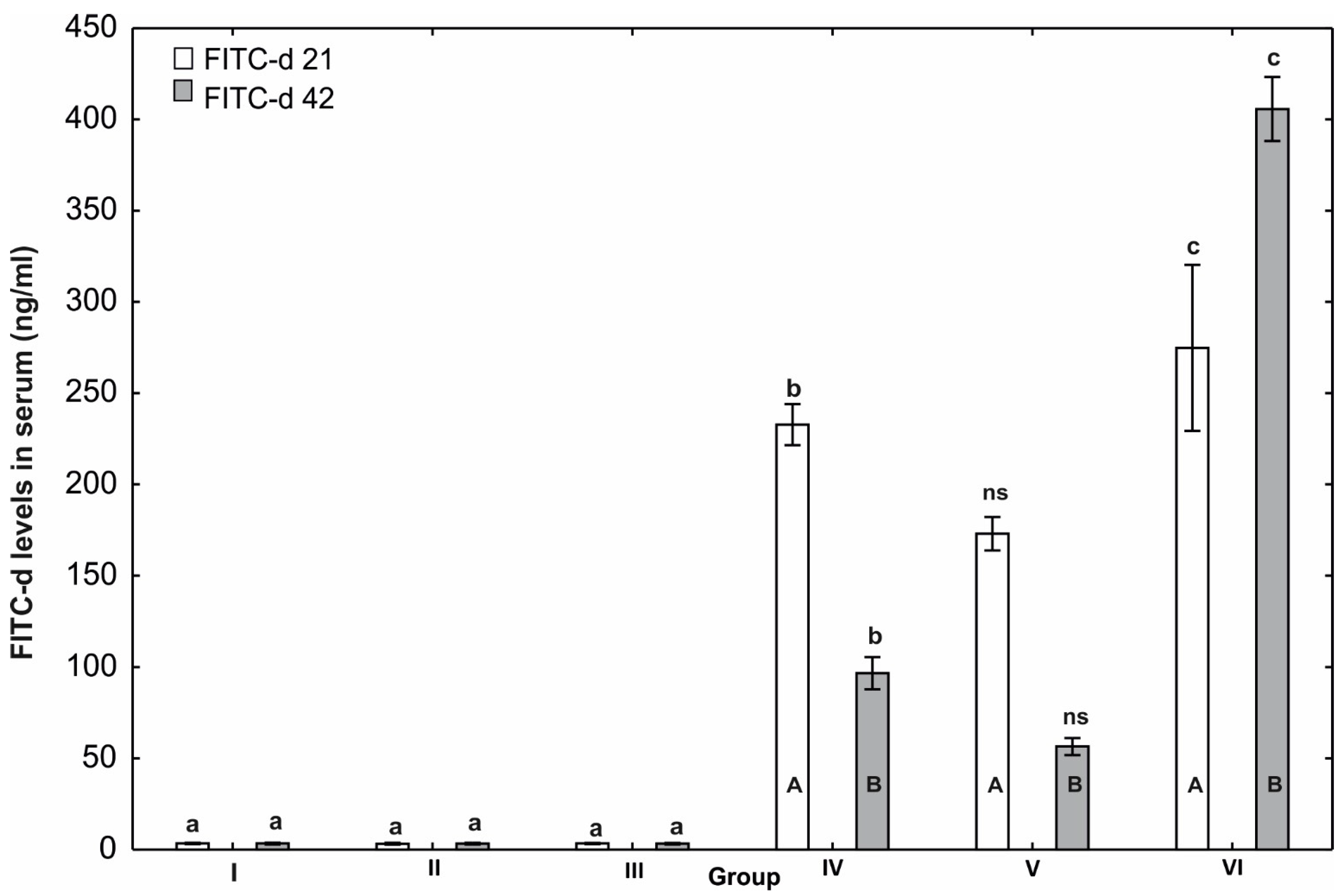
3.5. Evaluation of Gastrointestinal Permeability

The highest level of FITC-d was observed on day 21 of this study in the group receiving the basal diet and infected with E. tenella (group VI) compared to the other groups. Similar results were seen on day 42. Increased FITC recovery on study day 42 was observed in the E. tenella-infected groups (IV, V, and VI) compared to sera from the control birds and the groups supplemented but not infected with E. tenella. Moreover, the FITC-d level in the sera of birds from groups IV and V on day 42 of the experiment was lower than on day 21. Conversely, in birds from group VI, the FITC-d level in the serum on day 42 of this study was significantly higher (p ≤ 0.05) than on day 21 (Figure 4).

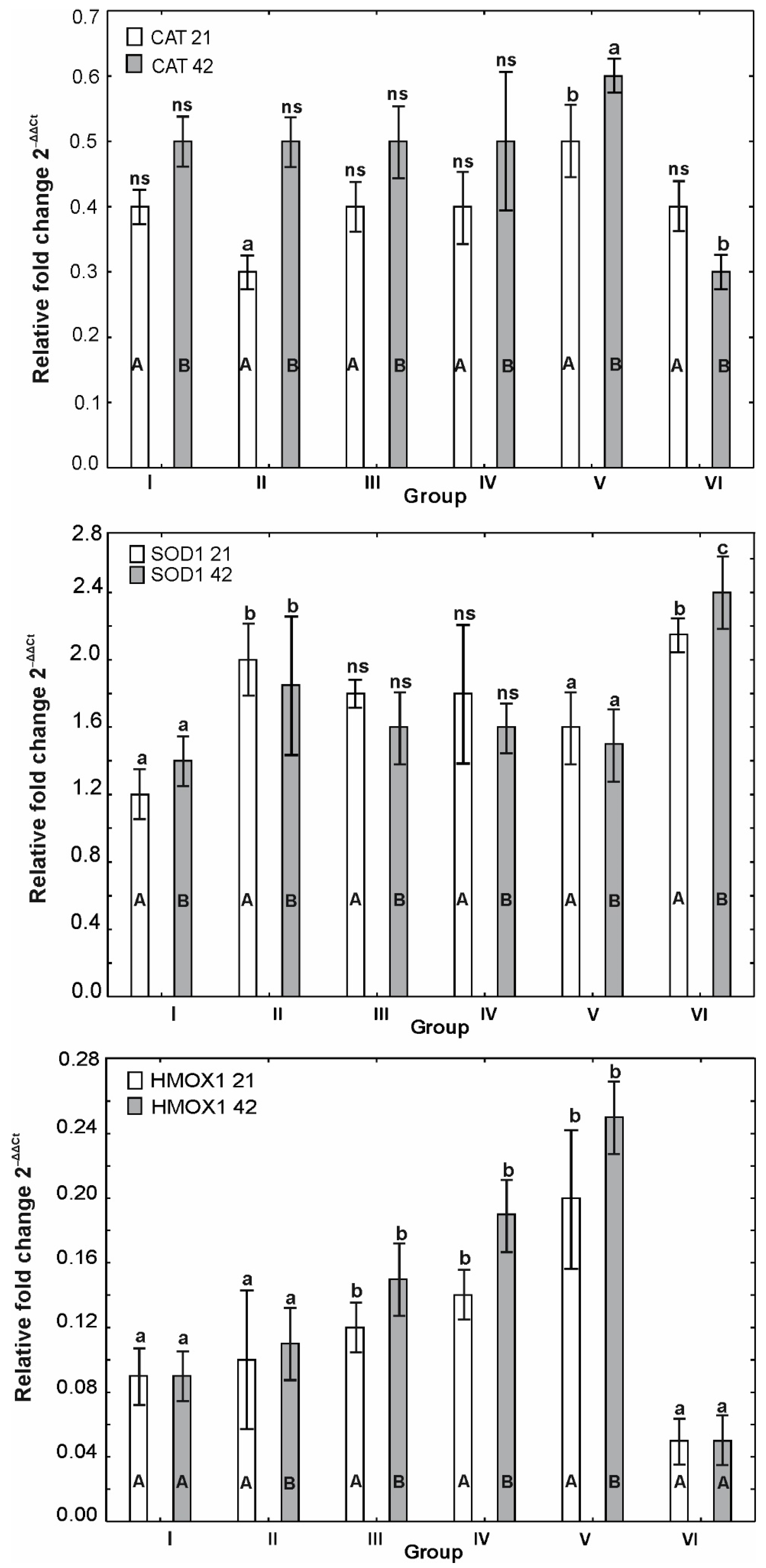
3.6. mRNA Expression of OCLDN, CLDN1, CLDN2, MUC2, ZO1, ZO2, JAM2, CAT, SOD1 and HMOX1

A comparison of the mRNA expression of OCLDN between the groups on day 21 of this study showed the highest levels of expression in the control group and group VI (1.0 and over 1.2, respectively).
In groups IV, V, and VI, the level of OCLDN expression was higher on day 21 of this study, while in group II it was significantly lower (p ≤ 0.05) on day 21 than on day 42 (Figure 5).
Higher mRNA expression of CLDN1 was observed on the 21st day of this study compared to day 42 in all groups except the control group. The analysis of the mRNA expression of CLDN1 on day 21 of this study showed statistically significant differences (p ≤ 0.05) between groups IV and V and groups II and III. CLDN1 expression was highest in group VI (infected with E. tenella), at 5.25, and differed significantly from the other groups (Figure 5).
Analysis of CLDN2 expression on the 21st day of this study showed statistically significantly higher (p ≤ 0.05) expression of CLDN2 in groups II, III, and VI compared to the control group. In group VI (infected with E. tenella), the expression of CLDN2 was statistically significantly higher (p ≤ 0.05) than in the control group and other experimental groups on both days of this study (Figure 5).
Statistically significant differences (p ≤ 0.05) in the level of MUC2 expression, depending on the measurement day, were observed for groups IV and V, in which it was more than twice as high on day 42 as on day 21. Comparison of MUC2 expression between groups on day 21 of this study showed that it was statistically significantly (p ≤ 0.05) higher in the control group. On day 42 of this study, the mRNA expression level of MUC2 was the highest in group V and the lowest in group VI (Figure 6).
The level of mRNA of ZO1 expression on the 21st day of the experiment was highest in the control group and differed significantly from the levels in groups II, III, and VI. The level of this protein expression on the 42nd day of this study in the control group was statistically significantly higher (p ≤ 0.05) than in groups II, III, IV, and VI (Figure 6).
A comparison of the ZO2 expression level between groups on day 21 showed that it was statistically significantly higher (p ≤ 0.05) in the control group than in groups II, III, and IV but did not differ significantly from the level in groups V and VI. On day 42, the ZO2 expression level was higher in groups I and V than in groups II and VI. The differences obtained in groups III and IV were not statistically significant (Figure 6).
JAM2 expression on the 21st day of the experiment was statistically significantly higher (p ≤ 0.05) in groups II and VI than in group V. The expression of this protein on the 42nd day of the experiment was the highest in group VI and statistically significantly higher (p ≤ 0.05) in groups II and III than in groups I, IV, and V (Figure 6).
The level of CAT expression on the 21st day of this study in group V was statistically significantly higher (p ≤ 0.05) than in the other groups. Similarly, on the 42nd day of this study, the level of CAT expression in group VI (infected with E. tenella) was statistically significantly lower (p ≤ 0.05) than in the other groups. In experimental groups II, III, IV, and V and in the control group, a statistically significant increase (p ≤ 0.05) in CAT expression was observed between days 21 and 42 of this study. In contrast, in group VI, the expression of mRNA of this protein was statistically significantly lower (p ≤ 0.05) on day 42 of the experiment than on day 21 (Figure 7).
The expression of mRNA of SOD1 on the 21st day of the experiment was highest in group VI (infected with E. tenella). The results obtained for groups III and IV did not differ significantly from the other groups (Figure 7).
The comparison of HMOX1 expression between groups on day 21 of the experiment showed statistically significant values in groups III, IV, and V compared to groups II and VI. Similar results were obtained on day 42, but the level of HMOX1 expression in groups III, IV, and V was higher than on the first day of this study. HMOX1 expression in groups I, II, and VI were statistically significantly lower (p ≤ 0.05) than in the other groups (Figure 7).

4. Discussion

Maintenance of the integrity of the intestinal barrier is dependent on tight connections between intestinal epithelial cells [46,47,48]. This involves structures of tight junctions (TJ) composed of multiprotein complexes of transmembrane proteins, claudins and occludins, adhesion proteins (JAM—junctional adhesion molecules), tricellulin, and zonulin [49,50]. Malfunctioning of the barrier of the intestinal epithelium and the associated increase in intestinal permeability predispose the birds to the development of numerous diseases with symptoms of gastroenteritis, accompanied by malabsorption and decreased weight gains [49,50,51].
In the experiment, the integrity of the intestinal barrier was tested in vivo by determining the concentration of fluorescein isothiocyanate dextran (FITC-d) in the serum of chickens following oral administration of FITC-d. During the experiment, damage to the tight junctions between enterocytes was demonstrated in a group of birds aged 21 and 42 days infected with E. tenella, which increased the permeability of the intestinal barrier. Our findings indicate that feed supplementation with a multi-strain probiotic and clinoptilolite strengthens the intestinal barrier and stimulates cell regeneration. This is also supported by the reduction in the amount of excreted Eimeria oocysts observed in the experiment (Figure 3), as well as by the mRNA expression of genes responsible for the functioning of TjJ. The use of a multi-strain probiotic in the diet of chickens also leads to modification of the composition of the intestinal microbiome, which helps to inhibit the multiplication of conditionally pathogenic bacteria and stimulates the local immune system, improving the repair and regeneration mechanisms of the intestinal epithelium [23,24,25,26,27,28,29,30,31,32,33,34,35,36,37,38,39,40,41,42,43,44,45,46,47,48,49,50,51,52,53]. The reduction in intestinal permeability is not only due to the mitigation of damage to the mucosa caused by the multiplication of protozoa in the enterocytes, but is also the effect of the probiotic and clinoptilolite on the synthesis of proteins making up tight junctions, which regulate paracellular permeability. Clinoptilolite supplementation reduces zonulin synthesis and hence improves intestinal barrier integrity, perhaps by interacting with intestinal bacteria [54,55,56]. The transmembrane protein occludin performs an important function in the intestinal barrier, stabilising tight junctions (TJ) and thereby ensuring their structural integrity [57]. When microorganisms selectively disturb tight junction complexes formed by the plasma membrane proteins occludin and zonula occludens (ZO), the transepithelial electrical resistance (TER) of the epithelial cell layer rapidly decreases, resulting in increased paracellular permeability [58]. This hypothesis may be supported by Teng et al. [11], who showed that E. maxima infection decreases the mRNA expression of OCLDN, ZO_1, and CLDN2. Similar observations were made by Utech et al. [59] in a model of intestinal inflammation caused by Eimeria spp. infection, which was accompanied by an increase in TNF-α and IFN-γ concentrations and a decrease in the expression of OCLDN and ZO-1. These results were not confirmed in our experiment, in which birds infected with E. tenella (group VI) showed higher (p ≤ 0.05) mRNA expression of OCLDN. The results of research conducted in a human model indicate that TNF-α and IFN-γ reduce gene expression of tight junction proteins, but the composition of the tight junction is highly regulated, and the loss or redistribution of one member of the intercellular junction may be compensated for through the upregulation of another tight junction protein [60]. Therefore, the increase in OCLDN mRNA expression observed in group VI may be due to the activation of the intracellular transcription pathway of genes for tight junction proteins dependent on TNF-α and IFN-γ. These results, in combination with the clinical course of coccidial infection, enteritis observed in necropsied birds, and high lesion scores (LS) (Table 4 and Table 7), suggest the promotion of the Th1 cellular immune response phenotype during E. tenella infection. Synthesis of cytokines, e.g., TNF-α, resulting from local intestinal inflammation induced by Eimeria, can be assumed to stimulate the body’s defence mechanisms and promote regeneration of the intestinal epithelium, leading to increased expression of OCLDN. Similar results were obtained by Wickramasuriya et al. [61], who reported that chickens infected with E. acervulina that received a probiotic containing B. subtilis in their feed showed increased expression of OCLDN, which helps to stabilise the intestinal barrier. In our study, however, chickens infected with E. tenella that received EM Bokashi® and clinoptilolite with their feed showed reduced expression of OCLDN in the caecum. Therefore, it can be assumed that both components of the feed additive take part in regulating the anti-parasitic response, stimulating mechanisms responsible for the regeneration of caecal epithelial cells, and leading to the restoration of the function of the intestinal barrier and the preservation of homeostasis within the intestinal wall.
Paracellular, they take part in cell polarization, cellular adhesion, and migration of cells, including leukocytes [62,63,64,65] (p ≤ 0.05). Our results showed that the expression of JAM-2 mRNA was also significantly increased (p ≤ 0.05) during E. tenella infection in the groups of infected birds receiving a probiotic and clinoptilolite in their feed. However, the level of its expression in these groups was lower by half compared to infected birds that received a basal diet, which demonstrates the protective function of the probiotic and clinoptilolite for the intestinal epithelium. These results demonstrate not only the protective role of these formulations for the intestinal epithelium but also the preservation of intact tight junctions between the enterocytes, which prevent large molecules that can disturb homeostasis from passing through the intestinal barrier. The results for the CLDN-1 gene were similar. (p ≤ 0.05). The CLDN family of proteins, such as JAM-2, is responsible for the formation of tight junctions between cells, the apposition of cell membranes, and paracellular transport [62]. The low expression of the CLDN family and JAM-2 genes noted in the groups of birds receiving the probiotic and clinoptilolite is indicative of the neutralising effects of these compounds on Eimeria tenella in the caecum. In contrast, the significant increase (p ≤ 0.05) in the expression of this gene in the group of infected birds (group VI) suggests that repair processes are activated in the junctions between enterocytes, which were damaged by E. tenella sporozoites. Increased expression of CLDN-1 may also be associated with an excessive inflammatory response to infection by E. tenella, which leads to a sharp increase in the concentrations of pro-inflammatory cytokines TNF-α and IFN-γ [63,64]. Poritz [65] and Utech [59] showed that during the inflammatory process, these cytokines increase the expression of CLDN1 while decreasing the expression of OCLDN and ZO1. These results were partially confirmed in our study, in which the birds infected with E. tenella (group VI) showed an increase in the expression of CLDN-1 and JAM-2 and a decrease in that of ZO-1. The increase in expression of the CLDN2 gene in the group of infected birds (group VI) indicates a leaky intestinal epithelium and the presence of inflammation induced by E. tenella infection. Similar observations were made by Pham and Hatabu [6], who showed high mRNA expression of CLDN-2 in chickens infected with E. tenella, indicating inflammation in the intestine and disturbances of transport by enterocytes.
Another protein, zonulin, is a physiological modulator of the function of tight junctions and is responsible for the transepithelial transport of ions and fluids between the intestinal lumen and the bloodstream, thereby regulating the permeability of the intestines [66,67,68,69]. Teng et al. [8] reported reduced mRNA expression of ZO-1 and ZO-2 in chickens infected with E. maxima. Similar results were obtained in our study. The decrease in mRNA expression of ZO-1 and ZO-2 in chickens infected with E. tenella (group VI) suggests a physiological loss of junctions between enterocytes. The low expression of genes noted in the present study may also be linked to the bloody diarrhoea observed in chickens infected with E. tenella, confirming the occurrence of intestinal inflammation, which was also reported by Pham and Hatabu [6].
The low mRNA expression of MUC-2 encoding the major mucin produced by goblet cells, shown in the caecal mucosa of the chickens infected with E. tenella (group VI), is associated with increased intestinal inflammation and impaired regeneration of the mucus layer [70,71,72]. The correlation of mRNA expression of MUC-2, ZO-1, ZO-2, CLDN-1, and CLDN-2 in this group of birds indicates a total loss of intercellular junctions and impeded transport between cells as well as between cells and the intestinal lumen, which is conducive to bacterial infections exacerbating intestinal inflammation induced by E. tenella. The chickens receiving a feed additive consisting of EM Bokashi®, together with clinoptilolite, showed a gradual increase in the expression of the MUC-2 gene, which reached its highest level at 42 days of age. It should be noted that probiotic bacteria, e.g., Lactobacillus or Bacillus spp., can bind to specific receptor sites on enterocytes and stimulate MUC2 synthesis [70,73,74]. It can be hypothesised that the EM Bokashi® and clinoptilolite formulation used in the diet of poultry contributed to increased resistance to enteric pathogens, including Eimeria spp., and improved feed conversion, in part by upregulating MUC2 expression, which increased mucin production and protected the intestine against morphological changes following infection with E. tenella.
One of the effects of damage to enterocytes by coccidia is the release of reactive oxygen species (ROS), which impair the function of the intestinal barrier [75,76,77]. Preservation of the balance between the production and removal of free oxygen radicals depends in part on enzymes with antioxidant properties, such as superoxide dismutase (SOD), catalase (CAT), and haem oxygenase (HMOX1/HO-1) [78,79]. In the present study, the highest mRNA expression (p ≤ 0.05) of SOD was noted in chickens infected with E. tenella (group VI), while in the birds receiving the preparation containing EM Bokashi® and clinoptilolite, its expression did not differ from that noted in control. Different results were obtained by Elmahallawy et al. [80]. The results of our study show that the use of a feed additive containing a probiotic and clinoptilolite in the diet of poultry with experimentally induced coccidiosis reduces ROS release by reducing the severity of the infection, intestinal inflammation, and the degree of tissue damage in the caecum, which improves the overall health of birds. It is worth noting that probiotic bacteria used in feed additives, e.g., lactic acid bacteria, owing to their ability to adhere to the intestinal mucosa, can supply exogenous antioxidant enzymes to the inflamed tissue lying below and, in this way, inactivate ROS by transforming superoxide anion radicals into the less toxic H2O2, thereby limiting inflammation [81,82]. The low SOD concentrations obtained in the present study may therefore indicate homeostasis of mechanisms responsible for combating oxidative stress in E. tenella infection. A similar relationship has been shown for the degree of expression of the SOD1 and CAT genes in the duodenal mucosa of chickens infected with E. acervulina receiving a diet with B. subtilis-cNK-2 [61].
The reverse relationship was shown for the mRNA expression of CAT, which was highest in poultry infected with E. tenella and receiving a diet with a probiotic together with clinoptilolite. The results are supported by research by Wickramasuriya et al. [61], who showed no statistically significant differences in CAT expression in the intestines of poultry, while in poultry infected with E. acervulina and fed a diet supplemented with B. subtilis-cNK-2, expression of CAT and HO-1 increased in the spleen. Interesting results were obtained in the analysis of the mRNA expression of haem oxygenase HO-1. Increased expression of HO-1 is usually noted in conditions of cell exposure to oxidative stress [83]. The protective activity of this enzyme involves limiting the damage caused by oxidative stress, ischaemia, or inflammation, while the mechanism of protective activity is varied and multifaceted [77]. These results are consistent with those published by Wickramasuriya et al. [61], who also showed increased expression of the HMOX1 gene in poultry infected with E. acervulina. These findings confirm the cytoprotective effect of haem oxygenases on intestinal cells, expressed as a reduction in oxidative stress and the severity of intestinal inflammation as well as the maintenance of enterocyte integrity, which is also evidenced by the clinical course of infection in the birds. Our results showed that the formulation consisting of EM Bokashi® and clinoptilolite in the diet of poultry enhanced antioxidant processes in the caecal mucosa and reduced oxidative stress induced by E. tenella infection, reducing the risk of damage to the enterocytes.
Eimeria spp., through invasive activity and destruction of the intestinal epithelium, leads to malabsorption in poultry and clinical gastrointestinal symptoms, including bloody diarrhoea. In infected birds, there is a secondary reduction in feed intake and conversion and a decrease in body weight gains, which ultimately leads to developmental disorders and death [64,84,85,86]. The chickens infected with Eimeria tenella had a low growth rate, expressed as a reduction in BWG and FI in comparison to the uninfected control and the other experimental groups. Our study showed that the use of EM Bokashi® and clinoptilolite in the diet significantly (p ≤ 0.05) affected the growth rate of chicken broilers infected with E. tenella, expressed as an increase in BWG and FI. However, previously published data on the effect of probiotics in the diet of poultry on the health and production parameters of birds infected with Eimeria spp. are conflicting [87,88,89,90]. Similarly, divergent results have been published for zeolite [28,29,91]. Our results are largely consistent with those published by Giannenas et al. [92,93], Zhou et al. [91], and Timmerman et al. [94] and indicate that body weight gains in broilers receiving a diet with probiotics and zeolite and infected with E. tenella are significantly higher than in birds fed a standard diet. In addition, the use of the composite probiotic formulation in our experiment in the first period after hatching favours competitive colonisation of the intestinal epithelium with probiotic microbes, even in birds infected with E. tenella at 14 days of age. The microbes contained in the probiotic prevent the adhesion of pathogenic agents to the intestinal epithelium and modulate the expression of genes in epithelial cells, taking part in the immune response against the development of infections that negatively affect production and health parameters [95]. The use of the multi-strain and multi-species probiotic formulation allows the effective microorganisms contained in it to act in different parts of the intestine and present varied mechanisms of action. This makes it possible to eliminate the negative effects of an Eimeria infection. In addition, the clinoptilolite used in the experiment, together with the probiotic preparation, is known to be rich in macro- and microelements essential for the growth and development of the body. In ionised form, these elements have been shown to be quickly absorbed and distributed to various organs, beneficially affecting the metabolic processes taking place in them and their biological functions [96,97], which translates to improved production parameters.
The use of the feed supplement composed of a probiotic and clinoptilolite in the diet of poultry caused a significant (p ≤ 0.05) reduction in the number of E. tenella oocysts in the faeces and caecum in comparison to the group of infected birds, the other experimental groups, and the control. Similar observations were reported by Giannenas et al. [93], who showed that the body weight of chickens infected with E. tenella receiving feed with a probiotic was higher than or similar to that of infected birds that received a coccidiostat, while the number of oocysts in the faeces was much lower than in the control group of infected birds fed a standard diet. Similar results were obtained by Lee et al. [98] in a study of poultry fed a diet with a probiotic supplement containing Pediococcus acidilactici. The beneficial effect of the probiotic as a feed additive was expressed in part as increased resistance to experimental infection with E. acervulina, increased body weight gains in comparison with infected birds fed the basal diet, and a decrease in excretion of oocysts. Similar relationships were demonstrated in our study. The mechanism of coccidiostatic action of the compounds most likely relies on competition between probiotic organisms and oocysts for access to specific sites of colonisation of the intestinal epithelium. In our opinion, the inclusion of clinoptilolite in the feed supplement plays a role as well. These aluminosilicates are known to have a strong adsorption capacity and are able to adsorb bacteria, toxic substances, and other harmful compounds in the intestines, which promotes the excretion of oocysts [99,100,101]. Another advantage of clinoptilolite added to feed is its uniform distribution over the surface of the intestinal mucosa, owing to which it forms a kind of protective layer that reduces colonisation of the intestine by oocysts and damage to the mucosa by Eimeria spp. Nevertheless, the exact mechanism of action of probiotics and clinoptilolite at the molecular level and their effect on the colonisation of the intestinal epithelium by oocysts and the growth of animals are not fully understood and require further research.
The analysis of lesion scores (LS) in the experiment showed that replication of the parasite and the pathological changes found in the intestines, especially in the caecum, were statistically significantly (p ≤ 0.05) more severe in the chickens infected with E. tenella (group VI) in comparison with the other experimental groups and the control. This was confirmed by the clinical observations of the birds in the infected group, in which symptoms of bloody diarrhoea and inhibition of body weight gains and feed intake were noted. The development of pathological changes in the caecum following infection with E. tenella is complex and is the combined effect of the accumulation of parasites mechanically damaging the enterocytes and the inflammatory immune response. Conway et al. [102] and Soutter et al. [103] showed that 4000 oocysts of E. tenella are sufficient to induce visible anatomopathological changes in the intestines affecting the production and health parameters of birds, while higher levels also cause an increase in mortality [102,103]. We made similar observations in our study, in which the number of oocysts in both the faeces and the caecum exceeded 4000 in the control group infected with E. tenella.
The synergistic activity of the probiotic formulation in combination with clinoptilolite, affecting digestion, nutrient utilisation, and nutrient metabolism, limits colonisation of the intestinal mucosa by E. tenella and the pathogenic effects of oocysts [104], thereby improving the survival of birds and reducing the extent of damage caused by the parasite in the gastrointestinal tract [104]. The reduced severity of lesions in this group of birds can be linked to the antimicrobial properties of the feed additive, based on its competitive capacity to eliminate microorganisms from the intestinal lumen [105,106] and a reduction in the effects of conditionally pathogenic bacteria on the intestinal epithelium [107]. These processes effectively inhibit the adhesion of oocysts to the intestinal barrier, their penetration of the epithelium, and their pathogenic effects. The improvement in growth parameters and overall health observed in this study additionally indicates mitigation of the clinical course of E. tenella infection in birds fed a diet supplemented with a probiotic and clinoptilolite.

5. Conclusions

The combined use of effective microorganisms (EM) in a multi-strain probiotic (Bokashi®) and clinoptilolite helps to maintain the intestinal barrier in chickens. The use of these compounds in E. tenella-infected birds is indicative of their neutralising effects on the parasite in the caecum. This prevents damage to the intestinal epithelium and demonstrates the protective role of enterocytes. Moreover, they inhibit the development of inflammation and prevent pro-inflammatory molecules from penetrating the intestinal mucosa. An additional benefit is improved feed conversion and stimulation of growth in broilers, partly due to the increase in mucin production and protection of the intestine against morphological changes following infection with E. tenella. The formulation used in this study also enhanced antioxidant processes in the caecal mucosa and reduced oxidative stress induced by E. tenella infection. The use of effective microorganisms (EM) in a multi-strain probiotic (Bokashi®) and clinoptilolite in the diet of poultry with experimentally induced coccidiosis limits ROS release by reducing the severity of the infection, intestinal inflammation, and the degree of tissue damage in the caecum, thereby improving the overall health of birds.

Author Contributions

Conceptualization, A.C., Ł.S.J. and M.H.; methodology, Ł.S.J., M.K., A.M., M.H. and S.N.; software, S.G. and A.R.; validation, A.R.; formal analysis, Ł.S.J. and Z.G.; investigation, A.C., Ł.S.J. and M.K.; resources, S.G. and A.R.; data curation, Ł.S.J.; writing—original draft preparation, A.C. and Ł.S.J.; writing—review and editing, Ł.S.J. and A.M.; visualization, A.R.; supervision, Z.G.; project administration, Ł.S.J. and Z.G. All authors have read and agreed to the published version of the manuscript.

Funding

This research was funded by the University of Life Sciences in Lublin, Poland, project No. WKE/MN-1/WET/20.

Institutional Review Board Statement

All procedures involving animals were in compliance with the ethical standards of the institution at which the experiment was conducted. All procedures used during the research were approved by the Local Ethics Committee for Animal Testing at the University of Life Sciences in Lublin, Poland (approval no. 11/2021 of 1 March 2021).

Data Availability Statement

All data generated or analyzed during this study are included in this published article and are available on request from the corresponding author.

Conflicts of Interest

The authors certify that they have no affiliations with or involvement in any organization or entity with any financial interest (such as honoraria, educational grants, participation in speakers’ bureaus, membership, employment, consultancies, stock ownership, or other equity interest, expert testimony, or patent-licensing arrangements) or non-financial interest (such as personal or professional relationships, affiliations, knowledge, or beliefs) in the subject matter or materials discussed in this manuscript.

References

  1. Al-Quraishy, S.; Abdel-Baki, A.S.; Dkhil, M.A. Eimeria tenella infection among broiler chicks Gallus domesticus in Riyadh city, Saudi Arabia. J. King Saud Univ. Sci. 2009, 21, 191–193. [Google Scholar] [CrossRef] [Green Version]
  2. Attree, E.; Sanchez-Arsuaga, G.; Jones, M.; Xia, D.; Marugan-Hernandez, V.; Blake, D.; Tomley, F. Controlling the causative agents of coccidiosis in domestic chickens; an eye on the past and considerations for the future. CABI Agric. Biosci. 2021, 2, 37. [Google Scholar] [CrossRef] [PubMed]
  3. Jeurissen, S.H.; Janse, E.M.; Vermeulen, A.N.; Vervelde, L. Eimeria tenella infections in chickens: Aspects of host-parasite: Interaction. Vet. Immunol. Immunopathol. 1996, 54, 231–238. [Google Scholar] [CrossRef] [PubMed]
  4. Macdonald, S.E.; Nolan, M.J.; Harman, K.; Boulton, K.; Hume, D.A.; Tomley, F.M.; Stabler, R.A.; Blake, D.P. Effects of Eimeria tenella infection on chicken caecal microbiome diversity, exploring variation associated with severity of pathology. PLoS ONE 2017, 12, e0184890. [Google Scholar] [CrossRef] [PubMed]
  5. Madlala, T.; Okpeku, M.; Adeleke, M.A. Understanding the interactions between Eimeria infection and gut microbiota, towards the control of chicken coccidiosis: A review. Parasite 2021, 28, 48. [Google Scholar] [CrossRef]
  6. Pham, H.H.S.; Hatabu, T. Eimeria tenella infection modulates the expression levels of intestinal epithelial barrier-related genes in chicken. J. Environ. Sci. Sustain. Soc. 2021, 10, 13–16. [Google Scholar] [CrossRef]
  7. Pham, H.H.S.; Matsubayashi, M.; Tsuji, N.; Hatabu, T. Relationship between Eimeria tenella associated-early clinical signs and molecular changes in the intestinal barrier function. Vet. Immunol. Immunopathol. 2021, 240, 110321. [Google Scholar] [CrossRef]
  8. Teng, P.Y.; Choi, J.; Tompkins, Y.; Lillehoj, H.; Kim, W. Impacts of increasing challenge with Eimeria maxima on the growth performance and gene expression of biomarkers associated with intestinal integrity and nutrient transporters. Vet. Res. 2021, 52, 81. [Google Scholar] [CrossRef]
  9. Su, S.; Miska, K.B.; Fetterer, R.H.; Jenkins, M.C.; Wong, E.A. Expression of digestive enzymes and nutrient transporters in Eimeria-challenged broilers. Exp. Parasitol. 2015, 150, 13–21. [Google Scholar] [CrossRef]
  10. Zaiss, M.M.; Harris, N.L. Interactions Between the Intestinal Microbiome and Helminth Parasites. Parasite. Immunol. 2016, 38, 5–11. [Google Scholar] [CrossRef]
  11. Teng, P.Y.; Yadav, S.; Castro, F.; Tompkins, Y.H.; Fuller, A.L.; Kim, W.K. Graded Eimeria challenge linearly regulated growth performance, dynamic change of gastrointestinal permeability, apparent ileal digestibility, intestinal morphology, and tight junctions of broiler chickens. Poult. Sci. 2020, 99, 4203–4216. [Google Scholar] [CrossRef] [PubMed]
  12. Cheng, Y.H.; Horng, Y.B.; Chen, W.J.; Hua, K.F.; Dybus, A.; Yu, Y.H. Effect of fermented products produced by Bacillus licheniformis on the growth performance and cecal microbial community of broilers under coccidial challenge. Animals 2021, 11, 1245. [Google Scholar] [CrossRef] [PubMed]
  13. Fatoba, A.J.; Adeleke, M.A. Diagnosis and control of chicken coccidiosis: A recent update. J. Parasit. Dis. 2018, 42, 483–493. [Google Scholar] [CrossRef] [PubMed]
  14. Giannenas, I.; Florou-Paneri, P.; Papazahariadou, M.; Christaki, E.; Botsoglou, N.A.; Spais, A.B. Effect of dietary supplementation with oregano essential oil on performance of broilers after experimental infection with Eimeria tenella. Arch. Tierernahr. 2003, 57, 99–106. [Google Scholar] [PubMed]
  15. Quiroz-Castañeda, R.E.; Dantán-González, E. Control of avian coccidiosis: Future and present natural alternatives. Biomed. Res. Int. 2015, 2015, 430610. [Google Scholar] [CrossRef] [Green Version]
  16. Muthamilselvan, T.; Kuo, T.F.; Wu, Y.C.; Yang, W.C. Herbal remedies for coccidiosis control: A review of plants, compounds, and anticoccidial actions. Evid. Based Complement Alternat. Med. 2016, 2016, 2657981. [Google Scholar] [CrossRef] [Green Version]
  17. Mohsin, M.; Zhang, Z.; Yin, G. Effect of probiotics on the performance and intestinal health of broiler chickens infected with Eimeria tenella. Vaccines 2022, 10, 97. [Google Scholar] [CrossRef]
  18. Atsbeha, A.T.; Hailu, T.G. The impact of Effective Microorganisms (EM) on egg quality and laying performance of chickens. Int. J. Food Sci. 2021, 2021, 8895717. [Google Scholar] [CrossRef]
  19. Esatu, W.; Adey, S.; Dessie, T. Effect of effective microorganisms on growth parameters and serum cholesterol levels in broilers. Afr. J. Agric. Res 2011, 6, 3841–3846. [Google Scholar]
  20. Jwher, D.M.T.; Abd, S.K.; Mohammad, A.G. The study of using effective microorganisms (EM) on health and performance of broiler chicks. Iraqi J. Vet. Sci. 2013, 27, 73–78. [Google Scholar] [CrossRef]
  21. Jha, R.; Das, R.; Oak, S.; Mishra, P. Probiotics (Direct-Fed Microbials) in poultry nutrition and their effects on nutrient utilization, growth and laying performance, and gut health: A ystematic review. Animals 2020, 10, 1863. [Google Scholar] [CrossRef] [PubMed]
  22. Elbaz, A.M.; Ibrahim, N.S.; Shehata, A.M.; Mohamed, N.G.; Abdel-Moneim, A.M.E. Impact of multistrain probiotic, citric acid, garlic powder or their combinations on performance, ileal histomorphometry, microbial enumeration and humoral immunity of broiler chickens. Trop. Anim. Health Prod. 2021, 53, 115. [Google Scholar] [CrossRef] [PubMed]
  23. Andronikashvili, T.G.; Urushadze, T.F.; Eprikashvili, L.G.; Kordzakhia, T.N.; Zautashvili, M.G.; Pirtzkhalava, N.V.; Dzagania, M.A. Natural zeolites in poultry farming. Ann Agra Sci. 2014, 12, 1–17. [Google Scholar]
  24. Shariatmadari, F. The application of zeolite in poultry production. World’s Poult. Sci. J. 2008, 64, 76–84. [Google Scholar] [CrossRef]
  25. Wlazło, Ł.; Nowakowicz-Dębek, B.; Kapica, J.; Kwiecień, M.; Pawlak, H. Removal of ammonia from poultry manure by aluminosilicates. J. Environ. Manag. 2016, 183, 722–725. [Google Scholar] [CrossRef] [PubMed]
  26. Wawrzyniak, A.; Kapica, M.; Stępień-Pyśniak, D.; Szewerniak, R.; Olejarska, A.; Jarosz, Ł. Effect of feeding Transcarpathian Zeolite on gastrointestinal morphology and function in broiler chickens. Rev. Bras. Cienc. Avic. 2017, 19, 737–746. [Google Scholar] [CrossRef] [Green Version]
  27. Olver, M.D. The effect of feeding clinoptilolite (zeolite) to laying hens. S. Afr. J. Anim. Sci. 1983, 13, 107–110. [Google Scholar]
  28. Wu, Q.J.; Wang, L.C.; Zhou, Y.M.; Zhang, J.F.; Wang, T. Effects of clinoptilolite and modified clinoptilolite on the growth performance, intestinal microflora, and gut parameters of broilers. Poult Sci. 2013, 92, 684–692. [Google Scholar] [CrossRef]
  29. Wu, Q.J.; Zhou, Y.M.; Wu, Y.N.; Wang, T. Intestinal development and function of broiler chickens on diets supplemented with clinoptilolite. Asian-Australas. J. Anim. Sci. 2013, 26, 987–994. [Google Scholar] [CrossRef] [Green Version]
  30. Mumpton, F.A.; Fishman, P.H. The Application of Natural Zeolites in Animal Science and Aquaculture. J. Anim. Sci. 1977, 45, 1188–1203. [Google Scholar] [CrossRef]
  31. Papaioannou, D.S.; Kyriakis, C.S.; Alexopoulos, C.; Tzika, E.D.; Polizopoulou, Z.S.; Kyriakis, S.C. A field study on the effect of the dietary use of a clinoptilolite-rich tuff, alone or in combination with certain antimicrobials, on the health status and performance of weaned, growing and finishing pigs. Res. Vet. Sci. 2004, 76, 19–29. [Google Scholar] [CrossRef] [PubMed]
  32. Khambualai, O.; Ruttanavut, J.; Kitabatake, M.; Goto, H.; Erikawa, T.; Yamauchi, K. Effects of dietary natural zeolite including plant extract on growth performance and intestinal histology in Aigamo ducks. Br. Poult. Sci. 2009, 50, 123–130. [Google Scholar] [CrossRef] [PubMed]
  33. Serek, P.; Oleksy-Wawrzyniak, M. The effect of bacterial infections, probiotics and zonulin on intestinal barrier integrity. Int. J. Mol. Sci. 2021, 22, 11359. [Google Scholar] [CrossRef] [PubMed]
  34. Huang, L.; Luo, L.; Zhang, Y.; Wang, Z.; Xia, Z. Effects of the dietary probiotic, Enterococcus faecium NCIMB11181, on the intestinal barrier and system immune status in Escherichia coli O78-challenged broiler chickens. Probiotics Antimicro. Prot. 2019, 11, 946–956. [Google Scholar] [CrossRef] [Green Version]
  35. Chang, C.H.; Teng, P.Y.; Lee, T.T.; Yu, B. Effects of multi-strain probiotic supplementation on intestinal microbiota, tight junctions, and inflammation in young broiler chickens challenged with Salmonella enterica subsp. enterica. Asian Australas. J. Anim. Sci. 2020, 33, 1797–1808. [Google Scholar] [CrossRef] [Green Version]
  36. Alcala-Canto, Y.; Gutierrez-Olvera, L.; Gutierrez-Olvera, C.; Sumano-Lopez, H. Effects of clinoptilolite on Eimeria spp. infection in sheep. Small Rum. Res. 2011, 100, 184–188. [Google Scholar] [CrossRef]
  37. Aviagen. Ross Broiler Management Handbook; Aviagen: Newbridge, UK, 2014. [Google Scholar]
  38. Hauck, R.; Pacheco, W.J. Detection of coccidia oocysts in litter and feces of broilers in a floor pen trial. J Parasitol. 2021, 107, 878–881. [Google Scholar] [CrossRef]
  39. Raether, W.; Hofmann, J.; Uphoff, M.; Eckert, H.S. In vitro cultivation of avian Eimeria species: Eimeria tenella. In Biotechnology. Guidelines on Techniques in Coccidiosis Research; Eckert, J., Braun, R., Shirley, M.W., Coudert, P., Eds.; European Commission: Brussels, Belgium, 1995; pp. 79–84. [Google Scholar]
  40. Lee, H.A.; Hong, S.; Chung, Y.; Kim, O. Sensitive and specific identification by polymerase chain reaction of Eimeria tenella and Eimeria maxima, important protozoan pathogens in laboratory avian facilities. Lab. Anim. 2011, 27, 255–258. [Google Scholar] [CrossRef] [Green Version]
  41. Shirley, M.W. Eimeria and Isospora. In Biotechnology, Guidelines on Techniques in Coccidiosis Research; Eckert, J., Braun, R., Shirley, M.W., Coudert, P., Eds.; European Commission: Brussels, Belgium, 1995; pp. 4–7. [Google Scholar]
  42. Hodgson, J.N. Coccidiosis: Oocyst-counting technique for coccidiostat evaluation. Exp. Parasitol. 1970, 28, 99–102. [Google Scholar] [CrossRef]
  43. Johnson, J.; Reid, W.M. Anticoccidial drugs: Lesion scoring techniques in battery and floor-pen experiments with chickens. Exp. Parasitol. 1970, 28, 30–36. [Google Scholar] [CrossRef]
  44. Kuttappan, V.A.; Berghman, L.R.; Vicuña, E.A.; Latorre, J.D.; Menconi, A.; Wolchok, J.D.; Wolfenden, A.D.; Faulkner, O.B.; Tellez, G.I.; Hargis, B.M.; et al. Poultry enteric inflammation model with dextran sodium sulfate mediated chemical induction and feed restriction in broilers. Poult Sci. 2015, 94, 1220–1226. [Google Scholar] [CrossRef] [PubMed]
  45. Lammers, A.; Wieland, W.H.; Kruijt, L.; Jansma, A.; Straetemans, T.; Schots, A.; den Hartog, G.; Parmentier, H.K. Successive immunoglobulin and cytokine expression in the small intestine of juvenile chicken. Dev. Comp. Immunol. 2010, 34, 1254–1262. [Google Scholar] [CrossRef] [PubMed]
  46. Romero, E.S.; Cotoner, C.A.; Camacho, C.P.; Bedmar, M.C.; Vicario, M. The intestinal barrier function and its involvement in digestive disease. Rev. Esp. Enferm. Dig. 2015, 107, 686–696. [Google Scholar]
  47. Takiishi, T.; Fenero, C.; Câmara, N. Intestinal barrier and gut microbiota: Shaping our immune responses throughout life. Tissue Barriers 2017, 5, e1373208. [Google Scholar] [CrossRef] [PubMed]
  48. Chelakkot, C.; Ghim, J.; Ryu, S.H. Mechanisms regulating intestinal barrier integrity and its pathological implications. Exp. Mol. Med. 2018, 50, 1–9. [Google Scholar] [CrossRef] [PubMed] [Green Version]
  49. Umeda, K.; Matsui, T.; Nakayama, M.; Furuse, K.; Sasaki, H.; Furuse, M.; Tsukita, S. Establishment and characterization of cultured epithelial cells lacking expression of ZO-1. J. Biol. Chem. 2004, 279, 44785–44794. [Google Scholar] [CrossRef] [Green Version]
  50. Lee, S.H. Intestinal permeability regulation by tight junction: Implication on inflammatory bowel diseases. Intest. Res. 2015, 13, 11–18. [Google Scholar] [CrossRef] [Green Version]
  51. Macelline, S.P.; Wickramasuriya, S.S.; Cho, H.M.; Kim, E.; Shin, T.K.; Hong, J.S.; Kim, J.C.; Pluske, J.R.; Choi, H.J.; Hong, Y.G.; et al. Broilers fed a low protein diet supplemented with synthetic amino acids maintained growth performance and retained intestinal integrity while reducing nitrogen excretion when raised under poor sanitary conditions. Poult. Sci. 2020, 99, 949–958. [Google Scholar] [CrossRef]
  52. Nakphaichit, M.; Thanomwongwattana, S.; Phraephaisarn, C.; Sakamoto, N.; Keawsompong, S.; Nakayama, J.; Nitisinprasert, S. The effect of including Lactobacillus reuteri KUB-AC5 during post-hatch feeding on the growth and ileummicrobiota of broiler chickens. Poult. Sci. 2011, 90, 2753–2765. [Google Scholar] [CrossRef]
  53. Wang, Y.; Sun, J.; Zhong, H.; Li, N.; Xu, H.; Zhu, Q.; Liu, Y. Effect of probiotics on the meat flavour and gut microbiota of chicken. Sci Rep 2017, 7, 6400. [Google Scholar] [CrossRef] [Green Version]
  54. Lamprecht, M.; Bogner, S.; Steinbauer, K.; Schuetz, B.; Greilberger, J.F.; Leber, B.; Wagner, B.; Zinser, E.; Petek, T.; Wallner-Liebmann, S.; et al. Effects of zeolite supplementation on parameters of intestinal barrier integrity, inflammation, redoxbiology and performance in aerobically trained subjects. J. Int. Soc. Sports Nutr. 2015, 12, 40. [Google Scholar] [CrossRef] [PubMed] [Green Version]
  55. Pavelić, K.; Hadzija, M.; Bedrica, L.; Pavelić, J.; Dikić, I.; Katić, M.; Kralj, M.; Bosnar, M.H.; Kapitanović, S.; Poljak-Blazi, M.; et al. Natural zeolite clinoptilolite: New adjuvant in anticancer therapy. J. Mol. Med. 2001, 78, 708–720. [Google Scholar] [CrossRef] [PubMed]
  56. Pavelic, K.; Katic, M.; Sverko, V.; Marotti, T.; Bosnjak, B.; Balog, T.; Stojkovic, R.; Radacic, M.; Colic, M.; Poljak-Blazi, M. Immunostimulatory effect of natural clinoptilolite as a possible mechanism of its antimetastatic ability. J. Cancer Res. Clin. Oncol. 2002, 128, 37–44. [Google Scholar] [CrossRef] [PubMed]
  57. Buckley, A.; Turner, J.R. Cell biology of tight junction barrier regulation and mucosal disease. Cold Spring Harb. Perspect. Biol. 2018, 10, a029314. [Google Scholar] [CrossRef] [PubMed]
  58. Li, E.; Stenson, W.F.; Kunz-Jenkins, C.; Swanson, P.E.; Duncan, R.; Stanley, S.L., Jr. Entamoeba histolytica interactions with polarized human intestinal Caco-2 epithelial cells. Infect. Immun. 1994, 62, 5112–5119. [Google Scholar] [CrossRef] [Green Version]
  59. Utech, M.; Ivanov, A.I.; Samarin, S.N.; Bruewer, M.; Turner, J.R.; Mrsny, R.J.; Parkos, C.A.; Nusrat, A. Mechanism of IFN-gamma-induced endocytosis of tight junction proteins: Myosin II-dependent vacuolarization of the apical plasma membrane. Mol. Biol. Cell 2005, 16, 5040–5052. [Google Scholar] [CrossRef] [Green Version]
  60. Mankertz, J.; Tavalali, S.; Schmitz, H.; Mankertz, A.; Riecken, E.O.; Fromm, M.; Schulzke, J.D. Expression from the human occludin promoter is affected by tumor necrosis factor alpha and interferon gamma. J. Cell Sci. 2000, 113, 2085–2090. [Google Scholar] [CrossRef]
  61. Wickramasuriya, S.S.; Park, I.; Lee, Y.; Kim, W.H.; Przybyszewski, C.; Gay, C.G.; van Oosterwijk, J.G.; Lillehoj, H.S. Oral delivery of Bacillus subtilis expressing chicken NK-2 peptide protects against Eimeria acervulina infection in broiler chickens. Front. Vet. Sci. 2021, 8, 684818. [Google Scholar] [CrossRef]
  62. Heinemann, U.; Schuetz, A. Structural features of Tight-Junction proteins. Int. J. Mol. Sci. 2019, 20, 6020. [Google Scholar] [CrossRef] [Green Version]
  63. Yun, C.H.; Lillehoj, H.S.; Lillehoj, E.P. Intestinal immune responses to coccidiosis. Dev. Comp. Immunol. 2000, 24, 303–324. [Google Scholar] [CrossRef]
  64. Allen, P.C.; Fetterer, R.H. Recent advances in biology and immunobiology of Eimeria species and in diagnosis and control of infection with these coccidian parasites of poultry. Clin. Microbiol. Rev. 2002, 15, 58–65. [Google Scholar] [CrossRef] [PubMed] [Green Version]
  65. Poritz, L.S.; Harris, L.R., 3rd; Kelly, A.A.; Koltun, W.A. Increase in the tight junction protein claudin-1 in intestinal inflammation. Dig. Dis. Sci. 2011, 56, 2802–2809. [Google Scholar] [CrossRef] [PubMed] [Green Version]
  66. Rahner, C.; Mitic, L.L.; Anderson, J.M. Heterogeneity in expression and subcellular localization of claudins 2, 3, 4, and 5 in the rat liver, pancreas, and gut. Gastroenterol. 2001, 120, 411–422. [Google Scholar] [CrossRef] [PubMed]
  67. Fujita, H.; Sugimoto, K.; Inatomi, S.; Maeda, T.; Osanai, M.; Uchiyama, Y.; Yamamoto, Y.; Wada, T.; Kojima, T.; Yokozaki, H.; et al. Tight junction proteins claudin-2 and -12 are critical for vitamin D-dependent Ca2+ absorption between enterocytes. Mol. Biol. Cell 2008, 19, 1912–1921. [Google Scholar] [CrossRef] [PubMed]
  68. Fasano, A. Zonulin and its regulation of intestinal barrier function: The biological door to inflammation, autoimmunity, and cancer. Physiol. Rev. 2011, 91, 151–175. [Google Scholar] [CrossRef] [Green Version]
  69. Fasano, A. Regulation of intercellular tight junctions by zonula occludens toxin and its eukaryotic analogue zonulin. Ann. N. Y. Acad. Sci. 2000, 915, 214–222. [Google Scholar] [CrossRef]
  70. Aliakbarpour, H.R.; Chamani, M.; Rahimi, G.; Sadeghi, A.A.; Qujeq, D. The Bacillus subtilis and lactic acid bacteria probiotics influences intestinal mucin gene expression, histomorphology and growth performance in broilers. Asian. Aust. J. Anim. Sci. 2012, 25, 1285. [Google Scholar] [CrossRef] [Green Version]
  71. Chen, J.; Tellez, G.; Richards, J.D.; Escobar, J. Identification of potential biomarkers for gut barrier failure in broiler chickens. Front. Vet. Sci. 2015, 2, 14. [Google Scholar] [CrossRef] [Green Version]
  72. Xie, Z.; Zhao, Q.; Wang, H.; Wen, L.; Li, W.; Zhang, X.; Lin, W.; Li, H.; Xie, Q.; Wang, Y. Effects of antibacterial peptide combinations on growth performance, intestinal health, and immune function of broiler chickens. Poult. Sci. 2020, 99, 6481–6492. [Google Scholar] [CrossRef]
  73. Mack, D.R.; Michail, S.; Wei, S.; McDougall, L.; Hollingsworth, M.A. Probiotics inhibit enteropathogenic E. coli adherence in vitro by inducing intestinal mucin gene expression. Am. J. Physiol. 1999, 276, 941–950. [Google Scholar]
  74. Mattar, A.F.; Teitelbaum, D.H.; Drongowski, R.A.; Yongy, F.; Harmon, C.M.; Coran, A.G. Probiotics up-regulate MUC-2 mucin gene expression in a Caco-2 cell-culture model. Pediatr. Surg. Int. 2002, 18, 586–590. [Google Scholar]
  75. Surai, P.F.; Kochish, I.I.; Fisinin, V.I.; Kidd, M.T. Antioxidant defence systems and oxidative stress in poultry biology: An update. Antioxidants 2019, 8, 235. [Google Scholar] [CrossRef] [PubMed] [Green Version]
  76. Çam, Y.; Atasever, A.; Eraslan, G.; Kibar, M.; Atalay, Ö.; Beyaz, L.; Inci, A.; Liman, B.C. Eimeria stiedae: Experimental infection in rabbits and the effect of treatment with toltrazuril and ivermectin. Exp. Parasitol. 2008, 119, 164–172. [Google Scholar] [CrossRef]
  77. Mittal, M.; Siddiqui, M.R.; Tran, K.; Reddy, S.P.; Malik, A.B. Reactive oxygen species in inflammation and tissue injury. Antioxid Redox Signal. 2014, 20, 1126–1167. [Google Scholar] [CrossRef] [PubMed] [Green Version]
  78. Gonzales, S.; Perez, M.J.; Perazzo, J.C.; Tomaro, M.L. Antioxidant role of heme oxygenase-1 in prehepatic portal hypertensive rats. World J. Gastroenterol. 2006, 12, 4149–4155. [Google Scholar] [CrossRef]
  79. Minatel, L.; Carfagnini, J.C. Copper deficiency and immune response in ruminants. Nutr. Res. 2000, 20, 1519–1529. [Google Scholar] [CrossRef]
  80. Elmahallawy, E.K.; Fehaid, A.; El-Shewehy, D.M.M.; Ramez, A.M.; Alkhaldi, A.A.M.; Mady, R.; Nasr, N.E.; Arafat, N.; Hassanen, E.A.A.; Alsharif, K.F.; et al. S-methylcysteine ameliorates the intestinal damage induced by Eimeria tenella infection via targeting oxidative stress and inflammatory modulators. Front. Vet. Sci. 2022, 8, 754991. [Google Scholar] [CrossRef]
  81. Rahman, K. Studies on free radicals, antioxidants, and co-factors. Clin. Interv. Aging. 2007, 2, 219–236. [Google Scholar] [PubMed]
  82. Leblanc, J.G.; del Carmen, S.; Miyoshi, A.; Azevedo, V.; Sesma, F.; Langella, P.; Bermúdez-Humarán, L.G.; Watterlot, L.; Perdigon, G.; de Moreno de LeBlanc, A. Use of superoxide dismutase and catalase producing lactic acid bacteria in TNBS induced Crohn’s disease in mice. J. Biotechnol. 2011, 151, 287–293. [Google Scholar] [CrossRef]
  83. Lee, M.T.; Lin, W.C.; Lee, T.T. Potential crosstalk of oxidative stress and immune response in poultry through phytochemicals—A review. Asian-Australas. J. Anim. Sci. 2019, 32, 309–319. [Google Scholar] [CrossRef]
  84. Dalloul, R.A.; Lillehoj, H.S. Poultry coccidiosis: Recent advancements in control measures and vaccine development. Expert. Rev. Vaccines 2006, 5, 143–163. [Google Scholar] [CrossRef] [PubMed]
  85. Assis, R.C.; Luns, F.D.; Beletti, M.E.; Assis, R.L.; Nasser, N.M.; Faria, E.S.; Cury, M.C. Histomorphometry and macroscopic intestinal lesions in broilers infected with Eimeria acervulina. Vet. Parasitol. 2010, 168, 185–189. [Google Scholar] [CrossRef]
  86. Dalloul, R.A.; Lillehoj, H.S. Recent advances in immunomodulation and vaccination strategies against coccidiosis. Avian Dis. 2005, 49, 1–8. [Google Scholar] [CrossRef]
  87. Ignatova, M.; Sredkova, V.; Marasheva, V. Effect of dietary inclusion of probiotic on chickens performance and some blood indices. Biotech. Anim. Husb. 2009, 25, 1079–1085. [Google Scholar]
  88. Sen, S.; Ingale, S.L.; Kim, Y.W.; Kim, J.S.; Kim, K.H.; Lohakare, J.D.; Kim, E.K.; Kim, H.S.; Ryu, M.H.; Kwon, I.K.; et al. Effect of supplementation of Bacillus subtilis LS 1-2 to broiler diets on growth performance, nutrient retention, caecal microbiology and small intestinal morphology. Res. Vet. Sci. 2012, 93, 264–268. [Google Scholar] [CrossRef] [PubMed]
  89. Rahimi, S.; Kathariou, S.; Grimes, J.L. Siletzky, R.M. Effect of direct-fed microbials on performance and Clostridium perfringens colonization of turkey poults. Poult. Sci. 2011, 90, 2656–2662. [Google Scholar] [CrossRef] [PubMed]
  90. Wolfenden, R.E.; Pumford, N.R.; Morgan, M.J.; Shivaramaiah, S.; Wolfenden, A.D.; Pixley, C.M.; Green, J.; Tellez, G.; Hargis, B.M. Evaluation of selected direct-fed microbial candidates on live performance and Salmonella reduction in commercial turkey brooding houses. Poult. Sci. 2011, 90, 2627–2631. [Google Scholar] [CrossRef] [PubMed]
  91. Zhou, P.; Tan, Y.Q.; Zhang, L.; Zhou, Y.M.; Gao, F.; Zhou, G.H. Effects of dietary supplementation with the combination of zeolite and attapulgite on growth performance, nutrient digestibility, secretion of digestive enzymes and intestinal health in broiler chickens. Asian Australas. J. Anim. Sci. 2014, 27, 1311–1318. [Google Scholar] [CrossRef]
  92. Giannenas, I.; Papadopoulos, E.; Tsalie, E.; Triantafillou, E.; Henikl, S.; Teichmann, K.; Tontis, D. Assesment of dietary supplementation with probiotics on performance, intestinal morphology and microflora of chickens infected with Eimeria tenella. Vet. Parasitol. 2012, 188, 31–40. [Google Scholar] [CrossRef]
  93. Giannenas, I.; Tsalie, E.; Triantafillou, E.; Hessenberger, S.; Teichmann, K.; Mohnl, M.; Tontis, D. Assessment of probiotics supplementation via feed or water on the growth performance, intestinal morphology and microflora of chickens after experimental infection with Eimeria acervulina, Eimeria maxima and Eimeria tenella. Avian Pathol. 2014, 43, 209–216. [Google Scholar] [CrossRef] [Green Version]
  94. Timmerman, H.M.; Veldman, A.; van den Elsen, E.; Rombouts, F.M.; Beynen, A.C. Mortality and growth performance of broilers given drinking water supplemented with chicken-specific probiotics. Poult. Sci. 2006, 85, 1383–1388. [Google Scholar] [CrossRef] [PubMed]
  95. Hooper, L.V.; Wong, M.H.; Thelin, A.; Hansson, L.; Falk, P.G.; Gordon, J.I. Molecular analysis of commensal host-microbial relationships in the intestine. Science 2001, 291, 881–884. [Google Scholar] [CrossRef] [PubMed] [Green Version]
  96. Slamova, R.; Trckova, M.; Vondruskova, H.; Zraly, Z.; Pavlik, I. Clay minerals in animal nutrition. Appl. Clay Sci. 2011, 51, 395–398. [Google Scholar] [CrossRef]
  97. Zhang, J.; Lv, Y.; Tang, C.; Wang, X. Effects of dietary supplementation with palygorskite on intestinal integrity in weaned piglets. Appl. Clay Sci. 2013, 86, 185–189. [Google Scholar] [CrossRef]
  98. Lee, S.H.; Lillehoj, H.S.; Dalloul, R.A.; Park, D.W.; Hong, Y.H.; Lin, J.J. Influence of Pediococcus-based probiotic on coccidiosis in broiler chickens. Poult. Sci. 2007, 86, 63–66. [Google Scholar] [CrossRef]
  99. Ward, T.L.; Watkins, K.L.; Southern, L.L. Interactive effects of sodium zeolite A (Ethacal) and monensin in uninfected and Eimeria acervulina-infected chicks. Poult. Sci. 1990, 69, 276–280. [Google Scholar] [CrossRef]
  100. Nesic, V.; Aleksic, Z.; Dimitrijevic, S.; Knezevic, M.; Ilic, T.; Resanovic, R. The influence of a diet of mixed feed containing zeolite on the course of cecal coccidiosis in broilers. Acta Vet. 2003, 53, 377–383. [Google Scholar]
  101. Trckova, M.; Matlova, L.; Dvorska, L.; Pavlik, I. Kaolin, bentonite and zeolites as feed supplements for animals: Health advantages and risks. Vet. Med. 2004, 49, 389–399. [Google Scholar] [CrossRef]
  102. Conway, D.P.; Sasai, K.; Gaafar, S.M.; Smothers, C.D. Effects of different levels of oocyst inocula of Eimeria acervulina, E. tenella, and E maxima on plasma constituents, packed cell volume, lesion scores, and performance in chickens. Avian Dis. 1993, 37, 118–123. [Google Scholar] [CrossRef]
  103. Soutter, F.; Werling, D.; Kim, S.; Pastor-Fernández, I.; Marugán-Hernández, V.; Tomley, F.M.; Blake, D.P. Impact of Eimeria tenella oocyst dose on parasite replication, lesion score and cytokine transcription in the caeca in three breeds of commercial layer chickens. Front. Vet. Sci. 2021, 8, 640041. [Google Scholar] [CrossRef]
  104. Apajalahti, J.; Kettunen, A.; Graham, H. Characteristics of the gastrointestinal microbial communities, with special reference to the chicken. Worlds Poult. Sci. J. 2004, 60, 223–232. [Google Scholar] [CrossRef]
  105. Fernandez, F.; Hinton, M.; Van Gils, B. Dietary mannan-oligosaccharides and their effect on chicken caecal microflora in relation to Salmonella enteritidis colonization. Avian Pathol. 2002, 31, 49–58. [Google Scholar] [CrossRef] [PubMed] [Green Version]
  106. Mountzouris, K.C.; Paraskevas, V.; Tsirtsikos, P.; Palamidi, I.; Steiner, T.; Schatzmayr, G.; Fegeros, K. Assessment of a phytogenic feed addtive effect on broiler growth performance, nutrient digestibility and caecal microflora composition. Anim. Feed Sci. Technol. 2011, 168, 223–231. [Google Scholar] [CrossRef]
  107. Bedford, M.R. Exogenous enzymes in monogastric nutrition—Their current value and future benefits. Anim. Feed Sci. Technol. 2000, 86, 1–13. [Google Scholar] [CrossRef]
Figure 1. Experimental flowchart.
Agriculture 12 02176 g001
Figure 2. The pathological findings in ceca of E. tenella infected broiler chickens. Gross appearance of ceca showing thickening of the intestinal wall and congestion.
Agriculture 12 02176 g002
Figure 3. Results of the Kruskal–Wallis one-way ANOVA test (lowercase letters—a, b, c) and the Mann–Whitney U test (uppercase letters—A, B) for the number of oocysts per gram of faeces and the number of oocysts in the caecum of chickens at 21 and 42 days of age. Statistical differences (p ≤ 0.05) are marked with different letters; ns—statistically non-significant differences. Group I (control group)—basal diet; group II—basal diet + 0.5% multi-strain probiotic formulation EM Bokashi® per tonne of feed + 3% clinoptilolite as a feed additive; group III—basal diet + 0.8% multi-strain probiotic formulation EM Bokashi® per tonne of feed + 3% clinoptilolite; group IV—basal diet + 0.5% multi-strain probiotic formulation EM Bokashi® per tonne of feed + 3% clinoptilolite + E. tenella infection; group V—basal diet +  0.8% multi-strain probiotic formulation EM Bokashi® per tonne of feed + 3% clinoptilolite + E. tenella infection; and group VI—basal diet + E. tenella infection.
Agriculture 12 02176 g003
Figure 4. Results of the Kruskal–Wallis one-way ANOVA test (lowercase letters—a, b, c) and the Mann–Whitney U test (uppercase letters—A, B) for FITC-d (fluorescein isothiocyanate dextran) levels in the serum (ng/mL) of chickens at 21 and 42 days of age. Statistical differences (p ≤ 0.05) are marked with different letters; ns—statistically non-significant differences. Group I (control group)—basal diet; group II—basal diet + 0.5% multi-strain probiotic formulation EM Bokashi® per tonne of feed + 3% clinoptilolite as a feed additive; group III—basal diet + 0.8% multi-strain probiotic formulation EM Bokashi® per tonne of feed + 3% clinoptilolite; group IV—basal diet + 0.5% multi-strain probiotic formulation EM Bokashi® per tonne of feed + 3% clinoptilolite + E. tenella infection; group V—basal diet +  0.8% multi-strain probiotic formulation EM Bokashi® per tonne of feed + 3% clinoptilolite + E. tenella infection; and group VI—basal diet + E. tenella infection.
Agriculture 12 02176 g004
Figure 5. Results of the Kruskal–Wallis one-way ANOVA test (lowercase letters—a, b, c) and the Mann–Whitney U test (uppercase letters—A, B) for 2 OCLDN—occludin, CLDN1—claudin 1 and CLDN2—claudin 2 gene expression in the chicken intestine (caecum) at 21 and 42 days of age. Statistical differences (p ≤ 0.05) are marked with different letters; ns—statistically non-significant differences. Group I (control group)—basal diet; group II—basal diet + 0.5% multi-strain probiotic formulation EM Bokashi® per tonne of feed + 3% clinoptilolite as a feed additive; group III—basal diet + 0.8% multi-strain probiotic formulation EM Bokashi® per tonne of feed + 3% clinoptilolite; group IV—basal diet + 0.5% multi-strain probiotic formulation EM Bokashi® per tonne of feed + 3% clinoptilolite + E. tenella infection; group V—basal diet +  0.8% multi-strain probiotic formulation EM Bokashi® per tonne of feed + 3% clinoptilolite + E. tenella infection; and group VI—basal diet + E. tenella infection.
Agriculture 12 02176 g005
Figure 6. Results of the Kruskal–Wallis one-way ANOVA test (lowercase letters—a, b, c) and the Mann–Whitney U test (uppercase letters—A, B) for 3 JAM2—junctional adhesion molecule 2, MUC2—mucin, ZO1– zonula occludens 1, and ZO2—zonula occludens 2 gene expression in the caecum of chickens at 21 and 42 days of age. Statistical differences (p ≤ 0.05) are marked with different letters; ns—statistically non–significant differences. Group I (control group)—basal diet; group II—basal diet + 0.5% multi-strain probiotic formulation EM Bokashi® per tonne of feed + 3% clinoptilolite as a feed additive; group III—basal diet + 0.8% multi-strain probiotic formulation EM Bokashi® per tonne of feed + 3% clinoptilolite; group IV—basal diet + 0.5% multi-strain probiotic formulation EM Bokashi® per tonne of feed + 3% clinoptilolite + E. tenella infection; group V—basal diet +  0.8% multi-strain probiotic formulation EM Bokashi® per tonne of feed + 3% clinoptilolite + E. tenella infection; and group VI—basal diet + E. tenella infection.
Agriculture 12 02176 g006
Figure 7. Results of the Kruskal–Wallis one-way ANOVA test (lowercase letters—a, b, c) and the Mann–Whitney U test (uppercase letters—A, B) for CAT—catalase, SOD1—superoxide dismutase 1, and HMOX1—haem oxygenase 1 gene expression in the caecum of chickens at 21 and 42 days of age. Statistical differences (p ≤ 0.05) are marked with different letters; ns—statistically non-significant differences. Group I (control group)—basal diet; group II—basal diet + 0.5% multi-strain probiotic formulation EM Bokashi® per tonne of feed + 3% clinoptilolite as a feed additive; group III—basal diet + 0.8% multi-strain probiotic formulation EM Bokashi® per tonne of feed + 3% clinoptilolite; group IV—basal diet + 0.5% multi-strain probiotic formulation EM Bokashi® per tonne of feed + 3% clinoptilolite + E. tenella infection; group V—basal diet +  0.8% multi-strain probiotic formulation EM Bokashi® per tonne of feed + 3% clinoptilolite + E. tenella infection; and group VI—basal diet + E. tenella infection.
Agriculture 12 02176 g007
Table 1. Experimental design.
GroupBasal DietAddition to Basal DietInfection
14 Days of Age
Multi-Strain Probiotic Formulation EM Bokashi®Clinoptilolite
I+---
II+0.5%3%-
III+0.8%3%-
IV+0.5%3%1.7 × 10⁴ E. tenella *
V+0.8%3%1.7 × 10⁴ E. tenella *
VI+--1.7 × 10⁴ E. tenella *
* Number of sporulated oocysts inoculated into the crop.
Table 2. Composition and nutrient value of basal diet (%).
Percentage %
ComponentStarter
(Days 1–21)
Grower
(Days 22–35)
Finisher
(Days 36–42)
Wheat (group I, VI)35.0240.0049.73
Wheat (group II, IV)31.5236.5046.23
Wheat (group III, V)31.2236.2045.93
Soybean meal34.0229.4022.88
Maize22.5417.2110.03
Rapeseed oil2.012.504.11
Lard2.002.973.50
Rapeseed meal1.005.007.00
Premix without coccidiostat 11.001.001.00
Monocalcium phosphate0.720.550.41
Calcium carbonate0.620.440.26
L-Methionine0.300.240.18
L-Lysine0.220.180.20
Sodium chloride0.200.200.20
NaHCO30.120.160.12
Threonine0.120.080.07
L-valine0.120.060.03
Optiphos (0.01%) 20.010.010.01
AMEN12.4712.7713.39
Crude protein22.721.9420.14
Crude fat6.027.369.33
P available0.480.430.4
Ca0.960.870.79
Na0.160.160.16
Cl0.160.160.16
Lys dig.1.251.151.05
Met dig.0.60.540.46
Thr dig.0.840.770.7
Val dig0.940.870.79
1 Vitamin–mineral premix provided per kg diet: Mn, 55 mg; Zn, 50 mg; Fe, 80 mg; Cu, 5 mg; Se, 0.1 mg; I, 0.36 mg; Na, 1.6 g, retinol, 2.48 mg; cholecalciferol, 25 µg; DL-α-tocopherol, 60 mg; cyanocobalamin, 0.012 mg; menadione sodium bisulphite, 1.1 mg; niacin, 53 mg; choline chloride, 1020 mg; folic acid, 0.75 mg; biotin, 0.25 mg; riboflavin, 5.5 mg; and xylanase (Econase HCP 4000; AB Vista, Marlborough, UK), 4 mg. 2 Optiphos—6-phytase derived from E. coli.
Table 3. Composition of EM Bokashi® composition.
Microbial CompositionStrain NumberContent per Gram of Product
1.Saccharomyces cerevisiaeY2000075 × 104 CFU/g
2.Lactobacillus caseiATCC 74695 × 108 CFU/g
3.Lactobacillus plantarumATCC 80145 × 108 CFU/g
4.Enterococcus faecalisUC-100 (CGMCC No.1.0130)2.5 × 10⁶ CFU/g
5.Enterococcus faeciumNCIMB SF685 × 109 CFU/g
Table 4. Evaluation of health parameters.
ItemGroup IGroup IIGroup IIIGroup IVGroup VGroup VI
Mortality rate (%)8.00%
(8 birds)
6.00%
(6 birds)
5.00%
(5 birds)
5.00%
(5 birds)
4.00%
(4 birds)
11.00%
(11 birds)
Gastrointestinal symptomsDiarrhoea lasting 5–6 days and remitting spontaneously (n = 16).Diarrhoea lasting 2 days and remitting spontaneously (n = 22).NoneDiarrhoea with mucus and blood lasting 6 days and remitting spontaneously (n = 45).Diarrhoea with mucus and blood lasting 4 days and remitting spontaneously (n = 52).Diarrhoea with mucus and blood lasting 7 days (n = 18) andbloody diarrhoea (n = 39).
Respiratory symptomsCough n = 15-Cough n = 15Cough n = 8--
Sneezing n = 23Sneezing n = 25Sneezing n = 26Sneezing n = 16Sneezing n = 8Sneezing n = 26
Conjunctivitis n = 36Conjunctivitis n = 28Conjunctivitis n = 42Conjunctivitis n = 31Conjunctivitis n = 11Conjunctivitis n = 38
Anatomopathological changes in dead birdsIntestinal hyperaemia, petechiae in the mucosa of the small intestine, and catarrhal enteritis.Intestinal hyperaemia petechiae in the mucosa of the small intestine.Intestinal hyperaemia petechiae in the mucosa of the small intestine.Intestinal hyperaemia, isolated pinpoint petechiae in the mucosa of the small intestine, and catarrhal enteritis.Intestinal hyperaemia, isolated pinpoint petechiae in the mucosa of the small intestine, and catarrhal enteritis.Intestinal hyperaemia, isolated pinpoint petechiae in the mucosa of the small intestine, andhaemorrhagic enteritis.
Group I (control group)—basal diet; group II—basal diet + 0.5% multi-strain probiotic formulation EM Bokashi® per tonne of feed + 3% clinoptilolite as a feed additive; group III—basal diet + 0.8% multi-strain probiotic formulation EM Bokashi® per tonne of feed + 3% clinoptilolite; group IV—basal diet + 0.5% multi-strain probiotic formulation EM Bokashi® per tonne of feed + 3% clinoptilolite + E. tenella infection; group V—basal diet +  0.8% multi-strain probiotic formulation EM Bokashi® per tonne of feed + 3% clinoptilolite + E. tenella infection; and group VI—basal diet + E. tenella infection.
Table 5. Growth parameters.
Starter
(0–10)
Grower
(11–24)
Finisher
(25–42)
Total
(0–42)
GroupAdditionInfectionBWGFIFCRBWGFIFCRBWGFIFCRBWGFIFCR
I--2062221.08117015461.31166625931.533060 bc43841.43
II0.5%X1 + 3%X2-2142221.04119816211.33176426891.563077 bc45181.45
III0.8%X1 + 3%X2-2132291.06122616271.35180728731.503291a47941.44
IV0.5%X1 + 3%X2+2152291.05123516471.34178728081.573237 a46841.45
V0.8%X1 + 3%X2+2112301.09119216061.38178728141.543143 ab46501.45
VI-+2022081.03116315021.33159626311.622965 c42351.47
SEM 1.7202.0790.00610.61013.5940.00620.03124.7780.01024.8730.0060.222
p value 0.19310.0140.05680.29540.01190.07110.00930.0020.01570.00050.00010.6452
Starter
(0–10)
Grower
(11–24)
Finisher
(25–42)
Total
(0–42)
AdditionInfectionBWGFIFCRBWGFIFCRBWGFIFCRBWGFIFCR
Main effects
- 204 b215 b1.0611671524 b1.32 b1631 b2612 b1.57 a30134309 b1.45
0.5%X1 + 3%X2 215 a226 a1.0412161634 a1.34 ab1776 a2749 a1.57 a31574601a1.45
0.8%X1 + 3%X2 212 ab229 a1.0712091617 a1.36 a1797 a2843 a1.52 b32174722 a1.45
-2112241.06119815981.33174627181.53b314345661.44
+2102221.06119715851.35172327511.57 a311545231.46
p value
Addition 0.0390.01050.17210.13370.00170.01530.0010.00040.03970.0009<0.00010.9778
Infection 0.66950.55610.94140.94570.60290.28760.53560.4520.01970.51370.53590.2244
Addition × Infection 0.78930.06630.13740.37380.5090.6660.57860.2420.17950.00850.1060.3972
X1—multi-strain probiotic formulation EM Bokashi®, X2—clinoptilolite; group I (control group)—basal diet; group II—basal diet + 0.5% multi-strain probiotic formulation EM Bokashi® per tonne of feed + 3% clinoptilolite as a feed additive; group III—basal diet + 0.8% multi-strain probiotic formulation EM Bokashi® per tonne of feed + 3% clinoptilolite; group IV—basal diet + 0.5% multi-strain probiotic formulation EM Bokashi® per tonne of feed + 3% clinoptilolite + E. tenella infection; group V—basal diet + 0.8% multi-strain probiotic formulation EM Bokashi® per tonne of feed + 3% clinoptilolite + E. tenella infection; and group VI—basal diet + E. tenella infection. a, b, c—statistical differences, SEM—standard error of the mean
Table 6. Primers used for quantification of mRNA expression of tight junction proteins, mucin, and antioxidant genes by qRT-PCR.
RNA TargetPrimerSequence (5′→3′)Source of ReferenceAccession No.
CLDN1For
Rev
5′- TGGAGGATGACCAGGTGAAGA -3′
5′- CGAGCCACTCTGTTGCCATA -3′
Teng et al., 2021NM_001013611.2
CLDN2For
Rev
5′- CCTGCTCACCCTCATTGGAG -3′
5′- GCTGAACTCACTCTTGGGCT -3′
Teng et al., 2021 NM_001277622.1
OCLDNFor
Rev
5′- ACGGCAGCACCTACCTCAA -3′
5′- GGCGAAGAAGCAGATGAG -3′
Teng et al., 2021 NM_205128.1
ZO1For
Rev
5′- CAACTGGTGTGGGTTTCTGAA -3′
5′- TCACTACCAGGAGCTGAGAGGTAA -3′
Teng et al., 2021 XM_015278981.2
ZO2For
Rev
5′- ATCCAAGAAGGCACCTCAGC -3′
5′- CATCCTCCCGAACAATGC -3′
Teng et al., 2021 NM_204918.1
JAM2For
Rev
5′- AGCCTCAAATGGGATTGGATT -3′
5′- CATCAACTTGCATTCGCTTCA -3′
Teng et al., 2021 NM_001006257.1
MUC2For
Rev
5′- ATGCGATGTTAACACAGGACTC -3′
5′- GTGGAGCACAGCAGACTTTG -3′
Teng et al., 2021 JX284122.1
SOD1For
Rev
5′- ATTACCGGCTTGTCTGATGG -3′
5′- CCTCCCTTTGCAGTCACATT -3′
Wickramasuriya et al., 2021 NM205064.1
CATFor
Rev
5′- ACTGCAAGGCGAAAGTGTTT -3′
5′- GGCTATGGATGAAGGATGGA -3′
Wickramasuriya et al., 2021 NM001031215.1
HMOX1For
Rev
5′- CTGGAGAAGGGTTGGCTTTCT -3′
5′- GAAGCTCTGCCTTTGGCTGTA -3′
Wickramasuriya et al., 2021 NM205344
GAPDH aFor
Rev
5′- CCTCTCTGGCAAAGTCCAAG -3′
5′- GGTCACGCTCCTGGAAGATA -3′
Teng et al., 2021 NM_204305.1
GAPDH—glyceraldehyde-3-phosphate dehydrogenase; CLDN1—claudin 1; CLDN2—claudin 2; OCLDN—occludin; ZO1—zonula occludens 1; ZO2—zonula occludens 2; JAM2—junctional adhesion molecule 2; MUC2—mucin; HMOX1—haem oxygenase 1; SOD1—superoxide dismutase 1; CAT—catalase; F—forward primer; R—reverse primer; and a housekeeping gene.
Table 7. Lesion scoring in chicken intestine.
Part of Intestine
Duodenum (I)Jejunum (II)Ileum (III)Ceca and Rectum (IV)
Day of Life
Group2142214221422142
I0.17
±0.41 A
0.33
±0.52
0.50
±0.55
0.67
±0.52
0.33
±0.52
1.83
±0.75
1.00
±0.63 B
2.17
±0.75
II0.33
±0.52
0.33
±0.52 A
0.50
±0.84
0.50
±0.84
1.17
±0.75
1.17
±0.75
1.67
±0.52
1.67
±0.52 B
III0.17
±0.41
0.17
±0.41 A
0.33
±0.52
0.33
±0.52
0.83
±0.41
0.83
±0.41
1.00
±0.63
1.00
±0.63 Ba
IV0.33
±0.52
0.33±0.52 a0.17
±0.41 A
0.17
±0.41
1.17
±0.75
1.17
±0.75
1.50
±0.55 B
1.50
±0.55 Ba
V0.17
±0.41
0.17
±0.41
0.17
±0.41
0.17
±0.41
0.83
±0.75
0.83
±0.75
0.83
±0.75 a
0.83
±0.75 b
VI0.33
±0.52 A
0.17
±0.41 A
0.67
±0.82 A
0.33
±0.52 A
1.50
±0.55
2.00
±0.63
2.67
±0.82 Bb
3.00
±0.63 B
a, b—statistical differences (p ≤ 0.05) in groups of birds. A, B—statistical differences (p ≤ 0.05) for individual sections of the intestine. Group I (control group)—basal diet; group II—basal diet + 0.5% multi-strain probiotic formulation EM Bokashi® per tonne of feed + 3% clinoptilolite as a feed additive; group III—basal diet + 0.8% multi-strain probiotic formulation EM Bokashi® per tonne of feed + 3% clinoptilolite; group IV—basal diet + 0.5% multi-strain probiotic formulation EM Bokashi® per tonne of feed + 3% clinoptilolite + E. tenella infection; group V—basal diet +  0.8% multi-strain probiotic formulation EM Bokashi® per tonne of feed + 3% clinoptilolite + E. tenella infection; and group VI—basal diet + E. tenella infection.
Publisher’s Note: MDPI stays neutral with regard to jurisdictional claims in published maps and institutional affiliations.

