Article

Combined Reproductive Effects of Imidacloprid, Acetochlor and Tebuconazole on Zebrafish (Danio rerio)

State Key Laboratory for Biology of Plant Disease and Insect Pests, Institute of Plant Protection, Chinese Academy of Agricultural Sciences, Beijing 100193, China
*
Authors to whom correspondence should be addressed.
Agriculture 2022, 12(12), 1979; https://doi.org/10.3390/agriculture12121979
Submission received: 29 September 2022 / Revised: 13 November 2022 / Accepted: 17 November 2022 / Published: 23 November 2022
(This article belongs to the Special Issue Impacts of Pesticides on Soil and Environment)

Abstract

:
Pesticides usually occur as mixtures of multiple chemicals in the natural aquatic ecosystem, so research based on the toxicity data of a single compound on aquatic organisms is not enough to accurately assess the actual toxicity risk of pesticides. There is still a gap in the research on the reproductive toxicity of combined insecticides, herbicides and fungicides on zebrafish (Danio rerio). In this study, zebrafish were used to systematically investigate the separate and combined reproductive toxicity of imidacloprid (IMI), acetochlor (ACT) and tebuconazole (TBZ), which are commonly used in rice fields. Adult zebrafish were exposed to the three pesticides individually and in combination for 28 days, and the number, heartbeat, deformation rate, body length, and swim bladder development of F1 offspring embryos were observed and the reproductive hormones testosterone (T), estradiol (E2), and vitellogenin (VTG) contents and the expressions of nine reproductive genes (ar, esr2a, vtg1, gr, star, fshr, hmgcrb, 3βhsd and vasa) in the testes of the male and the ovaries of the female F0 zebrafish adults were measured to evaluate the individual and combined effects. The results showed that exposure to the mixtures of IMI, ACT and TBZ resulted in a decrease in heartbeat, body length and swim bladder development and an increase in the deformity rate of F1 offspring embryos compared to the individual exposure groups. In the combined exposure group, the content of T decreased significantly and the content of VTG increased significantly in the testes of the males; the content of T significantly increased, while the content of E2 and VTG significantly decreased in the ovaries of the females, indicating that combined exposure showed a more obvious endocrine-disrupting effect compared to the individual exposures. In addition, the expression of nine reproductive genes was significantly altered compared to the individual exposure groups. Therefore, our results indicated that the mixture of IMI, ACT and TBZ caused fewer number of F1 embryos, higher developmental defects of F1, greater disruption in the content of reproductive hormones and the expression of reproductive genes compared to the individual pesticides at the corresponding doses. Therefore, the presence of pesticides in mixtures in the real water environment is likely to increase the toxic reproductive effects on zebrafish and cause more serious impacts on aquatic ecosystems.

1. Introduction

Pesticides will inevitably enter the aquatic ecosystem environment through various pathways after application [1], causing adverse effects on aquatic non-target organisms and even threatening human health [2,3]. In most cases, pesticides are applied in the form of mixtures in agricultural fields, and they usually occur as mixtures rather than individual compounds in the natural aquatic ecosystem [4]. In earlier years, scientists used individual compounds to assess the potential risk to the environment, however, the combined toxicity values of chemical mixtures obtained from individual compound toxicity data alone may be higher or lower than the true value of combined toxicity [5]. Nowadays, more and more studies have focused on the combined toxicity of pesticides on aquatic organisms.
As an important aquatic model organism in toxicological investigations, zebrafish have many advantages, such as their high reproductive capacity, transparent embryos, short sexual maturation cycle and ease of breeding [6]. An increasing number of studies have used zebrafish to study the toxic effects of combined exposure [7]. However, most of the previous studies on combined toxicity on zebrafish mainly focused on the same types of pesticides. For example, two fungicides, fludioxonil and triadimefon, showed synergistic effects in toxicity tests on zebrafish embryos [8]. Similarly, the fungicides cyprodinil and kresoxim-methyl had synergistic effects on zebrafish toxicity and affected carboxylesterase and cytochrome P450 activities more than the individual exposure groups [9]. In addition, two insecticides, permethrin and cypermethrin, significantly induced superoxide dismutase activity in zebrafish embryos [10]. However, pesticides, such as insecticides, herbicides and fungicides are usually used simultaneously to prevent pathogens, insects and weeds during the cultivation of crops [11,12]. Therefore, it is necessary to study the combined toxicity of different types of pesticides on zebrafish to assess the potential risks of pesticide mixtures to non-target organisms in a natural aquatic ecosystem environment.
Imidacloprid (IMI) is a neonicotinoid insecticide and is widely used in rice growing areas [13,14]; according to recent studies, the predicted environmental concentration of IMI was greater than 0.1 μg/L in 20% of northern China [15], and the maximum concentration observed for IMI in California was up to 41.1 µg/L [16]. IMI is of environmental concern because there is much evidence that shows that it can affect the nervous system, oxidative stress and immune system of zebrafish [17,18,19]. Acetochlor (ACT) is a chloroacetanilide herbicide with a high efficiency, low cost and broad spectrum [20]; more than 10 million kilograms of ACT are used in China each year [21], and concentrations of ACT were as high as 0.311 mg/L in water samples from sugar planted fields in Guangxi, China [22]. ACT can induce estrogen production, reduce ovarian oxidative stress and affect ovarian development [23]. Tebuconazole (TBZ) is a widely used triazole fungicide that is considered to be a low acute toxicity pesticide [24]. TBZ has a half-life of nearly 600 days in soil [25], and it can migrate from the soil to the aquatic ecosystem environment because of its strong mobility [26]. Therefore, TBZ is frequently detected in the soil and water environment, and previous studies reported that TBZ was detected at a concentration of 9.1 μg/L in the streams of Braunschweig, Germany [27]. TBZ can alter zebrafish acetylcholinesterase activity [28] and induce oxidative stress [29]. Furthermore, TBZ can be transferred from exposed parents to offspring, resulting in thyroid endocrine disruption and developmental toxicity [24]. Although many studies have reported the toxicity of these single pesticides to aquatic organisms, limited knowledge is available regarding the combined reproductive toxicity of IMI, ACT and TBZ to zebrafish. Therefore, it is necessary to improve our understanding of the harmful effects on zebrafish of exposures to these three pesticide mixtures.
In the present study, individual and combined exposures of zebrafish to IMI, ACT and TBZ for 28 days were conducted to investigate the effect of combined exposure on adult zebrafish reproduction. The combined exposure ratio was 1:4:4 (IMI:ACT:TBZ), which is the maximum acute toxicity increase ratio [30]. The changes in indicators (deformity rate, heartbeat, body length and swim bladder development) of F1 offspring embryos were observed to evaluate the effect of pesticide mixtures on zebrafish reproductive function. Additionally, the contents of androgen testosterone (T), estrogen estradiol (E2) and vitellogenin (VTG) and the expression of reproductive genes (ar, esr2a, vtg1, gr, star, fshr, hmgcrb, 3βhsd and vasa) in the testes of F0 males and ovaries of the females were measured. All the selected genes play an important role in maintaining the reproductive function of zebrafish. Overall, we aimed to reveal the potential environmental toxic effects of IMI, ACT, TBZ and their mixtures on zebrafish.

2. Materials and Methods

2.1. Test Chemicals and Reagents

IMI (Technical (TC): 98%; CAS Number: 138261-41-3), ACT (TC:93.5%; CAS Number: 34256-82-1) and TBZ (TC: 95%; CAS Number: 107534-96-3) were supplied by Yuanye Bio-Technology Co., Ltd. (Shanghai, China). Solvent acetone was supplied by Neovander Science & Technology Co., Ltd. (Beijing, China).
The contents of E2, T and VTG in the gonads of adult zebrafish were measured with ELISA kits from Meimian Industrial Co., Ltd. (Jiangsu, China). The TRNzol Universal Reagent kits were obtained from Tiangen Biotech (Beijing, China). The TaKaRa TaqTM kits were obtained from Takara Bio (Beijing, China). The Hifair® III 1st Strand cDNA Synthesis SuperMix for qPCR kits and the Hieff UNICON® Universal Blue qPCR SYBR Green Master MIX kits were obtained from Yeasen Biotechnology (Shanghai, China).

2.2. Zebrafish Maintenance

Adult zebrafish (AB wild-type) that were 4 months old were purchased from Beijing Hongda Gaofeng Aquarium Products Company, China. Before the experiment, all zebrafish were separated according to sex and domesticated in the laboratory for two weeks under formal test conditions, with a temperature of 26 ± 1 °C, 16 h: 8 h light/dark cycle, pH 7.5 to 8.0 and a dissolved oxygen concentration of 7–9 mg L−1. The zebrafish were fed moderate amounts of newly hatched brine shrimp (Artemia sinica) twice daily during the domestication. The water used during the test was circulating water.

2.3. Zebrafish Combined Chronic Exposure

According to the combined acute toxicity results [30], 1/1300, 1/650 and 1/130 of the 96 h–LC50 of IMI, ACT and TBZ were set as low, medium and high concentrations; the adult zebrafish were exposed to IMI, ACT and TBZ individually and in combination for 28 d by a semi-static method, and the combined exposure ratio was 1:4:4 (IMI: ACT:TBZ). The low, medium and high concentrations of IMI were 0.213 mg a.i. L−1, 0.426 mg a.i. L−1 and 2.13 mg a.i. L−1, respectively; ACT concentrations were 0.005 mg a.i. L−1, 0.009 mg a.i. L−1 and 0.047 mg a.i. L−1, respectively; TBZ concentrations were 0.025 mg a.i. L−1, 0.050 mg a.i. L−1 and 0.251 mg a.i. L−1, respectively. The low, medium and high concentrations in the IMI + ACT + TBZ combined exposure group (MIX) were designed based on a sum of IMI, ACT and TBZ with low, medium and high concentrations, respectively. The blank control and solvent control group were also performed. The female and male zebrafish were exposed separately and fed with newly hatched fungus shrimp twice daily. Each treatment was tested in three replicates and all experimental liquids were replaced once every two days during the experiment.

2.4. Adult Fish Producing F1 Offspring Embryos

The adult male and female zebrafish were separated and exposed for 28 d, then one male and one female zebrafish from each treatment group were removed and placed in a spawning tank with clearwater, and the male and female fish were separated by a divider. The spawning tanks were placed in dark conditions for 8 h and then they were illuminated to stimulate zebrafish to produce embryos. After that, embryos were transferred into new circulating water without any drugs. The F1 offspring embryos produced by all treatment groups were observed under the BX60 of an microscope (Olympus, Tokyo, Japan), and the heartbeat, deformity rate, body length and swim bladder development were measured.

2.5. Histology Microphotographs of Testes and Ovaries

After 28 days of chronic exposure, the testes and ovaries of zebrafish were fixed with 10% formalin solution for 48 h. The tissues were dehydrated with ethanol gradient, and embedded in paraffin and sectioned, stained with hematoxylin and eosin (H&E), and they were observed and photographed under an Olympus microscope (Olympus, Japan).

2.6. Reproductive Hormones Assay

After 28 days of exposure and spawning, the ovaries and testes were taken separately in centrifuge tubes and stored in a −80 °C freezer. A certain amount of PBS was added to the samples and ground sufficiently. The reproductive hormone (T, E2 and VTG) levels were measured in both the testes and ovaries by using enzyme-linked immunosorbent assay (ELISA) kits according to the manufacturer’s instructions. The detection limits of T, E2 and VTG were 1.0 pg/mL, 0.1 pmol/L and 1.0 ng/mL, respectively. The inter- and intra-assay variation for the reproductive hormone assay was less than 10%.

2.7. Reproductive Genes Expression Assay

Total RNA was extracted from the testes and ovaries using Trizol, and RNA samples of each treatment were collected in triplicate for quantitative real time PCR (RT-PCR). Reverse transcription of RNA to cDNA was conducted using Hifair ® III 1st Strand cDNA Synthesis SuperMix for qPCR kits. Primers were designed by NCBI for 2 internal reference genes (rpl8 and ef1α) and nine reproductive genes of the zebrafish, and PCR amplification was conducted using TaKaRa Taq™ kits. The size of the target fragment was verified by 1% agarose gel electrophoresis, and the accuracy of the amplified fragment was further verified by sequencing; the amplification efficiencies of all primers were between 90% and 110%. Fluorescence quantification was conducted using the Hieff UNICON® Universal Blue qPCR SYBR Green Master Mix kit, using rpl8 and ef1a as internal reference genes, and the reproductive gene expression levels were calculated using the 2 −ΔΔCt method. Primer sequences are shown in Table 1.

2.8. Statistical Analysis

All treatments were set up with three replicates. The experimental data were analyzed by SPSS 22.0 (SPSS, Chicago, IL, USA) and one-way ANOVA and two-way ANOVA were used for evaluating the effects. All statistical analyses are expressed as mean ± standard deviation and p < 0.05 was considered statistically significant. All figures were created using OriginPro 2018 (OriginLab, MA, USA). In this paper, p < 0.05 was considered significant and p < 0.01 was considered extremely significant.

3. Results

3.1. Reproductive Effects

The solvent control showed no significant difference in zebrafish reproductive effects compared to the control. The development of F1 offspring embryos to 48 h post-fertilization (hpf) and 72 hpf is shown in Figure 1. The results of the effects on the number of F1 embryos is shown in Figure 2A. Compared to the control, the number of F1 embryos was significantly decreased in the TBZ individual exposure group and was extremely significantly decreased in the MIX exposure group.
The effect on the heartbeat of the F1 offspring embryos is shown in Figure 2B. Compared with the control, there was no significant difference in the embryonic heartbeat in the IMI and TBZ exposure groups, but the heartbeat at 48 hpf in the ACT exposure group was significantly inhibited and subsequently recovered. However, compared with the individual ACT exposure group, the heartbeat in the MIX exposure group was significantly inhibited with no following recovery. The difference was most significant at 96 hpf, where the average heartbeat of the embryos was 43.3 times per 20 s in the control, while that of the embryos at a high concentration of the MIX exposure group was only 30.7 times per 20 s.
The effect on the deformity rate of embryos is shown in Figure 2C. The main deformity symptom of the embryo was yolk sac edema. The deformity rate of embryos in the IMI, ACT and TBZ individual exposure groups was not significantly different compared to the control. The deformity rate of embryos in the MIX exposure group was significantly increased (the deformity rate was 58%) compared to the individual exposure groups and the control. Apparently, the highest rate of deformity was observed at 48 hpf in all exposure groups, and the deformity completely recovered at 72 hpf.
The effect on the body length of embryo is shown in Figure 2D. In the individual exposure groups, compared with the control, no significant difference was found in the IMI exposure group while ACT and TBZ significantly inhibited body length. The body length was significantly inhibited in the MIX exposure group compared to the ACT and TBZ individual exposure groups and the control.
The effect on swim bladder development is shown in the Figure 2E. At 144 hpf, 80% of the embryos developed swim bladders in the control. There was no significant difference in swim bladder development in the individual exposure groups compared to the control, while swim bladder development was significantly inhibited in the MIX exposure group and only 38% of the embryos developed swim bladders at the high concentration.
According to the results of the effects on F1 development, it was obvious that the MIX exposure group had a greater effect on zebrafish reproduction than the individual exposure groups.

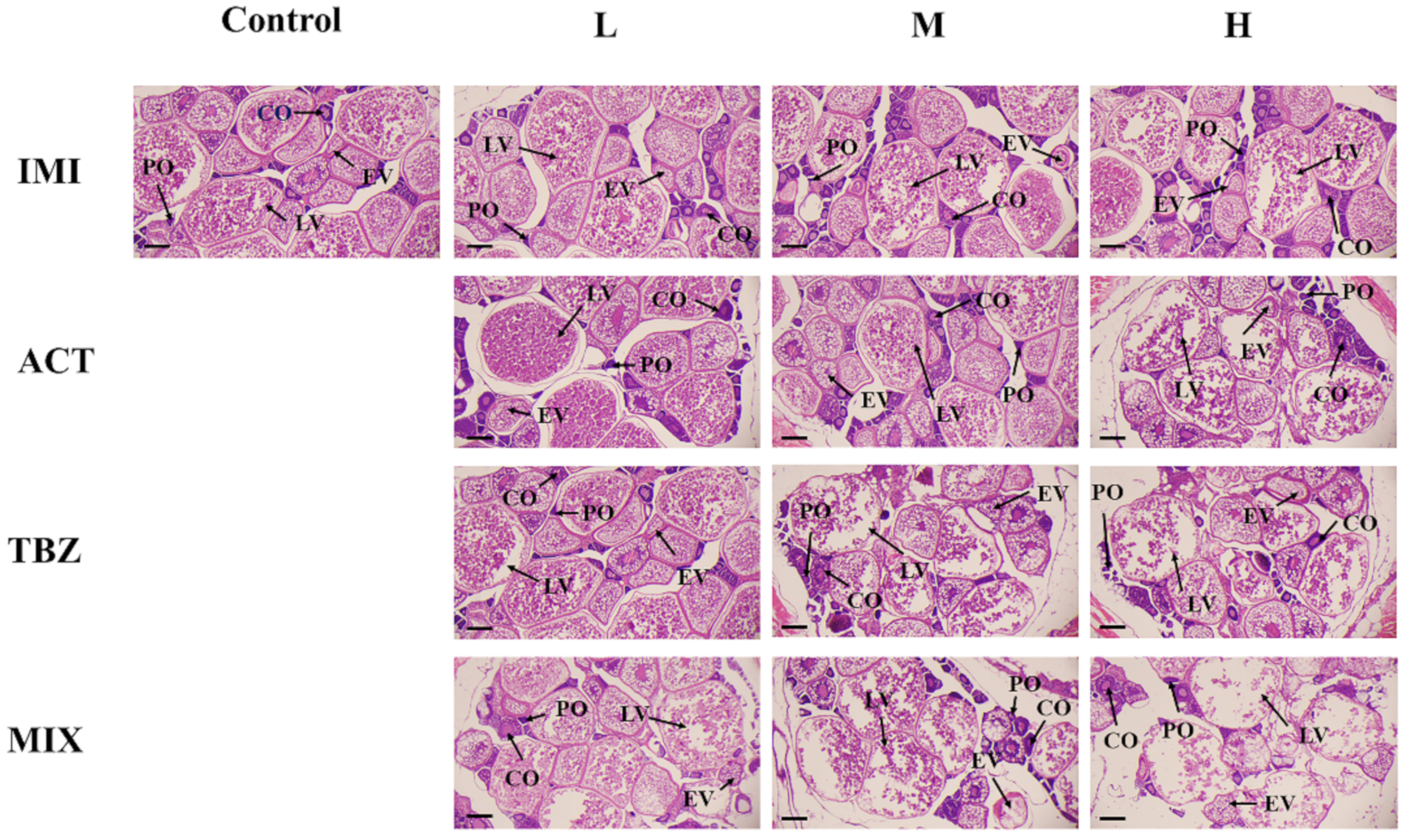
3.2. Histological Alterations in the Gonads

The histology microphotographs of the testes showed that the proportion of mature spermatids (St) was high in the control group, and the number of immature spermatocytes (Sc) increased and the number of mature St decreased significantly with the increase in concentration in individual and combined exposures (Figure 3).
The histology microphotographs of the ovaries showed that the cells of the ovaries were mainly composed of cells in early vitellogenic oocytes (EV) and late vitellogenic oocytes (LV) in the control group, in contrast, the amount of EV and LV decreased in the individual and combined exposure groups. With the increase in concentration, vacuolar damage was observed in the oocytes, and this was more obvious in the combined exposure group (Figure 4).

3.3. Reproductive Hormones Effects

The results of reproductive hormones in the testes of F0 male and the ovaries of F0 females after being exposed for 28 days are shown in Figure 5. In the testes, compared with the control, the content of T was significantly inhibited in the IMI, ACT and MIX exposure groups (Figure 5A), the content of E2 was only significantly increased in the TBZ exposure group (Figure 5B) and the content of VTG was significantly increased in the TBZ and MIX exposure groups. The increase in VTG in the MIX exposure group was more significant compared to the TBZ exposure group (Figure 5C).
In the ovaries, compared with the control, all exposure groups significantly affected the contents of the three reproductive hormones. Compared with the individual exposure groups, the content of T was significantly increased (Figure 5D) and the content of VTG was significantly inhibited (Figure 5F) in the MIX exposure group. Additionally, the content of E2 was significantly inhibited in the MIX exposure group compared to the IMI and ACT exposure groups (Figure 5E).

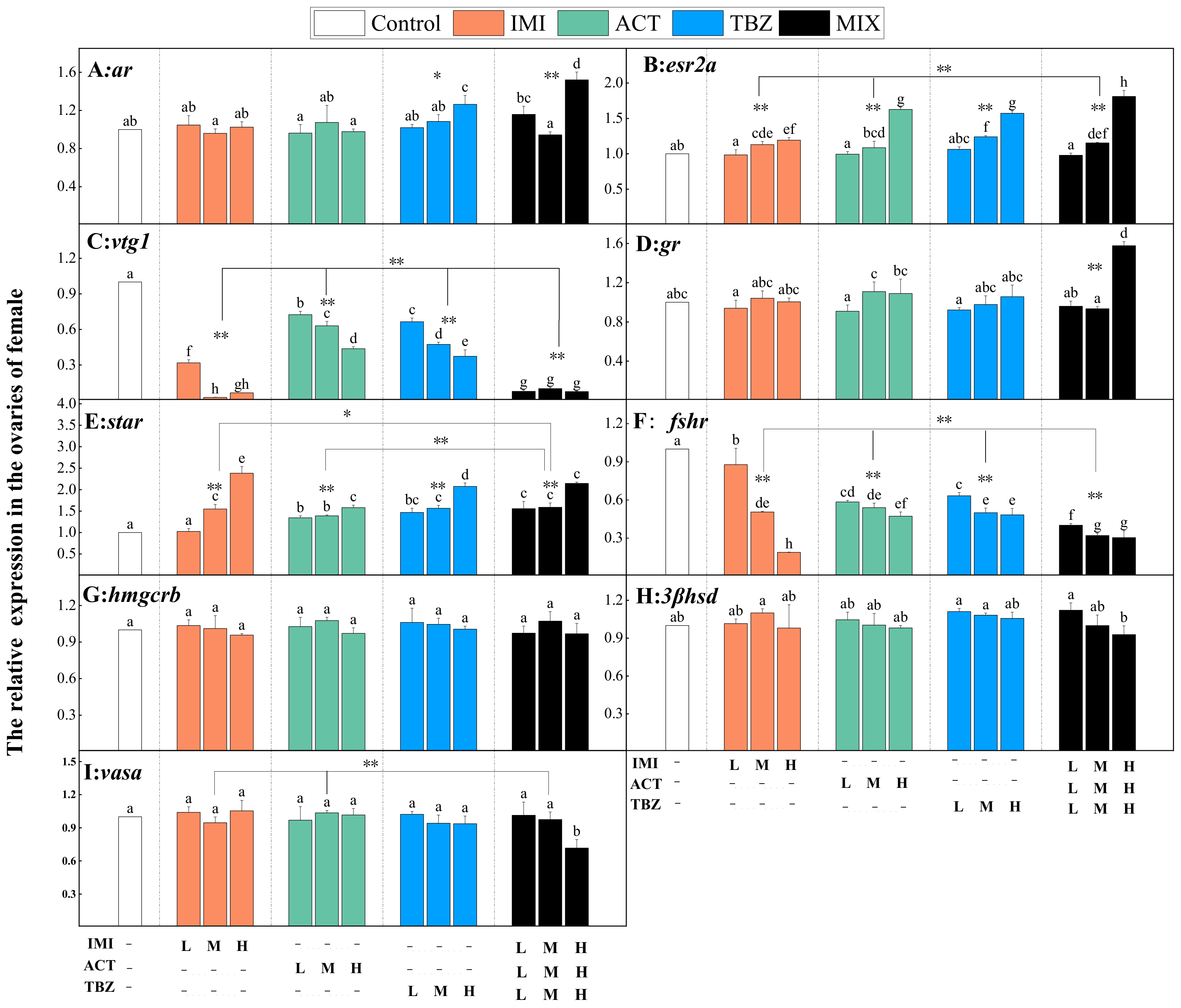
3.4. Reproductive Genes Expression

After 28 days of exposure to adult zebrafish, the relative expressions of reproductive genes (ar, esr2a, vtg1, gr, star, fshr, hmgcrb, 3βhsd and vasa) in the testes of F0 males are shown in Figure 6. The expression of ar was significantly downregulated in the IMI and MIX exposure groups compared to the control, and the downregulation was more significant in the MIX exposure group compared to the IMI exposure group (Figure 6A). The expression of esr2a was significantly upregulated in the IMI and MIX exposure groups compared to the control, and the upregulation was more significant in the MIX exposure group compared to IMI exposure group (Figure 6B). The expressions of vtg1 and star were significantly upregulated and downregulated in the ACT, TBZ and MIX exposure groups compared to the control, and the expressions were significantly affected in the MIX exposure group compared to the ACT and TBZ exposure groups (Figure 6C,E). The expression of gr was significantly downregulated in all exposure groups compared to the control, and the expression was significantly downregulated in the MIX exposure group compared to the individual exposure groups (Figure 6D). The expression of hmgcrb was not significantly different in the individual exposure groups, and the expression was significantly downregulated in the MIX exposure group compared to the control and individual exposure groups (Figure 6G). The expressions of 3βhsd and vasa were significantly downregulated in the TBZ and MIX exposure groups compared to the control, and the expressions were significantly downregulated in the MIX exposure group compared to TBZ exposure group (Figure 6H,I).
The relative expression levels of reproductive genes in the ovaries of F0 females are shown in the Figure 7. The expression of ar was significantly upregulated in the TBZ and MIX exposure groups compared to the control, and the expression was significantly upregulated in the MIX exposure group compared to the TBZ exposure group (Figure 7A). The expressions of esr2a and star were significantly upregulated in all exposure groups compared to the control, and the expressions were significantly upregulated in the MIX exposure group compared to the individual exposure groups (Figure 7B,E). The expressions of vtg1 and fshr were significantly downregulated in all exposure groups compared to the control, and the expressions were significantly downregulated in the MIX exposure group compared to the individual exposure groups (Figure 7C,F). The expressions of gr and vasa were not significantly different in the individual exposure groups and the expressions were significantly upregulated in the MIX exposure group compared to the control and individual exposure groups (Figure 7D,I).

4. Discussion

The results of the study revealed that exposure to the mixtures of IMI, ACT and TBZ significantly inhibited the number, heartbeat, body length and swim bladder development and increased the deformity rate of F1 offspring embryos, while the development of the F0 gonad was impaired. Furthermore, the contents of reproductive hormones and the expression of reproductive genes in the testes of F0 males and the ovaries of F0 females were affected. The effects of the concentration factor on zebrafish showed that a high concentration brought more significant reproductive toxicity, and the analysis results on zebrafish reproduction analyzed by two-way ANOVA are provided in the Supplementary Materials. Previous studies have also shown that exposure to pesticides can affect the reproduction of zebrafish, and the combined effect of multiple pesticides on zebrafish reproduction is more pronounced [31,32,33].
The developmental defects of F1 offspring embryos indicated that pesticides pose a reproductive risk to zebrafish [24,34]. In this study, the number of F1 offspring was significantly reduced; the F1 offspring embryos produced by zebrafish following exposure to IMI had no significant difference in phenotype, demonstrating the low reproductive toxicity of IMI to zebrafish. However, some studies have shown that IMI has reproductive effects on rats [35,36]. ACT has been reported to affect the reproduction of zebrafish, inhibiting embryonic heartbeat and body length. Moreover, ACT can interfere with the endocrine system and impair the innate immunity of zebrafish embryos [37]. TBZ significantly inhibited the body length of the F1 offspring embryos, and it has been demonstrated to cause developmental and reproductive toxicity in zebrafish [38,39,40]. However, the F1 offspring’s embryonic heartbeat, deformity rate, body length and swim bladder development inhibition rate were significantly affected in the MIX exposure group, which means that combined exposure is more reproductively toxic to zebrafish and causes more severe intergenerational effects on F1 offspring embryos. Similarly, research showed that IMI, TBZ and imazalil exhibited a combined genotoxicity in exposure tests on mammalian bone marrow erythrocytes (CD-1 mice) [41]. In addition, exposure of adult zebrafish to Bisphenol F for 150 days resulted in a reduced heart rate and shorter body length in F1 offspring embryos [42]. According to the histology microphotographs of the testes and ovaries, individual and combined exposures adversely affected the development of zebrafish gonads and the damage caused by combined exposure is more significant; it is inferred that the reproductive function of adult zebrafish was impaired.
Several studies have shown that IMI [43,44], ACT [45] and TBZ [46] had endocrine-disrupting effects. Therefore, we further investigated the effects of combined exposure on the contents of the reproductive hormones T, E2 and VTG and the expression of reproductive genes in the testes of F0 males and the ovaries of F0 females. Therefore, it is necessary to study the combined reproductive toxicity effects of these three pesticides on zebrafish.
The reproductive system plays an important role in biological life and the continuation of the species [47]. Follicle-stimulating hormone (FSH) and luteinizing hormone (LH) regulate the production of gonadal steroids and gametogenesis in fish [48], and gonadotropins (GTHs) stimulate gonads produce sex steroid hormones, such as T and E2 [49], which are the major androgens and estrogens in vertebrates, and their main functions are to maintain normal gonadal development. T and E2 play a very important role in maintaining the reproductive function of zebrafish.
The Imprint of T sets the long-term growth regulation of the prostate and seminal vesicles in response to androgenic stimulation in adulthood. T is the main hormone of androgenic steroid and is involved in physiological processes, such as sexual differentiation in females, especially during pregnancy [50]. T cooperates with the androgen receptor (ar) to form androgen receptor complexes for their relevant physiological effects. In this study, the expression of the ar gene was significantly downregulated in the MIX exposure group in the testes. In contrast, in the ovaries of the females, the expression of ar was significantly upregulated resulting in a significant increase in T in the MIX exposure group, moreover, T content was already significantly increased at the low concentration of MIX exposure group and started to increase significantly at the medium concentration of the individual exposure groups.
Estradiol (E2) is an important estrogen which is produced in the ovaries of maturing females [51]. In males, the testes and adrenal glands also produce small amounts of E2 which play an important role in regulating the proliferation of spermatogonia and the physiological function of Sertoli cells [47]. Maternal estrogen receptor 2a (esr2a) exists in scattered granular and filamentous structures [50,52]. In this study, in the testes, the expression of esr2a was significantly upregulated in the IMI and MIX exposure groups, and the content of E2 was significantly increased in the TBZ exposure group. In the ovaries, compared with the individual exposure groups, the expression of esr2a was significantly downregulated and the content of E2 was significantly reduced in the MIX exposure group.
The effects of sex hormones on the gonads of the female and male were different and the impact was greater on the females. However, the effects of the sex hormones were significantly different in the MIX exposure group compared to the individual exposure groups. The results are consistent with the published literature, which indicate that the contents of E2 and T, as well as expression levels of ar and esr2a in male or female fish will change when exposed to pollutants [53,54]. The E2/T ratio is an indication of sex steroid hormone disorders in fish [55]. Changes in T and E2 levels may affect the development of fish gonads and even sex differentiation [47]. In the MIX exposure group, changes in ar and esr2a expressions and T and E2 content probably cause reproductive dysfunction in zebrafish.
VTG is essential for follicle development, yolk biosynthesis and oocyte maturation [56]. Normally, VTG is mainly found in maturing females, but some amount of VTG can also be produced in males after stimulation by pollutants [57]. The expression of type I vtg (vtg1, vtg4-7) was the highest among the seven vtg genes identified [58]. The results of this study showed that in the testes, vtg1 expression was significantly upregulated at high concentrations in the ACT and TBZ individual exposure groups, and the content of VTG was significantly increased in the high concentrations of ACT, while it was significantly increased in the medium concentrations of the MIX exposure group. In the ovaries, compared with the individual exposure groups, vtg1 was barely expressed and the content of VTG was also significantly reduced in the MIX exposure group. Therefore, the combined exposure caused a higher reproductive toxicity by affecting the content of VTG, and the reproductive toxicity was greater in females. Historical studies show that many pesticides can affect the expression of vtg1 [59,60], which is consistent with the results of this test.
Glucocorticoid also belongs to the steroid hormones, which have effects on reproduction, immunity and other functions in animals. Gonadotropin-releasing hormone (GnRH) is synthesized by the hypothalamus and induces the release of FSH and LH, which triggers the release of T from the testis and E2 and progesterone in the ovary, glucocorticoids affect reproduction by interfering with the release of GnRH from the hypothalamus [61]. Moreover, it can only achieve their physiological functions by combining with the glucocorticoid receptor (gr) to form a complex [62]. The results of gr expression showed that compared with the individual exposure groups, the expression of gr was significantly downregulated in the male testes and upregulated in the female ovaries in the MIX exposure group. In historical studies, the expression of gr in zebrafish was also affected by pesticide pollution [63], and thus affected the reproductive function of zebrafish.
Steroidogenic acute regulatory (star) protein is a key limiting point in the synthesis of corticosteroids and sex steroids [64]. The results of the expression of the star gene show that the star expression was significantly downregulated in the MIX exposure group compared to the ACT and TBZ exposure groups in the testes, conversely, star expression was significantly upregulated in the MIX exposure group compared to the individual exposure groups in the ovaries. Some studies illustrated that the function of steroidogenic acute regulatory proteins (star) is to transport cholesterol [65,66], therefore, the change in star expression affects the level of cortisol. However, the expression of star was significantly downregulated in both male and female zebrafish gonads after beta-cypermethrin exposure [67]. Combined exposure interferes with the expression of star and impacts the normal synthesis of corticosteroids and sex steroids, thereby affecting reproductive function.
The follicle-stimulating hormone receptor (fshr) affects follicle production in the ovaries; knockout of the fshr gene in females may lead to sex reversal. Severe inhibition of fshr expression also delays male puberty [68,69]. The results showed that some pesticides may promote the expression of fshr [70] but also may inhibit the expression of fshr [71]. In the present study, the expression of fshr in the testes was not significantly affected, but in the ovaries, it was significantly downregulated in all exposure groups, especially in the MIX exposure group. Thus, combined exposure can prevent the normal development of follicles in the ovaries and ultimately affect their reproductive function.
The rate of conversion of 3-hydroxy-3-methylglutaryl coenzyme A to cholesterol is catalyzed and controlled by 3-hydroxy-3-methylglutaryl coenzyme A reductase (HMGCR) in fish ovaries, ultimately producing E2 [72]. The trend of hmgrb expression in the testes and ovaries of zebrafish after Tris (2-butoxyethyl) phosphate exposure at 500 μg/L was suppressed and both were significantly upregulated [73]. In the present study, the expression of hmgrb was significantly downregulated in the testes of males in the MIX exposure group, while the expression of hmgrb n the ovaries of female was not significantly affected.
In addition, 3β-hydroxysteroid dehydrogenase (3βhsd) is the final step in catalyzing the synthesis of steroid hormones [74,75]. In this study, the expression of 3βhsd in the testis was significantly downregulated at the high concentrations of the TBZ and MIX exposure groups, especially in the MIX exposure group, while the expression of 3βhsd in the ovaries was not significantly affected. In fact, 3βhsd mainly regulates the synthesis of androgens, and the downregulation of 3βhsd expression will reduce the content of T [76], which is consistent with the results of the downregulation of 3βhsd expression and the decrease in T content in the testes.
Vasa plays a crucial role in germ cell development [77]. In this study, the expression of vasa in the testis was significantly downregulated in the TBZ and MIX exposure groups. In the TBZ exposure group, vasa expression began to be significantly downregulated at high concentrations, while in the MIX exposure group, vasa expression began to be significantly downregulated at medium concentrations in the testes. In the ovaries, the expression was only significantly downregulated in the MIX exposure group. Historical studies show that vasa genes can affect reproduction in mice [78], fish [79,80] and other organisms. Therefore, downregulation of vasa expression could affect the normal development of germ cells.
According to data from the China Pesticide Information Network, IMI 21–42 g a.i./hm, the actual field usage of IMI, ACT and TBZ was 21–42 g a.i./ha, 90–120 g a.i./ha, 77.4–96.75 mL a.i./hm, respectively. The 1:4:4 (IMI:ACT:TBZ) ratio is similar to that used in the field, therefore, the effects of pesticides on zebrafish reproduction in real water environments could not be evaluated by an individual pesticide.

5. Conclusions

In summary, the combination of IMI, ACT and TBZ affected the expression of reproductive genes of zebrafish, which will further affect the normal synthesis of steroid hormones (T and E2) and VTG in the zebrafish gonads and may interfere more with the germ cell development of germ cells, thus leading to impaired reproductive function in zebrafish, as expressed in the inhibition of the heartbeat and the increase in deformity rate of the F1 offspring embryos produced by the exposed zebrafish. Moreover, the individual exposure group affected only a few of these reproductive gene expressions and reproductive hormone content in the males or females, but the combined exposure groups affected most of the reproductive gene expression and reproductive hormone content measured in each of the males and females. Thus, it has been shown that the assessment of the toxicity effects on zebrafish in the natural aquatic ecosystem environment cannot be described by an individual pesticide and the impact of mixed pesticide pollution on the water environment should be further considered in the ecotoxicological risk assessment of the pesticide environment.

Supplementary Materials

The following supporting information can be downloaded at: https://www.mdpi.com/article/10.3390/agriculture12121979/s1.

Author Contributions

Conceptualization, J.Y.; Methodology, J.Y. and Y.C.; Validation, L.Z. (Lizhen Zhu), L.M. and L.Z. (Lan Zhang); Investigation, J.Y. and L.Z. (Lizhen Zhu); Resources, H.J.; Data curation, Y.C. and L.M.; Writing—original draft, J.Y., Y.C., L.Z. (Lizhen Zhu) and L.Z. (Lan Zhang); Writing—review & editing, Y.Z., X.L. and H.J.; Visualization, L.Z. (Lan Zhang) and X.L.; Supervision, X.L.; Project administration, Y.Z. and L.M.; Funding acquisition, Y.Z. All authors have read and agreed to the published version of the manuscript.

Funding

This research received no external funding.

Institutional Review Board Statement

The animal study protocol was approved by the Ethics Committee of Institute of Plant Protection, Chinese Academy of Agricultural Sciences (protocol code is IPP2022-05 and date of approval is 8th April, 2022).

Acknowledgments

This work was supported by the Chinese National Natural Science Foundation (32102268).

Conflicts of Interest

The authors declare no conflict of interest.

Ethics Statements

All the experimental procedures and sample collection were carried out in accordance with the Experimental Animal Welfare and Ethical of Institute of Plant Protection, Chinese Academy of Agricultural Sciences.

References

  1. Hladik, M.L.; Kolpin, D.W.; Kuivila, K.M. Widespread occurrence of neonicotinoid insecticides in streams in a high corn and soybean producing region, USA. Environ. Pollut. 2014, 193, 189–196. [Google Scholar] [CrossRef] [PubMed]
  2. Tan, H.; Li, Q.; Zhang, H.; Wu, C.; Zhao, S.; Deng, X.; Li, Y. Pesticide residues in agricultural topsoil from the Hainan tropical riverside basin: Determination, distribution, and relationships with planting patterns and surface water. Sci. Total Environ. 2020, 722, 137856. [Google Scholar] [CrossRef] [PubMed]
  3. Meng, Z.; Liu, L.; Yan, S.; Sun, W.; Jia, M.; Tian, S.; Huang, S.; Zhou, Z.; Zhu, W. Gut microbiota: A key factor in the host health effects induced by pesticide exposure? J. Agric. Food Chem. 2020, 68, 10517–10531. [Google Scholar] [CrossRef] [PubMed]
  4. Grung, M.; Lin, Y.; Zhang, H.; Steen, A.O.; Huang, J.; Zhang, G.; Larssen, T. Pesticide levels and environmental risk in aquatic environments in China—A review. Environ. Int. 2015, 81, 87–97. [Google Scholar] [CrossRef] [PubMed] [Green Version]
  5. Uwizeyimana, H.; Wang, M.; Chen, W.; Khan, K. The eco-toxic effects of pesticide and heavy metal mixtures towards earthworms in soil. Environ. Toxicol. Pharmacol. 2017, 55, 20–29. [Google Scholar] [CrossRef]
  6. Shahjahan, M.; Rahman, M.S.; Islam, S.M.; Uddin, M.; Al-Emran, M. Increase in water temperature increases acute toxicity of sumithion causing nuclear and cellular abnormalities in peripheral erythrocytes of zebrafish Danio rerio. Environ. Sci. Pollut. Res. 2019, 26, 36903–36912. [Google Scholar] [CrossRef]
  7. Luo, T.; Weng, Y.; Huang, Z.; Zhao, Y.; Jin, Y. Combined hepatotoxicity of imidacloprid and microplastics in adult zebrafish: Endpoints at gene transcription. Comp. Biochem. Physiol. Part C Toxicol. Pharmacol. 2021, 246, 109043. [Google Scholar] [CrossRef]
  8. Wang, Y.; Xu, C.; Wang, D.; Weng, H.; Yang, G.; Guo, D.; Yu, R.; Wang, X.; Wang, Q. Combined toxic effects of fludioxonil and triadimefon on embryonic development of zebrafish (Danio rerio). Environ. Pollut. 2020, 260, 114105. [Google Scholar] [CrossRef]
  9. Wang, Y.; Dai, D.; Yu, Y.; Yang, G.; Shen, W.; Wang, Q.; Weng, H.; Zhao, X. Evaluation of joint effects of cyprodinil and kresoxim-methyl on zebrafish, Danio rerio. J. Hazard. Mater. 2018, 352, 80–91. [Google Scholar] [CrossRef]
  10. Yang, Y.; Ma, H.; Zhou, J.; Liu, J.; Liu, W. Joint toxicity of permethrin and cypermethrin at sublethal concentrations to the embryo-larval zebrafish. Chemosphere 2013, 96, 146–154. [Google Scholar] [CrossRef]
  11. Kongtip, P.; Nankongnab, N.; Kallayanatham, N.; Pundee, R.; Yimsabai, J.; Woskie, S. Longitudinal study of metabolic biomarkers among conventional and organic farmers in Thailand. Int. J. Environ. Res. Public Heal. 2020, 17, 4178. [Google Scholar] [CrossRef]
  12. Graham, K.K.; Milbrath, M.O.; Zhang, Y.; Soehnlen, A.; Baert, N.; McArt, S.; Isaacs, R. Identities, concentrations, and sources of pesticide exposure in pollen collected by managed bees during blueberry pollination. Sci. Rep. 2021, 11, 16857. [Google Scholar] [CrossRef]
  13. Bradford, B.Z.; Huseth, A.; Groves, R.L. Widespread detections of neonicotinoid contaminants in central Wisconsin groundwater. PLoS ONE 2018, 13, e0201753. [Google Scholar] [CrossRef]
  14. Sánchez-Bayo, F.; Hyne, R.V. Detection and analysis of neonicotinoids in river waters—Development of a passive sampler for three commonly used insecticides. Chemosphere 2014, 99, 143–151. [Google Scholar] [CrossRef]
  15. Anderson, J.; Dubetz, C.; Palace, V. Neonicotinoids in the Canadian aquatic environment: A literature review on current use products with a focus on fate, exposure, and biological effects. Sci. Total Environ. 2015, 505, 409–422. [Google Scholar] [CrossRef]
  16. Mineau, P. Neonicotinoids in California. Their Use and Threats to the State’s Aquatic Ecosystems and Pollinators, with a Focus on Neon-ic-Treated Seeds. Pierre Mineau Consulting. 2020. Available online: https://www.nrdc.org/sites/default/files (accessed on 29 September 2022).
  17. Zhang, J.-G.; Ma, D.-D.; Xiong, Q.; Qiu, S.-Q.; Huang, G.-Y.; Shi, W.-J.; Ying, G.-G. Imidacloprid and thiamethoxam affect synaptic transmission in zebrafish. Ecotoxicol. Environ. Saf. 2021, 227, 112917. [Google Scholar] [CrossRef]
  18. Luo, T.; Wang, X.; Jin, Y. Low concentrations of imidacloprid exposure induced gut toxicity in adult zebrafish (Danio rerio). Comp. Biochem. Physiol. Part C Toxicol. Pharmacol. 2021, 241, 108972. [Google Scholar] [CrossRef]
  19. Baysal, M.; Atlı-Eklioğlu, Ö. Comparison of the toxicity of pure compounds and commercial formulations of imidacloprid and acetamiprid on HT-29 cells: Single and mixture exposure. Food Chem. Toxicol. 2021, 155, 112430. [Google Scholar] [CrossRef]
  20. Kolpin, D.W.; Nations, B.K.; Goolsby, D.A.; Thurman, E.M. Acetochlor in the hydrologic system in the midwestern United States, 1994. Environ. Sci. Technol. 1996, 30, 1459–1464. [Google Scholar] [CrossRef] [Green Version]
  21. Li, L.; Wang, M.; Chen, S.; Zhao, W.; Zhao, Y.; Wang, X.; Zhang, Y. A urinary metabonomics analysis of long-term effect of acetochlor exposure on rats by ultra-performance liquid chromatography/mass spectrometry. Pestic. Biochem. Physiol. 2016, 128, 82–88. [Google Scholar] [CrossRef]
  22. Li, H.; Feng, Y.; Li, X.; Zeng, D. Analytical confirmation of various herbicides in drinking water resources in sugarcane production regions of Guangxi, China. Bull. Environ. Contam. Toxicol. 2018, 100, 815–820. [Google Scholar] [CrossRef] [PubMed]
  23. Zhang, Y.; Xue, W.; Long, R.; Yang, H.; Wei, W. Acetochlor affects zebrafish ovarian development by producing estrogen effects and inducing oxidative stress. Environ. Sci. Pollut. Res. 2020, 27, 27688–27696. [Google Scholar] [CrossRef] [PubMed]
  24. Liu, N.; Dong, F.; Xu, J.; Liu, X.; Zheng, Y. Chiral bioaccumulation behavior of tebuconazole in the zebrafish (Danio rerio). Ecotoxicol. Environ. Saf. 2016, 126, 78–84. [Google Scholar] [CrossRef] [PubMed]
  25. Cui, N.; Xu, H.; Yao, S.; He, Y.; Zhang, H.; Yu, Y. Chiral triazole fungicide tebuconazole: Enantioselective bioaccumulation, bioactivity, acute toxicity, and dissipation in soils. Environ. Sci. Pollut. Res. 2018, 25, 25468–25475. [Google Scholar] [CrossRef] [PubMed]
  26. Yu, L.; Chen, M.; Liu, Y.; Gui, W.; Zhu, G. Thyroid endocrine disruption in zebrafish larvae following exposure to hexaconazole and tebuconazole. Aquat. Toxicol. 2013, 138–139, 35–42. [Google Scholar] [CrossRef] [Green Version]
  27. Berenzen, N.; Lentzen-Godding, A.; Probst, M.; Schulz, H.; Schulz, R.; Liess, M. A comparison of predicted and measured levels of runoff-related pesticide concentrations in small lowland streams on a landscape level. Chemosphere 2005, 58, 683–691. [Google Scholar] [CrossRef]
  28. Azevedo, R.D.S.; Falcão, K.V.G.; Assis, C.R.D.; Martins, R.M.G.; Araújo, M.C.; Yogui, G.T.; Neves, J.L.; Seabra, G.M.; Maia, M.B.S.; Amaral, I.P.G.; et al. Effects of pyriproxyfen on zebrafish brain mitochondria and acetylcholinesterase. Chemosphere 2021, 263, 128029. [Google Scholar] [CrossRef]
  29. Vieira, R.S.F.; Venâncio, C.A.S.; Félix, L.M. Behavioural impairment and oxidative stress by acute exposure of zebrafish to a commercial formulation of tebuconazole. Environ. Toxicol. Pharmacol. 2022, 91, 103823. [Google Scholar] [CrossRef]
  30. Chang, Y.; Mao, L.; Zhang, L.; Zhang, Y.; Jiang, H. Combined toxicity of imidacloprid, acetochlor, and tebuconazole to zebrafish (Danio rerio): Acute toxicity and hepatotoxicity assessment. Environ. Sci. Pollut. Res. 2020, 27, 10286–10295. [Google Scholar] [CrossRef]
  31. Brander, S.M.; Jeffries, K.M.; Cole, B.J.; DeCourten, B.M.; White, J.W.; Hasenbein, S.; Fangue, N.A.; Connon, R.E. Transcriptomic changes underlie altered egg protein production and reduced fecundity in an estuarine model fish exposed to bifenthrin. Aquat. Toxicol. 2016, 174, 247–260. [Google Scholar] [CrossRef]
  32. Chu, S.-H.; Liao, P.-H.; Chen, P.-J. Developmental exposures to an azole fungicide triadimenol at environmentally relevant concentrations cause reproductive dysfunction in females of medaka fish. Chemosphere 2016, 152, 181–189. [Google Scholar] [CrossRef]
  33. Cao, F.; Zhu, L.; Li, H.; Yu, S.; Wang, C.; Qiu, L. Reproductive toxicity of azoxystrobin to adult zebrafish (Danio rerio). Environ. Pollut. 2016, 219, 1109–1121. [Google Scholar] [CrossRef]
  34. Wang, H.; Dong, F.; Zhao, Y.; Fu, S.; Zhao, H.; Liu, S.; Zhang, W.; Hu, F. Exposure to diclofenac alters thyroid hormone levels and transcription of genes involved in the hypothalamic-pituitary-thyroid axis in zebrafish embryos/larvae. Comp. Biochem. Physiol. Part C Toxicol. Pharmacol. 2022, 257, 109335. [Google Scholar] [CrossRef]
  35. Lonare, M.; Kumar, M.; Raut, S.; More, A.; Doltade, S.; Badgujar, P.; Telang, A. Evaluation of ameliorative effect of curcumin on imidacloprid-induced male reproductive toxicity in wistar rats. Environ. Toxicol. 2015, 31, 1250–1263. [Google Scholar] [CrossRef]
  36. Bal, R.; Naziroğlu, M.; Türk, G.; Yilmaz, Ö.; Kuloğlu, T.; Etem, E.; Baydas, G. Insecticide imidacloprid induces morphological and DNA damage through oxidative toxicity on the reproductive organs of developing male rats. Cell Biochem. Funct. 2012, 30, 492–499. [Google Scholar] [CrossRef] [Green Version]
  37. Wang, Y.; Wu, S.; Chen, J.; Zhang, C.; Xu, Z.; Li, G.; Cai, L.; Shen, W.; Wang, Q. Single and joint toxicity assessment of four currently used pesticides to zebrafish (Danio rerio) using traditional and molecular endpoints. Chemosphere 2018, 192, 14–23. [Google Scholar] [CrossRef]
  38. Altenhofen, S.; Nabinger, D.D.; Wiprich, M.T.; Pereira, T.C.B.; Bogo, M.R.; Bonan, C.D. Tebuconazole alters morphological, behavioral and neurochemical parameters in larvae and adult zebrafish (Danio rerio). Chemosphere 2017, 180, 483–490. [Google Scholar] [CrossRef]
  39. Li, S.; Wu, Q.; Sun, Q.; Coffin, S.; Gui, W.; Zhu, G. Parental exposure to tebuconazole causes thyroid endocrine disruption in zebrafish and developmental toxicity in offspring. Aquat. Toxicol. 2019, 211, 116–123. [Google Scholar] [CrossRef]
  40. Li, S.; Jiang, Y.; Sun, Q.; Coffin, S.; Chen, L.; Qiao, K.; Gui, W.; Zhu, G. Tebuconazole induced oxidative stress related hepatotoxicity in adult and larval zebrafish (Danio rerio). Chemosphere 2019, 241, 125129. [Google Scholar] [CrossRef]
  41. Ilyushina, N.A.; Egorova, O.V.; Masaltsev, G.V.; Averianova, N.S.; Revazova, Y.A.; Rakitskii, V.N.; Goumenou, M.; Vardavas, A.; Stivaktakis, P.; Tsatsakis, A. Genotoxicity of mixture of imidacloprid, imazalil and tebuconazole. Toxicol. Rep. 2020, 7, 1090–1094. [Google Scholar] [CrossRef]
  42. Mu, X.; Qi, S.; Liu, J.; Wang, H.; Yuan, L.; Qian, L.; Li, T.; Huang, Y.; Wang, C.; Guo, Y.; et al. Environmental level of bisphenol F induced reproductive toxicity toward zebrafish. Sci. Total Environ. 2021, 806, 149992. [Google Scholar] [CrossRef] [PubMed]
  43. Paten, A.M.; Colin, T.; Coppin, C.W.; Court, L.N.; Barron, A.B.; Oakeshott, J.G.; Morgan, M.J. Non-additive gene interactions underpin molecular and phenotypic responses in honey bee larvae exposed to imidacloprid and thymol. Sci. Total Environ. 2022, 814, 152614. [Google Scholar] [CrossRef] [PubMed]
  44. Mzid, M.; Ghlissi, Z.; Ben Salem, M.; Ben Khedir, S.; Chaabouni, K.; Ayedi, F.; Sahnoun, Z.; Hakim, A.; Rebai, T. Chemoprotective role of ethanol extract of Urtica urens L. against the toxicity of imidacloprid on endocrine disruption and ovarian morphometric in female rats, GC/MS analysis. Biomed. Pharmacother. 2018, 97, 518–527. [Google Scholar] [CrossRef]
  45. Zhou, J.; Wang, D.; Yang, H.; Wang, F. Specific detection of acetamiprid with aptamer based on flexible and adhesive SERS membrane. Spectrochim. Acta Part A Mol. Biomol. Spectrosc. 2021, 270, 120801. [Google Scholar] [CrossRef] [PubMed]
  46. Li, S.; Sun, Q.; Wu, Q.; Gui, W.; Zhu, G.; Schlenk, D. Endocrine disrupting effects of tebuconazole on different life stages of zebrafish (Danio rerio). Environ. Pollut. 2019, 249, 1049–1059. [Google Scholar] [CrossRef] [Green Version]
  47. Li, M.; Cao, J.; Chen, J.; Song, J.; Zhou, B.; Feng, C.; Wang, J. Waterborne fluoride exposure changed the structure and the expressions of steroidogenic-related genes in gonads of adult zebrafish (Danio rerio). Chemosphere 2016, 145, 365–375. [Google Scholar] [CrossRef]
  48. Kazeto, Y.; Kohara, M.; Miura, T.; Miura, C.; Yamaguchi, S.; Trant, J.M.; Adachi, S.; Yamauchi, K. Japanese eel follicle-stimulating hormone (Fsh) and luteinizing hormone (Lh): Production of biologically active recombinant Fsh and Lh by Drosophila S2 cells and their differential actions on the reproductive biology. Biol. Reprod. 2008, 79, 938–946. [Google Scholar] [CrossRef] [Green Version]
  49. Kime, D.E. ‘Classical’ and ‘non-classical’ reproductive steroids in fish. Rev. Fish Biol. Fish. 1993, 3, 160–180. [Google Scholar] [CrossRef]
  50. Vivar, O.I.; Zhao, X.; Saunier, E.F.; Griffin, C.; Mayba, O.S.; Tagliaferri, M.; Cohen, I.; Speed, T.P.; Leitman, D.C. Estrogen receptor β binds to and regulates three distinct classes of target genes. J. Biol. Chem. 2010, 285, 22059–22066. [Google Scholar] [CrossRef] [Green Version]
  51. Cui, J.; Shen, Y.; Li, R. Estrogen synthesis and signaling pathways during aging: From periphery to brain. Trends Mol. Med. 2013, 19, 197–209. [Google Scholar] [CrossRef]
  52. Celeghin, A.; Benato, F.; Pikulkaew, S.; Rabbane, G.; Colombo, L.; Valle, L.D. The knockdown of the maternal estrogen receptor 2a (esr2a) mRNA affects embryo transcript contents and larval development in zebrafish. Gen. Comp. Endocrinol. 2011, 172, 120–129. [Google Scholar] [CrossRef]
  53. Wu, S.M.; Tseng, Y.-J.; Chen, T.-H. Maternal effect and dietary supplementation of estradiol-17β on female zebrafish (Danio rerio) affects the swimming behavior and stress-coping styles of its offspring. Comp. Biochem. Physiol. Part C Toxicol. Pharmacol. 2021, 252, 109211. [Google Scholar] [CrossRef]
  54. Zhou, H.; Chen, A.; Lu, W. Corticotropin-releasing hormone reduces basal estradiol production in zebrafish follicular cells. Mol. Cell. Endocrinol. 2021, 527, 111222. [Google Scholar] [CrossRef]
  55. Orlando, E.F.; Kolok, A.S.; Binzcik, G.A.; Gates, J.L.; Horton, M.K.; Lambright, C.S.; Gray, L.E., Jr.; Soto, A.M.; Guillette, L.J., Jr. Endocrine disrupting effects of cattle feedlot effluent on an aquatic sentinel species, the fathead minnow. Environ. Health Perspect. 2004, 112, 353–358. [Google Scholar] [CrossRef] [Green Version]
  56. Yuan, H.-X.; Xu, X.; Sima, Y.-H.; Xu, S.-Q. Reproductive toxicity effects of 4-nonylphenol with known endocrine disrupting effects and induction of vitellogenin gene expression in silkworm, Bombyx mori. Chemosphere 2013, 93, 263–268. [Google Scholar] [CrossRef]
  57. Aoki, J.-Y.; Nagae, M.; Takao, Y.; Hara, A.; Lee, Y.-D.; Yeo, I.-K.; Lim, B.-S.; Park, C.-B.; Soyano, K. Survey of contamination of estrogenic chemicals in Japanese and Korean coastal waters using the wild grey mullet (Mugil cephalus). Sci. Total Environ. 2010, 408, 660–665. [Google Scholar] [CrossRef] [Green Version]
  58. Jun, I.; Ryu, C.; Park, C.; Cho, H.; Kim, Y. 5α-reductase inhibition results in decreases of DHT, E2 and VTG in zebrafish larvae. Toxicol. Lett. 2021, 350, S70. [Google Scholar] [CrossRef]
  59. Wu, S.; Li, X.; Liu, X.; Yang, G.; An, X.; Wang, Q.; Wang, Y. Joint toxic effects of triazophos and imidacloprid on zebrafish (Danio rerio). Environ. Pollut. 2018, 235, 470–481. [Google Scholar] [CrossRef]
  60. Darvishi, M.; Safari, R.; Hoseinifar, S.H.; Shabani, A.; Dadar, M.; Jarayedi, Z.; Paolucci, M. Sublethal doses of diazinon affected reproductive, immune, and oxidative status in female zebrafish (Danio rerio). Aquac. Rep. 2021, 22, 100944. [Google Scholar] [CrossRef]
  61. Whirledge, S.; Cidlowski, J.A. Glucocorticoids and reproduction: Traffic control on the road to reproduction. Trends Endocrinol. Metab. 2017, 28, 399–415. [Google Scholar] [CrossRef]
  62. Faught, E.; Vijayan, M.M. Mechanisms of cortisol action in fish hepatocytes. Comp. Biochem. Physiol. Part B Biochem. Mol. Biol. 2016, 199, 136–145. [Google Scholar] [CrossRef] [PubMed]
  63. Wang, Y.; Chen, C.; Yang, G.; Wang, X.; Wang, Q.; Weng, H.; Zhang, Z.; Qian, Y. Combined lethal toxicity, biochemical responses, and gene expression variations induced by tebuconazole, bifenthrin and their mixture in zebrafish (Danio rerio). Ecotoxicol. Environ. Saf. 2021, 230, 113116. [Google Scholar] [CrossRef] [PubMed]
  64. Kusakabe, M.; Zuccarelli, M.D.; Nakamura, I.; Young, G. Steroidogenic acute regulatory protein in white sturgeon (Acipenser transmontanus): cDNA cloning, sites of expression and transcript abundance in corticosteroidogenic tissue after an acute stressor. Gen. Comp. Endocrinol. 2009, 162, 233–240. [Google Scholar] [CrossRef] [PubMed]
  65. Alderman, S.L.; Bernier, N.J. Ontogeny of the corticotropin-releasing factor system in zebrafish. Gen. Comp. Endocrinol. 2009, 164, 61–69. [Google Scholar] [CrossRef] [PubMed]
  66. Alderman, S.L.; Leishman, E.; Fuzzen, M.L.; Bernier, N.J. Corticotropin-releasing factor regulates caspase-3 and may protect developing zebrafish from stress-induced apoptosis. Gen. Comp. Endocrinol. 2018, 265, 207–213. [Google Scholar] [CrossRef]
  67. Lu, J.; Wu, Q.; Yang, Q.; Li, G.; Wang, R.; Liu, Y.; Duan, C.; Duan, S.; He, X.; Huang, Z.; et al. Molecular mechanism of reproductive toxicity induced by beta-cypermethrin in zebrafish. Comp. Biochem. Physiol. Part C Toxicol. Pharmacol. 2020, 239, 108894. [Google Scholar] [CrossRef]
  68. Chu, L.; Li, J.; Liu, Y.; Cheng, C.H.K. Gonadotropin signaling in zebrafish ovary and testis development: Insights from gene knockout study. Mol. Endocrinol. 2015, 29, 1743–1758. [Google Scholar] [CrossRef] [Green Version]
  69. Ferraresso, S.; Bargelloni, L.; Babbucci, M.; Cannas, R.; Follesa, M.C.; Carugati, L.; Melis, R.; Cau, A.; Koutrakis, M.; Sapounidis, A.; et al. fshr: A fish sex-determining locus shows variable incomplete penetrance across flathead grey mullet populations. iScience 2020, 24, 101886. [Google Scholar] [CrossRef]
  70. Yang, R.; Wang, X.; Wang, J.; Chen, P.; Liu, Q.; Zhong, W.; Zhu, L. Insights into the sex-dependent reproductive toxicity of 2-ethylhexyl diphenyl phosphate on zebrafish (Danio rerio). Environ. Int. 2021, 158, 106928. [Google Scholar] [CrossRef]
  71. Cao, J.; Wang, G.; Wang, T.; Chen, J.; Wenjing, G.; Wu, P.; He, X.; Xie, L. Copper caused reproductive endocrine disruption in zebrafish (Danio rerio). Aquat. Toxicol. 2019, 211, 124–136. [Google Scholar] [CrossRef]
  72. Yu, M.; Zhang, X.; Guo, L.; Tian, H.; Wang, W.; Ru, S. Anti-estrogenic effect of semicarbazide in female zebrafish (Danio rerio) and its potential mechanisms. Aquat. Toxicol. 2016, 170, 262–270. [Google Scholar] [CrossRef]
  73. Xu, Q.; Wu, D.; Dang, Y.; Yu, L.; Liu, C.; Wang, J. Reproduction impairment and endocrine disruption in adult zebrafish (Danio rerio) after waterborne exposure to TBOEP. Aquat. Toxicol. 2017, 182, 163–171. [Google Scholar] [CrossRef]
  74. Maharajan, K.; Muthulakshmi, S.; Karthik, C.; Nataraj, B.; Nambirajan, K.; Hemalatha, D.; Jiji, S.; Kadirvelu, K.; Liu, K.-C.; Ramesh, M. Pyriproxyfen induced impairment of reproductive endocrine homeostasis and gonadal histopathology in zebrafish (Danio rerio) by altered expression of hypothalamus-pituitary-gonadal (HPG) axis genes. Sci. Total Environ. 2020, 735, 139496. [Google Scholar] [CrossRef]
  75. Zhang, S.; Mo, J.; Wang, Y.; Ni, C.; Li, X.; Zhu, Q.; Ge, R.-S. Endocrine disruptors of inhibiting testicular 3β-hydroxysteroid dehydrogenase. Chem. Interact. 2019, 303, 90–97. [Google Scholar] [CrossRef]
  76. Ma, Y.; Han, J.; Guo, Y.; Lam, P.K.; Wu, R.S.; Giesy, J.P.; Zhang, X.; Zhou, B. Disruption of endocrine function in in vitro H295R cell-based and in in vivo assay in zebrafish by 2,4-dichlorophenol. Aquat. Toxicol. 2012, 106–107, 173–181. [Google Scholar] [CrossRef]
  77. Liu, Y.; Xue, W.; Zhu, L.; Ye, D.; Zhu, X.; Wang, H.; Sun, Y.; Deng, F. Nanog suppresses the expression of vasa by directly regulating nlkl in the early zebrafish embryo. Biochimie 2017, 142, 93–101. [Google Scholar] [CrossRef]
  78. Fujiwara, Y.; Komiya, T.; Kawabata, H.; Sato, M.; Fujimoto, H.; Furusawa, M.; Noce, T. Isolation of a DEAD-family protein gene that encodes a murine homolog of Drosophila vasa and its specific expression in germ cell lineage. Proc. Natl. Acad. Sci. USA 1994, 91, 12258–12262. [Google Scholar] [CrossRef] [Green Version]
  79. Kobayashi, T.; Kajiura-Kobayashi, H.; Nagahama, Y. Differential expression of vasa homologue gene in the germ cells during oogenesis and spermatogenesis in a teleost fish, tilapia, Oreochromis niloticus. Mech. Dev. 2000, 99, 139–142. [Google Scholar] [CrossRef]
  80. Yoshizaki, G.; Sakatani, S.; Tominaga, H.; Takeuchi, T. Cloning and characterization of a vasa-like gene in rainbow trout and its expression in the germ cell lineage. Mol. Reprod. Dev. 2000, 55, 364–371. [Google Scholar] [CrossRef]
Figure 1. Effects on F1 offspring embryos after individual and combined exposure to adult zebrafish. (A) F1 offspring embryos developed to 48 hpf when the middle concentration of the combined exposure group started to have yolk sac edema deformation (within the yellow dashed line). (B) At 72 hpf, the yolk sac edema deformation recovered, and the red lines indicate body length. Scale bar = 200 μm. Here L: low concentration, M: medium concentration and H: high concentration.
Agriculture 12 01979 g001
Figure 2. F1 offspring embryo indicators: (A) the number of F1 embryos; (B) the number of heartbeats per 20 s (48–96 hpf); (C) deformity rate (48–96 hpf); (D) body length (96–168 hpf); (E) swim bladder development (144 hpf). Here L: low concentration, M: medium concentration, and H: high concentration. In the figure, “a, b, c, …” indicate that the results were analyzed by one-way ANOVA; “* and **” indicate the significant differences between the exposure groups and the control and the results were analyzed by two-way ANOVA (*: p < 0.05; **: p < 0.01).
Agriculture 12 01979 g002
Figure 3. Histology microphotographs of the testes of F0 zebrafish after 28 d exposure. The cells of the testes mainly included spermatocytes (Sc) and spermatids (St), (200 magnification). Scale bar = 100 μm. Here L: low concentration, M: medium concentration and H: high concentration.
Agriculture 12 01979 g003
Figure 4. Histology microphotographs of the ovaries of F0 zebrafish after 28 d exposure. The oocytes in the ovaries included perinucleolar oocytes (PO), cortical alveolar oocytes (CO), early vitellogenic oocytes (EV) and late vitellogenic oocytes (LV) (50 magnification). Scale bar = 400 μm. Here L: low concentration, M: medium concentration and H: high concentration.
Agriculture 12 01979 g004
Figure 5. Contents of reproductive hormones T, E2 and VTG in the testes and ovaries: (AC) the content of T, E2 and VTG in the testes; (DF) the content T, E2 and VTG in the ovaries. Values are presented as mean ± standard deviation (n = 3, replicates of 50 embryos each) (p < 0.05). Here L: low concentration, M: medium concentration and H: high concentration. In the figure, “a, b, c, …” indicate that the results were analyzed by one-way ANOVA; “* and **” indicate the significant differences between the exposure groups and the control and the results were analyzed by two-way ANOVA (*: p < 0.05; **: p < 0.01).
Agriculture 12 01979 g005
Figure 6. Expressions of reproductive genes (A) ar, (B) esr2a, (C) vtg1, (D) gr, (E) star, (F) fshr, (G) hmgcrb, (H) 3βhsd and (I) vasa in the testes of the males. Values are presented as mean ± standard deviation (n = 3) (p < 0.05). Here L: low concentration, M: medium concentration and H: high concentration. In the figure, “a, b, c, …” indicate that the results were analyzed by one-way ANOVA; “* and **” indicate the significant differences between the exposure groups and the control and the results were analyzed by two-way ANOVA (*: p < 0.05; **: p < 0.01).
Agriculture 12 01979 g006
Figure 7. Expressions of reproductive genes (A) ar, (B) esr2a, (C) vtg1, (D) gr, (E) star, (F) fshr, (G) hmgcrb, (H) 3βhsd and (I) vasa in the ovaries of the females. Values are presented as mean ± standard deviation (n = 3) (p < 0.05). Here L: low concentration, M: medium concentration and H: high concentration. In the figure, “a, b, c, …” indicate that the results were analyzed by one-way ANOVA; “* and **” indicate the significant differences between the exposure groups and the control and the results were analyzed by two-way ANOVA (*: p < 0.05; **: p < 0.01).
Agriculture 12 01979 g007
Table 1. Gene primer sequences.
GeneSequence of the Primers (5′ →3′)Accession No.
rpl8F: TTGTTGGTGTTGTTGCTGGTNM_200713.1
R: GGATGCTCAACAGGGTTCAT
ef1αF: GATCACTGGTACTTCTCAGGCTGANM_131263.1
R: GGTGAAAGCCAGGAGGGC
arF: GCGAATGGATGGATGTAACNM_001083123.1
R: TCATCAGAGCAGATTAGGC
esr2aF: CTCTGAACTCATCCGCCTTCNM_180966.2
R: AGCAGAGCGGGACTGTAAAA
vtg1F: CTCCCGAGTTCATTCAGANM_001044897.2
R: ATGACAACTTCACGCAGA
grF: GAGCCAGACACCCTCTATGCNM_001020711.3
R: CCAGCCCAGTCCAAAAGACA
starF: GAATGCCTGAGCAGAAGGGANM_131663.1
R: CGTCTATACCCCCACCGGAT
fshrF: GTCTGTCTGGGCAACAAGGTNM_001001812.1
R: CACCACTATTCTCTTCAGCTCGT
hmgrbF: CCCGCCCAAAGCCATAAAAGNM_001014292
R: GCTTCCAGACGGTATCCCAG
3βhsdF: AACAAGCCCATTCTGCCCATAY279108
R: TCCTCCCAGTCATACCGAGG
vasaF: CAACAGGCTTCAACCCACGBC129275.1
R: GCCAGTTATTCCCATTCCTCA
Publisher’s Note: MDPI stays neutral with regard to jurisdictional claims in published maps and institutional affiliations.

