Article

Evaluation of Pyrenophora tritici-repentis Infection of Wheat Heads

Centre for Crop and Disease Management, School of Molecular Life Sciences, Curtin University, Bentley, Western Australia 6102, Australia
*
Author to whom correspondence should be addressed.
Agriculture 2020, 10(9), 417; https://doi.org/10.3390/agriculture10090417
Submission received: 27 August 2020 / Revised: 16 September 2020 / Accepted: 18 September 2020 / Published: 21 September 2020
(This article belongs to the Section Crop Protection, Diseases, Pests and Weeds)

Abstract

:
The incidence of wheat head infection by Pyrenophora tritici-repentis (Ptr), the etiological agent of tan spot disease, was evaluated during grain development in a glasshouse experiment. Heads artificially inoculated with a Ptr spore suspension developed widespread brown spots across the spikelets, and mycelia and conidophores were observed on glumes and awns. Seeds of heavily infected heads were darkened and shrivelled, but no red smudge symptoms were apparent. The recovery rate of Ptr isolates from the inoculated wheat heads was low, and colonies that were re-isolated displayed an irregular morphology with reddish mycelia when grown on agar plates. The presence of Ptr on inoculated wheat heads was assessed directly via PCR detection, and a limitation of Ptr hyphae to proliferate beyond the point of contact of spore inoculum on floret tissues was observed. The systemic transmission of Ptr from infected seeds was minimal; however, saprophytic growth of the pathogen occurred on the senescing leaves of wheat plants grown from inoculated seeds. Thus, even though Ptr seed infection is not as common as foliar infection, infected seeds are still a source of disease inoculum and screening for pathogen contamination is advisable.

1. Introduction

The major wheat disease tan spot is caused by the fungus Pyrenophora tritici-repentis (Died.) abbreviated to Ptr. Although Ptr is mainly known as a foliar pathogen that causes tan coloured lesions often surrounded by a chlorotic halos, Ptr has also been associated as a seed-borne pathogen, with infected grain reported to be a source of inoculum [1,2,3,4]. The most common symptom associated with seed infection by Ptr is red smudge, a pink to bright red discolouration of the wheat kernel [1,5]. Other seed symptoms have also been reported to be associated with Ptr infection and include black point [1], a black discolouration of the grain coat overlying the germ, and dark smudge, which is characterised by darkening at the germ end [5,6]; however, other fungal pathogens such as Alternaria species and Cochiliobolus sativus are generally associated with such darkening symptoms [1,3,7,8]. Although the biosynthetic pathways leading to the development of the pink discolouration on the kernel has not been understood, oxidation of phenolic compounds has been suggested to be the cause [9].
Since grain grade and quality are highly important traits, blemished or discoloured kernels result in economic losses due to the downgrading of the grade or even rejection, as per strict receival standards. In Australia, grain staining limits are a maximum of 5% for high protein milling grades and up to 50% for feed wheat, while pink fungal staining limits are 2% for milling wheat and 5% for general purpose and feed grades [10].
The detrimental effect of fungal infected seeds is not limited to profitability but also affects crop plant productivity and ultimately yield if the infected seeds are used as source for sowing. A slower rate of seedling emergence, a decrease in overall seed germination and seedling vigour [5,11], as well as a reduced numbers of tillers and heads [2,8,12,13] are some of the negative effects that have been observed as a result of Ptr infected seeds. In plants grown from heavily infected seed, up to 20% or fewer heads have been reported, which resulted in a 17% lower grain yield per plot [2]. However, discrepancies as to the effect of red smudge on seed germination and the resulting number of tillers have been described. Although a 20% decrease in germination was recorded across the discoloured kernels, the effect of red smudge on the numbers of tillers has been shown to be more variable in comparison to black point or dark smudge under different experimental environments [8].
The occurrence of black point caused by Alternaria alternata [7,14,15] and the recovery of Ptr from pink grain in storage [15] has been reported, however, the extent of the occurrence of wheat seed discolouration or infection caused by Ptr in Australia is unknown. The majority of the incidence of red smudge reported in Canada occurred on durum wheat, and similar symptoms appeared to be less apparent on common wheat [9]. Since tan spot is a major wheat disease in Australia, seed infection by this pathogen is an important consideration which may have an impact on the epidemiology of the disease in the field and grower returns. Therefore, the objectives of this study were to (1) assess the occurrence of Ptr infection on wheat heads during grain development of an Australian bread wheat variety, (2) examine the appearance of grain from infected wheat heads for discolouration and (3) evaluate the occurrence of tan spot disease transmission via seed.

2. Materials and Methods

2.1. Plant Growth and Inoculation

The common Australian bread wheat (Triticum aestivum) variety Yitpi, which is rated as susceptible to very susceptible (SVS) to tan spot disease [16] was used in this study. Six seeds were sown per pot, and plants were grown in Richgro potting mix (Richgro, Lansdale, WA, Australia) and placed in an ambient temperature glasshouse with natural light. Pots were fertilized initially with Nitrophoska Perfekt (Bayer, Leverkusen, Germany) compound fertilizer followed by Thrive all-purpose water soluble fertilizer (Yates, Clayton, Victoria, Australia) fortnightly. After two months, Thrive flowering water soluble fertilizer (Yates) was applied and followed with monthly application of Nitrophoska Perfekt (Bayer) compound fertilizer. After three months, wheat heads were inoculated at either ear emergence/heading (GS50-59), flowering (GS60-69) or ripening (GS90-99) by spraying with a conidial suspension of Ptr at the concentration of 2500 spores per ml. Twenty five ml of spore suspension in 0.25% gelatin was sprayed onto the wheat heads for each pot, and for each growth stage, 10 pots were inoculated. To ensure that the inoculum level was not a limitation for the development of grain discolouration symptoms, a separate batch of pots were grown for multiple inoculations with the same plants being sprayed at all three growth stages (ear emergence/heading, flowering and ripening). The Ptr spore suspension was prepared from the West Australian Ptr isolate M4 (race 1) as described previously [17]. Inoculated plants were kept at 100% humidity for 24 h. A gelatin solution of 0.25% was sprayed on the wheat heads of uninfected control plants (10 pots).

2.2. Re-Isolation of Ptr from Inoculated Wheat Heads

The heads of wheat plants were sampled at 1, 3, 6 and 9 weeks post-inoculation and assessed for symptoms. A total of 3 biological replicates (where each biological replicate consisted of three wheat heads) were sampled for each time point at each growth stage. From each replicate, 5 randomly selected spikelets were sampled, totalling 60 spikelets per time point each for the heading, flowering and ripening growth stages. In order to minimise the contamination of surface saprophytic fungi, all wheat heads sampled were surface sterilised in 10% of bleach for 1 min, followed by rinsing twice with sterile water and allowed to air dry. Grains were then separated from the glume, palea and lemma (collectively referred to as the husk thereafter). Surface sterilised grain and husk materials were plated on water agar plates (15% agar) containing antibiotics (ampicillin 100 mg L−1, neomycin 50 mg L−1 and streptomycin 30 mg L−1) and incubated at 22 °C for 5 days under a 12 h photoperiod. Any fungal colonies growing on the water agar were then transferred to V8PDA media plates (Campbell’s V8 juice 150 mL L−1, potato dextrose agar 10 g L−1, CaCO3 3 g L−1 and agar 15 g L−1) by excising and transferring a 5 mm × 5 mm agar plug containing fungal mycelia. Plates were then incubated at 22 °C for 7 days. A Nikon SMZ800 stereomicroscope imaging system (Nikon, Chiyoda, Japan) was used for closer examination and documentation of infected wheat heads. Fungal colonies were confirmed as Ptr via PCR using the Ptr specific primers PtrUnique_F2 and PtrUnique_R2 [18]. Genomic DNA was extracted from each fungal colony using a Qiagen BioSprint 15 DNA extraction kit according to the manufacturer’s instruction (Qiagen, Hilden, Germany). PCR was performed in a 20 µL reaction consisting of 1 × MyTaq Reaction Buffer Red (Bioline, London, UK), 250 nM of each forward and reverse primer, 100 ng of DNA and 1 U of MyTaq Red DNA polymerase (Bioline) with thermal cycling conditions of 94 °C/3 min, (94 °C/30 s, 57 °C/30 s, 72 °C/30 s) × 35 cycles. The genomic DNA of Ptr strain M4 [19] was used as the positive control for PCR amplification. PCR products were analysed via agarose (1.5%) gel electrophoresis.

2.3. PCR Detection of Ptr from Inoculated Wheat Heads

The single inoculated wheat heads collected 6 and 9 weeks post-inoculation at each of the three growth stages (heading, flowering and ripening) were assessed for Ptr presence via PCR. A total of three biological replicates were analysed (where each replicate consisted of 3 wheat heads). For each replicate, the grain and husk were separated and DNA was extracted using a Qiagen Biosprint 15 DNA extraction kit according to the manufacturer’s instructions (Qiagen). The presence or absence of Ptr in the plant material was confirmed in a standard PCR-based assay using Ptr specific primers (PtrMulti_F and PtrMulti_R) [20]. Briefly, each PCR assay was performed in a 20 µL reaction containing 1 × MyTaq Reaction Buffer Red (Bioline), 250 nM of each forward and reverse primer, 1 U of MyTaq Red DNA polymerase (Bioline) and 100 ng of DNA template, with the following cycling conditions: 94 °C/3 min, (94 °C/30 s, 59 °C/30 s, 72 °C/30 s) × 35 cycles. PCR products were separated via agarose (1.2%) gel electrophoresis. The genomic DNA from Ptr isolate M4 [19] was used as a positive control in the PCR assays.

2.4. Germination of Seedlings from Recovered Grain

Seeds from the triple inoculated wheat heads were threshed manually and those with visible discolouration or shrivelling were sown in vermiculate and grown in a controlled growth chamber at 22 °C with a 12 h photoperiod. In total, 60 seeds were sown (5 seeds from each biological replicate) for disease evaluation. Individual seedlings were visually assessed for disease 4 weeks after sowing. To verify the presence of Ptr on senescing leaves, leaf DNA was extracted and assayed via PCR using the PtrMulti_F and PtrMulti_R primer set as described above. Single inoculated wheat grains had little or no visual symptoms and therefore were not selected for evaluation of disease transmission.

3. Results

3.1. Wheat Head Symptoms and Seed Discoloration

Wheat heads inoculated with Ptr developed small brown spots that were visible on the entire spikelet (Figure 1A–D). Brown spots were observed on spikelets that were inoculated once during flowering, grain-filling and ripening stages (Figure 1A–C) as well as on those heads that were inoculated sequentially three times (Figure 1D). Upon visual assessment, brown spotting was more prevalent on the triple inoculated plants compared to those single inoculated. Symptoms were not restricted to the glume but were also observed on the awns (Figure 2A). Closer examination of the spots on the spikelets showed presence of fungal mycelia (Figure 2B,C). Conidiophores were also observed on some of the lesions (Figure 2D).
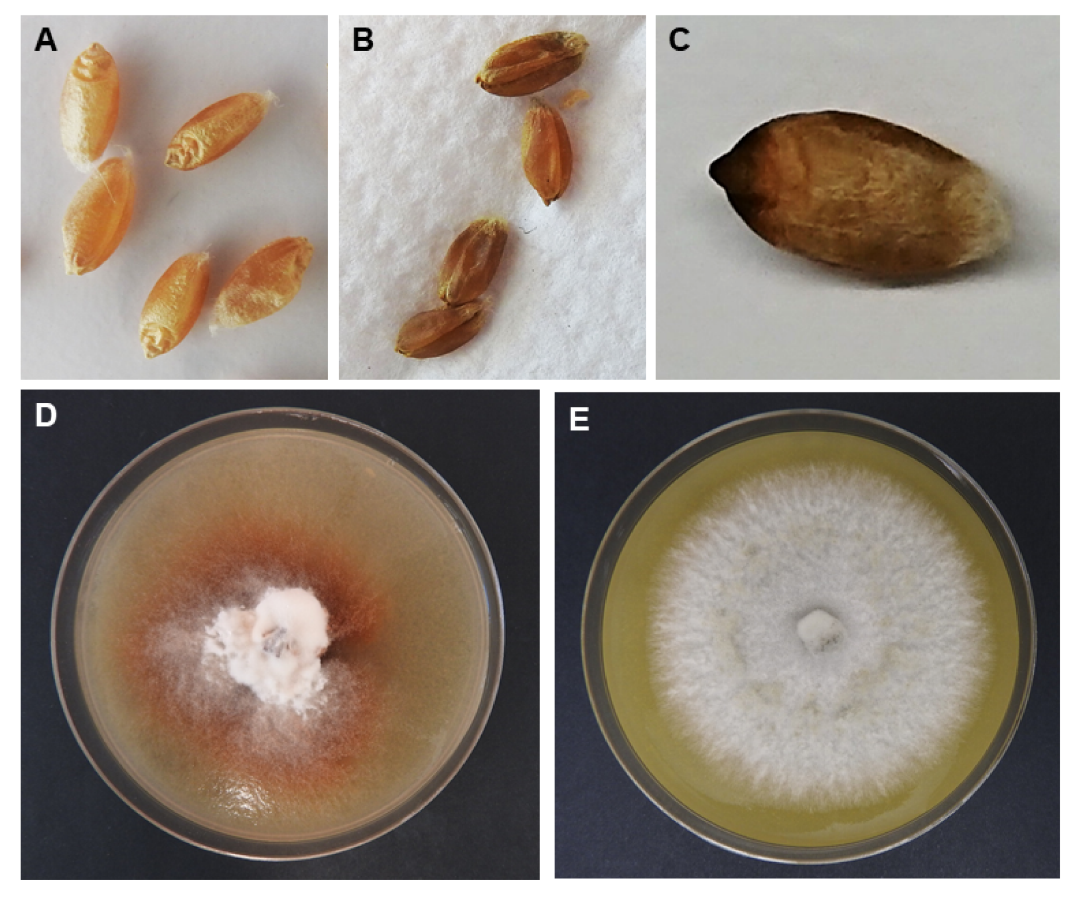
No evidence of red smudge was observed on the seeds harvested from the heads for any of the inoculations, including those that had been inoculated three times. However, the triple inoculated seeds displayed symptoms of shrivelling and darkening, reminiscent of black point and dark smudge (Figure 3B,C).

3.2. Re-Isolation of Ptr Colonies from Inoculated Wheat Heads

Of the 180 samples of inoculated heads across the three growth stages (60 samples per stage), Ptr colonies were recovered from 34 (18.9%), of which 11 were obtained at the heading stage, 6 from the flowering stage and 17 from the ripening stage. Of the 34 re-isolated Ptr colonies, 28 (82.3%) of the Ptr colonies exhibited irregular morphology, with a tint of reddish mycelia that lacked filamentous morphology and an incapacitated sporulation capacity (Figure 3D). These type of Ptr colonies were obtained from both the husks and grains and were confirmed as Ptr using PCR-based detection with Ptr species-specific primers (Figure S1).

3.3. PCR Detection of Ptr from Inoculated Wheat Heads

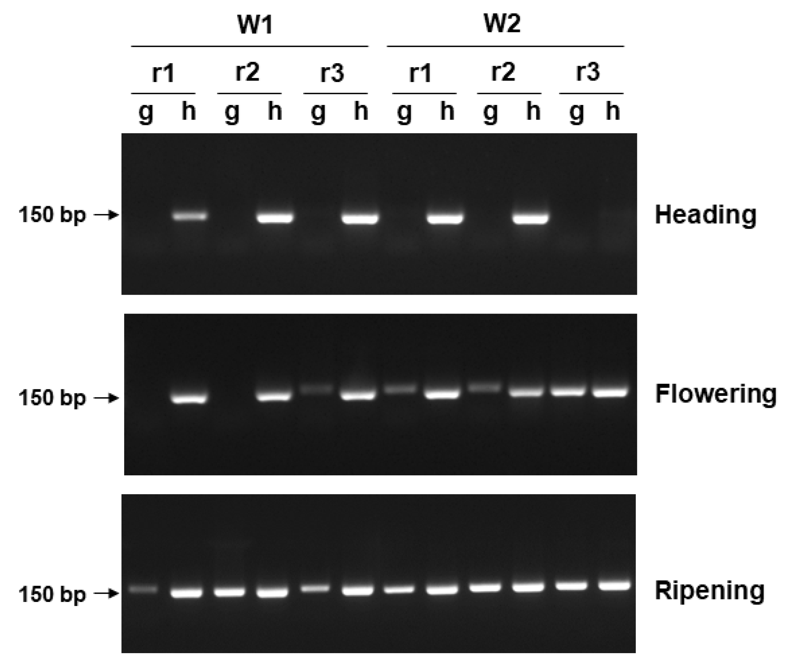
Due to the low recovery of Ptr colonies using conventional culturing methods on water agar, PCR assays were conducted to detect the presence of Ptr directly from inoculated wheat heads. Ptr was detected on the head samples as shown by the presence of a Ptr species-specific PCR band that was amplified from the DNA samples collected across the three growth stages (Figure 4). Ptr fungal DNA was predominantly detected in the husk in comparison to the grain. Ptr was only detected in the husk for samples inoculated at the heading stage. For samples that were inoculated at the flowering stage, Ptr was consistently present in the husk samples, while less intense PCR bands or absence of amplification were observed for the grain samples. When inoculated at the ripening stage, Ptr DNA was detected across all the biological replicates and both post-inoculation time points (six and nine weeks) in both grain and husk.

3.4. Systemic Transmission of Tan Spot Disease via Seeds

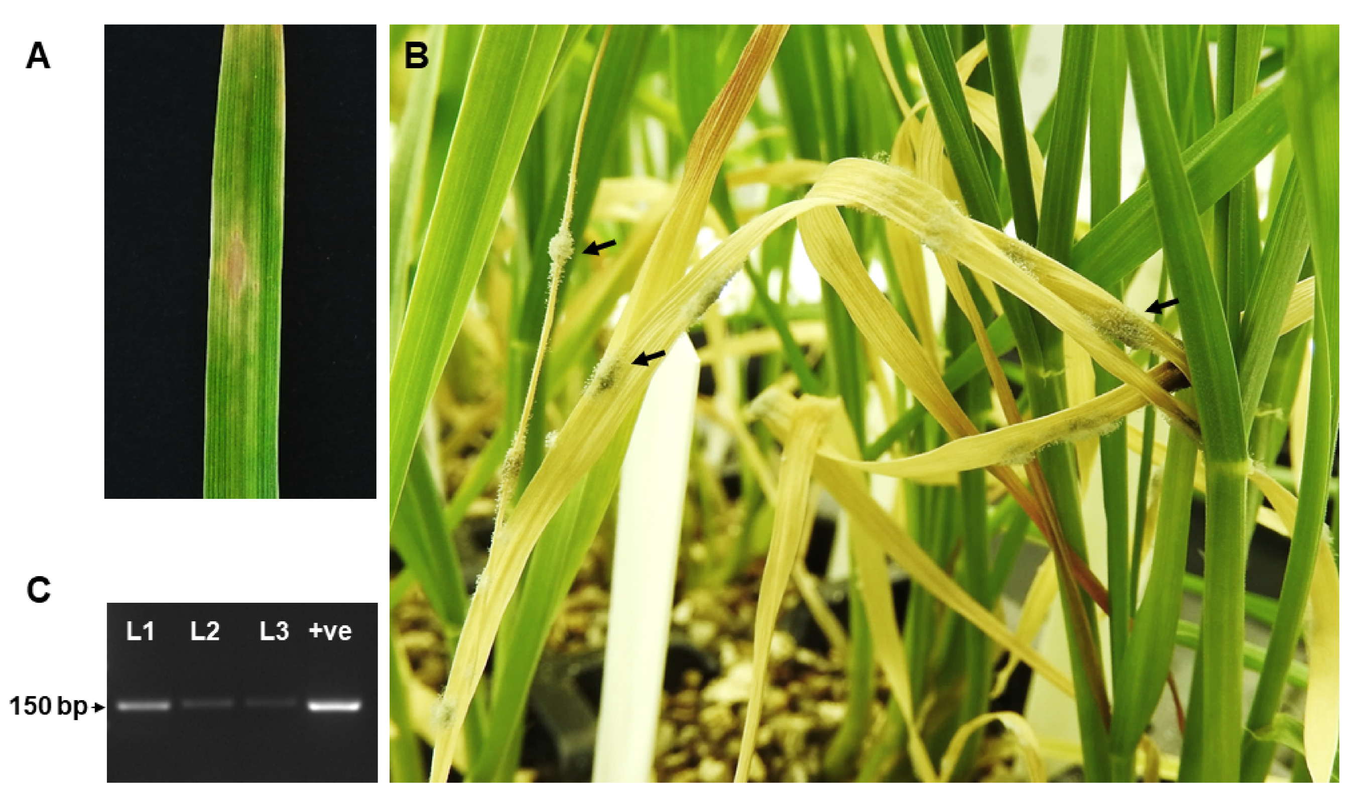
To investigate if tan spot disease can be transmitted from seeds, seeds collected from the triple inoculated wheat heads were germinated and monitored for the presence of foliar tan spot symptoms. No delay of seed germination was recorded. Out of the 60 plantlets, only 1 seedling developed a tan spot lesion (Figure 5A). However, fungal mycelia were observed on the senescing leaves (Figure 5B) and confirmed as Ptr via PCR (Figure 5C).

4. Discussion

Symptoms of Ptr infection on wheat heads were distinct from the typical foliar symptoms of tan spot, with brown spots present on the spikelets. Although a susceptible wheat variety Yitpi was inoculated, no red smudge symptoms were observed on the grain. Since the wheat heads were artificially inoculated, the inoculum level could not have been the limiting factor to red smudge development, but rather other factors such as the environment, genotype or wheat type may be the cause. Prolonged high humidity and temperature have been found to have an effect on grain discolouration [6], therefore, it is probable that the conditions in the glasshouse of this study did not favour red smudge development. The incidence of red smudge may also be related to the grain type, since pigmented phenolic compounds such as catechins, proanthocyanidins and anthocyanins accumulate in the grain coat [21,22] resulting in coloured grain. White grain wheats are only produced in Australia; however, the evaluation of other varieties, including the red grain wheats produced in America and Europe, would be of interest for further work.
Intriguingly, some Ptr colonies re-isolated from wheat heads had a reddish appearance which does not conform to the standard morphology of the pathogen when cultured in vitro. Although the recovery of Ptr from infected or discoloured glumes and kernel has previously been reported [6,8,23], no study has reported an irregular Ptr colony morphology isolated from wheat heads. While the case for such changes in morphology is unknown, it is possible that the reddish pigmentation observed in the colonies may be responsible for the red smudge symptoms, if in high abundance under the right conditions. Ptr is known to produce several mycotoxins from the anthraquinone class such as emodin, islandicin and catenarin [24,25,26], and both catenarin and emodin have been detected in Ptr-infected grain. Catenarin has been hypothesized to cause red smudge due to the red pigmentation property of the compound [27]. Thus, an understanding of the biosynthesis of these natural pigmented compounds in Ptr under in vitro and in vivo conditions would shed some light on the development of grain discolouration. Furthermore, some fungal pathogens are associated with the capability to produce mycotoxins in infected discoloured grain. For example, in black point disease, Alternaria species is associated with the production of alternariol (AOH), alternariol-monomethyl ether (AME), tenuazonic acid (TA) and altenuene (ALT) in grains [28]. The ability of Ptr to produce toxic secondary metabolite compounds in the kernel remains to be explored.
While a significant body of work has focused on plant host resistance to foliar diseases, the inheritance of resistance to grain disease is a less explored topic. Several wheat genetic studies have been published with the aim to improve the resistance of breeding lines to black point [29,30,31,32] and wheat spike diseases [33,34,35,36,37,38]. However, the complexity of studying grain infection is often exacerbated with the influence of foliar disease on the host. This is particularly the case with fungal pathogens that have the ability to infect both leaves and wheat heads, such as Fusarium species. The observation that Ptr infection of wheat heads can result in grain shrivelling and discolouration on a wheat variety that is susceptible to tan spot, poses the question as to the incidence of grain symptoms on more resistant varieties. In this study, Ptr was detected on the husk but not on the developing grains at the earlier growth stage, while presence of Ptr on the grains only occurred at the later growth stages. This demonstrates that grain infection occurred via direct contact of inoculum when grains were no longer shielded within the glume, lemma and palea. This observation is supported by an earlier study [39] which also showed that the thick-walled epidermides of these structures was sufficient to inhibit further hyphal penetration. Restricted growth of Ptr mycelia between the different tissues and absence of mass proliferation of saprophytic growth on wheat seeds has previously been reported [3] with Ptr shown to produce appressoria on the pericarp layers of wheat seeds; however, no evidence of mycelia growth was found in the seed coat or embryo. A study of the infection of Pyrenophora semeniperda (a pathogen that infects seed and causes leaf spot on grasses) on wheat florets, showed that hyphae grew saprophytically on the surface of anther, stigma and stylar tissues [40]. On the developing seed, hyphae were generally confined to the epidermal layers and were not observed in the endosperm [40]. Altogether, these results suggest that seed infection by Pyrenophora species may be limited to the point of inoculation contact or confined to the outer layer of the seeds, during a phase of a saprophytic growth.
A single tan spot lesion on a seedling grown from Ptr infected seed was observed in this study, thus the incidence of seed transmission was very low. However, what appears to be more detrimental is the presence of Ptr hyphae on the seeds that progressed to saprophytic growth of mycelia on senescing leaves. Such a mass of fungal mycelia could act as a source of inoculum that may lead to the proliferation of fungal load under the right environmental conditions. Therefore, seed screening for the presence of Ptr would be advisable to reduce the inoculum load as well as the biosecurity risk of an incursion by an exotic Ptr race from imported wheat seeds. Molecular detection of the fungal pathogen in grain samples using PCR-based assays would be able to diagnose and potentially circumvent the transmission of pathogen propagules via seed.

Supplementary Materials

The following are available online at https://www.mdpi.com/2077-0472/10/9/417/s1, Figure S1: PCR amplification using Ptr specific primers (PtrUnique_F2/R2) confirming the fungal colonies with atypical morphology as Ptr.

Author Contributions

Conceptualization, P.T.S. and C.S.M.; methodology, P.T.S.; investigation, N.S.; writing—original draft preparation, P.T.S.; writing—review and editing, C.S.M.; All authors have read and agreed to the published version of the manuscript.

Funding

This research was funded by Grains Research and Development Corporation (GRDC) and Curtin University (project code CUR00023).

Acknowledgments

The authors thank the National variety trials (NVT) for the provision of seeds.

Conflicts of Interest

The authors declare no conflict of interest. The funders had no role in the design of the study; in the collection, analyses, or interpretation of data; in the writing of the manuscript, or in the decision to publish the results.

References

  1. Fernandez, M.R.; Clarke, J.M.; Depauw, R.; Irvine, R.B.; Knox, R. Black point and red smudge in irrigated durum wheat in southern Saskatchewan in 1990–1992. Can. J. Plan Pathol. 1994, 16, 221–227. [Google Scholar] [CrossRef]
  2. Fernadez, M.R.; Clarke, J.M.; de Pauw, R.M.; Lefkovitch, L.P. Emergence and growth of durum wheat derived from red smudge-infected seed. Crop Sci. 1997, 37, 510–514. [Google Scholar] [CrossRef]
  3. Schilder, A.M.C.; Bergstrom, G.C. Pyrenophora-tritici-repentis as a component of the fungal flora of winter-wheat seed in New-York. Seed Sci. Technol. 1994, 22, 285–297. [Google Scholar]
  4. Carmona, M.; Ferrazini, M.; Barreto, D.E. Tan spot of wheat caused by Drechslera tritici-repentis: Detection, transmission, and control in wheat seed. Cereal Res. Commun. 2006, 34, 1043–1049. [Google Scholar] [CrossRef]
  5. Fernandez, M.R.; de Pauw, R.M.; Clarke, J.M.; Fox, S.L. Discoloration of wheat kernels by Pyrenophora tritici-repentis. Can. J. Plant Pathol. Rev. Can. Phytopathol. 1998, 20, 380–383. [Google Scholar] [CrossRef]
  6. Fernandez, M.R.; Clarke, J.M.; de Pauw, R.M. Effect of environmental variables on the development of kernel discoloration by Pyrenophora tritici-repentis in durum wheat. Can. J. Plant Pathol. Rev. Can. Phytopathol. 1998, 20, 104–110. [Google Scholar] [CrossRef]
  7. Rees, R.G.; Martin, D.J.; Law, D.P. Black point in bread wheat: Effects on quality and germination, and fungal associations. Aust. J. Exp. Agric. 1984, 24, 601–605. [Google Scholar] [CrossRef]
  8. Fernandez, W.; Singh, A. Impact of seed discolouration on emergence and early plant growth of durum wheat at different soil gravimetric water contents. Can. J. Plant Pathol. 2014, 36, 509–516. [Google Scholar] [CrossRef]
  9. Fernandez, M.R.; de Pauw, R.M.; Clarke, J.M. Reaction of common and durum wheat cultivars to infection of kernels by Pyrenophora tritici-repentis. Can. J. Plant Pathol. 2001, 23, 158–162. [Google Scholar] [CrossRef]
  10. Grain Trade Australia: Wheat Standards 2020/2021—Statement of Standards. Available online: www.graintrade.org.au (accessed on 24 August 2020).
  11. Fox, S.L.; Fernandez, M.R.; de Pauw, R.M. Red smudge infection modifies sprouting response in four wheat lines. Can. J. Plant Sci. 2003, 83, 163–169. [Google Scholar] [CrossRef]
  12. Fernandez, M.R.; de Pauw, R.M.; Clarke, J.M.; Lefkovitch, L.P. Red smudge in durum wheat reduces seedling vigour. Can. J. Plant Sci. 1996, 76, 321–324. [Google Scholar] [CrossRef]
  13. Schilder, A.M.C.; Bergstrom, G.C. Seed transmission of Pyrenophora tritici-repentis, causal fungus of tan spot of wheat. Eur. J. Plant Pathol. 1995, 101, 81–91. [Google Scholar] [CrossRef]
  14. Wilson, J.M. Analysis of Black Point in Wheat; Technical Bulletin 88; Department of Agriculture and Food: Perth, Australia, 1993. [Google Scholar]
  15. Klein, T.A. Fungi associated with discoloured wheat grain in northern New South Wales. Australas. Plant Pathol. 1987, 16, 69–71. [Google Scholar] [CrossRef]
  16. Shackley, B.; Paynter, B.; Troup, G.; Bucat, J.; Seymour, M.; Blake, A. 2020 Western Australia Crop Sowing Guide; Grains Research and Development Corporation: Perth, Australia, 2020. [Google Scholar]
  17. Moffat, C.S.; See, P.T.; Oliver, R.P. Generation of a ToxA knockout strain of the wheat tan spot pathogen Pyrenophora tritici-repentis. Mol. Plant Pathol. 2014, 15, 918–926. [Google Scholar] [CrossRef]
  18. Moffat, C.S.; See, P.T.; Oliver, R.P. Leaf yellowing of the wheat cultivar mace in the absence of yellow spot disease. Australas. Plant Pathol. 2015, 44, 161–166. [Google Scholar] [CrossRef]
  19. Moolhuijzen, P.; See, P.T.; Hane, J.K.; Shi, G.; Liu, Z.; Oliver, R.P.; Moffat, C.S. Comparative genomics of the wheat fungal pathogen Pyrenophora tritici-repentis reveals chromosomal variations and genome plasticity. BMC Genom. 2018, 19, 279. [Google Scholar] [CrossRef]
  20. See, P.T.; Moffat, C.S.; Morina, J.; Oliver, R.P. Evaluation of a multilocus indel DNA region for the detection of the wheat tan spot pathogen Pyrenophora tritici-repentis. Plant Dis. 2016, 100, 2215–2225. [Google Scholar] [CrossRef]
  21. Lachman, J.; Martinek, P.; Kotíková, Z.; Orsák, M.; Šulc, M. Genetics and chemistry of pigments in wheat grain—A review. J. Cereal Sci. 2017, 74, 145–154. [Google Scholar] [CrossRef]
  22. Böhmdorfer, S.; Oberlerchner, J.T.; Fuchs, C.; Rosenau, T.; Grausgruber, H. Profiling and quantification of grain anthocyanins in purple pericarp × blue aleurone wheat crosses by high-performance thin-layer chromatography and densitometry. Plant Methods 2018, 14, 29. [Google Scholar] [CrossRef] [Green Version]
  23. Fernandez, M.R.; Pearse, P.G.; Holzgang, G.; Basnyat, P.; Zentner, R.P. Impacts of agronomic practices on the leaf spotting complex of common wheat in eastern Saskatchewan. Can. J. Plant Sci. 2009, 89, 717–730. [Google Scholar] [CrossRef]
  24. Bouras, N.; Kim, Y.M.; Strelkov, S.E. Influence of water activity and temperature on growth and mycotoxin production by isolates of Pyrenophora tritici-repentis from wheat. Int. J. Food Microbiol. 2009, 131, 251–255. [Google Scholar] [CrossRef] [PubMed]
  25. Bouras, N.; Strelkov, S.E. Influence of carbon source on growth and mycotoxin production by isolates of Pyrenophora tritici-repentis from wheat. Can. J. Microbiol. 2010, 56, 874–882. [Google Scholar] [CrossRef] [PubMed]
  26. Bouras, N.; Holtz, M.D.; Aboukhaddour, R.; Strelkov, S.E. Influence of nitrogen sources on growth and mycotoxin production by isolates of Pyrenophora tritici-repentis from wheat. Crop J. 2016, 4, 119–128. [Google Scholar] [CrossRef] [Green Version]
  27. Bouras, N.; Strelkov, S.E. The anthraquinone catenarin is phytotoxic and produced in leaves and kernels of wheat infected by Pyrenophora tritici-repentis. Physiol. Mol. Plant Pathol. 2008, 72, 87–95. [Google Scholar] [CrossRef]
  28. Masiello, M.; Somma, S.; Susca, A.; Ghionna, V.; Logrieco, A.F.; Franzoni, M.; Ravaglia, S.; Meca, G.; Moretti, A. Molecular identification and mycotoxin production by alternaria species occurring on durum wheat, showing black point symptoms. Toxins 2020, 12, 275. [Google Scholar] [CrossRef] [Green Version]
  29. Lehmensiek, A.; Campbell, A.W.; Sutherland, M.W.; Williamson, P.M.; Michalowitz, M.; Daggard, G.E. QTLs for black-point resistance in wheat and the identification of potential markers for use in breeding programmes. Plant Breed. 2004, 123, 410–416. [Google Scholar] [CrossRef]
  30. Li, Q.; Niu, H.; Xu, K.; Xu, Q.; Wang, S.; Liang, X.; Jiang, Y.; Niu, J. GWAS for resistance against black point caused by Bipolaris sorokiniana in wheat. J. Cereal Sci. 2020, 91, 102859. [Google Scholar] [CrossRef]
  31. Liu, J.; He, Z.; Wu, L.; Bai, B.; Wen, W.; Xie, C.; Xia, X. Genome-wide linkage mapping of QTL for black point reaction in bread wheat (Triticum aestivum L.). Theor. Appl. Genet. 2016, 129, 2179–2190. [Google Scholar] [CrossRef]
  32. Liu, J.; He, Z.; Rasheed, A.; Wen, W.; Yan, J.; Zhang, P.; Wan, Y.; Zhang, Y.; Xie, C.; Xia, X. Genome-wide association mapping of black point reaction in common wheat (Triticum aestivum L.). BMC Plant Biol. 2017, 17, 220. [Google Scholar] [CrossRef] [Green Version]
  33. Cai, J.; Wang, S.; Li, T.; Zhang, G.; Bai, G. Multiple minor QTLs are responsible for fusarium head blight resistance in chinese wheat landrace haiyanzhong. PLoS ONE 2016, 11, e0163292. [Google Scholar] [CrossRef] [Green Version]
  34. Czembor, P.C.; Arseniuk, E.; Radecka-Janusik, M.; Piechota, U.; Słowacki, P. Quantitative trait loci analysis of adult plant resistance to Parastagonospora nodorum blotch in winter wheat cv. Liwilla (Triticum aestivum L.). Eur. J. Plant Pathol. 2019, 155, 1001–1016. [Google Scholar] [CrossRef] [Green Version]
  35. Shatalina, M.; Messmer, M.; Feuillet, C.; Mascher, F.; Paux, E.; Choulet, F.; Wicker, T.; Keller, B. High-resolution analysis of a QTL for resistance to Stagonospora nodorum glume blotch in wheat reveals presence of two distinct resistance loci in the target interval. Theor. Appl. Genet. 2014, 127, 573–586. [Google Scholar] [CrossRef] [PubMed] [Green Version]
  36. Yi, X.; Cheng, J.; Jiang, Z.; Hu, W.; Bie, T.; Gao, D.; Li, D.; Wu, R.; Li, Y.; Chen, S.; et al. Genetic analysis of fusarium head blight resistance in CIMMYT bread wheat line C615 using traditional and conditional QTL mapping. Front. Plant Sci. 2018, 9, 573. [Google Scholar] [CrossRef] [PubMed]
  37. Zhu, Z.; Chen, L.; Zhang, W.; Yang, L.; Zhu, W.; Li, J.; Liu, Y.; Tong, H.; Fu, L.; Liu, J.; et al. Genome-wide association analysis of fusarium head blight resistance in Chinese elite wheat lines. Front. Plant Sci. 2020, 11, 206. [Google Scholar] [CrossRef] [PubMed] [Green Version]
  38. Zhuang, Y.; Gala, A.; Yen, Y. Identification of functional genic components of major fusarium head blight resistance quantitative trait loci in wheat cultivar Sumai 3. Mol. Plant Microbe Interact. 2013, 26, 442–450. [Google Scholar] [CrossRef] [Green Version]
  39. Schilder, A.M.C.; Bergstrom, G.C. Infection of wheat seed by Pyrenophora tritici-repentis. Can. J. Bot. 1994, 72, 510–519. [Google Scholar] [CrossRef]
  40. Campbell, M.A.; Medd, R.W.; Brown, J.B. Optimizing conditions for growth and sporulation of Pyrenophora semeniperda. Plant Pathol. 2003, 52, 448–454. [Google Scholar] [CrossRef]
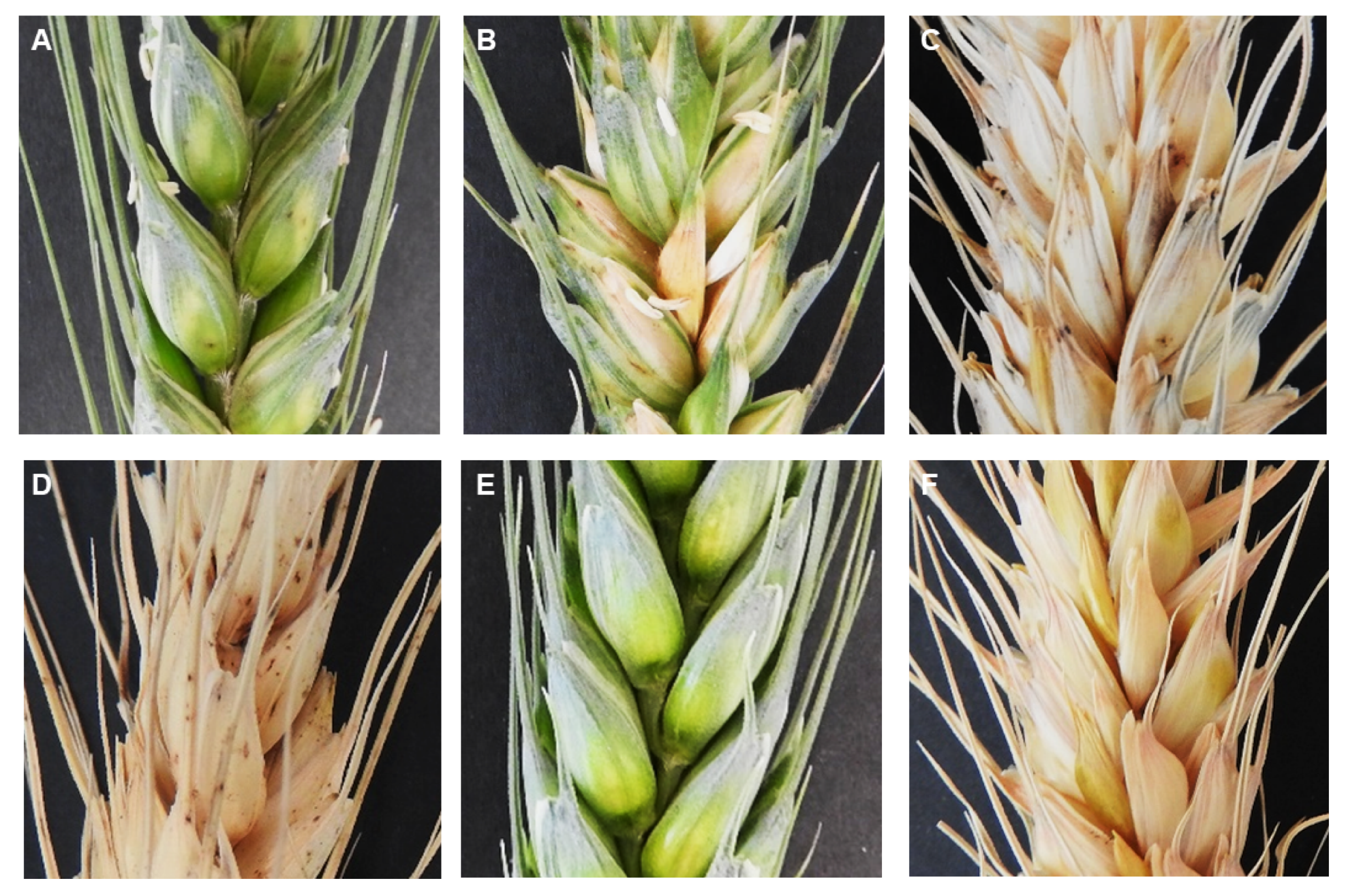
Figure 1. Representative images of wheat heads infected with Ptr upon single inoculation at: (A) Heading; (B) Flowering; (C) Ripening; (D) Multiple inoculations at heading, flowering and ripening; (E,F) Wheat heads sprayed with gelatin only as control.
Agriculture 10 00417 g001
Figure 2. Representative images of diseased wheat spikelets as indicated by the white arrows: (A) Lesions on glumes and awns; (B) Mycelia on lemma; (C) Mycelia on glume; (D) Conidiophores on glume. Scale bar, 1 mm.
Agriculture 10 00417 g002
Figure 3. Representative images of ripened seeds from Ptr infected wheat heads: (A) Seeds with no visual symptoms; (B) Shrivelled and discoloured seed; (C) Black tip symptoms. Images of Ptr colonies recovered from inoculated wheat heads: (D) Irregular colony morphology lacking aerial hyphae; (E) Colony with typical filamentous morphology.
Agriculture 10 00417 g003
Figure 4. Agarose gel analysis of Ptr species-specific PCR amplification for the detection of the pathogen on developing grain (g) and husk (h) inoculated at the heading, flowering and ripening stage. Samples were obtained from wheat heads collected at 6 (W1) and 9 (W2) weeks post inoculation. DNA samples are of three biological replicates (r1, r2 and r3) extracted from independent grain and husk samples. PCR was performed using the PtrMulti_F/R primer set.
Agriculture 10 00417 g004
Figure 5. Wheat seedlings germinated from Ptr infected seeds: (A) Tan spot disease symptom on the leaf; (B) Saprophytic growth of fungus on the senescing leaves; (C) Agarose gel electrophoresis of Ptr species-specific bands amplified from DNA extracted from three senescing leaves (L1, L2 and L3; three biological replicates of independent senescent leaves). +ve; positive control of PCR amplification using M4 DNA and PtrMulti_F/R primers.
Agriculture 10 00417 g005