Next Article in Journal
Modeling the Dynamic Response of Plant Growth to Root Zone Temperature in Hydroponic Chili Pepper Plant Using Neural Networks
Previous Article in Journal
Will Yellow Mealworm Become a Source of Safe Proteins for Europe?
 
 
Erratum published on 14 October 2020, see Agriculture 2020, 10(10), 474.
Review

Microflora in the Reproductive Tract of Cattle: A Review

1
Joint Laboratory of Modern Agricultural Technology International Cooperation, Ministry of Education, Jilin Agricultural University, Changchun 130118, China
2
Key Laboratory of Animal Production, Product Quality, and Security, Ministry of Education, Jilin Agricultural University, Changchun 130118, China
3
College of Animal Science and Technology, Jilin Agricultural University, 2888 Xincheng Street, Changchun 130118, China
*
Authors to whom correspondence should be addressed.
Agriculture 2020, 10(6), 232; https://doi.org/10.3390/agriculture10060232
Submission received: 5 May 2020 / Revised: 7 June 2020 / Accepted: 9 June 2020 / Published: 17 June 2020
(This article belongs to the Section Farm Animal Production)

Abstract

:
There are microbial communities in and on the bodies of all multicellular organisms, and this microbiota can have a significant impact on the biology of the host. Most studies have focused on the microbiome of the skin, mouth, and gut, whereas relatively little is known about the reproductive microbiome. From the perspective of the bovine reproductive tract, uterine diseases such as metritis and endometritis are traditionally viewed to result only from interactions occurring between the host animal and pathogens originating from either the environment or ascension from the vagina. This outdated opinion has been refuted by recent advanced studies that propose that, in addition to bacteria colonization through the extrinsic and ascending pathways to the vagina, bacteria can also move from the gut to the uterus, which is also associated with reproductive tract disorders. This has led to the concept of the “endogenous route hypothesis”, which has vital inferences for comprehending the etiology of metritis and endometritis. Furthermore, it has opened up the possibility of developing new prophylactic and therapeutic agents as alternatives to antimicrobial agents. In addition, the unveiling of next-generation sequencing technology makes it more convenient to perform detailed sequencing and analysis of data on the cervical, vaginal, and uterine flora and to further study uncultured bacteria in these niches—most importantly, the cervical niche, which previously was thought to have lower bacterial complexity. Research conducted to date has proven that the composition of microflora in a community varies widely between environmental sites, host niches, and health status. Furthermore, it has also been suggested that the occurrence of endometritis in the dairy and beef cattle reproductive tract is neither casual nor indirect but multifactorial. Whether disturbance in the variety of the microflora in the reproductive tract (dysbiosis) has a role in determining the sensitivity to metritis and endometritis is not yet known. This article outlines the current progress in understanding the microflora with regards to the bovine reproductive tract. The compositions of microflora in various niches of the reproductive tract are briefly elucidated. In addition, the functional role of these microflora communities in the reproductive tract is discussed, with particular emphasis on the association of bacterial flora with reproductive disorders and failures. Finally, prophylaxis and therapeutic approaches based on the new comprehension of the effects of antimicrobials, probiotics, and bacteriophages on the composition of the reproductive tract microflora are also considered.

1. Introduction

Animals and plants live and thrive in a microbial-dominated world and contain these various microbial communities in and on the bodies. Recently, the study on host-associated microbial communities (i.e., microbiota and/or microbiomes) has undergone explosive variations, triggering a biological revolution. Although previous studies typically looked at microorganisms from a pathological point of view, it is now generally believed that the host-microorganism relationship covers continuity from harmful to useful. Owing to the influence of the microbiome on the health, physiology, development, and behavior of the host [1,2], the microbiome can be considered as a vital component of the host phenotype and potentially also of the host genome (e.g., the hologenome concept) [2].
Though the importance of the skin, oral and gut microbiome in host ecosystems, evolution, and adaptation have been emphasized, little information however exists regarding the reproductive tract microbiome. This is startling considering the long-term understanding of microorganisms in the reproductive systems of the male and female (e.g., [3], and DNA sequencing studies conducted currently; suggesting the presence of dynamic microbial communities in reproductive tissues [4], particularly the vagina [5]. Therefore, the reproductive microbiome epitomizes a considerable task in the study of host-related microbial communities.
For many years, it was generally recognized that the female reproductive tract of mammals was a sterile organ [4,6]. Culture-based technologies have traditionally been used to identify the presence of microbes, but this technology has limitations since many microorganisms are difficult to culture in vitro. With the start of the Human Microbiome Project (HMP) in 2007, the use of sequencing technologies to identify the microbiome in healthy people has increasingly gained attention. Data from this project have depicted that body sites that were historically believed to be sterile are now colonized with their unique microbiome [7,8]. There is an increasing amount of information available that supports the idea that the human reproductive tract, as well as veterinary species, harbors their microbiome, which is distinct.
Furthermore, modern culture-independent molecular methods focused on sequencing have also widened our present understanding of the microbiome of the uterus in cattle with metritis, pyometra, and endometritis [9,10]. There is a growing interest in the microflora of the reproductive tract, and its relationship to disease and health is a new and rapidly evolving field of study. Even though the microbiome can have significant influences on host biology, relatively little is known about the microbial communities in the reproductive tract of the dairy and beef cattle. Hence, this current review seeks to summarize the current progress in understanding the reproductive tract microflora—its possible origins and functional roles as well as the prophylaxis and therapeutic approaches modulating this flora.

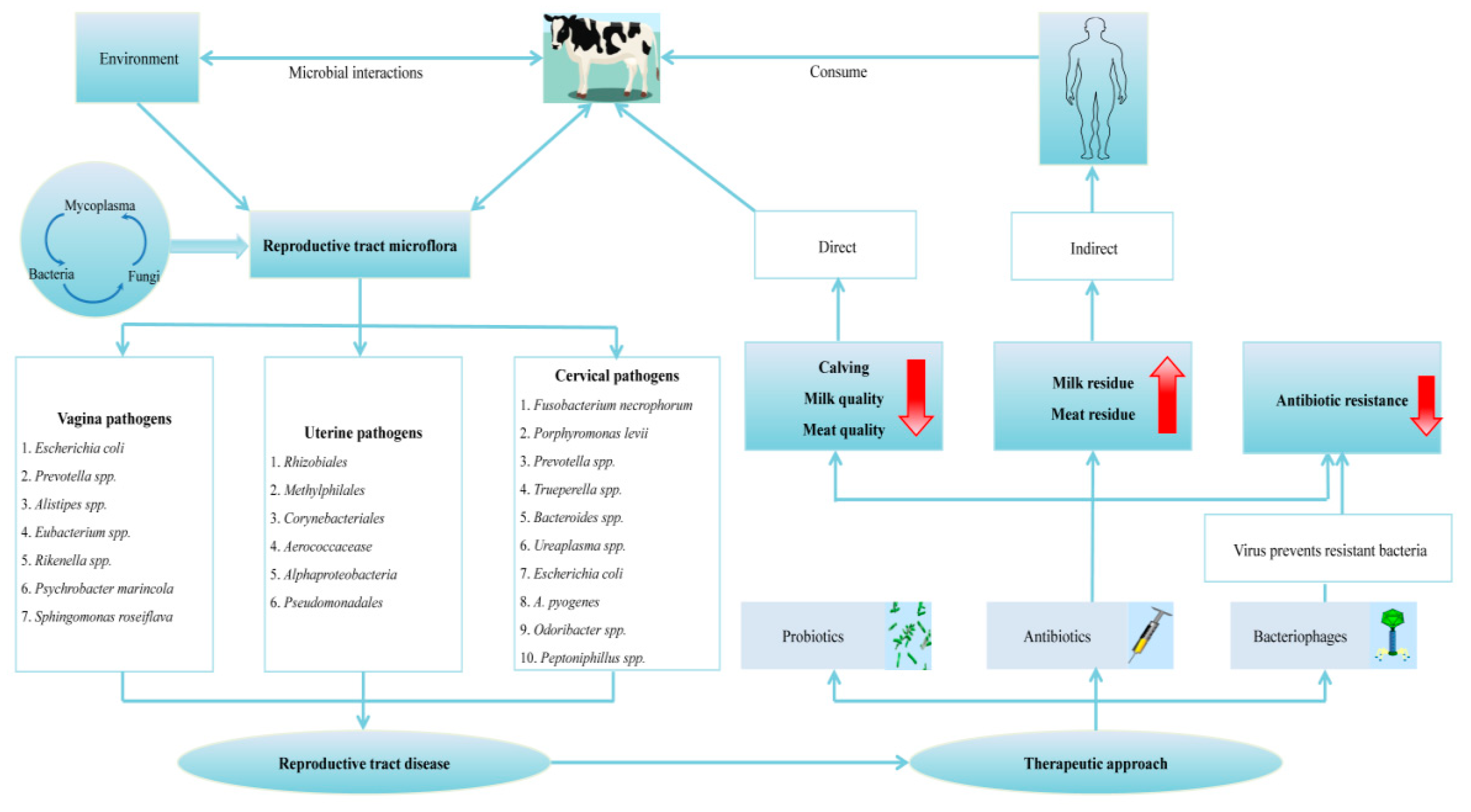
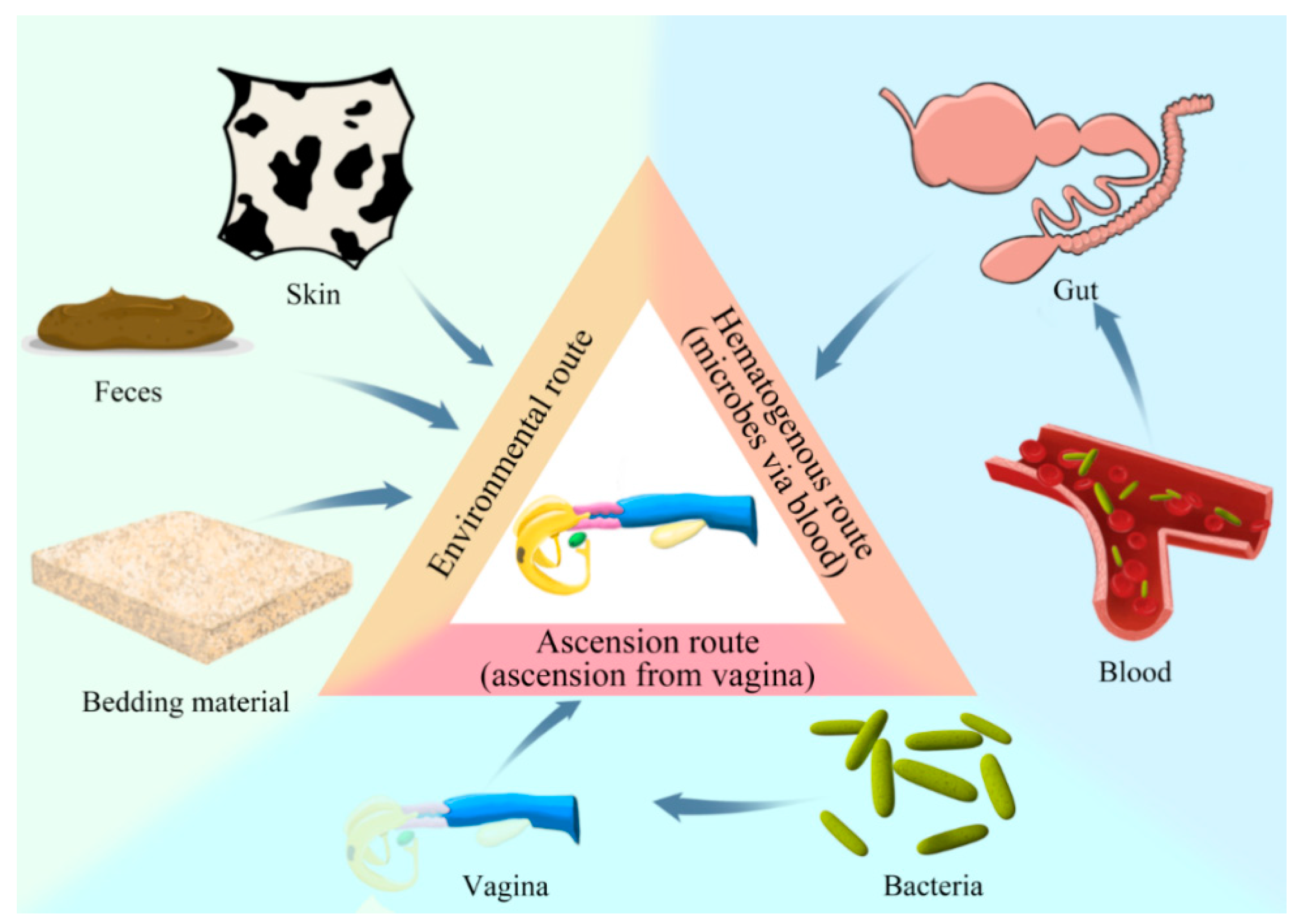
2. Prospective Origin(s) of the Reproductive Tract Microflora

From an ecological point of view, the ecological niches in the host microflora are not separate environments but are a network of interconnected communities that are continually exchanging [11]. Therefore, microorganisms can enter the reproductive tract from other anatomical sites. At birth, the physical cervical barrier is compromised, allowing bacteria to be transported from the vagina or from the environment via the vagina, and also from the feces and animal skin to the genital tract [12,13]. For example, pathogens such as Porphyromonas, Fusobacterium, and Bacteroides [9,14] inhabit portions of the cattle rumen typical flora [15] and are excreted in feces. Thus, increased infection of the uterus because of the environment (Figure 1) will possibly add to the occurrence of metritis. The flora of the vagina is also known to contain the same uterine pathogens, as described previously [16]. Therefore, contamination of the uterus coming from the vagina is likewise plausible (Figure 1). Among the various bacteria that cause uterine infections, the most common microbes include Prevotella, Fusobacterium necrophorum, Escherichia coli, and Arcanobacterium pyogenes [17], which are also linked to the vagina as described previously. All of these bacteria also come from fecal contamination of the fur, environment, and bedding material (Figure 1).
Several studies also support the hypothesis that the presence of bacteria in the dairy and beef cattle reproductive tract is not the mere result of external colonization but also occurs through a hematogenous transmission pathway (Figure 1). Galvao and colleagues [18] recently established in their studies that the hematogenous route is a viable pathway for uterine infection with uterine pathogens. Most importantly, it was found that pathogens isolated from the uterus—namely Trueperella pyogenes and F. Necrophorum [19], as described previously—are responsible for abscess of the liver, while valvular endocarditis is caused by Helcococcus ovis [20]; hence, the blood route of the colonization of bacteria in the uterus cannot be disregarded. In some previous studies, the uterus had a microbiome established within 60 min of delivery [9].
This demonstrated that colonization occurred before or immediately after parturition either from or to the vagina via the bloodstream or both as a result of increased contamination. Similarly, in a study led by Jeon et al. [21], the authors reported that the blood had a unique microbiota and that the occurrence of these uterine pathogenic microorganisms in blood and feces is due to hematogenous route spread. It has also been discovered that there is a microbial invasion from the gut to the uterus through the blood (Figure 1), and this gut affects both the vagina and uterine microflora [22]. Bacteroides heparinolyticus is an example of such bacteria resident in the dairy cow’s feces and blood and it is assumed that the uterus is more likely to be inhabited by this bacterial species.
Furthermore, blood is a common component of lochia. For that reason, the blood of the maternal will naturally spread to the uterus after birth. In dairy cows, this is mainly due to degenerative vascular changes described by apoptosis and necrosis, and accompanied by the karyorrhexis of endothelial cells as well as the cells of the tunica media of small blood vessels. These variations were noticed within 24 h after delivery [23]. In the first 24 h after birth, the endometrium becomes very edematous [17], which may be the result of the above-mentioned blood vessel changes that cause the infiltration of blood components into the uterine cavity. In the dairy and beef cattle and like other reported species, there is a high degree of leukocyte migration into the uterus, where delivery is imminent and then enters the uterus after birth [24]. As a result, free-floating bacteria can quickly move into the endometrium after birth. A particularly important fact is that Coxiella was part of the blood network of the pathogens of the uterus, and this bacterium infects monocytes, grows, and reacts to the uterus [9]. Hence, the flow of leukocytes into the uterine lumen after delivery may contribute to a higher incidence of Coxiella.
However, the association between Coxiella and uterine pathogens means that uterine pathogens can also be transferred to the lumen of the uterus by monocytes. Though microbes in the gut have been found in blood leukocytes in middle-lactation cows, uterine pathogens have not been detected in all cows [25]; hence, shortly after calving, the presence of uterine pathogens in blood leukocytes should be studied. These different possible origins of bacteria entering the bovine reproductive tract either through the ascension path (ascend via the vagina), environmental route (i.e., via feces, bedding material, animal skin, etc.), or, finally, through the hematogenous route (via blood) of bacterial transmission from the gut to the uterus is depicted pictographically in Figure 1.

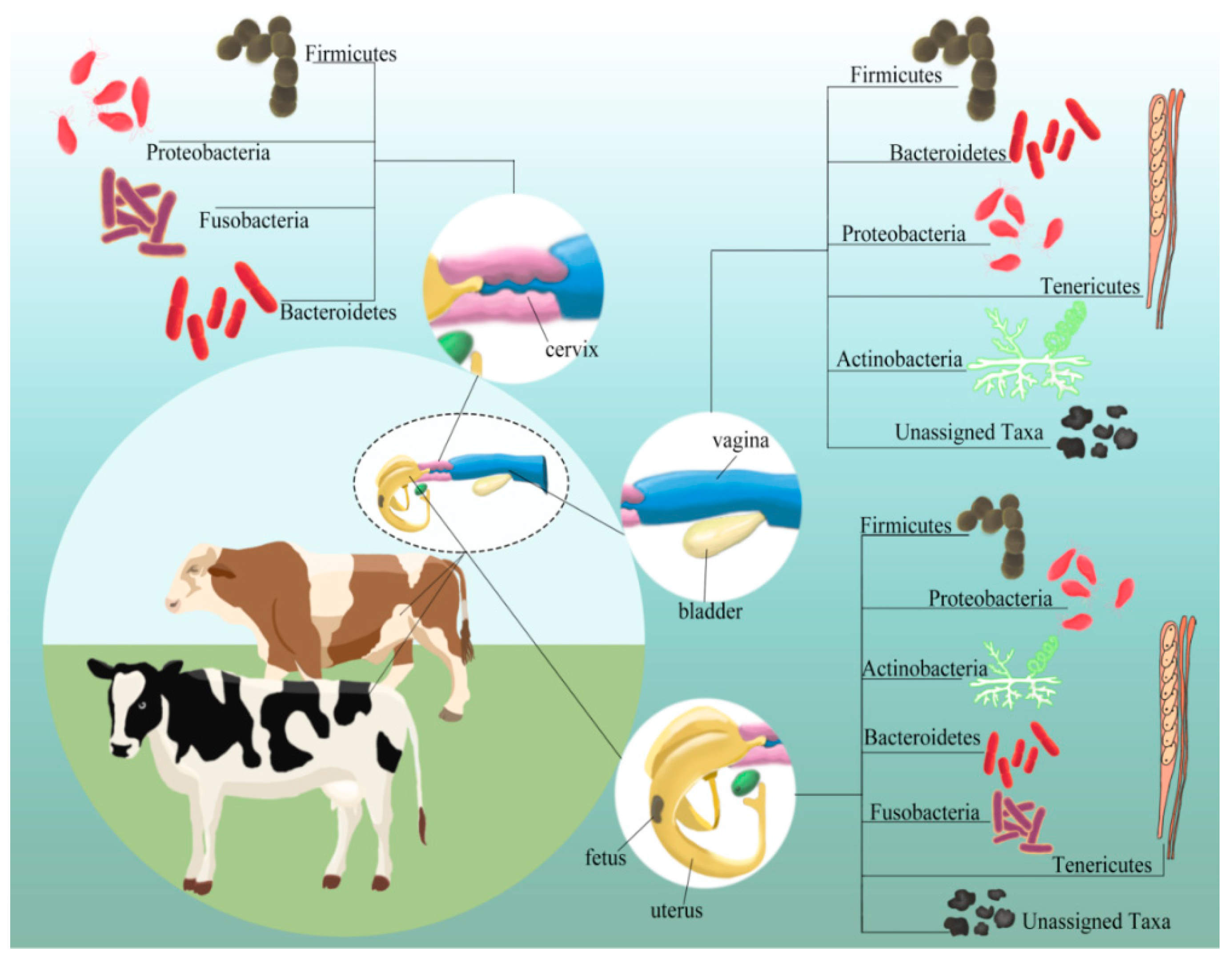
3. Microflora Composition(s) in Various Niches of the Reproductive Tract

In recent years, microbial research has shifted from an individual in vitro focus to a more ecological viewpoint by centering on how these organisms interact within and between species in definite niches called microbiomes [26]. The term microbiome refers to an array of microorganisms that live peacefully and co-exist in species, but disturbances of these natural populations can have a detrimental effect on health and normal physiological conditions [27]. The reproductive system of the bovine consists of a complex dynamic example of microbes residing in different niches of the reproductive tract of dairy and beef cattle and varying in communities or their populations (Figure 2).

3.1. Vaginal Microflora

The microflora in the vagina of cattle has been estimated based on a disease effect using a culture-dependent technique [28]. However, with the advent of advanced technologies such as metagenomics, similar groups of bacteria have been revealed in the Nellore, Holstein, and Fleckvieh cattle. Such bacteria at the phylum level specifically comprised Firmicutes, Bacteroidetes, and Proteobacteria [22,29] as depicted in Figure 2, and that constitute about 30–40% of the total bacteria present in the vaginal niche [30]. Recent studies by Giannattasio-Ferraz et al. [31] have also revealed a substantial similarity existing between the vaginal microbiota of Nellore cattle and Gyr (which is also another essential breed) and the main bacterial species described were mainly Aeribacillus, Bacillus, Clostridium, Bacteroides, and Ruminococcus. Mycosphaerella was the dominant fungus in this microbiota, while Archaea were poorly represented, and Methanobrevibacter were the most common. These data start to unravel the bovine vaginal microbiota, and a broad picture is being defined with the primary microbial agents from this tract, which permit studies of probiotics and prebiotics.
Besides these similar groups of microflora reported in different cattle breeds, pathogenic fungi have specifically also been reported recently as a colonizer of the cervical–vaginal fluids of Holstein dairy cattle, mainly yeast and penicillium [32]. However, the function of these opportunistic fungal pathogens, which play a role in the various theriogenological processes, requires further investigation. The microflora in the cattle vagina tract has also recently been revealed to play a significant role as biomarkers of reproductive success and failure [33]. Primarily, in an investigation conducted on beef heifer samples, the presence of Histophilus, Clostridiaceae, and Campylobacter in the vagina, as well as Bacteroidales and Dorea in the feces, was recently revealed using Random Forest as important indicators for pregnancy [33]. However, more experimentation is needed to validate these biomarkers and to study their role in a female’s ability to regulate pregnancy. Additionally, a shift in the diversity and phylogenetic relationship of these diverse microbial communities up to the time of breeding can also lead to a good pregnancy in cattle [34,35]. Therefore, the description and the importance of the diverse vaginal microbiota in cattle are crucial for a better comprehension of its physiology and its effects on the maintenance of health.
As mentioned above, although the vaginal microbiome makes a significant contribution to aspects of reproduction with a large microbial diversity, there is still very limited knowledge about the composition of this microbiome during different phases of the estrus cycle. The influence of the vaginal microbiota during the various phases of the buffalo (a genus of bovine) estrus cycle, for example, was recently reviewed by Mahalingam and colleagues [36]. As representative 16S rRNA gene sequences from this prominent operational taxonomic unit (OTUs) were integrated into a phylogenetic tree alongside those of neighboring bacterial strains, it became clear that the vaginal fluid of buffaloes was predominantly colonized by bacteria that are most closely related to Corynebacterium, Porphyromonas, Helcococcus, Anaerococcus, and Fastidiosipila. The Firmicutes phylum was particularly significant during the estrus phase. This means that the identification of bacterial communities during the different phases of the estrus cycle can lead to the development of new approaches such as treatment with probiotics and the introduction of microbial strains, which would have an affirmative effect on reproductive efficiency.
Research has also recounted recently that the bovine reproductive tract microflora populations differ on average between the luteal and follicular stages [37]. For instance, in the comparison of follicular and luteal phases, the vaginal microflora of beef cattle was dominated by E. coli, Aerococcus vaginalis, Aerococcus viridans, Haemophilus Somnus, Streptococcus pluranimalium, Sphingomonas roseiflava, Psychrobacter marincola, and Lactobacillus spp. as revealed in our laboratory using PCR-DGGE [37]. These bacterial populations, however, differed from that in the dairy cattle, and this mismatch in the microbial composition may be due to geographical location, the methods used, and breed effects. Noticeably, the vaginal Streptococcus spp. in the luteal phase was significantly larger than that in the follicular phase, while the number of vaginal Lactobacillus spp. in the follicular phase was considerably higher than in the luteal phase. However, to better comprehend the differences between the vaginal microflora between the follicular and luteal phases, it is recommended that future studies should use Metagenomic Shotgun Sequencing (MSS) analysis, as this enables a detailed analysis of the bacteria within the genome community [38,39].

3.2. Cervical Microflora

The cervix, composed of a series of mucous-lined collagenous rings, is one of the significant anatomical barriers protecting the uterus from pathogens in the environment [13,40]. The cervical mucus functions as a biological and physical barrier against germs that may otherwise ascend the reproductive tract from the lower genital tract [13]. Conversely, because of its anatomical position, the cervix is also exposed to inflammation generated by luminal debris and bacteria expelled by uterine contractions [41]. Although the cervix has long been viewed as a physical barrier protecting the uterus, the cervix must be assessed individually during the postpartum period to predict subsequent reproductive success [42]. Currently accepted ‘’uterine health’’ includes not only inflammation in uterine compartments but also inflammation of the cervix as well. These mechanisms linking endocervical inflammation with delay to conception may be of interest for therapy and should, therefore, be explored in future studies.
Previous studies have agreed and confirmed that the bacterial population’s diversity observed in the dairy cow’s cervix would even be more sophisticated than that reported by traditional culture-dependent methods used in earlier research work [43]. Furthermore, with the emergence of next-generation sequencing, it has become easier to explore non-cultured bacteria in this niche by performing thorough sequencing and data analysis of the cervical flora. For example, with the help of this advanced technique, the bacterial pathogens present in the cervix are revealed to be more common than their existence in vaginal swabs. Notably, in cows that have aborted, the frequency of S. aureus isolated from their cervical swabs was higher compared to that in those animals without any record of abortion [44]. These differences in the prevalence of such bacteria in the cattle cervix may be attributed to geographical region and the difference in the breeds of cattle used in that study.
Furthermore, with the advent of these advanced techniques as aforementioned, the various phases in the dairy cow’s cervix were also investigated by Wang and coworkers [45]. The authors found that the communities of bacteria in the cervix in the Clinical formative (CF), Clinical gestation (CG), and Clinical postpartum (CP) phases of cows consisted mainly of Proteobacteria, Bacteroidetes, and Firmicutes as previously described in the vagina flora. On the other hand, the bacterial community in the cervical niche of metritis-afflicted cows consisted mainly of higher expression of Bacteroidetes and Fusobacteria as previously described than that in dairy cows in the CF, CG, and CP phases. This indicated that the community of microflora in the cervical niche altered as metritis infected the cows. Nevertheless, as a result of the intricate development and extremely active changing course of metritis, additional research on the association between bacterial diversity in the cervical flora and metritis warrants further investigation.
Besides the cervical bacterial population diversity revealed through these advanced techniques, as well as its relation to some cattle reproductive disorders, the cervical microbiome population in the context of the human reproductive tract was also recently examined by Curtis and coworkers [46], in which the authors described that fluctuations in the cervical microbiota could be used as potential biomarkers to recognize the development of cervical intraepithelial neoplasia (CIN) and invasive cervical cancer associated with human papillomavirus (HPV) infection. Unfortunately, there is currently a lack of tools that can permanently alter cervical communities. However, the measurement of inflammatory biomarkers for the cervical and vaginal mucosa early after birth could also be used as an alternative method to predict genital pathology in the dairy and beef cows and other species [47]. In addition, Chen and colleagues [5] have provided insights into the nature of the vaginal–cervical microbiome and suggested that the examination of the vaginal or cervical microbiota may be useful in detecting common diseases of the upper reproductive tract.

3.3. Uterine Microflora

In the last 20 years, the comprehension of the microflora in the postpartum reproductive tract has changed, and recently, its role (uterine flora) in diseases has been explored by Galvao and coworkers [18]. Previously, the uterus was believed to be sterile during pregnancy and contaminated with non-specific bacteria from animals and the environment after parturition. However, there is now evidence from the HMP that the uterus is not sterile [7,48,49], and that specific microorganisms (commensal bacteria) adapt to the endometrium and can also have an influence on the uterine cells present in the micro-environment.
Similarly, the findings of Chen and colleagues [5] indicated a continuity of the microbiota along the female reproductive tract, suggesting that the environment was non-sterile. For that reason, we can assume that the uterine flora and the local immune system have a cross-regulating effect [50]. Fluorescent probes that image bacteria and 16S ribosomal RNA sequencing of the endometrium have provided evidence that a sparse microbiota still exists in the uterus even during pregnancy [51,52]. Bacteria recognized in these studies include endometrial pathogens such as T. pyogenes, Fusobacteria species, and Prevotella spp as previously described. Nonetheless, the uterine microbiota is far less abundant than that of the gut or vagina in terms of its microbial composition, and only a small part of this bacterial load contributes to postpartum uterine disease. However, other studies [53,54] disagree with this information, and the difference in the methods used and the breeds of cattle could explain these conflicting results. However, the bacteria related to the uterus, irrespective of the cow’s health, have been revealed to comprise the leading representatives at the phylum level as Proteobacteria, Tenericutes, Firmicutes, Bacteroidetes, Fusobacteria, Actinobacteria and Unassigned taxa [54], as previously described in the vagina and cervical niche but in greater diversity in the uterine niche (Figure 2). This may be attributed to the dispersal and mixing of vaginal and uterine microbial communities due to the compartmentalization loss in the reproductive tract upon calving [55]. These various bacteria usually contaminate the uterine lumen of early postpartum dairy cows [44], and they can be classified as opportunistic pollutants, potential pathogens, and uterine pathogens according to their potential pathogenicity [56], as shown in Figure 3.
In addition, many of these bacteria mentioned above in the postpartum uterus, apart from the dispersal and mixing of the vaginal and uterine microbes [55], they can also originate from the vagina, skin, and gut, as well as from the environment (Figure 1). The massive growth of these pathogenic bacteria from the uterine microbiota after parturition can also help in the establishment of disease. For example, according to a recent study, exposure to these pathogenic microbes might result in reduced uterine health, which may also have a significant effect on subsequent reproductive metrics as currently reported in pasture-based Holstein–Friesian cows [57]. Likewise, findings from a current study clearly emphasize a strong association between uterine health, the status of the corpus luteum, the fertility sub-index, and reproductive function in lactating dairy cows [58]. Interestingly, it is noteworthy that a similar relationship was not observed during lactation in the dairy cow fertility sub-index, as found in another recent study by Rojas and coworkers [59]. These differences may be related to the methods investigated in both studies as well as to the genetic factors of the host or the effect of the breed of cattle.
Most importantly, T. pyogenes and E. coli, as previously mentioned, have been observed to be major regulators of these uterine diseases. But, no genetic sequences associated with these organisms have been identified using culture-independent methods. Collectively, this evidence suggests that E. coli, T. pyogenes, and anaerobic bacteria are significant pathogens that cause clinical signs of uterine disease, but other pathogens can also cause or contribute to endometrial pathology. In addition, some metagenomic studies have also shown that E. coli and T. pyogenes are associated with the disease, whereas others do not. Although the community of bacteria varies from animal to animal, between diseases, and with time postpartum, some bacteria, such as Peptostreptococcus and Propionibacterium, are also associated with the health status of the uterus [17]. However, there is a lack of understanding of which bacteria cause the pathogenesis of the uterine disease. Similarly, evidence from Gilbert and Santos [60] reported increased fertility yields in cows with alpha-hemolytic streptococci at 7 days postpartum (DPP). In addition, culture-based studies have even displayed that streptococci spp, and more specifically, alpha-hemolytic streptococci, can improve uterine health. There were also signs that the subsequent detection of E. coli during lactation may also promote uterine health [61], but these findings are inconsistent [62]. Therefore, we can conclude from this information that bacteria in the reproductive tract of cattle do not always cause infection and disease, but most importantly, they can also serve other purposes, such as improving the health condition of the uterus.
Furthermore, a recent study has also revealed that beside some beneficial microorganisms contributing to the healthy status of the uterus, as aforementioned, the emergence of new methods to improve uterine health will also come from an advanced comprehension of tolerance and resistance in the postpartum reproductive tract [17]. This resistance mechanism includes innate and adaptive immune-driven inflammation, with the goal to kill and eliminate pathogenic bacteria, thereby contributing to a healthy uterus [63]. Furthermore, the defense mechanisms that contribute to postpartum uterine health depend heavily on the innate immune response [64,65]. After parturition, a physiological, robust, and controlled inflammatory response is required to ensure the timely removal of uterine bacterial contamination and mediate uterine restoration. However, recent scientific studies conducted by Konig et al. [66] have provided new information that shows that the long-term panacea to preventing these uterine diseases is to breed resilient cows that not only have a strong immune system but can tolerate uterine pathogens.

4. Functional Role of Microflora in the Reproductive Tract

Colonization by microbial infections in the reproductive tract can adversely affect reproduction in postpartum dairy and beef cattle. For example, uterine diseases caused by pathogenic bacteria in dairy cows are ubiquitous and affect fertility by mediating anovulation and damaging developing oocytes in a way that prevents fertilization or interferes with normal development [67]. Most periparturient dairy and beef cattle experience bacterial contamination in the uterus after parturition, but only a small portion develop subclinical or clinical diseases. Understanding the functional role of these bacterial communities in the reproductive tracts of beef and dairy cattle provides vital data to determine how these factors are linked with reproductive success. In addition, a better understanding of these communities can lead to new approaches to enhance reproduction, such as treatment with probiotics, antibiotics, and bacteriophages, as well as the emergence of microflora communities that contribute to positive results for reproductive tract health.

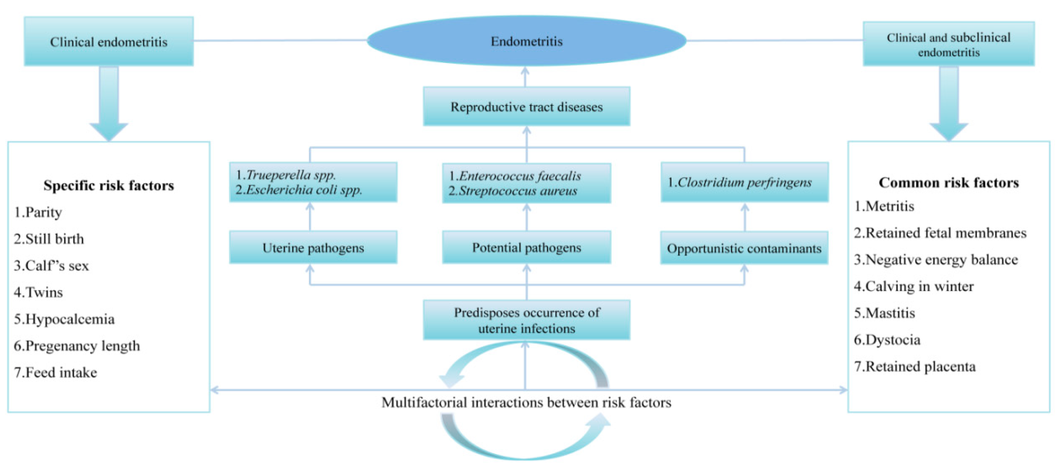
4.1. Bacterial Microflora Association with Reproductive Disorders and Failures

There is not a very clear relationship between infection with one pathogen and postpartum uterine disease, but some bacteria are commonly linked to uterine disease or a healthy uterus, while others are ubiquitous. Disruptions in the microbiome result in dysbiosis, which increases inflammation; this increased inflammation is seen as an important part of the pathogenesis of infectious diseases of the reproductive tract [68]. The postpartum uterine diseases endometritis, metritis, and pyometra are a significant concern for dairy farmers, due to both welfare and economic reasons. These diseases result in uterine contamination that occurs during and after calving [44], causing uterine inflammation in dairy and beef cattle, and are also one of the most important causes of infertility in dairy herds [69,70].

4.1.1. Metritis

To date, metritis and endometritis were not regarded as two clinical diseases that were distinct [71]. At present, nonetheless, it would be intolerable not to differentiate between these two diseases associated with the uterus [12,72]. There seems to be a significant connection between the level of metritis clinical signs and the possibility of endometritis. In 303 dairy cows investigated in a study [73], it was revealed that uterine inflammatory cows had a higher risk of acquiring clinical endometritis (CE) than clinical metritis cows in terms of the odds ratio (i.e., OR: 2.2 vs. 1.4, correspondingly). Because the connection between metritis and endometritis seems to be strong, any risk of infection with metritis immediately after delivery will indirectly intensify the likelihood of endometritis development, as summarized in Figure 4. Hossein and his co-workers [74] unraveled that calving in winter (OR: 2.4), primiparity (OR: 1.7), placental retention (OR: 27.7), twins (OR: 6.6), dystocia (OR: 4.3), male calves (OR: 2.4), and stillbirth (OR: 6.6) influenced the possibility of infection with metritis.
However, recent studies have also revealed that besides these factors mentioned above, E. coli, as previously described, and some anaerobic bacteria were classified as general isolates [76]. In addition, Galvao and coworkers [18], in their recent findings, also showed an association of metritis with a dysbiosis of the uterine microbiota characterized by decreased richness and an increase in Bacteroidetes and Fusobacteria, particularly Bacteroides, Porphyromonas, and Fusobacterium. However, in other studies, Helcococcus ovis was rather recently revealed as an emerging pathogen implicated in the pathogenesis of metritis in Holstein Dairy Cows [77]. Taken together, this information will enable a better understanding of the bacterium’s pathogenicity and antimicrobial resistance.
Some scientists further investigated the incidence of a high comparative frequency of certain genera that are low in abundance, as has been reported in a group infected with metritis, such as Sneathia and Peptostreptococcus; meanwhile, Peptostreptococcus has already been linked to uterine infections in some previous studies [78]. Conversely, a recent study conducted by Sicsic and colleagues [79] reported an association existing between Sneathia and metritis. However, these findings dispute those reported in a previous investigation by Jeon et al. [9], which instead linked Sneathia to the health of the uterus. These inconsistent results can be attributed to the fact that in each study, environmental bacteria contributed to the occurrence of the populations of microflora in the uterus of postpartum cattle. These bacterial genera with high relative prevalence may play a more vital role in the frequency of metritis than the predicted relative incidence.
Apart from these microbes aforementioned as linked to metritis, disorders that can affect the function of the immune system and intake of feed also pose significant risk factors for metritis. Conflicting results, however, exist regarding the efficacy of prepartum immunization against uterine pathogens in reducing the incidence of metritis in dairy cows; hence, the uterine protective role of adaptive immunity has not been completely established [64]. Nonetheless, to reduce uterine pathogens associated with the occurrence of metritis, new promising results have been reported by Singh and coworkers [80] in a recent study that indicated the potential use of mesenchymal stem cells (MSc) from the adipose tissue of cattle (Bos indicus) for the treatment of metritis through wound healing and also decreasing microbial infection.

4.1.2. Clinical and Sub-Clinical Endometritis

Endometritis is considered to be a multifactorial disease, with many factors having a direct or indirect determining or predisposing influence; the summary for the risk factors of endometritis associated with the reproductive tract of the bovine culminating in diseases is depicted in Figure 4. However, because cows have multiple confounding postpartum conditions, it is difficult to comprehend the mechanisms that link fertility decline to endometritis. This may be because most of the risk factors for endometritis are the same, but some are distinctly different, implying that in some instances, the methods used diagnoses separate components of the reproductive tract disease (RTD) complex [81]. Piersanti and colleagues [82] recently created an in vivo model that allows the use of pathogenic E. coli and S. pyogenes to study the mechanisms of reproductive failure associated with endometritis in Holstein heifers. The authors described the isolation of the effects of uterine disease on fertility from the confounding factors that can occur in cows during the postpartum period.
Besides, some numerous studies have established E. coli as one of the significant endometrial pathogens in beef cattle [83] and dual-purpose cattle [84,85]. However, in other studies conducted—even though endometritis is regarded as a multifactorial disease with the E. coli pathogen usually viewed as a causal agent—there has, however, been reported incidence where no bacteria were isolated from cows with subclinical endometritis (SCE) [86], but rather coagulase-negative staphylococci (CoNS) were commonly isolated from healthy cows, and T. pyogenes—as previously described—was frequently isolated from cows with CE in most cases [87]. Furthermore, in recent years, a novel Corynebacterium endometrii sp. nov., has also been isolated from the uterus of a cow with endometritis [88], whereas data in other studies according to Miranda-Casoluengo et al. [55] and Wang et al. [89] also confirmed a higher relative abundance of Bacteroidetes and Fusobacterium in cows that developed CE.
One possible explanation is that by the time CE and SCE are diagnosed, the host defense mechanism that occurs while the inflammatory response is still in progress removes the bacteria from the uterus and restores the normal uterine environment. It is also possible that the reduced number of cows having SCE could have diminished the chances of getting bacterial isolates from those cows [86]. Finally, because the study was conducted using traditional culture methods instead of more advanced culture-independent methods [53], the bacteria may have been hidden behind inconspicuous cell morphology in SCE cows. Thus, the number of uterine samples without isolation was increased. Therefore, it is suggested that future studies using culture-independent methods be conducted to reveal the unseen bacterial population in cows with SE.
On the other hand, current investigations have also confirmed an interaction existing between endometritis and cystic ovarian disease (COD) in Japanese black cattle [90]. According to the authors of the study, COD also has potential effects for endometritis, beside E. coli at 40–60 DPP, compared with cows with a normal ovarian cycle [90]. Similarly, a research report by Mohammed and colleagues [91] further demonstrated that endometritis has a significant adverse effect on the postpartum ovarian cycle of Holstein dairy cows. In particular, cows with endometritis have an increased chance of prolonged corpus luteum (CL) activity, which, if not diagnosed, may extend the interval from calving to conception and may increase farmers’ costs. For instance, cows with severe vaginal discharge are particularly affected. This highlights the importance of monitoring the cyclicity of cows with endometritis to diagnose early symptoms for treatment. However, the CL on the cow’s ovary and its fate may affect the recovery from severe clinical endometritis [88]. Therefore, the size and location of the reproductive tract affected self-healing from clinical endometritis. However, the relationship between endometritis and COD is still unclear, which warrants further study.
In addition, recent data by Miranda-Casoluengo et al. [55] also suggested that the development of postpartum endometritis can also be associated with a delayed differentiation of vaginal and uterine microbiomes at the beginning of the postpartum period, while that from other studies also demonstrated that an altered histopathological endometrial and hematological profile might also be associated with endometritis in about 90% of repeat breeder buffaloes, which exhibited signs of moderate or severe endometritis that could contribute to the low fertility recounted in repeat breeder buffaloes [92,93]. Taken together, these numerous causes of endometritis depict the complex nature of the disease, and as such, it requires future studies to explore its underlying mechanism of action. However, although all of the above risk factors are associated with the decreased fertility reported in dairy and beef cattle suffering from endometritis and metritis, it has recently been proven that the use of ozone therapy for intrauterine treatment is beneficial for cows suffering from endometritis and metritis. This novel therapy has subsequently resulted in improving the efficiency of the reproductive tract as well as decreasing treatment costs with no opposing effects [94,95,96].

5. Prophylaxis and Therapeutic Approaches Modulating Reproductive Tract Microflora

Diseases related to microbial infections are becoming a major global health problem. In addition to using conventional antibiotics to treat these infections, there is also a motivation to develop versatile antimicrobial agents that are at low risk of inducing resistance [97]. The production of novel bacterial proteins through transgenic technology has been shown to be essential for the prevention of mastitis, another microbial infectious disease during reactive oxygen species (ROS) pathogenesis [98]. Additionally, in recent years, the emergence of ozone therapy has been revealed to have no adverse effect following intrauterine, intravaginal, and intramammary applications after it was administered on dairy cows and goats according to all relevant cited reports in a current review [96]. Remarkably, there are many facts in these reports that emphasize the benefits of ozone preparations over antibiotics used for the same preventive and therapeutic purposes [94,95,99]. In the treatment of various pathological conditions, some of the merits of using ozone compared to the use of antibiotics are over-the-counter use; no adverse effects; no residues in milk, meat, and other tissues; and, therefore, no microbial resistance [94,99].
Furthermore, because the cause of these reproductive problems (diseases) in the bovine is multifactorial (Figure 4), not a single preventive measure will be universally effective. Based on this information, optimization of peripartum immune function, principally through management to encourage feed intake in the transition period for the prevention of microbial infections, has also become indispensable [100]. During the dry-off period and early lactation, cows should be challenge-fed to avoid metabolic disease prevalence during the puerperal period, such as fat cow syndrome, milk fever, acidosis, and ketosis. These can influence the frequency of diseases linked to reproduction and decrease reproductive performance. However, in a current overview by Szenci et al. [101], it was pointed out that, despite high milk production during this period, if the perinatal period is cautiously monitored to advance the reproductive performance of dairy cows, satisfactory fertility results can still be accomplished even in large dairy farms.
The reproductive tract microflora has also been revealed as a possible target for both animal and human health. In the near future, microbiota manipulation is likely to be used to achieve a balanced immune reaction to increase resistance to diseases and enhance animal performance [102]. In the next section, we review effective central strategies that modulate the microbial ecology of the reproductive tract for the improvement of cattle health and, indirectly, of human health, because according to the One Health concept [103], there is a connection between human, animal, and environmental health, as depicted in Figure 5. Hence, environmental and agricultural policy should not be approached with a silo mentality [104].

5.1. Antibiotics

Antibiotic resistance (ABR) is a danger to human and animal health. Public health officials have, therefore, stepped up their communication efforts to reduce the reckless use of antibiotics. Much attention to antimicrobial resistance (AMR) in food animals is related to human health as aforementioned (Figure 5), but AMR is also a veterinary problem. Despite the negative impact of the development of AMR, there are few useful strategies for reducing AMR in food-producing animals. The antimicrobial responsibility for the selection, dosage, and duration of antimicrobial treatment is crucial to reduce the occurrence of AMR resistance in the animal industry [108]. In order to minimize the use of antimicrobial agents and AMR, scientifically based measures must be taken, checked, and, if necessary, adjusted, taking into account the accompanying variables [109].
In the treatment of dairy cows infected with acute puerperal metritis (APM), antibiotics such as penicillin and third-generation cephalosporins are systematically administered. In addition, in beef and dairy cattle infected with APM, systemic penicillin, or ampicillin in combination with intrauterine oxytetracycline or ampicillin and cloxacillin is also administered [110]. It is well known that after this antibacterial treatment, not all infections are removed [111], and the definitions of self-healing and cure rates vary widely [112]. Therefore, there is an urgent need to use antibiotics and alternative therapies with caution [113]. Such strategies include selecting the most appropriate drug at the optimal dose and duration to cure infection while minimizing its side effects and stress when selecting resistant strains [114]. Meta-analysis studies have also depicted that commonly used ceftiofur antibiotics have a beneficial effect in the treatment of bovine metritis [115]. Similarly, Oliveira and colleagues [116] recently showed that treating cows for metritis with ceftiofur increased cure rates but did not decrease culling in the first 60 d postpartum, but instead, it increased milk yield and fertility up to 300 d postpartum.
However, in contrast to other findings by Jeon et al. [117], a decrease in the efficacy of ceftiofur in the control of Bacteroidetes, specifically Porphyromonas, (as previously described to be linked to the vagina and uterus), increased significantly following treatment with ceftiofur but did not change after treatment with ampicillin. Therefore, it can be concluded that members of the phylum Bacteroidetes seem to be more resilient to ceftiofur and the reason for these differences may be the continued availability of mechanisms of antibiotic resistance (e.g., through the production of a broad spectrum of beta-lactamases), which give Bacteroidetes higher resistance to third-generation cephalosporins. However, it reduces the sensitivity of Bacteroidetes to ampicillin. It is also plausible that metritis-associated inflammation may affect the ability of ceftiofur rather than ampicillin to reach the uterus [118]. Hence, the ability of ceftiofur to reach the minimum inhibitory concentration for Bacteroidetes may be impaired and still merits further investigation. However, irrespective of antibiotic treatment, the prevailing trend is to increase the bacterial counts of Bacteroidetes. Hence, it is recommended that new treatment approaches that better control this important phylum should be considered as it improves the rate of metritis recovery, which is worth investigating further.
On the other hand, a meta-analytic comparison of the effectiveness of antibiotic and non-antibiotic treatment options was not possible because the benefits or demerits of other antibiotics could not be investigated and the number of comparable trials used in the study was limited. Nonetheless, a recent study investigated the efficacy of non-antibiotic treatment administration with prostaglandin αF2 (αPGF2) in cows affected by severe clinical endometritis [119], and the authors found that PGF2 could better treat severe clinical endometritis in dairy cows with CL in comparison with other treatments. Similarly, Bandai and colleagues [120] further demonstrated that a convenient synchronization protocol used including the administration of prostaglandin F2a (PGF) and estradiol benzoate (EB) 24 h apart instead of 56 h between PGF and Gonadotropin-releasing hormone (GnRH) had a significant potential to allow for the accurate detection of functional CL and thus increased the pregnancy rate of lactating dairy cows after timed artificial insemination (TAI). This beneficial effect of the EB protocol has been demonstrated in multiparous cows.
Prospective research has also shown that, with the increasing threat of endometritis from E. coli and T. pyogenes in cattle, a new method of treatment, “Samia-Treat”, for SCE has emerged, which gave the highest cumulative pregnancy rate of 83.3%, while other groups had lower pregnancy rates of 40% and 50%, respectively [121]. Samia-treat (SAT) is an entirely new and effective treatment for cases of ‘repeat breeder’ by SCE in dairy cows. It caused an increase in reproductive performance and the cumulative pregnancy rate without the excessive use of antibiotics. Besides this new treatment, another study was recently conducted on the efficacy of Aditoprim, a novel dihydrofolate reductase inhibitor, based on ex vivo pharmacodynamic analysis using cattle uterine fluid, as a novel antimicrobial agent to combat endometritis caused by T.pyogenes and thus reduce antibiotic resistance [122]. Furthermore, some other authors also showed that the intrauterine infusion of hyperimmune serum could also replace antibiotics, treat repeat breeder cows, reduce losses from the reproductive syndrome, and prevent the elimination of valuable cows [123]. In addition, a summary of the latest knowledge of the preventive and therapeutic effects of ozone preparations has been outlined for the treatment of genital tract diseases, metritis, endometritis, urovagina, fetal membrane retention, and mastitis, as well as the recovery of the endometrium in functional dairy cows [96]. There is also sufficient confirmation that ozone therapy is superior to antibiotic use. Nevertheless, there are some limitations to the use of ozone in veterinary medicine and buiatrics, such as the lack of activity against intercellular microbes and selective activity against the same species of bacteria, as well as the initiation of inflammation in tissue due to the improper use of the preparation. Summarily, these different emerging alternative therapies aforementioned have proved beneficial in improving the reproductive tract microflora in cattle when compared to the adverse effects resulting from antibiotics usage. Hence it is recommended that future studies explore their mechanism of action during pathogenic microbes’ control.
On the other hand, there have also been reports of varying degrees of effectiveness of alternative drugs or agents for the prevention and treatment of metritis. These include probiotics usage [124], bacteriophages [79], non-steroidal anti-inflammatory drugs [125], botanical essential oils [126], Sheng Hua Tang [127], and chitosan microparticles (CMs) [116,128,129]. For example, it has been demonstrated that the intrauterine administration of CMs can be a viable alternative to traditional antibiotics for the prevention of metritis [128] as well as their effectiveness against the treatment of bacterial infections in animals [130]. Recently, nano-antibiotics (nAbts) has also been of great interest to overcome the drug resistance developed by several pathogenic microorganisms against the most commonly used antibiotics [131]. Nanoparticle-based carriers provide antibiotics with better biofilm penetration and less toxicity, thereby combating bacterial resistance. However, the successful adaptation of nanoformulations to clinical practice requires a detailed assessment of their safety profiles and their potential immunotoxicity.
With reference to this novel rational approach to antibiotic-resistant infections as mentioned above, there are also other alternative therapies for the treatment of CE instead of antibiotics such as hypertonic dextrose (DEX) solution and paraffin, which have no residues in milk and are also usually inexpensive [132,133,134,135]. However, studies conducted by both Machado et al. [136] and Makki et al. [132] revealed no difference in terms of the cytological and clinical cure rate between the DEX group and the two groups receiving -common treatments for CE (PG and CB). This inconsistency in the results concerning DEX’s efficacy on the clinical endometritis cure rate may be attributed to the difference in the routes of administration for the diagnosis of CE between the two studies. Future extensive studies are, however, required for the assessment of DEX and LP as alternative therapies for the treatment of CE instead of antibiotics. It was, however, reported in other studies that the use of the intrauterine hypertonic solution of 50% DEX alone improved clinical cure, the resumption of the postpartum estrous cycle, and the subsequent reproductive performance of purulent vaginal discharge (PVD) cows, compared with untreated CON cows [137]. It may be possible that the intrauterine infusion of DEX (200 mL) can flush the uterus and, with the likely reflux into the vaginal canal, control infection but how DEX functions to control these infectious bacteria requires detailed study.
However, according to some researchers, it is believed a 50% DEX infusion in cows affected by endometritis improves their uterine health by inhibiting local bacterial growth, increasing uterine tone, and providing natural uterine protection (e.g., macrophages and neutrophils) to control a bacterial infection, thereby reducing the water activity of bacteria and nurturing endometrial cells to improve overall reproductive function in cows treated with DEX [133]. DEX can also control uterine infections by changing the bacterial environment (due to its hypertonicity) at the site of infection, affecting the attachment of bacteria to the endometrial mucosa, or increasing uterine contractions (thus evacuation). DEX, in addition, provides energy for cell healing along with tissue contraction, which promotes wound healing [138]. This tissue contraction can help by increasing the uterine tone [137]. Vishnu [135] and Ahmadi [134] further showed that the use of intrauterine DEX in cows with endometritis could promote faster uterine recovery by locally inhibiting bacterial growth, increasing uterine tone, or maintaining endometrial cells. Sugar can help wound healing because it can control bacterial infections by causing osmotic drainage from the affected area, thereby reducing water activity [139], which is necessary for bacterial survival. The osmotic removal of fluid from tissues would aid tissue contraction and thus cause increased uterine tone. Taken together, the use of intrauterine DEX infusion for the treatment of PVD in lactating dairy cows has provided valuable information, and this useful information will help develop new hypotheses for future research to elucidate the underlying mechanisms by which the infection is controlled and its relationship with clinical recovery and reproductive performance.

5.2. Probiotics: A New Approach to Modulate the Reproductive Tract Microflora

Probiotics are defined as “live microorganisms which, when administered in adequate amounts confers a health benefit on the host” [140]. The most commonly used are those belonging to the Lactobacillus and Bifidobacterium genera, which are commensal bacteria that live in or on human bodies [141]. They can regulate the balance of gut microbes, promote the growth and development of animals, and also improve the host’s resistance to diseases [142] and thus improve the host’s health. Due to their competitive exclusion capacity and immunomodulatory function, probiotics are an ideal replacement for antimicrobial agents in animal nutrition [143]. Therefore, the use of probiotics is one of the options proposed by the scientific community as an alternative to antibiotics [144,145].

5.2.1. Lactic Acid Bacteria

Lactic acid bacteria (LAB) are a big group of diverse Gram-positive bacteria that produce lactic acid as the primary end product of carbohydrate fermentation and are therefore very tolerant to acidic conditions. Consequently, they represent bacteria from many taxa but mostly from Lactobacillales in the phylum of Firmicutes as previously described. The most mentioned genera of LAB are Lactobacillus, Pediococcus, Leuconostoc, and Weissella (LPLW), which are phylogenetically close to each other [146] and usually measured together as the Lactobacillus group [147]. Bacterial isolates from cattle vaginal samples have been examined on their properties to develop probiotics to avert or treat uterine infections. The application of these probiotic strains of bacteria has been proposed as an alternative to prevent postpartum uterine infections and inflammation in vivo [148]. Pellegrino et al. [149] further demonstrated that the LAB strain isolated from the tract of the vagina has a strong ability to produce H2O2.
Consistently, recent studies have also indicated that Lactobacillus strain SQ0048, present in the vaginal microflora of healthy cows, serves as a significant microbiological barrier to infection with genital pathogens, playing a protective role in the reproductive tract, and thus adhering to the specific epithelium, and producing inhibitors [150]. Probiotic strains can affect pathogenic microorganisms through different mechanisms, such as enhancing the intestinal barrier’s function, increasing mucin production, and modulating the immune system’s activity [151]. Genis et al. [152], in their recent work, also demonstrated that a LAB combination consisting of L. rhamnosus, P. acidilactici, and L. reuteri in a ratio of 25:25:2 showed the best potential to regulate inflammation in the uterus as well as reduce in vitro and ex vivo infection caused by E. coli [152,153] compared with the individual LAB strains separately. Remarkably, when there was intra-vaginal administration of LAB, it reformed the microflora in the vagina and tended to restrain the number of bacteria moving to the uterus, even though LAB did not make their way to the endometrium [153]. However, cows treated with LAB had low expression of B-defensins and MUC1 in the endometrium, and these were two indicators of uterine infection.
This can be explained by the regulation of the pathogenic environment in the vaginal tract by the action of the LAB and the subsequent decrease in pathogenic bacteria reaching the endometrium. Besides, studies conducted in our laboratory by Wang et al. [28] revealed that the healthy postpartum cow’s vaginal flora is dominated by W. koreensis and L. sakei subsp. which are all LAB, and the waning of these vaginal flora dominant species may lead to the emergence of endometritis, but in another study conducted in our laboratory by Zhao et al. [154], we screened four different LAB strains (Leuconostoc lactis, Ilyobacter polytropus, E. hirae, and Weissella confusa) from the healthy cow’s vaginas, which also seem to demonstrate probiotic properties. The difference in the levels of bacterial diversity between these two studies can be attributed to the experimental methods or techniques used in the two studies. In addition, the intra-vaginal LAB administration was able to lessen the incidence of metritis compared to that in CON cows [148]. This combination of LAB applied through the intra-vaginal route twice per week during a 3-week period proceeding to calving appeared to be a favorable approach to prevent infections by metritis. On the other hand, a recent meta-analysis also showed that females with bacterial communities dominated by L. iners or non-Lactobacillus species had an approximately three-to-five-fold increased risk of HPV infection and progression to cervical cancer compared to females with L. crispatus-dominant communities [155]. This also demonstrates the inhibitory effect of LAB on the growth of pathogenic bacteria that cause infection.
Similarly, the use of probiotics in vivo and in vitro for HPV clearance and significant CIN regression is shown and has recently been described by Curty and coworkers [46]. This suggests that manipulating the microbiota with probiotics or vaginal microbiota transplant (VMT) could be a viable option to control HPV infection clearance, CIN regression, and cervical cancer progression related to the microbiota of the human vagina. Results obtained using an in vitro model of the human cervix confirmed that supernatants from both probiotics have a beneficial effect on cervical cells and that both strains could grow in the biofilm and show aggregation and adhesion properties on biotic or abiotic surfaces [156]. These properties are modulated and even increased by lactoferrin, which shows the beneficial effects of this combination of biotherapeutics for the prevention and treatment of urogenital tract infections in women.
Some studies, on the other hand, have also reported that Candida albicans connects with the presence of high diversity anaerobic bacterial communities that are poor in Lactobacillus [157], and some in vitro studies support the idea that Lactobacillus before infection with C. albicans on the host could protect by physical colonization, the lowering of pH, and the modulation of the vaginal immune response [158,159], but conversely, other studies disagree with these results. These contradicting results can be attributed to possible colonization by other microbial agents such as fungi. Therefore, considering fungi, along with bacteria and viruses, would allow a broader view of the microbiome as a whole and would likely improve the microbiome’s associations with clinical features.
In addition, results from Koedooder and coworkers [160] further demonstrated that a high incidence of Lactobacillus appeared to be beneficial for in vitro fertilization (IVF) and the effect of an intracytoplasmic sperm injection (IVF-ICSI), but a high frequency of (>60%) L. crispatus was not beneficial. A third PCR-based study [161] contradicted these results, and the difference in the methods used could explain these conflicting findings. Overall, studies that generalize the potential positive effects of Lactobacilli on fertility must take into account that different types of Lactobacilli have different properties, such as the ability to produce lactic acid from the breakdown of glycogen conversion. The acidity produced by L. crispatus was recorded as high compared to that by L. iners, L. jensenii, and L. gasseri [162].
Although in vitro activity may not always be consistent with in vivo results, further clinical studies are needed to predict and select the real beneficial strains. Additionally, the biotechnological application of Lactobacillus strains as probiotics yield effective results in controlling microbial films formed by enteropathogenic bacteria, which is a viable alternative to medical use [163]. However, the results of some meta-analyses are still limited and inconclusive pertaining to the use of probiotics in the treatment of infectious diseases [164]. This is mainly due to the limited number of studies, the lack of standardization, and inconsistencies in reported results. Therefore, it is advisable that future studies should address these shortcomings and include an assessment of the overall use of prebiotics and probiotics.

5.2.2. Bacteriophages: Virus-Preventing Pathogens

In recent years, the use of microbes as a tool to defend against the antibiotic resistome is gathering momentum and it has also become a promising and novel area of emerging research [165,166]. A bacteriophage is a virus that infects and multiplies in a small number of host bacteria that breaks down when the phage enters the environment. Current data revealed a novel putative depolymerase-like protein, which is most likely to play an important role in phage–host interaction [167]. Phages have a very narrow range of host species, which makes them ideal for the targeted destruction of bacteria, but they also need therapeutic products consisting of phage cocktails. The use of antimicrobial agents that are specific for pathogenic bacteria is expected to decrease the development of ABR [168]. Bacteriophage therapy, which uses virulent (lytic) bacteriophages to control bacterial growth, has shown to be an attractive alternative to conventional drugs [169,170] and has been successful in animal trials against a broad range of bacterial pathogens such as enteropathogenic E. coli [171], Pseudomonas aeruginosa [172], S. aureus [173], Acinetobacter baumannii [172], and vancomycin-resistant Enterococcus faecium [174] as previously described. The increase in bacteria that are multi-drug resistant (MDR) in humans and animals has increased interest in bacteriophage therapy research [175,176].
Bovine metritis is likely to be successfully treated with bacteriophage therapy because of the following reasons: a large number of polyvalent bacteriophages from the environment are extremely active against E. coli [177], and a reduction in the intrauterine bacterial load is likely to reduce the severity of clinical disease [178]. Because intrauterine therapy is a viable option, the fast removal of phage from the circulatory system is not an issue. The bacteriophage cocktail had a remarkable ability to inhibit the in vitro growth of E. coli isolates, and it might also be an attractive substitute to the conventional treatment of metritis by reducing E. coli in the uteri of postpartum dairy cows and, consequently, the severity of clinical disease [178]. A ratio of 10 particles of bacteriophage per bacterial cell was proficient in constraining all isolates, which were at least 50%. However, to establish a ratio that entirely limits bacterial growth for extended periods, it is recommended that we test a higher multiplicity of infection (MOI) in a future in vitro study.
Bacteriophages are also known for their host specificity [177], so it is, therefore, possible that the phage cocktail used will not have similar antimicrobial characteristics when a different population of E. coli isolates is tested. There are presently no bacteriophage-based drugs permitted by the US Foods and Drug Authority (FDA) for treating bacterial diseases in humans and animals. However, the FDA recently changed the rules for using food additives to approve the use of the bacteriophage preparations on ready-to-eat meat and poultry [179]. The use of bacteriophages, which are naturally found in agriculture, for the treatment of environmental diseases, can be regarded as harmless to humans and the environment [180,181]. This is particularly important when bacteriophages isolated locally are used as they are more substantial during an interaction between the host and bacteria in the environment than those in the treated animal. For example, when evaluating the result of a single trial of bacteriophage preparations on the progression curves of 57 E. coli isolates, it was noted that all the 11 preparations of bacteriophages reduced the growth frequency of the isolates compared to their controls. Nevertheless, further in vitro and in vivo research is needed to assess the viability of these bacteriophages as therapeutic agents for metritis.

6. Conclusions and Future Recommendations

Overall, the area of the microbiota of the bovine reproductive tract is a rapidly developing area, and its physiological role in bovine reproduction remains to be understood. The importance of the interaction between the host and the flora it supports has been acknowledged. However, there are still many problems with the difference between this microbial environment within and between cattle in healthy and diseased conditions. In this overview, the composition of the microflora of the reproductive tract differs, contingent on many factors. Compared to healthy cattle, cattle with metritis and endometritis have a complex and dynamic bacterial flora that is occupied by various anaerobic and fastidious bacteria. Even though variations in the microbiome of the reproductive tract are prone to cause adverse health effects, molecular tools indicate that certain changes in the reproductive tract flora are normal and do not necessarily cause disease.
In the past few decades, the use of advanced metagenomics has led to an explosion in understanding the bacterial microflora compositions in various ecological niches. In addition, the recent rebirth of culture techniques, in addition to bacteria that have so far only been detected using molecular tools, has enabled the isolation of fastidious and new types of bacterial species in the vagina, cervix, and uterus. Comparing bacteria in the reproductive microbiome repertoire using advanced molecular tools with those isolated by culture-based methods showed that these two different techniques enabled an extensive and somewhat overlapping range of vaginal, uterine, and cervical bacteria species.
This repertoire is vital for microbiologists and clinicians and is also a starting point to better fathom the dysbiosis or infections caused by unsteadiness in the reproductive tract microflora and thus provide more appropriate treatments. Therefore, by combining culture and culture-independent methods to characterize the reproductive tract microflora, the role of bacteria in healthy and pathological conditions can definitely be shown. Nevertheless, it is recommended that enthusiastic work be continued in the future that aims at understanding the role of the microbiome in reproductive tract health.

Author Contributions

M.O.A. synthesized the literature and wrote the original draft of the manuscript. J.W. and W.L. reviewed and corrected the final manuscript. All authors have read and agreed to the published version of the manuscript.

Funding

This work was supported by the National Key R & D Program of China (2017YFD0501903).

Acknowledgments

The authors wish to express their sincere gratitude to Xu Ke and Lewei Guo (Jilin Agricultural University, College of Animal Science and Technology) and also to the anonymous reviewers for their valuable comments and suggestions to improve the quality of this manuscript.

Conflicts of Interest

The authors declare no conflict of interest.

Abbreviations

HMPHuman Microbiome Project;
OTUOperational Taxonomic Units;
SESubclinical endometritis;
CEClinical endometritis;
SCESubclinical endometritis;
AIArtificial Insemination;
OROdds Ratio;
OTCOperational Taxonomic Classification;
CFClinical Formative;
CPClinical Postpartum;
CGClinical Gestation;
DIMDays in Milk;
DPPDays Postpartum;
PVDPurulent Vaginal Discharge;
ABRAntibiotic Resistance;
MICminimum inhibitory concentration;
CCFAceftiofur crystalline free acid;
CMchitosan microparticles;
APMAcute Puerperal Metritis;
LABLactic Acid Bacteria;
CBCephapirin Benzathine;
DEXDextrose solution;
OTCOxytetracycline;
LPliquid paraffin;
CONcontrol;
PGprostaglandin;
PPPostpartum;
MOIMultiplicity of Infection;
S. aureusStaphylococcus aureus;
F. necrophorumFusobacterium necrophorum;
T. pyogenesTrueperella pyogenes;
CINcervical intraepithelial neoplasia;
HPVhuman papillomavirus;
CVMCervico-vaginal mucus;
CODcystic ovarian disease;
SATSamia-treat;
CoNScoagulase-negative staphylococci;
RTDreproductive tract disease;
AMUantimicrobial use;
AMRantimicrobial resistance;
ARMsantimicrobial resistant microorganisms;
EBestradiol benzoate;
CLcorpus luteum;
αPGF2prostaglandin αF2;
nAbtsnanoantibiotics;
VMTvaginal microbiota transplant;
FDAFoods and Drug Authority;
MScmesenchymal stem cells;
IVFin vitro fertilization
IVF-ICFSIin vitro fertilization-intracytoplasmic sperm injection
MSSMetagenomic Shotgun Sequencing;
RPretained placenta;
MDRmulti-drug resistant;
ROSreactive oxygen species.
GnRHGonadotropin-releasing hormone

References

  1. Parfrey, L.W.; Moreau, C.S.; Russell, J.A. Introduction: The host-associated microbiome: Pattern, process and function. Mol. Ecol. 2018, 27, 1749–1765. [Google Scholar] [CrossRef] [PubMed]
  2. Rosenberg, E.; Zilber-Rosenberg, I. Microbes drive evolution of animals and plants: The hologenome concept. MBio 2016, 7, e01395-15. [Google Scholar] [CrossRef] [Green Version]
  3. Koedooder, R.; Mackens, S.; Budding, A.; Fares, D.; Blockeel, C.; Laven, J.; Schoenmakers, S. Identification and evaluation of the microbiome in the female and male reproductive tracts. Hum. Reprod. Update 2018, 25, 298–325. [Google Scholar] [CrossRef]
  4. Schoenmakers, S.; Steegers-Theunissen, R.; Faas, M. The matter of the reproductive microbiome. Obst. Med. 2019, 12, 107–115. [Google Scholar] [CrossRef] [Green Version]
  5. Chen, C.; Song, X.; Wei, W.; Zhong, H.; Dai, J.; Lan, Z.; Li, F.; Yu, X.; Feng, Q.; Wang, Z. The microbiota continuum along the female reproductive tract and its relation to uterine-related diseases. Nat. Commun. 2017, 8, 1–11. [Google Scholar] [CrossRef] [Green Version]
  6. Scolari, F.; Attardo, G.M.; Aksoy, E.; Weiss, B.; Savini, G.; Takac, P.; Abd-Alla, A.; Parker, A.G.; Aksoy, S.; Malacrida, A.R. Symbiotic microbes affect the expression of male reproductive genes in Glossina m. morsitans. BMC Microbiol. 2018, 18, 169. [Google Scholar] [CrossRef]
  7. Baker, J.M.; Chase, D.M.; Herbst-Kralovetz, M.M. Uterine microbiota: Residents, tourists, or invaders? Front. Immunol. 2018, 9, 208. [Google Scholar] [CrossRef] [Green Version]
  8. Moreno, I.; Simon, C. Deciphering the effect of reproductive tract microbiota on human reproduction. Reprod. Med. Biol. 2019, 18, 40–50. [Google Scholar] [CrossRef] [Green Version]
  9. Jeon, S.J.; Vieira-Neto, A.; Gobikrushanth, M.; Daetz, R.; Mingoti, R.D.; Parize, A.C.B.; De Freitas, S.L.; Da Costa, A.N.L.; Bicalho, R.C.; Lima, S.; et al. Uterine microbiota progression from calving until establishment of metritis in dairy cows. Appl. Environ. Microbiol. 2015, 81, 6324–6332. [Google Scholar] [CrossRef] [Green Version]
  10. Knudsen, L.R.V.; Karstrup, C.C.; Pedersen, H.G.; Agerholm, J.S.; Jensen, T.K.; Klitgaard, K. Revisiting bovine pyometra—New insights into the disease using a culture-independent deep sequencing approach. Vet. Microbiol. 2015, 175, 319–324. [Google Scholar] [CrossRef]
  11. Neckovic, A.; van Oorschot, R.A.; Szkuta, B.; Durdle, A. Investigation of direct and indirect transfer of microbiomes between individuals. Forensic Sci. Int. Genet. 2020, 45, 102212. [Google Scholar] [CrossRef] [PubMed]
  12. Piersanti, R.L.; Bromfield, J.J. [AN354] The Consequence of Postpartum Uterine Disease on Dairy Cow Fertility. EDIS 2019, 2019, 1–4. [Google Scholar]
  13. Sheldon, I.M.; Dobson, H. Postpartum uterine health in cattle. Anim. Reprod. Sci. 2004, 82–83, 295–306. [Google Scholar] [CrossRef] [PubMed]
  14. Jeon, S.J.; Cunha, F.; Ma, X.; Martinez, N.; Vieira-Neto, A.; Daetz, R.; Bicalho, R.C.; Lima, S.; Santos, J.E.; Jeong, K.C. Uterine microbiota and immune parameters associated with fever in dairy cows with metritis. PLoS ONE 2016, 11, e0165740. [Google Scholar] [CrossRef] [Green Version]
  15. Lima, F.S.; Oikonomou, G.; Lima, S.F.; Bicalho, M.L.; Ganda, E.K.; de Oliveira Filho, J.C.; Lorenzo, G.; Trojacanec, P.; Bicalho, R.C. Prepartum and Postpartum Rumen Fluid Microbiomes: Characterization and Correlation with Production Traits in Dairy Cows. Appl. Environ. Microbiol. 2015, 81, 1327–1337. [Google Scholar] [CrossRef] [Green Version]
  16. Bicalho, M.; Santin, T.; Rodrigues, M.; Marques, C.; Lima, S.; Bicalho, R. Dynamics of the microbiota found in the vaginas of dairy cows during the transition period: Associations with uterine diseases and reproductive outcome. J. Dairy Sci. 2017, 100, 3043–3058. [Google Scholar] [CrossRef]
  17. Sheldon, I.M.; Cronin, J.G.; Bromfield, J.J. Tolerance and innate immunity shape the development of postpartum uterine disease and the impact of endometritis in dairy cattle. Ann. Rev. Anim. Biosci. 2019, 7, 361–384. [Google Scholar] [CrossRef] [Green Version]
  18. Galvão, K.N.; Bicalho, R.C.; Jeon, S.J. Symposium review: The uterine microbiome associated with the development of uterine disease in dairy cows. J. Dairy Sci. 2019, 102, 11786–11797. [Google Scholar] [CrossRef]
  19. Nagaraja, T.; Lechtenberg, K.F. Liver abscesses in feedlot cattle. Vet. Clin. N. Am. Food Anim. Pract. 2007, 23, 351–369. [Google Scholar] [CrossRef]
  20. Kutzer, P.; Schulze, C.; Engelhardt, A.; Wieler, L.H.; Nordhoff, M. Helcococcus ovis, an Emerging Pathogen in Bovine Valvular Endocarditis. J. Clin. Microbiol. 2008, 46, 3291–3295. [Google Scholar] [CrossRef] [Green Version]
  21. Jeon, S.J.; Cunha, F.; Vieira-Neto, A.; Bicalho, R.C.; Lima, S.; Bicalho, M.L.; Galvão, K.N. Blood as a route of transmission of uterine pathogens from the gut to the uterus in cows. Microbiome 2017, 5, 109. [Google Scholar] [CrossRef]
  22. Laguardia-Nascimento, M.; Branco, K.M.; Gasparini, M.R.; Giannattasio-Ferraz, S.; Leite, L.R.; Araujo, F.M.; Salim, A.C.; Nicoli, J.R.; Oliveira, G.C.; Barbosa-Stancioli, E.F. Vaginal microbiome characterization of Nellore cattle using metagenomic analysis. PLoS ONE 2015, 10, e0143294. [Google Scholar] [CrossRef] [PubMed]
  23. Amos, M.R.; Healey, G.D.; Goldstone, R.J.; Mahan, S.M.; Düvel, A.; Schuberth, H.-J.; Sandra, O.; Zieger, P.; Dieuzy-Labaye, I.; Smith, D.G. Differential endometrial cell sensitivity to a cholesterol-dependent cytolysin links Trueperella pyogenes to uterine disease in cattle. Biol. Reprod. 2014, 54, 1–13. [Google Scholar]
  24. Hansen, P.J. Physiology and Endocrinology Symposium: Maternal immunological adjustments to pregnancy and parturition in ruminants and possible implications for postpartum uterine health: Is there a prepartum-postpartum nexus? J. Anim. Sci. 2013, 91, 1639–1649. [Google Scholar] [CrossRef] [PubMed]
  25. Young, W.; Hine, B.C.; Wallace, O.A.; Callaghan, M.; Bibiloni, R. Transfer of intestinal bacterial components to mammary secretions in the cow. PeerJ 2015, 3, e888. [Google Scholar] [CrossRef] [PubMed]
  26. Yeoman, C.J.; Ishaq, S.L.; Bichi, E.; Olivo, S.K.; Lowe, J.; Aldridge, B.M. Biogeographical differences in the influence of maternal microbial sources on the early successional development of the bovine neonatal gastrointestinal tract. Sci. Rep. 2018, 8, 1–14. [Google Scholar] [CrossRef]
  27. Knight, R.; Callewaert, C.; Marotz, C.; Hyde, E.R.; Debelius, J.W.; McDonald, D.; Sogin, M.L. The microbiome and human biology. Ann. Rev. Genom. Hum. Genet. 2017, 18, 65–86. [Google Scholar] [CrossRef]
  28. Wang, J.; Sun, C.; Liu, C.; Yang, Y.; Lu, W. Comparison of vaginal microbial community structure in healthy and endometritis dairy cows by PCR-DGGE and real-time PCR. Anaerobe 2016, 38, 1–6. [Google Scholar] [CrossRef]
  29. Nesengani, L.T.; Wang, J.; Yang, Y.; Yang, L.; Lu, W. Unravelling vaginal microbial genetic diversity and abundance between Holstein and Fleckvieh cattle. RSC Adv. 2017, 7, 56137–56143. [Google Scholar] [CrossRef] [Green Version]
  30. El-Hayek, S.; Clarke, H.J. Control of oocyte growth and development by intercellular communication within the follicular niche. In Molecular Mechanisms of Cell Differentiation in Gonad Development; Springer: Berlin/Heidelberg, Germany, 2016; pp. 191–224. [Google Scholar]
  31. Giannattasio- Ferraz, S.; Laguardia-Nascimento, M.; Gasparini, M.R.; Leite, L.R.; Araujo, F.M.G.; de Matos Salim, A.C.; de Oliveira, A.P.; Nicoli, J.R.; de Oliveira, G.C.; da Fonseca, F.G. A common vaginal microbiota composition among breeds of Bos taurus indicus (Gyr and Nellore). Braz. J. Microbiol. 2019, 50, 1115–1124. [Google Scholar] [CrossRef]
  32. Saini, P.; Singh, M.; Kumar, P. Fungal endometritis in bovines. Open Vet. J. 2019, 9, 94–98. [Google Scholar] [CrossRef] [PubMed] [Green Version]
  33. Deng, F.; McClure, M.; Rorie, R.; Wang, X.; Chai, J.; Wei, X.; Lai, S.; Zhao, J. The vaginal and fecal microbiomes are related to pregnancy status in beef heifers. J. Anim. Sci. Biotechnol. 2019, 10, 1–13. [Google Scholar] [CrossRef] [PubMed] [Green Version]
  34. Serrano, M.G.; Parikh, H.I.; Brooks, J.P.; Edwards, D.J.; Arodz, T.J.; Edupuganti, L.; Huang, B.; Girerd, P.H.; Bokhari, Y.A.; Bradley, S.P. Racioethnic diversity in the dynamics of the vaginal microbiome during pregnancy. Nat. Med. 2019, 25, 1001–1011. [Google Scholar] [CrossRef] [PubMed]
  35. Ault, T.B.; Clemmons, B.A.; Reese, S.T.; Dantas, F.G.; Franco, G.A.; Smith, T.P.; Edwards, J.L.; Myer, P.R.; Pohler, K.G. Uterine and vaginal bacterial community diversity prior to artificial insemination between pregnant and nonpregnant postpartum cows. J. Anim. Sci. 2019, 97, 4298–4304. [Google Scholar] [CrossRef]
  36. Mahalingam, S.; Dharumadurai, D.; Archunan, G. Vaginal microbiome analysis of buffalo (Bubalus bubalis) during estrous cycle using high-throughput amplicon sequence of 16S rRNA gene. Symbiosis 2019, 78, 97–106. [Google Scholar] [CrossRef]
  37. Wang, J.; Liu, C.; Nesengani, L.T.; Gong, Y.; Yang, Y.; Yang, L.; Lu, W. Comparison of vaginal microbial community structure of beef cattle between luteal phase and follicular phase. Indian J. Anim. Res. 2019, 53, 1298–1303. [Google Scholar] [CrossRef]
  38. Fettweis, J.M.; Serrano, M.G.; Brooks, J.P.; Edwards, D.J.; Girerd, P.H.; Parikh, H.I.; Huang, B.; Arodz, T.J.; Edupuganti, L.; Glascock, A.L. The vaginal microbiome and preterm birth. Nat. Med. 2019, 25, 1012–1021. [Google Scholar] [CrossRef] [Green Version]
  39. Goltsman, D.S.A.; Sun, C.L.; Proctor, D.M.; DiGiulio, D.B.; Robaczewska, A.; Thomas, B.C.; Shaw, G.M.; Stevenson, D.K.; Holmes, S.P.; Banfield, J.F. Metagenomic analysis with strain-level resolution reveals fine-scale variation in the human pregnancy microbiome. Genome. Res. 2018, 28, 1467–1480. [Google Scholar] [CrossRef] [Green Version]
  40. Azawi, O. Postpartum uterine infection in cattle. Anim Reprod. Sci. 2008, 105, 187–208. [Google Scholar] [CrossRef]
  41. Sharma, A.; Singh, M.; Kumar, P.; Sharma, A.; Neelam, A.M.J.; Sharma, P. Postpartum Uterine Infections in Cows and Factors Affecting it–A Review. Int. J. Curr. Microbiol. App. Sci. 2017, 6, 1020–1028. [Google Scholar]
  42. Deguillaume, L.; Geffré, A.; Desquilbet, L.; Dizien, A.; Thoumire, S.; Vornière, C.; Constant, F.; Fournier, R.; Chastant-Maillard, S. Effect of endocervical inflammation on days to conception in dairy cows. J. Dairy Sci. 2012, 95, 1776–1783. [Google Scholar] [CrossRef] [Green Version]
  43. Singer, E.; Bushnell, B.; Coleman-Derr, D.; Bowman, B.; Bowers, R.M.; Levy, A.; Gies, E.A.; Cheng, J.-F.; Copeland, A.; Klenk, H.-P. High-resolution phylogenetic microbial community profiling. The ISME J. 2016, 10, 2020–2032. [Google Scholar] [CrossRef]
  44. Galvão, K. Postpartum uterine diseases in dairy cows. Anim Reprod. 2018, 9, 290–296. [Google Scholar]
  45. Wang, Y.; Wang, J.; Li, H.; Fu, K.; Pang, B.; Yang, Y.; Liu, Y.; Tian, W.; Cao, R. Characterization of the cervical bacterial community in dairy cows with metritis and during different physiological phases. Theriogenology 2018, 108, 306–313. [Google Scholar] [CrossRef] [PubMed]
  46. Curty, G.; de Carvalho, P.S.; Soares, M.A. The Role of the Cervicovaginal Microbiome on the Genesis and as a Biomarker of Premalignant Cervical Intraepithelial Neoplasia and Invasive Cervical Cancer. Int. J. Mol. Sci. 2020, 21, 222. [Google Scholar] [CrossRef] [PubMed] [Green Version]
  47. Adnane, M.; Meade, K.G.; O’Farrelly, C. Cervico-vaginal mucus (CVM)–an accessible source of immunologically informative biomolecules. Vet. Res. Commun. 2018, 42, 255–263. [Google Scholar] [CrossRef] [PubMed] [Green Version]
  48. Moreno, I.; Franasiak, J.M. Endometrial microbiota—new player in town. Fertil. Steril. 2017, 108, 32–39. [Google Scholar] [CrossRef] [Green Version]
  49. Pelzer, E.; Gomez-Arango, L.F.; Barrett, H.L.; Nitert, M.D. Maternal health and the placental microbiome. Placenta 2017, 54, 30–37. [Google Scholar] [CrossRef] [Green Version]
  50. Agostinis, C.; Mangogna, A.; Bossi, F.; Ricci, G.; Kishore, U.; Bulla, R. Uterine immunity and microbiota: A shifting paradigm. Front. Immunol. 2019, 10, 2387. [Google Scholar] [CrossRef] [Green Version]
  51. Moore, S.; Ericsson, A.; Poock, S.; Melendez, P.; Lucy, M. Hot topic: 16S rRNA gene sequencing reveals the microbiome of the virgin and pregnant bovine uterus. J. Dairy Sci. 2017, 100, 4953–4960. [Google Scholar] [CrossRef] [Green Version]
  52. Karstrup, C.C.; Klitgaard, K.; Jensen, T.K.; Agerholm, J.S.; Pedersen, H.G. Presence of bacteria in the endometrium and placentomes of pregnant cows. Theriogenology 2017, 99, 41–47. [Google Scholar] [CrossRef] [PubMed]
  53. Santos, T.M.A.; Gilbert, R.O.; Bicalho, R.C. Metagenomic analysis of the uterine bacterial microbiota in healthy and metritic postpartum dairy cows. J. Dairy Sci. 2011, 94, 291–302. [Google Scholar] [CrossRef] [PubMed]
  54. Santos, T.M.; Bicalho, R.C. Diversity and succession of bacterial communities in the uterine fluid of postpartum metritic, endometritic and healthy dairy cows. PLoS ONE 2012, 7, e53048. [Google Scholar] [CrossRef] [PubMed] [Green Version]
  55. Miranda-CasoLuengo, R.; Lu, J.; Williams, E.J.; Miranda-CasoLuengo, A.A.; Carrington, S.D.; Evans, A.C.; Meijer, W.G. Delayed differentiation of vaginal and uterine microbiomes in dairy cows developing postpartum endometritis. PLoS ONE 2019, 14, e0200974. [Google Scholar] [CrossRef] [Green Version]
  56. Williams, E.J.; Fischer, D.P.; Pfeiffer, D.U.; England, G.C.; Noakes, D.E.; Dobson, H.; Sheldon, I.M. Clinical evaluation of postpartum vaginal mucus reflects uterine bacterial infection and the immune response in cattle. Theriogenology 2005, 63, 102–117. [Google Scholar] [CrossRef] [PubMed]
  57. Ryan, N.J.; Meade, K.G.; Williams, E.J.; O’Farrelly, C.; Grant, J.; Evans, A.C.; Beltman, M.E. Purulent vaginal discharge diagnosed in pasture-based Holstein-Friesian cows at 21 days postpartum is influenced by previous lactation milk yield and results in diminished fertility. J. Dairy Sci. 2020, 103, 666–675. [Google Scholar] [CrossRef]
  58. Canadas, E.R.; Herlihy, M.; Kenneally, J.; Grant, J.; Kearney, F.; Lonergan, P.; Butler, S. Associations between postpartum phenotypes, cow factors, genetic traits, and reproductive performance in seasonal-calving, pasture-based lactating dairy cows. J. Dairy Sci. 2020, 103, 1016–1030. [Google Scholar] [CrossRef] [Green Version]
  59. Rojas Canadas, E.R.; Herlihy, M.M.; Kenneally, J.; Grant, J.; Kearney, F.; Lonergan, P.; Butler, S.T. Associations between ovarian cyclicity, uterine health, indicators of bioenergetic status, and genetic traits during early lactation in seasonal-calving, pasturebased lactating dairy cows. J. Dairy Sci. 2020, 103, 1002–1015. [Google Scholar] [CrossRef]
  60. Gilbert, R.O.; Santos, N.R. Dynamics of postpartum endometrial cytology and bacteriology and their relationship to fertility in dairy cows. Theriogenology 2016, 85, 1367–1374. [Google Scholar] [CrossRef] [Green Version]
  61. LeBlanc, S.; Duffield, T.; Leslie, K.; Bateman, K.; Keefe, G.P.; Walton, J.; Johnson, W. Defining and diagnosing postpartum clinical endometritis and its impact on reproductive performance in dairy cows. J. Dairy Sci. 2002, 85, 2223–2236. [Google Scholar] [CrossRef]
  62. De Boer, M.; Buddle, B.M.; Heuer, C.; Hussein, H.; Zheng, T.; LeBlanc, S.J.; McDougall, S. Associations between intrauterine bacterial infection, reproductive tract inflammation, and reproductive performance in pasture-based dairy cows. Theriogenology 2015, 83, 1514–1524. [Google Scholar] [CrossRef] [PubMed]
  63. Sheldon, I.; Molinari, P.C.; Ormsby, T.J.; Bromfield, J.J. Preventing postpartum uterine disease in dairy cattle depends on avoiding, tolerating and resisting pathogenic bacteria. Theriogenology 2020, 150, 158–165. [Google Scholar] [CrossRef] [PubMed]
  64. Machado, V.; Silva, T. Adaptive immunity in the postpartum uterus: Potential use of vaccines to control metritis. Theriogenology 2020, 150, 201–209. [Google Scholar] [CrossRef]
  65. Pascottini, O.B.; LeBlanc, S.J. Modulation of immune function in the bovine uterus peripartum. Theriogenology 2020, 150, 193–200. [Google Scholar] [CrossRef] [PubMed]
  66. König, S.; May, K. Invited review: Phenotyping strategies and quantitative-genetic background of resistance, tolerance and resilience associated traits in dairy cattle. Animal 2019, 13, 897–908. [Google Scholar]
  67. Gilbert, R.O. Symposium review: Mechanisms of disruption of fertility by infectious diseases of the reproductive tract. J. Dairy Sci. 2019, 102, 3754–3765. [Google Scholar] [CrossRef] [Green Version]
  68. Chase, C.; Kaushik, R.S. Mucosal Immune System of Cattle: All Immune Responses Begin Here. Vet. Clin. Food Anim. Pract 2019, 35, 431–451. [Google Scholar] [CrossRef]
  69. Helfrich, A.L.; Reichenbach, H.-D.; Meyerholz, M.M.; Schoon, H.-A.; Arnold, G.J.; Fröhlich, T.; Weber, F.; Zerbe, H. Novel sampling procedure to characterize bovine subclinical endometritis by uterine secretions and tissue. Theriogenology 2020, 141, 186–196. [Google Scholar] [CrossRef]
  70. Sheldon, I.M.; Owens, S.E. Postpartum uterine infection and endometritis in dairy cattle. Anim. Reprod. (AR) 2018, 14, 622–629. [Google Scholar] [CrossRef]
  71. Bruun, J.; Ersbøll, A.; Alban, L. Risk factors for metritis in Danish dairy cows. Prev. Vet. Med. 2002, 54, 179–190. [Google Scholar] [CrossRef]
  72. Sheldon, I.M.; Cronin, J.; Goetze, L.; Donofrio, G.; Schuberth, H.J. Defining postpartum uterine disease and the mechanisms of infection and immunity in the female reproductive tract in cattle. Biol. Reprod. 2009, 81, 1025–1032. [Google Scholar] [CrossRef] [PubMed]
  73. Giuliodori, M.J.; Magnasco, R.; Becu-Villalobos, D.; Lacau-Mengido, I.; Risco, C.; de la Sota, R.L. Metritis in dairy cows: Risk factors and reproductive performance. J. Dairy Sci. 2013, 96, 3621–3631. [Google Scholar] [CrossRef] [PubMed] [Green Version]
  74. Hossein-Zadeh, N.G.; Ardalan, M. Cow-specific risk factors for retained placenta, metritis and clinical mastitis in Holstein cows. Vet. Res. Commun. 2011, 35, 345–354. [Google Scholar] [CrossRef] [PubMed]
  75. Adnane, M.; Kaidi, R.; Hanzen, C.; England, G.C. Risk factors of clinical and subclinical endometritis in cattle: A review. Turk. J. Vet. Anim. Sci. 2017, 41, 1–11. [Google Scholar] [CrossRef]
  76. Imhof, S.; Luternauer, M.; Hüsler, J.; Steiner, A.; Hirsbrunner, G. Therapy of retained fetal membranes in cattle: Comparison of two treatment protocols. Anim. Reprod. Sci. 2019, 206, 11–16. [Google Scholar] [CrossRef]
  77. Cunha, F.; Jeon, S.J.; Kutzer, P.; Jeong, K.C.; Galvão, K.N. Draft Genome Sequences of Helcococcus ovis Strains Isolated at Time of Metritis Diagnosis from the Uterus of Holstein Dairy Cows. Microbiol. Resour. Announc. 2019, 8, e00402-19. [Google Scholar] [CrossRef] [Green Version]
  78. Peng, Y.; Wang, Y.; Hang, S.; Zhu, W. Microbial diversity in uterus of healthy and metritic postpartum Holstein dairy cows. Folia Microbiol. 2013, 58, 593–600. [Google Scholar] [CrossRef]
  79. Sicsic, R.; Goshen, T.; Dutta, R.; Kedem-Vaanunu, N.; Kaplan-Shabtai, V.; Pasternak, Z.; Gottlieb, Y.; Shpigel, N.Y.; Raz, T. Microbial communities and inflammatory response in the endometrium differ between normal and metritic dairy cows at 5–10 days post-partum. Vet Res. 2018, 49, 77. [Google Scholar] [CrossRef] [Green Version]
  80. Singh, R.; Saini, S.; Ansari, S.; Jamwal, S.; Malakar, D. 220 Exploring the use of mesenchymal stem cells for treatment of mastitis and metritis in cattle. Reprod. Fertil. Dev. 2020, 32, 238. [Google Scholar] [CrossRef]
  81. Kelly, E.; McAloon, C.; O’Grady, L.; Duane, M.; Somers, J.; Beltman, M. Cow-level risk factors for reproductive tract disease diagnosed by 2 methods in pasture-grazed dairy cattle in Ireland. J. Dairy Sci. 2020, 103, 737–749. [Google Scholar] [CrossRef]
  82. Piersanti, R.L.; Zimpel, R.; Molinari, P.C.; Dickson, M.J.; Ma, Z.; Jeong, K.C.; Santos, J.E.; Sheldon, I.M.; Bromfield, J.J. A model of clinical endometritis in Holstein heifers using pathogenic Escherichia coli and Trueperella pyogenes. J Dairy Sci. 2019, 102, 2686–2697. [Google Scholar] [CrossRef]
  83. Salah, N.; Yimer, N. Cytological endometritis and its agreement with ultrasound examination in postpartum beef cows. Vet. World. 2017, 10, 605. [Google Scholar] [CrossRef] [Green Version]
  84. Ricci, A.; Gallo, S.; Molinaro, F.; Dondo, A.; Zoppi, S.; Vincenti, L. Evaluation of subclinical endometritis and consequences on fertility in Piedmontese beef cows. Reprod. Domest. Anim. 2015, 50, 142–148. [Google Scholar] [CrossRef] [PubMed]
  85. De Cássia Bicudo, L.; Oba, E.; Bicudo, S.D.; da Silva Leite, D.; Siqueira, A.K.; de Souza Monobe, M.M.; Nogueira, M.; de Figueiredo Pantoja, J.C.; Listoni, F.J.P.; Ribeiro, M.G. Virulence factors and phylogenetic group profile of uterine Escherichia coli in early postpartum of high-producing dairy cows. Anim. Prod. Sci. 2019, 59, 1898–1905. [Google Scholar]
  86. Madoz, L.V.; Giuliodori, M.J.; Jaureguiberry, M.; Plöntzke, J.; Drillich, M.; de la Sota, R.L. The relationship between endometrial cytology during estrous cycle and cutoff points for the diagnosis of subclinical endometritis in grazing dairy cows. J. Dairy Sci. 2013, 96, 4333–4339. [Google Scholar] [CrossRef] [Green Version]
  87. Werner, A.; Suthar, V.; Plöntzke, J.; Heuwieser, W. Relationship between bacteriological findings in the second and fourth weeks postpartum and uterine infection in dairy cows considering bacteriological results. J. Dairy Sci. 2012, 95, 7105–7114. [Google Scholar] [CrossRef] [PubMed]
  88. Mogheiseh, A.; Ahmadi, M.R.; Nazifi, S.; Mirzaei, A.; Fallah, E. Destination of corpus luteum in postpartum clinical endometritis cows and factors affecting self-recovery. Vet. Anim Sci. 2020, 9, 100067. [Google Scholar] [CrossRef]
  89. Wang, M.; Liu, M.; Xu, J.; An, L.; Wang, J.; Zhu, Y. Uterine Microbiota of Dairy Cows With Clinical and Subclinical Endometritis. Front. Microbiol. 2018, 9, 2691. [Google Scholar] [CrossRef] [PubMed]
  90. Yamamoto, N.; Nishimura, R.; Gunji, Y.; Hishinuma, M. Research of postpartum endometritis in Japanese Black cattle with cystic ovarian disease by vaginal mucus test and endometrial cytology. Arch. Anim. Breed. 2020, 63, 1–8. [Google Scholar] [CrossRef] [Green Version]
  91. Mohammed, Z.; Mann, G.; Robinson, R. Impact of endometritis on post-partum ovarian cyclicity in dairy cows. Vet. J. 2019, 248, 8–13. [Google Scholar] [CrossRef]
  92. Salzano, A.; Pesce, A.; D’Andrea, L.; Paciello, O.; della Ragione, F.; Ciaramella, P.; Salzano, C.; Costagliola, A.; Licitra, F.; Neglia, G. Inflammatory response in repeat breeder buffaloes. Theriogenology. 2020, 145, 31–38. [Google Scholar] [CrossRef] [PubMed]
  93. Dash, S.; Basu, S.; Mishra, P.; Ray, K. Effect of immunomodulators on certain haematological parameters in ameliorating bovine Endometritis. IJCS 2019, 7, 1736–1739. [Google Scholar]
  94. Djuricic, D.; Vince, S.; Ablondi, M.; Dobranic, T.; Samardzija, M. Effect of preventive intrauterine ozone application on reproductive efficiency in Holstein cows. Reprod. Domest. Anim. 2012, 47, 87–91. [Google Scholar] [CrossRef]
  95. Đuričić, D.; Lipar, M.; Samardžija, M. Ozone treatment of metritis and endometritis in Holstein cows. Vet. Arhiv. 2014, 84, 103–110. [Google Scholar]
  96. Đuričić, D.; Valpotić, H.; Samardžija, M. Prophylaxis and therapeutic potential of ozone in buiatrics: Current knowledge. Anim. Reprod. Sci. 2015, 159, 1–7. [Google Scholar] [CrossRef]
  97. Roope, L.S.; Smith, R.D.; Pouwels, K.B.; Buchanan, J.; Abel, L.; Eibich, P.; Butler, C.C.; San Tan, P.; Walker, A.S.; Robotham, J.V. The challenge of antimicrobial resistance: What economics can contribute. Science. 2019, 364, eaau4679. [Google Scholar] [CrossRef]
  98. Kleczkowski, M.; Kluciński, W.; Czerski, M.; Kudyba, E. Association between acute phase response, oxidative status and mastitis in cows. Vet. Stn. 2017, 48, 177–186. [Google Scholar]
  99. Zobel, R.; Martinec, R.; Ivanović, D.; Rošić, N.; Stančić, Z.; Žerjavić, I.; Flajsig, B.; Plavec, H.; Smolec, O. Intrauterine ozone administration for improving fertility rate in Simmental cattle. Vet. Arhiv. 2014, 84, 1–8. [Google Scholar]
  100. Proudfoot, K.L. Maternal Behavior and Design of the Maternity Pen. Vet. Clin. Food Anim. Pract. 2019, 35, 111–124. [Google Scholar] [CrossRef]
  101. Szenci, O.; Szelényi, Z.; Lénárt, L.; Buják, D.; Kovács, L.; Fruzsina Kézér, L.; Han, B.; Horváth, A. Importance of monitoring the peripartal period to increase reproductive performance in dairy cattle. Vet. Stn. 2018, 49, 297–307. [Google Scholar]
  102. Gomez, D.E.; Galvão, K.N.; Rodriguez-Lecompte, J.C.; Costa, M.C. The Cattle Microbiota and the Immune System: An Evolving Field. Vet. Clin. Food Anim. Pract. 2019, 35, 485–505. [Google Scholar] [CrossRef] [PubMed]
  103. Van Bruggen, A.H.; Goss, E.M.; Havelaar, A.; van Diepeningen, A.D.; Finckh, M.R.; Morris, J.G. One Health-Cycling of diverse microbial communities as a connecting force for soil, plant, animal, human and ecosystem health. Sci. Total Environ. 2019, 664, 927–937. [Google Scholar] [CrossRef] [PubMed]
  104. Grout, A.H.; Goss, E.M.; Havelaar, A.; van Diepeningen, A.D.; Finckh, M.R.; Morris, J.G. A Review of Potential Public Health Impacts Associated with the Global Dairy Sector. GeoHealth 2020, 4, e2019GH000213. [Google Scholar] [CrossRef] [PubMed]
  105. White, A.; Hughes, J.M. Critical importance of a one health approach to antimicrobial resistance. EcoHealth 2019, 16, 404–409. [Google Scholar] [CrossRef] [Green Version]
  106. Jee, Y.; Carlson, J.; Rafai, E.; Musonda, K.; Huong, T.T.G.; Daza, P.; Sattayawuthipong, W.; Yoon, T. Antimicrobial resistance: A threat to global health. The Lancet. Infect. Dis. 2018, 18, 939–940. [Google Scholar] [CrossRef]
  107. Rochford, C.; Sridhar, D.; Woods, N.; Saleh, Z.; Hartenstein, L.; Ahlawat, H.; Whiting, E.; Dybul, M.; Cars, O. Global governance of antimicrobial resistance. Lancet 2018, 391, 1976–1978. [Google Scholar] [CrossRef]
  108. Ma, Z.; Lee, S.; Jeong, K.C. Mitigating antibiotic resistance at the livestock-environment interface: A review. J. Microbiol. Biotechnol. 2019, 29, 1683–1692. [Google Scholar] [CrossRef]
  109. Hommerich, K.; Ruddat, I.; Hartmann, M.; Werner, N.; Käsbohrer, A.; Kreienbrock, L. Monitoring Antibiotic Usage in German Dairy and Beef Cattle Farms—A Longitudinal Analysis. Front. Vet. Sci 2019, 6, 244. [Google Scholar] [CrossRef] [Green Version]
  110. Nak, Y.; Dagalp, S.; Cetin, C.; Nak, D.; Alkan, F.; Borum, E.; Tuna, B. Course and severity of postpartum metritis cases following antibiotic and PGF2α administration in postpartum metritis cows infected with BoHV-4. Transboud. Emerg. Dis. 2011, 58, 31–36. [Google Scholar] [CrossRef]
  111. El-Khadrawy, H.; Ahmed, W.; Zaabal, M.; Hanafi, E. Strategies for diagnosis and treatment of uterine infection in bovines. Global Vet. 2015, 15, 98–105. [Google Scholar]
  112. Lima, F.; Vieira-Neto, A.; Vasconcellos, G.; Mingoti, R.; Karakaya, E.; Solé, E.; Bisinotto, R.; Martinez, N.; Risco, C.; Galvao, K. Efficacy of ampicillin trihydrate or ceftiofur hydrochloride for treatment of metritis and subsequent fertility in dairy cows. J. Dairy Sci. 2014, 97, 5401–5414. [Google Scholar] [CrossRef] [Green Version]
  113. Ozawa, T.; Kiku, Y.; Mizuno, M.; Inumaru, S.; Kushibiki, S.; Shingu, H.; Matsubara, T.; Takahashi, H.; Hayashi, T. Effect of intramammary infusion of rbGM-CSF on SCC and expression of polymorphonuclear neutrophil adhesion molecules in subclinical mastitis cows. Vet. Res. Commun. 2012, 36, 21–27. [Google Scholar] [CrossRef] [PubMed]
  114. Weese, J.; Giguère, S.; Guardabassi, L.; Morley, P.; Papich, M.; Ricciuto, D.; Sykes, J.E. ACVIM consensus statement on therapeutic antimicrobial use in animals and antimicrobial resistance. J. Vet. Intern. Med. 2015, 29, 487–498. [Google Scholar] [CrossRef]
  115. Haimerl, P.; Arlt, S.; Borchardt, S.; Heuwieser, W.P. Antibiotic treatment of metritis in dairy cows—A meta-analysis. J. Dairy Sci. 2017, 100, 3783–3795. [Google Scholar] [CrossRef] [PubMed] [Green Version]
  116. De Oliveira, E.B.; Cunha, F.; Daetz, R.; Figueiredo, C.C.; Chebel, R.C.; Santos, J.E.; Galvão, K.N. Using chitosan microparticles to treat metritis in lactating dairy cows. J. Dairy Sci. 2019, 102 (Suppl. 1), 286. [Google Scholar]
  117. Jeon, S.J.; Lima, F.S.; Vieira-Neto, A.; Machado, V.S.; Lima, S.F.; Bicalho, R.C.; Santos, J.E.P.; Galvão, K.N. Shift of uterine microbiota associated with antibiotic treatment and cure of metritis in dairy cows. Vet. Microbiol. 2018, 214, 132–139. [Google Scholar] [CrossRef] [PubMed]
  118. Von Krueger, X.; Scherpenisse, P.; Roiger, S.; Heuwieser, W. Determination of ceftiofur derivatives in serum, endometrial tissue, and lochia in puerperal dairy cows with fever or acute puerperal metritis after subcutaneous administration of ceftiofur crystalline free acid. J. Dairy Sci. 2013, 96, 1054–1062. [Google Scholar] [CrossRef] [Green Version]
  119. Ahmadi, M.R.; Mogheiseh, A.; Mirzaei, A.; Nazifi, S.; Fallah, E. Treatment of cows with clinical endometritis III as cows affected by pyometra-Non antibiotic treatment of severe clinical endometritis. Asian Pac. J. Reprod. 2018, 7, 185. [Google Scholar]
  120. Bandai, K.; Kusaka, H.; Miura, H.; Kikuchi, M.; Sakaguchi, M. A simple and practical short-term timed artificial insemination protocol using estradiol benzoate with prostaglandin F2α in lactating dairy cows. Theriogenology 2020, 141, 197–201. [Google Scholar] [CrossRef]
  121. El-Rheem, S.M.A.; Ghallab, R.S.; El-Sharkawy, S. SAT, a New Approach in Understanding and Treatment of Subclinical Endometritis in Dairy Cows. Open J. Vet. Med. 2019, 9, 109. [Google Scholar] [CrossRef] [Green Version]
  122. Maan, M.K.; Sattar, A.; Mi, K.; Shabbir, M.A.B.; Xie, S.; Xin, L.; Ahmed, S.; Algharib, S.A.; Huang, L.; Yuan, Z. Integration of PK/PD for dose optimization of aditoprim against Trueperella pyogenes causing endometritis in bovines. Microb. Pathog. 2020, 142, 104097. [Google Scholar] [CrossRef] [PubMed]
  123. Aghamiri, S.M.; Ahmadi, M.R.; Haghkhah, M.; Derakhshandeh, A. Identification of pathogenic microorganisms of repeat breeder dairy cows and a hyperimmune treatment approach. Asian Pac. J. Reprod. 2020, 9, 44. [Google Scholar]
  124. Colombo, M.; Castilho, N.P.; Todorov, S.D.; Nero, L.A. Beneficial properties of lactic acid bacteria naturally present in dairy production. BMC Microbiol. 2018, 18, 219. [Google Scholar] [CrossRef] [PubMed]
  125. Pohl, A.; Bertulat, S.; Borchardt, S.; Burfeind, O.; Heuwieser, W. Randomized, controlled clinical trial on the efficacy of nonsteroidal antiinflammatory drugs for the treatment of acute puerperal metritis in dairy cows. J. Dairy Sci. 2016, 99, 8241–8249. [Google Scholar] [CrossRef] [PubMed] [Green Version]
  126. Pinedo, P.; Velez, J.; Bothe, H.; Merchan, D.; Piñeiro, J.; Risco, C. Effect of intrauterine infusion of an organic-certified product on uterine health, survival, and fertility of dairy cows with toxic puerperal metritis. J. Dairy Sci. 2015, 98, 3120–3132. [Google Scholar] [CrossRef] [Green Version]
  127. Cui, D.; Wang, S.; Wang, L.; Wang, H.; Li, X.; Liu, Y. Prophylactic strategy with herbal remedy to reduce puerperal metritis risk in dairy cows: A randomized clinical trial. Livest. Sci. 2015, 181, 231–235. [Google Scholar] [CrossRef]
  128. Daetz, R.; Cunha, F.; Bittar, J.; Risco, C.; Magalhaes, F.; Maeda, Y.; Santos, J.; Jeong, K.; Cooke, R.; Galvão, K. Clinical response after chitosan microparticle administration and preliminary assessment of efficacy in preventing metritis in lactating dairy cows. J. Dairy Sci. 2016, 99, 8946–8955. [Google Scholar] [CrossRef]
  129. Jeon, S.J.; Ma, Z.; Kang, M.; Galvao, K.N.; Jeong, K.C. Application of chitosan microparticles for treatment of metritis and in vivo evaluation of broad spectrum antimicrobial activity in cow uteri. Biomaterials 2016, 110, 71–80. [Google Scholar] [CrossRef] [Green Version]
  130. Riaz Rajoka, M.S.; Mehwish, H.M.; Wu, Y.; Zhao, L.; Arfat, Y.; Majeed, K.; Anwaar, S. Chitin/chitosan derivatives and their interactions with microorganisms: A comprehensive review and future perspectives. Crit. Rev. Biotechnol. 2020, 40, 365–379. [Google Scholar] [CrossRef]
  131. Engin, A.B.; Engin, A. Nanoantibiotics: A Novel Rational Approach to Antibiotic Resistant Infections. Curr. Drug Metab. 2019, 20, 720–741. [Google Scholar] [CrossRef]
  132. Makki, M.; Ahmadi, M.R.; Gheisari, H.R.; Nazifi, S. Cure rate of postpartum endometritis after different treatments in high produce dairy cows. Comp. Clin. Pathol. 2017, 26, 921–928. [Google Scholar] [CrossRef]
  133. Brick, T.A.; Schuenemann, G.M.; Bas, S.; Daniels, J.B.; Pinto, C.; Rings, D.M.; Rajala-Schultz, P.J. Effect of intrauterine dextrose or antibiotic therapy on reproductive performance of lactating dairy cows diagnosed with clinical endometritis. J. Dairy Sci. 2012, 95, 1894–1905. [Google Scholar] [CrossRef] [PubMed]
  134. Ahmadi, M.R.; Makki, M.; Mirzaei, A.; Gheisari, H.R. Effects of hypertonic dextrose and paraffin solution as non-antibiotic treatments of clinical endometritis on reproductive performance of high producing dairy cows. Reprod. Domest. Anim. 2019, 54, 762–771. [Google Scholar] [CrossRef] [PubMed]
  135. Vishnu, L.J. Bombay Veterinary College Parel, Mumbai-400 012. Ph.D. Thesis, Maharashtra Animal and Fishery Sciences University, Nagpur, India, 2016. [Google Scholar]
  136. Machado, V.; Oikonomou, G.; Ganda, E.; Stephens, L.; Milhomem, M.; Freitas, G.; Zinicola, M.; Pearson, J.; Wieland, M.; Guard, C. The effect of intrauterine infusion of dextrose on clinical endometritis cure rate and reproductive performance of dairy cows. J. Dairy Sci. 2015, 98, 3849–3858. [Google Scholar] [CrossRef] [PubMed]
  137. Maquivar, M.; Barragan, A.; Velez, J.; Bothe, H.; Schuenemann, G.M. Effect of intrauterine dextrose on reproductive performance of lactating dairy cows diagnosed with purulent vaginal discharge under certified organic management. J. Dairy Sci. 2015, 98, 3876–3886. [Google Scholar] [CrossRef] [PubMed] [Green Version]
  138. Doehring, C.; Sundrum, A. Efficacy of homeopathy in livestock according to peer-reviewed publications from 1981 to 2014. Vet. Rec. 2016, 179, 628. [Google Scholar] [CrossRef] [PubMed] [Green Version]
  139. Moghayedi, M.; Ahmadzadeh, H.; Ghazvini, K.; Goharshadi, E. Neglected antibacterial activity of ethylene glycol as a common solvent. Microb. Pathog. 2017, 107, 457–461. [Google Scholar] [CrossRef]
  140. Plaza-Diaz, J.; Ruiz-Ojeda, F.J.; Gil-Campos, M.; Gil, A. Mechanisms of action of probiotics. Adv. Nutr. 2019, 10, S49–S66. [Google Scholar]
  141. Novik, G.; Savich, V. Beneficial microbiota. Probiotics and pharmaceutical products in functional nutrition and medicine. Microbes Infect. 2020, 22, 8–18. [Google Scholar] [CrossRef]
  142. Shen, Z.-H.; Zhu, C.-X.; Quan, Y.-S.; Yang, Z.-Y.; Wu, S.; Luo, W.-W.; Tan, B.; Wang, X.-Y. Relationship between intestinal microbiota and ulcerative colitis: Mechanisms and clinical application of probiotics and fecal microbiota transplantation. World J. Gastroenterol. 2018, 24, 5. [Google Scholar] [CrossRef]
  143. Cameron, A.; McAllister, T. Could probiotics be the panacea alternative to the use of antimicrobials in livestock diets? Benef. Microbes. 2019, 10, 773–799. [Google Scholar] [CrossRef] [PubMed]
  144. VT Nair, D.; Venkitanarayanan, K.; Kollanoor Johny, A. Antibiotic-resistant Salmonella in the food supply and the potential role of antibiotic alternatives for control. Foods 2018, 7, 167. [Google Scholar] [CrossRef] [PubMed] [Green Version]
  145. Hossain, M.I.; Sadekuzzaman, M.; Ha, S.-D. Probiotics as potential alternative biocontrol agents in the agriculture and food industries: A review. Food Res. Int. 2017, 100, 63–73. [Google Scholar] [CrossRef]
  146. Abdul-Abbas, S.J.; Al-Badran, A.E.; Al-bayyar, A.H.A.; Al-Sherifi, H.R. Isolation and identification of alocalstrain of probiotic bacterial Lactobacillus plantarum and studied the tolerance ability for different levels of ph. Basrah J. Vet. Res. 2016, 15, 329–345. [Google Scholar] [CrossRef]
  147. Gorzelak, M.A.; Gill, S.K.; Tasnim, N.; Ahmadi-Vand, Z.; Jay, M.; Gibson, D.L. Methods for improving human gut microbiome data by reducing variability through sample processing and storage of stool. PLoS ONE 2015, 10, e0134802. [Google Scholar] [CrossRef]
  148. Deng, Q.; Odhiambo, J.F.; Farooq, U.; Lam, T.; Dunn, S.M.; Ametaj, B.N. Intravaginal lactic acid bacteria modulated local and systemic immune responses and lowered the incidence of uterine infections in periparturient dairy cows. PLoS ONE 2015, 10, e0124167. [Google Scholar] [CrossRef] [PubMed]
  149. Pellegrino, M.; Berardo, N.; Giraudo, J.; Nader-Macías, M.; Bogni, C. Bovine mastitis prevention: Humoral and cellular response of dairy cows inoculated with lactic acid bacteria at the dry-off period. Benef. Microbes. 2017, 8, 589–596. [Google Scholar] [CrossRef]
  150. Niu, C.; Cheng, C.; Liu, Y.; Huang, S.; Fu, Y.; Li, P. Transcriptome Profiling Analysis of Bovine Vaginal Epithelial Cell Response to an Isolated Lactobacillus Strain. MSystems 2019, 4, e00268-19. [Google Scholar] [CrossRef] [Green Version]
  151. Vieco-Saiz, N.; Belguesmia, Y.; Raspoet, R.; Auclair, E.; Gancel, F.; Kempf, I.; Drider, D. Benefits and inputs from lactic acid bacteria and their bacteriocins as alternatives to antibiotic growth promoters during food-animal production. Front. Microbiol. 2019, 10, 57. [Google Scholar] [CrossRef] [Green Version]
  152. Genís, S.; Bach, À.; Fàbregas, F.; Arís, A. Potential of lactic acid bacteria at regulating Escherichia coli infection and inflammation of bovine endometrium. Theriogenology 2016, 85, 625–637. [Google Scholar] [CrossRef]
  153. Genís, S.; Sánchez-Chardi, A.; Bach, À.; Fàbregas, F.; Arís, A. A combination of lactic acid bacteria regulates Escherichia coli infection and inflammation of the bovine endometrium. J. Dairy Sci. 2017, 100, 479–492. [Google Scholar] [CrossRef] [PubMed]
  154. Zhao, J.; Wang, J.; Yang, Y.; Li, X.; Sun, C. In vitro assessment of probiotic properties of lactic acid bacteria isolated from vaginas of healthy cows. Indian J. Anim. Res. 2015, 49, 355–359. [Google Scholar] [CrossRef]
  155. Norenhag, J.; Du, J.; Olovsson, M.; Verstraelen, H.; Engstrand, L.; Brusselaers, N. The vaginal microbiota, human papillomavirus and cervical dysplasia: A systematic review and network meta-analysis. BJOG Int. J. Obstet. Gynaecol. 2020, 127, 171–180. [Google Scholar]
  156. Superti, F.; De Seta, F. Warding Off Recurrent Yeast and Bacterial Vaginal Infections: Lactoferrin and Lactobacilli. Microorganisms 2020, 8, 130. [Google Scholar] [CrossRef] [PubMed] [Green Version]
  157. Pramanick, R.; Mayadeo, N.; Warke, H.; Begum, S.; Aich, P.; Aranha, C. Vaginal microbiota of asymptomatic bacterial vaginosis and vulvovaginal candidiasis: Are they different from normal microbiota? Microb. Pathog. 2019, 134, 103599. [Google Scholar] [CrossRef] [PubMed]
  158. Li, T.; Liu, Z.; Zhang, X.; Chen, X.; Wang, S. Local probiotic Lactobacillus crispatus and Lactobacillus delbrueckii exhibit strong antifungal effects against vulvovaginal candidiasis in a rat model. Front. Microbiol. 2019, 10, 1033. [Google Scholar] [CrossRef]
  159. Jang, S.J.; Lee, K.; Kwon, B.; You, H.J.; Ko, G. Vaginal lactobacilli inhibit growth and hyphae formation of Candida albicans. Sci. Rep. 2019, 9, 1–9. [Google Scholar] [CrossRef] [Green Version]
  160. Koedooder, R.; Singer, M.; Schoenmakers, S.; Savelkoul, P.H.; Morré, S.A.; de Jonge, J.D.; Poort, L.; Cuypers, W.-J.S.; Beckers, N.; Broekmans, F. The vaginal microbiome as a predictor for outcome of in vitro fertilization with or without intracytoplasmic sperm injection: A prospective study. Hum. Reprod. 2019, 34, 1042–1054. [Google Scholar] [CrossRef]
  161. Vergaro, P.; Tiscornia, G.; Barragán, M.; García, D.; Rodriguez, A.; Santaló, J.; Vassena, R. Vaginal microbiota profile at the time of embryo transfer does not affect live birth rate in IVF cycles with donated oocytes. Reprod. Biomed. Online 2019, 38, 883–891. [Google Scholar] [CrossRef]
  162. Hoang, T.; Toler, E.; DeLong, K.; Mafunda, N.A.; Bloom, S.M.; Zierden, H.C.; Moench, T.R.; Coleman, J.S.; Hanes, J.; Kwon, D.S. The cervicovaginal mucus barrier to HIV-1 is diminished in bacterial vaginosis. PLoS Pathog. 2020, 16, e1008236. [Google Scholar] [CrossRef] [Green Version]
  163. Fernandes, M.S.M.; Lourenço, M.L.M.C.; Vasconcelos, B.M.; Carneiro, V.A. Probiotics Lactobacillus strains: A promising alternative therapy against to biofilm-forming enteropathogenic bacteria? Biology 2019, 13, 544–551. [Google Scholar] [CrossRef]
  164. Bustamante, M.; Oomah, B.D.; Oliveira, W.P.; Burgos-Díaz, C.; Rubilar, M.; Shene, C. Probiotics and prebiotics potential for the care of skin, female urogenital tract, and respiratory tract. Folia Microbiol. 2019, 65, 1–20. [Google Scholar] [CrossRef] [PubMed] [Green Version]
  165. Jamal, M.; Bukhari, S.M.; Andleeb, S.; Ali, M.; Raza, S.; Nawaz, M.A.; Hussain, T.; Rahman, S.; Shah, S.S. Bacteriophages: An overview of the control strategies against multiple bacterial infections in different fields. J. Basic Microbiol. 2019, 59, 123–133. [Google Scholar] [CrossRef] [PubMed]
  166. Bhunia, A. Microbes as a tool to defend against antibiotic resistance in food animal production. Indian J. Anim. Health. 2019, 58, 01–18. [Google Scholar] [CrossRef]
  167. Cai, R.; Wang, Z.; Wang, G.; Zhang, H.; Cheng, M.; Guo, Z.; Ji, Y.; Xi, H.; Wang, X.; Xue, Y. Biological properties and genomics analysis of vB_KpnS_GH-K3, a Klebsiella phage with a putative depolymerase-like protein. Virus Genes. 2019, 55, 696–706. [Google Scholar] [CrossRef]
  168. Singh, S.B.; Young, K.; Silver, L.L. Silver, What is an “ideal” antibiotic? Discovery challenges and path forward. Biochem. Pharmacol. 2017, 133, 63–73. [Google Scholar] [CrossRef] [PubMed]
  169. Kortright, K.E.; Chan, B.K.; Koff, J.L.; Turner, P.E. Phage therapy: A renewed approach to combat antibiotic-resistant bacteria. Cell Host Microbe. 2019, 25, 219–232. [Google Scholar] [CrossRef] [Green Version]
  170. Amarillas, L.; Rubí-Rangel, L.; Chaidez, C.; González-Robles, A.; Lightbourn-Rojas, L.; León-Félix, J. Isolation and characterization of phiLLS, a novel phage with potential biocontrol agent against multidrug-resistant Escherichia coli. Front. Microbiol. 2017, 8, 1355. [Google Scholar] [CrossRef]
  171. Wang, L.; Qu, K.; Li, X.; Cao, Z.; Wang, X.; Li, Z.; Song, Y.; Xu, Y. Use of bacteriophages to control Escherichia coli O157: H7 in domestic ruminants, meat products, and fruits and vegetables. Foodborne Pathog. Dis. 2017, 14, 483–493. [Google Scholar] [CrossRef]
  172. Cao, F.; Wang, X.; Wang, L.; Li, Z.; Che, J.; Wang, L.; Li, X.; Cao, Z.; Zhang, J.; Jin, L. Evaluation of the efficacy of a bacteriophage in the treatment of pneumonia induced by multidrug resistance Klebsiella pneumoniae in mice. BioMed Res. Int. 2015, 2015. [Google Scholar] [CrossRef] [Green Version]
  173. Aldoori, A.A.; Mahdii, E.F.; Abbas, A.K.; Jassim, S.A. Bacteriophage biocontrol rescues mice bacteremic of clinically isolated mastitis from dairy cows associated with methicillin-resistant Staphyloccocus aureus. Adv. Microbiol. 2015, 5, 383. [Google Scholar] [CrossRef] [Green Version]
  174. Biswas, B.; Adhya, S.; Washart, P.; Paul, B.; Trostel, A.N.; Powell, B.; Carlton, R.; Merril, C.R. Bacteriophage therapy rescues mice bacteremic from a clinical isolate of vancomycin-resistant Enterococcus faecium. Infect. Immun. 2002, 70, 204–210. [Google Scholar] [CrossRef] [PubMed] [Green Version]
  175. Kakasis, A.; Panitsa, G. Bacteriophage therapy as an alternative treatment for human infections. A comprehensive review. Int. J. Antimicrob. Agents. 2019, 53, 16–21. [Google Scholar] [CrossRef] [PubMed]
  176. El Haddad, L.; Harb, C.P.; Gebara, M.A.; Stibich, M.A.; Chemaly, R.F. Systematic and critical review of bacteriophage therapy against multidrug-Resistant eskape organisms in humans. Clin. Infect. Dis. 2019, 69, 167–178. [Google Scholar] [CrossRef] [PubMed]
  177. Bryan, D.; El-Shibiny, A.; Hobbs, Z.; Porter, J.; Kutter, E.M. Bacteriophage T4 infection of stationary phase E. coli: Life after log from a phage perspective. Front. Microbiol. 2016, 7, 1391. [Google Scholar] [CrossRef] [PubMed] [Green Version]
  178. Lim, G.-Y.; Park, D.W.; Lee, Y.-D.; Park, J.-H. Isolation and characterization of bacteriophages for the control of Shiga Toxin-producing E. coli. Korean J. Food Sci.Technol. 2018, 50, 594–600. [Google Scholar]
  179. Domingo-Calap, P.; Georgel, P.; Bahram, S. Back to the future: Bacteriophages as promising therapeutic tools. Hla 2016, 87, 133–140. [Google Scholar] [CrossRef]
  180. Song, J.; Xia, F.; Jiang, H.; Li, X.; Hu, L.; Gong, P.; Lei, L.; Feng, X.; Sun, C.; Gu, J. Identification and characterization of HolGH15: The holin of Staphylococcus aureus bacteriophage GH15. J. Gen. Virol. 2016, 97, 1272–1281. [Google Scholar] [CrossRef] [Green Version]
  181. Kahn, L.H.; Bergeron, G.; Bourassa, M.W.; De Vegt, B.; Gill, J.; Gomes, F.; Malouin, F.; Opengart, K.; Ritter, G.D.; Singer, R.S. From farm management to bacteriophage therapy: Strategies to reduce antibiotic use in animal agriculture. Ann. N. Y. Acad. Sci. 2019, 1441, 31. [Google Scholar] [CrossRef]
Figure 1. Possible sources of microflora to the reproductive tract: the hematogenous pathway (via blood) of bacterial transmission from the gut to the uterus; the environmental pathway of microbes moving to the various niches of the reproductive tract; the ascension pathway of bacteria into the reproductive tract from the vagina.
Agriculture 10 00232 g001
Figure 2. Microflora compositions of various niches in the reproductive tract. Proportions of main bacterial phyla were estimated based on studies that used 16S RNA gene sequencing to explore reproductive tract microflora.
Agriculture 10 00232 g002
Figure 3. Possible pathogenicity of bacterial categories isolated from the lumen of the uterus [56].
Agriculture 10 00232 g003
Figure 4. Chart for subclinical and clinical endometritis and its risk agents. Some risk agents are particular for one kind of endometritis, but primarily, their interactive relationships with other risk agents turn endometritis into a disease that is multifactorial [75]
Agriculture 10 00232 g004
Figure 5. The reproductive tract microflora interactions in health and disease. Isolates of microbial flora from cervix, vagina, and uterine flora and their interactions with the host animal and environmental factors. Several determinants are also involved in the association of this microflora with various antimicrobials’ influence. In addition, information on the factors that facilitate antibiotic resistance in the host animal and indirectly affect human health through meat/milk residue after antibiotic administration in the host animal is elucidated. Specific isolates of probiotics and their natural products have shown promise as alternatives for therapeutics, including no side effects and the reversal of immunosuppression, while bacteriophage has also been explored for its ability to attack pathogenic bacteria, thereby limiting antibiotic resistance. RED arrows indicate whether there is an increase or decrease in presence or virulence [105,106,107].
Agriculture 10 00232 g005

