3. Results
Rural and urban study group demographics were not statistically different, and values were combined for maternal age, insurance status, race, length of hospital stay, employment status, marital status, prenatal vitamin use, alcohol or substance use, presence of hypertension or diabetes, mean gestational age, estimated blood loss, need for labor induction or cesarean section, mean hemoglobin or hematocrit, mean white blood cell (WBC) count, or mean platelet count (
Table 2). There were no differences in infants born to rural versus urban subjects regarding sex, mean 1 min and 5 min Apgar scores, mean birth weight, mean head circumference, mean length at birth, mean length of hospital stay, or percentage needing transferred to the neonatal intensive care unit (NICU). The combined data from urban and rural groups are provided (
Table 3 and
Table 4).
Mean umbilical cord blood thyroid hormone levels are also listed (
Table 5). Total T3, free T3, T3 uptake, total T4, free T4, and TSH umbilical cord blood levels were comparable between urban and rural newborns.
We found no significant effect of hypertension status on fetal umbilical cord thyroid hormone levels (
Table 6). Although only 10 patients had diabetes, maternal diabetic status was associated with significantly lower thyroid hormone levels T4 (
p = 0.0099) and free T4 (
p = 0.0025) (
Table 7). There was no significant association of nicotine with thyroid hormone levels, including total T3 (
p = 0.3893, 1.00 nmol/L), free T3 (
p = 0.1825, 0.78 ng/dL), T3 uptake (
p = 0.3712, 0.04%), T4 (
p = 0.3423, 0.09 mcg/dL), free T4 (
p = 0.5004, 0.74 ng/dL), or TSH (
p = 0.9723, −0.00 mU/L) (
Table 8). Cotinine did have an interaction with free T4 levels (
p = 0.0339, 49.58 mcg/dL) but had no significant effect on total T3 (
p = 0.1413, 36.48 nmol/L), free T3 (
p = 0.0238, 28.06 ng/dL), T3 uptake (
p = 0.7227, −0.36%), T4 (
p = 0.0822, 3.33 ng/dL), T4 (
p = 0.0822, 3.33 mcg/dL), or TSH (
p = 0.2358, −0.85 mU/L) (
Table 9).
In regard to maternal Body Mass Index (BM)I, our study showed an increase in total T3 as BMI increased (p = 0.0367) and no difference in free T3, T3 uptake, T4, free T4, or TSH.
There was a negative correlation between fetal TSH and 1 min Apgar scores (p = 0.0058). All other measured fetal outcomes (height, head circumference, birth weight, 5 min Apgar score, baby length of hospital stay) did not show any statistically significant correlation with thyroid hormones at the time of delivery.
Lead and cadmium have been implicated with tobacco smoking and have been shown to alter TSH levels, but no correlation was found in our study (r
2 = 0.0277) [
12,
13]. The lack of a correlation of metals may be attributed to mothers who were asked if they use tobacco but were not asked to identify the time lapse since the last use of a tobacco product. HPLC analysis for BaP-DNA adducts is a biomarker for sufficient exposure to BaP to result in DNA covalent binding. A total of 21 blood samples had detectable levels of BaP DNA adducts. There were no differences in BaP levels between urban (37.3 ± 10.3 fmol/ug DNA) and rural (70.5 ± 26.8 fmol/ug DNA) for DNA adducts (
p = 0.174).
4. Discussion
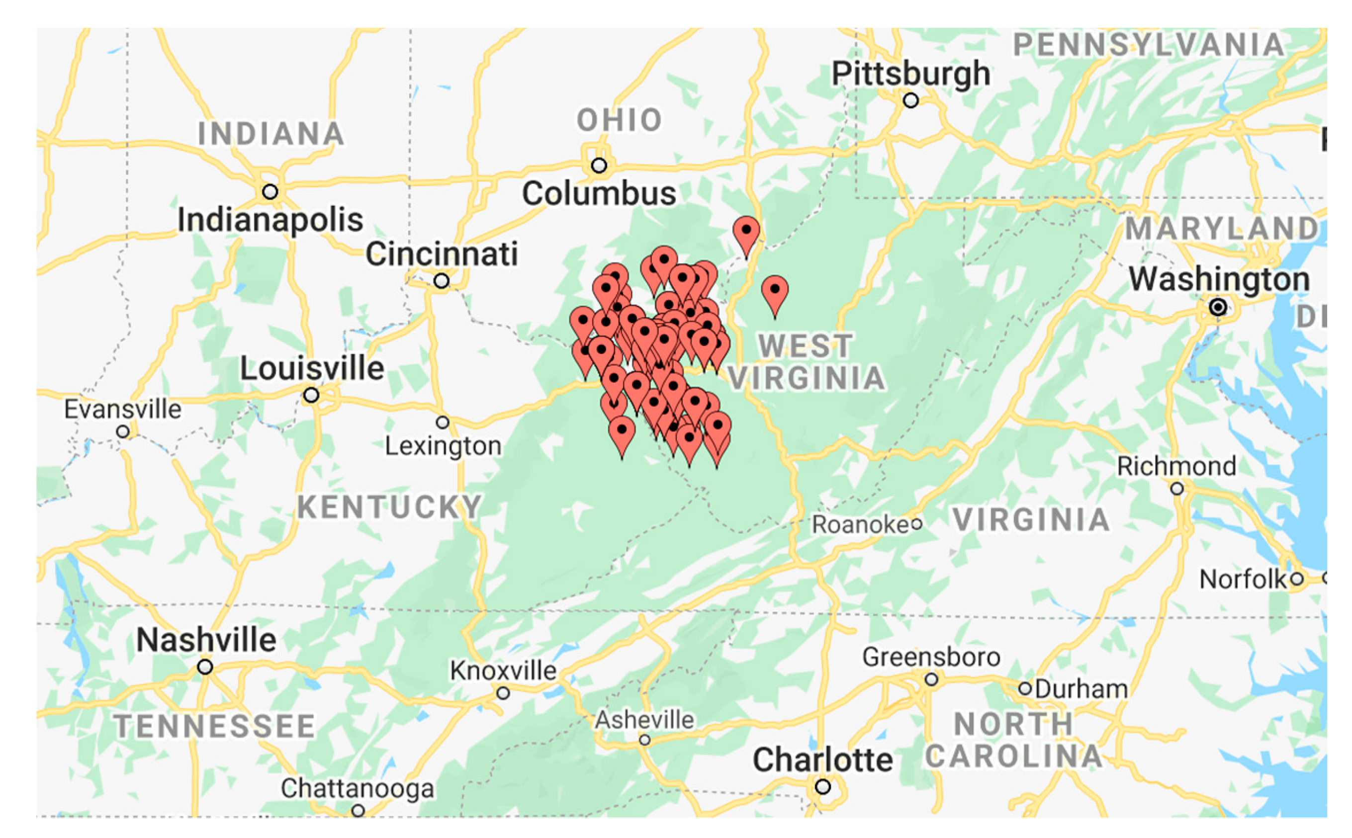
The Appalachian Region includes all of West Virginia and parts of 12 other surrounding states, covering 205,000 square miles and containing over 25 million people. Forty-two percent of the Appalachian Region population is rural, while only 20% of the nation’s population is rural. Across the region, poverty and unemployment rates, incomes, and education levels continue to fall behind the rest of the nation’s performance in these areas, despite efforts to bridge the gap. A few key indicators from a recent report on health disparities in Appalachia include higher mortality rates as a result of heart disease, cancer, stroke, diabetes, drug overdose, and suicide when compared to the rest of the nation, as well as a higher prevalence of depression, fewer healthcare professionals, and worse living conditions in the region [
15]. Despite recent improvements in several of these categories over the past two decades, our progress is insufficient when compared to that of the rest of the nation. We want to further investigate rural health disparities and how they affect neonatal health and thyroid function. A gap in the knowledge exists regarding whether or not babies born in the Appalachian region have an abnormal thyroid status, when comparing babies born to mothers from rural versus urban areas.
The thyroid gland plays a critical role in fetal development [
16,
17]. The developing fetus is reliant on transplacental passage of thyroid hormone until the 12th week of gestation, at which point thyroid hormones T3 and T4 begin to be produced by follicular cells of the thyroid gland. Clinically significant levels of T4 are produced by 18–20 weeks of gestation, while T3 significantly increases at 30 weeks of gestation. Postnatally, TSH, T4, and T3 levels rise abruptly, rapidly dwindle for the first five days of life, then gradually fall until about 1 month of life in both term and preterm infants. However, in preterm babies, the change in hormone levels is proportionally smaller [
18].
T3 is the metabolically active form of thyroid hormone and occupies nuclear receptors to mediate the actions of thyroid hormone, enabling proper brain, liver, and skeletal development [
19]. Without thyroid hormone, newborns may present with skeletal abnormalities due to a lack of endochondral and intramembranous ossification, longitudinal bone growth, and skeletal maturation. Thyroid hormones also regulate the expression of many genes important for embryonic central nervous system development, and newborns can develop permanent intellectual disability in the absence of timely and proper thyroid hormone supplementation [
19,
20,
21,
22].
Universal screening for congenital hypothyroidism obtained by a heel stick has helped to eliminate the devastating consequences of this condition, which affects 1 in 2000 to 1 in 4000 newborns [
23]. Additional screening tests are recommended for preterm infants to avoid false negative results from a delayed rise in serum TSH in these newborns [
24]. Reference intervals for thyroid hormone levels in infants between 1 day and 1 month old include 1.75–5 mIU/L TSH, 1.05–1.9 mg/dL T4, and 3.5–4.75 pg/mL T3 in females, and 3.7–4.5 pg/mL T3 in males. No reference intervals are available for infants less than 1 day old [
8]. Our neonate’s thyroid hormone levels were not comparable to these reference ranges, as mean TSH levels (8.64 mU/L) were higher and mean free T4 (0.97 ng/dL) and free T3 (1.44 ng/dL) levels were lower. The variability in results may be due to the recognized changes occurring at birth [
18]. However, TSH levels were comparable to a study by Shields and associates that reported TSH levels of 8.02 mIU/L in a cohort of their study examining blood levels during pregnancy and in umbilical cord blood [
25].
Attempting to account for thyroid disorder in relation to socioeconomic class, Talat, et al. did not show any association between income level and thyroid hormone levels [
26]. However, they did note that most of the subjects in their study had free access to healthcare and regular checkups, which may have affected the results. In another study, a Pakistani population’s prevalence of overt hypothyroidism was approximately 4%, while a study on northern Indian women showed a prevalence of 7%. The authors attributed differences in diet to partially account for this apparent disparity [
26,
27]. Furthermore, studies in China have shown variation in thyroid hormone levels in different races [
28].
A recent cross-sectional study by Velasco et al. examined the relationship between maternal and neonatal thyroid parameters, the effects of those hormone levels on birth outcomes, and the specific role of thyroid hormone in the regulation of fetal growth in low-risk pregnancies lacking any condition of obstetric or perinatal risk versus high-risk pregnancies. They found that maternal thyroid hormones may have a significant impact on fetal growth regulation. Thyroid function and iodine status at birth was affected by fetal iodine status, placental function, amniotic fluid levels, labor onset, and mode of delivery. Mean urinary iodine concentration was lower in high-risk pregnancies, and non-consumption of iodine supplements was associated with preterm births. However, iodine concentration in amniotic fluid was higher in high-risk pregnancies and had a negative correlation with birth weight. A negative association between maternal free T4 and neonatal birth weight could be explained by the hypothesized effect of compromised transplacental transfer of thyroid hormone: The more impeded the transfer of thyroid hormone is, the higher the maternal free T4 will be and the lower the neonatal thyroid hormone level will be. All thyroid parameters were higher in the low-risk pregnancy group besides free T4, which was similar between the low- and high-risk groups, with a positive correlation between maternal and neonatal thyroid hormone levels across the board. They concluded that an unfavorable environment in utero can limit the availability of free T4 in cord blood and limit fetal iodine metabolism, especially in small fetuses [
29]. Due to the limited number of neonatal thyroid studies available, our study examined whether or not specific maternal demographics impacted neonatal thyroid hormone levels. Our findings indicated that conditions of obesity and diabetes, which are more prevalent in the Appalachian region, were associated with an altered thyroid hormone status.
Previous studies have shown that maternal BMI may have an effect on thyroid function. Upon analysis of thyroid glands from electively terminated fetuses in the second trimester, Filis et al. found significant changes in thyroid function, increased fetal thyroid mass and plasma TSH levels, and changes in the expression of genes critical for proper thyroid development based upon maternal BMI [
30]. Another study conducted on Zhuang ethnic pregnant women did not find any statistically significant correlation between BMI and overt hypothyroidism, but a BMI > 25 was associated with a higher risk of overt hypothyroxinemia, defined as having a normal TSH with free T4 levels in the lower 2.5th percentile, based on the reference range for each trimester [
31]. However, our study did not show any association between BMI and thyroid disease.
In regard to fetal thyroid function and maternal diabetes, our study showed a significant difference with lower total T4 and free T4 in those with diabetes compared to those without diabetes (
Table 7). In 2000, the prevalence of thyroid disease in the general population and non-diabetics was 6.6% and 10.8%, respectively, with hypothyroidism being the most common type of thyroid dysfunction [
32]. Due to their increased risk of thyroid disorders, diabetics are screened regularly for thyroid abnormalities [
32,
33]. Additionally, a study in 2012 by Karakosta et al. showed a 4-fold increased risk of gestational diabetes associated with mothers who had a high TSH and thyroid antibodies in the first trimester of pregnancy [
34]. Recent studies have revealed a potential link between thyroid dysfunction and the development of type 2 diabetes mellitus due to dysregulated intestinal glucose absorption, alterations in gene expression, and aberrant hepatic processing of glucose that can occur in the setting of thyroid disease. In fact, hypothyroidism and hyperthyroidism have been associated with the development of insulin resistance [
35,
36]. We were limited by our number of diabetic patients included in our study, and further study is needed in this area. However, the lower total T4 and free T4 in the umbilical cord blood of mothers with diabetes suggests that newborns should be evaluated for thyroid status during well baby and pediatric examinations.
Smoking during pregnancy may also have an effect on thyroid function. Based upon urinary cotinine levels, cigarette smoking has been associated with decreased TSH levels and increased TPO titers in a dose-dependent manner [
37]. Proposed mechanisms to explain this finding are that smoke may interfere with the process of iodide transport and organification, or smoking may have a stimulatory effect on the thyroid gland, leading to higher levels of thyroxin-binding globulin and T3 concentrations and lower TSH concentrations due to negative feedback [
37]. Similarly, a 2007 Swedish study demonstrated that smoke exposure, assessed by urinary cotinine levels and questionnaires, was associated with higher free T3 and total T3 and lower concentrations of TSH during pregnancy [
38]. In our study, only cotinine levels affected T4. Cigarette smoking is associated with exposure to cadmium and lead, but in the present study, no differences were measured in lead or cadmium levels between smokers. The lack of a difference in metal levels in smokers may be due to the elapsed time between smoking and collection of umbilical cord blood. However, smoking was sufficient in some individuals to induce DNA adducts with BaP. This covalent binding reflects long-term exposure.
This is the largest study to date attempting to characterize the thyroid hormone levels at delivery of infants born in Appalachia. A strength of our study was the fairly homogenous patient population surveyed, robust numbers, and demographic information that was collected at the time of admission to labor and delivery. While our study was prospective in nature, due to the paucity of knowledge regarding the appropriate level of thyroid hormones at birth, more study is needed in this area. A follow-up of the infants whose cord blood was collected for this study is planned.


