Article

Palmitic Acid Targets Human Testicular Peritubular Cells and Causes a Pro-Inflammatory Response

1
Biomedical Center, Cell Biology, Anatomy III, Ludwig Maximilian University Munich, D-82152 Planegg-Martinsried, Großhaderner Straße 9, 82152 Munich, Germany
2
Division of Neuroscience, Oregon National Primate Research Center, Beaverton, OR 97006, USA
3
Andrologicum München, 80331 Munich, Germany
4
Urologie und Andrologie am Promenadeplatz, 80333 Munich, Germany
5
Division of Cardiometabolic Health, Oregon National Primate Research Center, Beaverton, OR 97006, USA
*
Author to whom correspondence should be addressed.
J. Clin. Med. 2020, 9(8), 2655; https://doi.org/10.3390/jcm9082655
Submission received: 27 July 2020 / Revised: 7 August 2020 / Accepted: 13 August 2020 / Published: 17 August 2020
(This article belongs to the Special Issue Updates in Male Infertility)

Abstract

:
Palmitic acid (PA) is a major fatty acid, derived from diet and endogenous production, which is being linked to inflammation. While such actions of PA at the level of the testis remain difficult to examine, we reasoned that studies in human testicular cells may be instructive. Human testicular peritubular cells (HTPCs) can be isolated from men and cultured. They have contractile properties but also produce Interleukin 6 (IL6), express the inflammasome member NLRP3, and via glia cell line derived neurotrophic factor (GDNF), they contribute to the spermatogonial stem cell niche. We found that PA at 100 µM significantly increased the levels of IL6, while NLRP3 or the related Interleukin 1 beta (IL1beta) were not affected. The contractility marker calponin (CNN1) and the growth factor GDNF were likewise not affected. ELISA studies confirmed the stimulatory PA actions on IL6. Hence, PA derived from diet and/or endogenous sources may be able to foster a pro-inflammatory milieu in the testis. A possible link of these results to diet and high fat intake and obesity is indicated by the about 12-fold elevated testicular levels of IL6 in testes of obese rhesus monkeys (n = 3), fed with a Western Style diet. They had elevated 2–5-fold increased body fat and increased circulating triglyceride levels. Further consequences of PA and obesity for testicular functions remain to be evaluated.

1. Introduction

There is evidence for a sterile type of inflammation in the testes of men suffering from idiopathic infertility [1,2,3]. The changes are typical for a pro-inflammatory tissue environment and are predominantly observed in the peritubular wall. This small testicular compartment in healthy, fertile men consists of elongated smooth muscle-like peritubular cells, i.e., contractile cells, which transport sperm and secrete extracellular matrix [3,4]. In contrast, in infertile men, fibrotic remodeling of the wall and immune cells, i.e., tryptase- and, in part, chymase-positive mast cells and CD68-positive macrophages, which also express tumor necrosis factor alpha (TNFalpha), are among the obvious changes. In addition, phenotypic changes of peritubular cells, namely a loss of smooth muscle characteristics occur [5,6,7,8,9,10]. The data suggest that the peritubular wall and its cells play roles in male infertility.
Peritubular cells are not well examined cells. However, they can be isolated from patients (HTPCs) and were previously characterized (e.g., [2,11,12,13]). It became clear that they are more than contractile cells. They produce e.g., glial cell lined derived factor (GDNF), required for spermatogonial stem cell (SSC) renewal [14], but also a number of pro-inflammatory factors, including interleukin 6 (IL6), and other cytokines, and they express the inflammasome family member NLRP3 (see [2,12,13,15]) and Toll-like receptors (TLRs; [1]). Thus, peritubular cells contribute to diverse testicular functions, including immunological properties of the testis and may fuel events that contribute to male sub/infertility.
Fatty acids (FAs) stem from ingested lipids and are produced by de novo-synthesis. Saturated FAs, particularly stearic, lauric, and palmitic acid (PA; C16:1), the latter being the main product of activity of fatty acid synthase, are known to activate TLR2 and 4. This activation was shown to drive pro-inflammatory responses and expression of pro-inflammatory genes (see [16,17,18,19]). Whether lipids and FAs/PA may target testicular peritubular cells and hence, whether they may be involved in the induction of testicular inflammation in man, is not known. In rodents, PA acts at the level of the male gonad, namely at Sertoli cells [20], where induction of dysfunction and apoptosis were reported [21,22].
Increased adiposity and high-fat diet (HFD) are linked to chronic systemic inflammation [23,24]. Increased circulating levels of pro-inflammatory cytokines, e.g., TNFalpha, Interleukin 1beta (IL1beta), and IL6 are reported [25,26]. In the context of the male gonad, IL6 is of interest, as it is involved in the regulation of the blood-testis barrier, formed by Sertoli cells. Elevated levels may lead to its breakdown, as shown in a rodent study [27]. The consequences may include exposure of spermatogenic cells to blood-borne harmful agents and induce autoimmune events [28], with deleterious consequences on sperm production.
While the mentioned data mainly stem from animal studies, there is evidence for adverse effects of obesity on reproduction in men [29,30]. They include lower testosterone levels and lower sperm counts, (see [31]). The mechanisms are under discussion and neuroinflammation causing a dysregulation of the hypothalamus–pituitary–gonadal axis, specifically at the level of the GnRH neurons (see [31]) was suggested as one possible reason.
Other possible actions, e.g., ones exerted by PA at the level of the human testis, are not well known and remain difficult to examine in man. We reasoned that studies employing HTPCs may be instructive and therefore we examined actions of PA in cultured HTPCs. In addition, expression levels of inflammatory genes in testes of three obese monkeys with elevated body fat and high circulating triglycerides were evaluated.

2. Materials and Methods

2.1. Human Samples: HTPCs

As described, the HTPCs studied were isolated from small testicular fragments [12]. The study was approved by the local ethical committee (Ethikkommission, Technische Universität München, Fakultät für Medizin, project number 309/14 and project 491/18S-KK). All patients provided written informed consent. The experiments were carried out in accordance with the relevant guidelines and regulations. Derived cells were cultured in DMEM High Glucose (Gibco, Paisley, UK) with 10% fetal bovine serum (Capricorn Scientific, Ebsdorfergrund, Germany), 1% penicillin/streptomycin (Biochrom, Berlin, Germany) at 37 °C and 5% (v/v) CO2. The cells studied in frame of this study stem from a total of 14 individual donors, with diagnoses ranging from obstructive azoospermia and normal spermatogenesis to non-obstructive idiopathic azoospermia. The age of the donors ranged from 29 to 55 years. All cells were initially characterized, and we verified that they express typical HTPCs markers, including androgen receptor (AR) and smooth musle actin (ACTA2).

2.2. Samples from Normal and Obese Monkeys

Rhesus monkey testis mRNA was obtained from unrelated studies approved by the Oregon National Primate Research Center (ONPRC) Institutional Animal Care and Use Committee. Obese animals were maintained on a high-fat diet (HFD) consisting of 36% fat (mostly saturated fats from lard; TAD Primate Diet—5LOP, Test Diet, St. Louis, MO, USA) that is supplemented with calorically dense treats (5LOP pellets, peanut butter, honey, banana, and cornstarch). Control animals were maintained on a diet consisting of 13% fat (from soybean oil, Monkey Diet 5045, Lab Diet, St. Louis, MO, USA).
Testicular RNA used for RT-PCR was extracted from 6 lean adult monkeys (age range 20–24 years, body weight range 9–11 kg) and 3 obese (age range 13–18 years, body weight range 16–24 kg).

2.3. Stimulation Experiments with Palmitic Acid (PA)

The stimulation with PA (Sigma Aldrich (P9767), St. Louis, MO, USA) was conducted as follows: 30.6 mg PA were dissolved in 150 mM NaCl and 0.17 mM BSA (Ultra Fatty Acid Free BSA, Roche 10775835001) to reach a 1 mM PA working solution (final ratio of 6:1). A solution of 150 mM NaCl/BSA served as solvent control. After performing pilot studies (with cells from 4 donors) for the experiments described here, HTPCs were incubated with/without 100 µM PA for 24 h in serum free medium. In all experiments a solvent control was included, which allowed us to compare the results of the treatment. The PA concentration of 100 µM was chosen in pilot experiments because it did not negatively affect viability of HTPCs and because the reported plasma concentration of FAs in a HFD can reach 100–400 µM or more (see [32]). Experiments (treatment and control) were done with cells from individual donors and then repeated with cells from other donors. For the qPCR studies cells from 6 donors were used, for the ELISA studies cells from additional 4 donors were studied.

2.4. Isolation of RNA, Reverse Transcription (RT-PCR) and Quantitative Real Time PCR (qPCR)

All procedures used were described previously [12]. In brief, RNeasy Plus Micro Kit (Qiagen, Hilden Germany) was used for total RNA isolation from HTPCs in passages 7–10 [12] and 200 ng RNA were reverse transcribed, using random 15mer primer and SuperScriptTM II (Invitrogen, Darmstadt, Germany). A LightCycler® 96 System (Roche) and QuantiFast® SYBR Green PCR Kit (Qiagen) were used. Primer information is given in Table 1. Unless indicated otherwise, primers matched human and rhesus monkey sequences. Non-reverse transcription control and non-template control were used as negative controls.

2.5. IL6 ELISA Measurements

The assay (Platinum Elisa Human IL6, #BMS213, affimetrix eBioscience, Bender MedSystems GmbH, Vienna, Austria) was performed, as described earlier (see [1,33]). Cellular supernatant of control and PA-treated cells (24 h) were derived from 4 donors. Samples were run in duplicates and values were normalized to cellular protein.

2.6. Statistical Analysis

The results of qPCRs were analyzed according to the 2−ΔΔCq method as described earlier (Walenta et al., 2018) and expression levels were normalized to RPL19, which served as endogenous reference.
Results were normalized to the controls (control treatment of HTPCs or non-obese samples). They are depicted as means ± SEM. Statistical analysis was done by t-tests (two-tailed) of ΔΔCq values and ELISA results, using GraphPad Prism 6.0 Software (GraphPad Software, San Diego, CA, USA).

3. Results

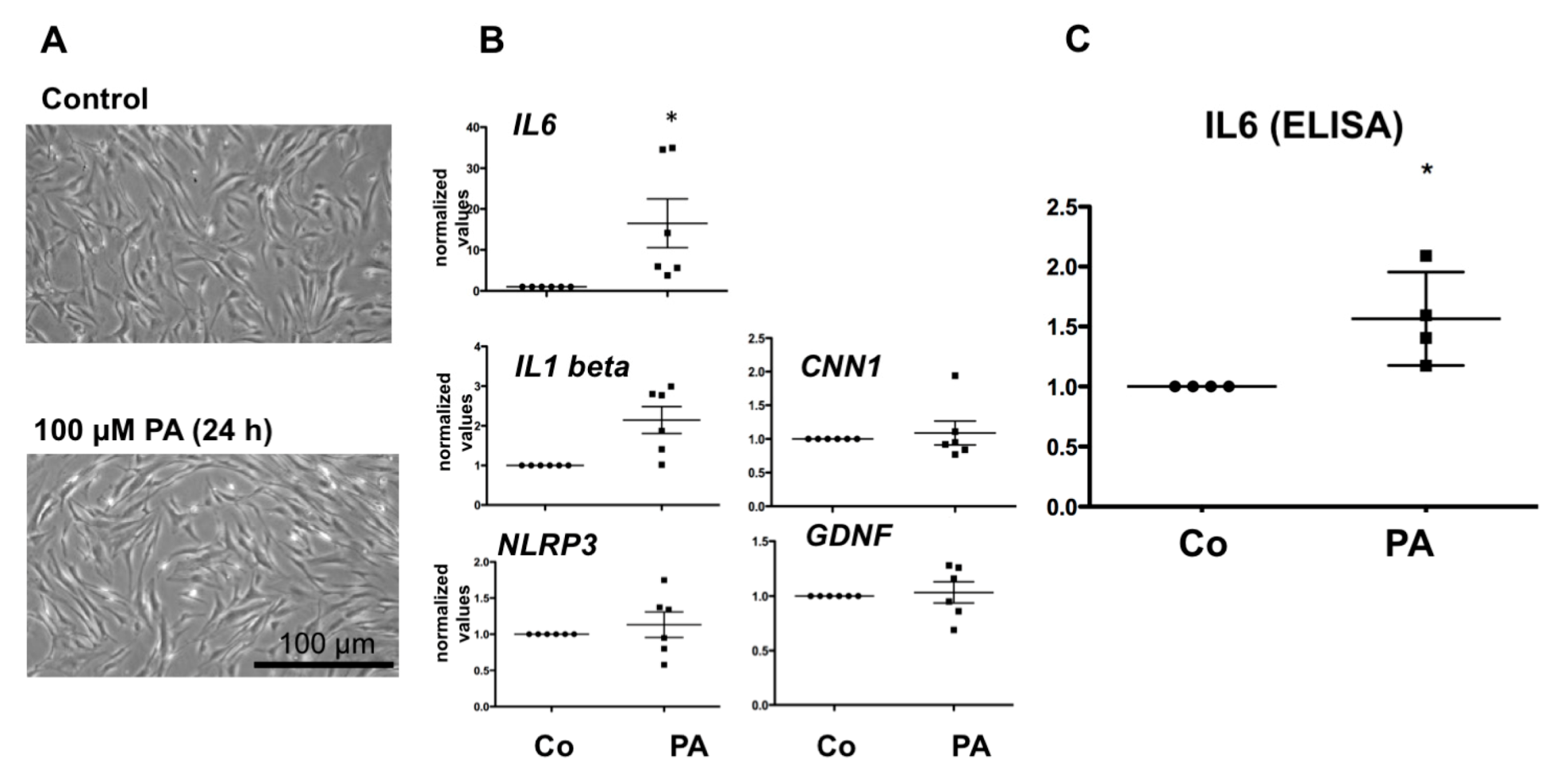
3.1. Effect of PA on HTPC mRNA Level of IL6, NLRP3, IL1beta and CNN1

As shown in Figure 1A, the cellular morphology of HTPCs cultured with 100 µM PA for 24 h did not change compared to cells cultured in the presence of the solvent control. Under both conditions, cells were characterized by a slim, spindle-like shape. Using qPCR, we found that PA at 100 µM significantly (p < 0.05) increased mRNA levels of IL6, while NLRP3 and IL1beta were not significantly affected (p > 0.05) (Figure 1B). The levels of the contractility marker CNN1 and the growth factor GDNF were likewise not changed.

3.2. Effect of PA on IL6 Secretion in HTPCs

ELISA measurements confirmed the stimulatory PA actions of IL6 in 4 samples (Figure 1C), thus supporting the qPCR results.

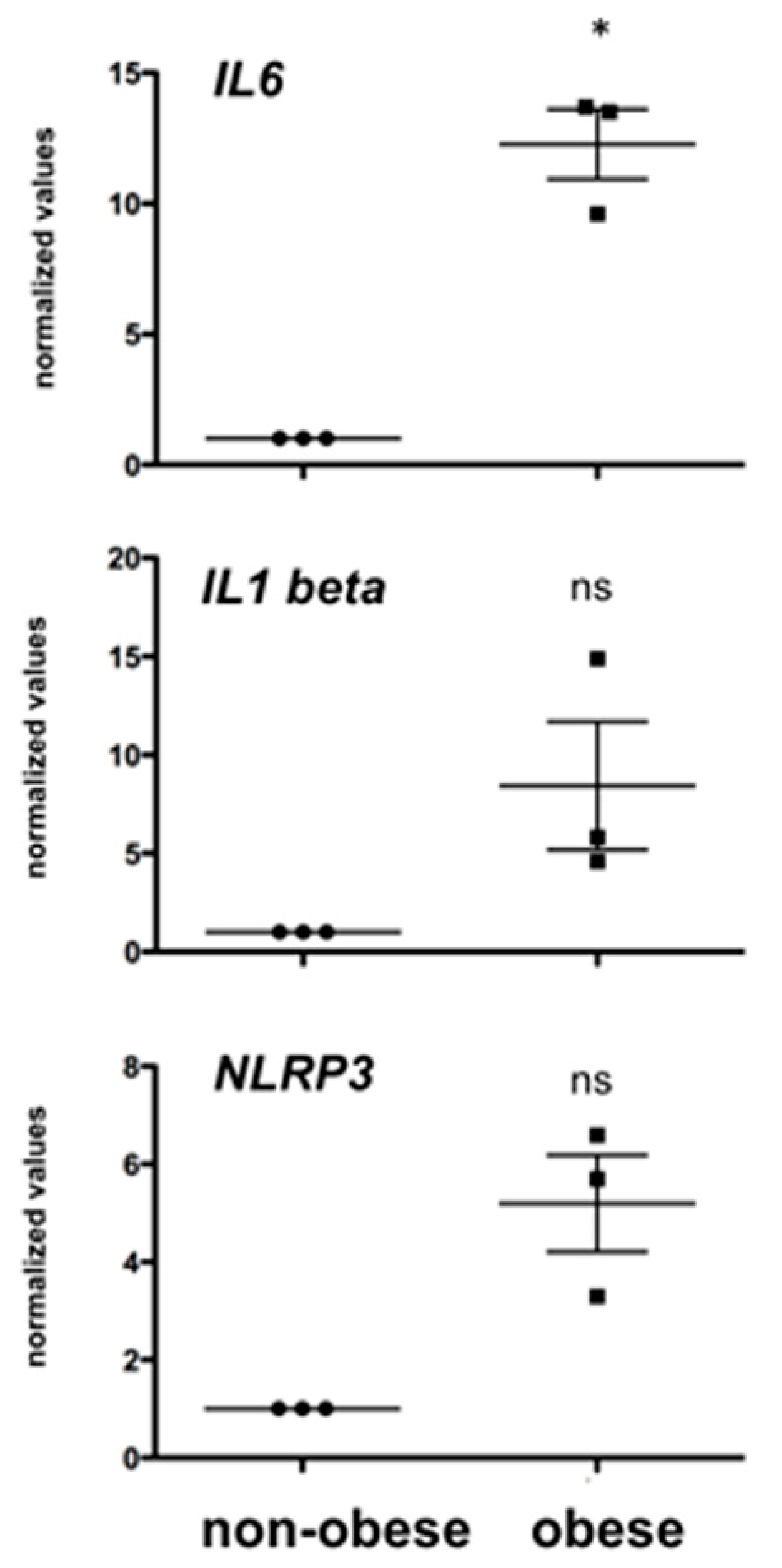
3.3. Obesity and Testicular IL6 Levels in Rhesus Monkeys

To explore consequences of obesity on testicular inflammatory markers, qPCR studies were performed using samples from obese monkeys and monkeys with normal body weight as controls. Levels of IL6 in the testes of the three obese adult Rhesus monkeys fed with a Western Style diet were almost 12-fold elevated (p < 0.05) compared to one in an age-matched control group (Figure 2). Levels of IL1beta and NLRP3 were also elevated but did not reach significance levels (p > 0.05).

4. Discussion

There is ample evidence for adverse effects of obesity on reproduction in men, namely lower testosterone levels and lower sperm counts [29,30,31,34]. A meta-analysis study in a large cohort of men, for example, came to the conclusion that obesity is positively correlated with a higher prevalence of azoospermia or oligozoospermia [35]. Another study reports that a high BMI in men is positively correlated with reduced semen parameters [36].
The underlying reasons for obesity-related impaired male reproductive function, specifically impaired spermatogenesis, are likely multifaceted and the chains of events remain difficult to examine in men, bearing in mind the complex regulation of testicular functions on one hand. On the other hand, obesity is also correlated with a host of diseases, including metabolic disease [16]. Medical conditions and medication, next to diet, may therefore also have a say and may result in impaired testis functions [37,38,39,40,41].
Rodent models are often used in biomedical research, including obesity research. Yet, results of a recent gene expression study of several organs clearly revealed that the testis, together with the liver, shows the largest differences in the expression of individual genes of all organs examined between the mouse and human species [42]. This emphasizes that in order to understand the human situation, adequate models for the human testis are required. Furthermore, the striking differences between the single-layered peritubular wall in rodents and the multilayered peritubular compartment in men and monkeys indicate that studies on peritubular cells require an adequate model.
In our study we therefore turned to an established cellular model, HTPCs, which are the only cells that can be isolated from the human testes, cultured and then examined in vitro. We employed them to explore the consequences of 100 µM PA. We tested possible actions on a smooth muscle marker, on GDNF, and inflammatory factors. PA is indeed well known to cause inflammation (e.g., [17,18,19]) and we found that PA elevates IL6 transcript and IL6 protein levels in HTPCs. Heterogeneity in basal and secreted IL6 levels of the human cells was observed, which is in line with the results of previous studies in HTPCs, in which IL6 was examined [1,6]. This may be due to the difference in age of the patients, differences in life style, and/or underlying reason for sub-/infertility. In contrast, PA did not significantly affect other HTPC-parameters under the experimental condition, including GDNF or the smooth muscle marker CNN1.
An HFD is linked to high circulating levels of FFA [32] and the reported plasma concentration of FFAs can reach up to 100–400 µM or even more. The concentration used in our study is in this range. Hence, provided that the cellular response is also present in the body, HTPCs may contribute to a pro-inflammatory milieu in the testis.
Evidence for such a milieu to exist in situ, in testes samples from three obese monkeys, with elevated blood triglyceride levels, was obtained, albeit in a limited set of samples. We found strikingly high expression levels of IL6 in the testes of obese monkeys. While elevated testicular IL6 in (whole) testes samples may stem from many testicular sources, peritubular cells are based on or result in HTPCs, likely included. Of note, other inflammatory parameters (NLRP3), also expressed by Sertoli cells; [2] and IL1beta showed a clear tendency to higher values, but due to the low number of available samples, they did not reach significance level.
Obesity in man is not only associated with impaired male reproductive function, but is also well known to be associated with low-grade, chronic inflammation, as reflected by alterations of immune cell populations and tissue remodeling in organs, such as liver, pancreas, and blood vessels [43]. Evidence for a sterile type of inflammation in the human testis was reported in cases of male idiopathic infertility due to impaired spermatogenesis [1,13]. The present results obtained in HTPCs and in a translational nonhuman primate model, suggest that obesity and/or increased dietary intake of PA, via induction of inflammation, could lead to a pro-inflammatory environment and possibly to impairments of testicular functions. Additional studies are now required to examine these points and elucidate the mechanisms and further consequences.

Author Contributions

K.-G.D. performed most of the cellular experiments, H.W. together with A.M. evaluated the data. P.K. and H.F.U. designed and executed the studies in monkeys, F.-M.K., U.P. and M.T. provided human samples. A.M. conceived of the study, directed the work. and supervised the experiments. A.M. and H.W. drafted the manuscript. All authors contributed to the manuscript and approved the final version. All authors have read and agreed to the published version of the manuscript.

Funding

Supported by DFG (MA 1080/27-1, 29-1 to A.M. and WE 4554/3- 1 to H.W.; common project number 427588170) and National Institutes of Health grants AG062220 and OD011092 to H.F.U.

Acknowledgments

The authors thank J.U. Schwarzer, Munich, for human samples, which allowed us to perform initial experiments. We gratefully acknowledge Astrid Tiefenbacher for skillful technical assistance and Karin Metzrath for editorial help.

Conflicts of Interest

The authors declare no competing interest.

References

  1. Mayer, C.; Adam, M.; Glashauser, L.; Dietrich, K.; Schwarzer, J.U.; Kohn, F.M.; Strauss, L.; Welter, H.; Poutanen, M.; Mayerhofer, A. Sterile inflammation as a factor in human male infertility: Involvement of Toll like receptor 2, biglycan and peritubular cells. Sci. Rep. 2016, 6, 37128. [Google Scholar] [CrossRef] [PubMed] [Green Version]
  2. Walenta, L.; Schmid, N.; Schwarzer, J.U.; Kohn, F.M.; Urbanski, H.F.; Behr, R.; Strauss, L.; Poutanen, M.; Mayerhofer, A. NLRP3 in somatic non-immune cells of rodent and primate testes. Reproduction 2018, 156, 231–238. [Google Scholar] [CrossRef] [PubMed]
  3. Mayerhofer, A. Human testicular peritubular cells: More than meets the eye. Reproduction 2013, 145, R107–R116. [Google Scholar] [CrossRef] [PubMed] [Green Version]
  4. Maekawa, M.; Kamimura, K.; Nagano, T. Peritubular myoid cells in the testis: Their structure and function. Arch. Histol. Cytol. 1996, 59, 1–13. [Google Scholar] [CrossRef] [Green Version]
  5. Welter, H.; Kohn, F.M.; Mayerhofer, A. Mast cells in human testicular biopsies from patients with mixed atrophy: Increased numbers, heterogeneity, and expression of cyclooxygenase 2 and prostaglandin D2 synthase. Fertil. Steril. 2011, 96, 309–313. [Google Scholar] [CrossRef]
  6. Welter, H.; Huber, A.; Lauf, S.; Einwang, D.; Mayer, C.; Schwarzer, J.U.; Kohn, F.M.; Mayerhofer, A. Angiotensin II regulates testicular peritubular cell function via AT1 receptor: A specific situation in male infertility. Mol. Cell. Endocrinol. 2014, 393, 171–178. [Google Scholar] [CrossRef]
  7. Meineke, V.; Frungieri, M.B.; Jessberger, B.; Vogt, H.; Mayerhofer, A. Human testicular mast cells contain tryptase: Increased mast cell number and altered distribution in the testes of infertile men. Fertil. Steril. 2000, 74, 239–244. [Google Scholar] [CrossRef]
  8. Schell, C.; Albrecht, M.; Mayer, C.; Schwarzer, J.U.; Frungieri, M.B.; Mayerhofer, A. Exploring human testicular peritubular cells: Identification of secretory products and regulation by tumor necrosis factor-alpha. Endocrinology 2008, 149, 1678–1686. [Google Scholar] [CrossRef]
  9. Frungieri, M.B.; Calandra, R.S.; Lustig, L.; Meineke, V.; Kohn, F.M.; Vogt, H.J.; Mayerhofer, A. Number, distribution pattern, and identification of macrophages in the testes of infertile men. Fertil. Steril. 2002, 78, 298–306. [Google Scholar] [CrossRef]
  10. Frungieri, M.B.; Weidinger, S.; Meineke, V.; Kohn, F.M.; Mayerhofer, A. Proliferative action of mast-cell tryptase is mediated by PAR2, COX2, prostaglandins, and PPARgamma: Possible relevance to human fibrotic disorders. Proc. Natl. Acad. Sci. USA 2002, 99, 15072–15077. [Google Scholar] [CrossRef] [Green Version]
  11. Flenkenthaler, F.; Windschuttl, S.; Frohlich, T.; Schwarzer, J.U.; Mayerhofer, A.; Arnold, G.J. Secretome analysis of testicular peritubular cells: A window into the human testicular microenvironment and the spermatogonial stem cell niche in man. J. Proteome Res. 2014, 13, 1259–1269. [Google Scholar] [CrossRef]
  12. Schmid, N.; Flenkenthaler, F.; Stockl, J.B.; Dietrich, K.G.; Kohn, F.M.; Schwarzer, J.U.; Kunz, L.; Luckner, M.; Wanner, G.; Arnold, G.J.; et al. Insights into replicative senescence of human testicular peritubular cells. Sci. Rep. 2019, 9, 15052. [Google Scholar] [CrossRef] [PubMed] [Green Version]
  13. Walenta, L.; Fleck, D.; Frohlich, T.; von Eysmondt, H.; Arnold, G.J.; Spehr, J.; Schwarzer, J.U.; Kohn, F.M.; Spehr, M.; Mayerhofer, A. ATP-mediated Events in Peritubular Cells Contribute to Sterile Testicular Inflammation. Sci. Rep. 2018, 8, 1431. [Google Scholar] [CrossRef] [PubMed] [Green Version]
  14. Spinnler, K.; Kohn, F.M.; Schwarzer, U.; Mayerhofer, A. Glial cell line-derived neurotrophic factor is constitutively produced by human testicular peritubular cells and may contribute to the spermatogonial stem cell niche in man. Hum. Reprod. 2010, 25, 2181–2187. [Google Scholar] [CrossRef] [PubMed]
  15. Eubler, K.; Herrmann, C.; Tiefenbacher, A.; Kohn, F.M.; Schwarzer, J.U.; Kunz, L.; Mayerhofer, A. Ca2+ Signaling and IL-8 Secretion in Human Testicular Peritubular Cells Involve the Cation Channel TRPV2. Int. J. Mol. Sci. 2018, 19, 2829. [Google Scholar] [CrossRef] [PubMed] [Green Version]
  16. Johnson, A.R.; Milner, J.J.; Makowski, L. The inflammation highway: Metabolism accelerates inflammatory traffic in obesity. Immunol. Rev. 2012, 249, 218–238. [Google Scholar] [CrossRef] [Green Version]
  17. Korbecki, J.; Bajdak-Rusinek, K. The effect of palmitic acid on inflammatory response in macrophages: An overview of molecular mechanisms. Inflamm. Res. 2019, 68, 915–932. [Google Scholar] [CrossRef] [Green Version]
  18. Nemecz, M.; Constantin, A.; Dumitrescu, M.; Alexandru, N.; Filippi, A.; Tanko, G.; Georgescu, A. The Distinct Effects of Palmitic and Oleic Acid on Pancreatic Beta Cell Function: The Elucidation of Associated Mechanisms and Effector Molecules. Front. Pharmacol. 2018, 9, 1554. [Google Scholar] [CrossRef] [Green Version]
  19. Fatima, S.; Hu, X.; Gong, R.H.; Huang, C.; Chen, M.; Wong, H.L.X.; Bian, Z.; Kwan, H.Y. Palmitic acid is an intracellular signaling molecule involved in disease development. Cell. Mol. Life Sci. 2019, 76, 2547–2557. [Google Scholar] [CrossRef]
  20. Jutte, N.H.; Eikvar, L.; Levy, F.O.; Hansson, V. Metabolism of palmitate in cultured rat Sertoli cells. J. Reprod. Fertil. 1985, 73, 497–503. [Google Scholar] [CrossRef] [Green Version]
  21. Hu, X.; Ge, X.; Liang, W.; Shao, Y.; Jing, J.; Wang, C.; Zeng, R.; Yao, B. Effects of saturated palmitic acid and omega-3 polyunsaturated fatty acids on Sertoli cell apoptosis. Syst. Biol. Reprod. Med. 2018, 64, 368–380. [Google Scholar] [CrossRef] [PubMed] [Green Version]
  22. Luo, D.; Zhang, M.; Su, X.; Liu, L.; Zhou, X.; Zhang, X.; Zheng, D.; Yu, C.; Guan, Q. High fat diet impairs spermatogenesis by regulating glucose and lipid metabolism in Sertoli cells. Life Sci. 2020, 257, 118028. [Google Scholar] [CrossRef] [PubMed]
  23. Dorfman, M.D.; Thaler, J.P. Hypothalamic inflammation and gliosis in obesity. Curr. Opin. Endocrinol. Diabetes Obes. 2015, 22, 325–330. [Google Scholar] [CrossRef] [PubMed] [Green Version]
  24. Osborn, O.; Olefsky, J.M. The cellular and signaling networks linking the immune system and metabolism in disease. Nat. Med. 2012, 18, 363–374. [Google Scholar] [CrossRef]
  25. Lainez, N.M.; Jonak, C.R.; Nair, M.G.; Ethell, I.M.; Wilson, E.H.; Carson, M.J.; Coss, D. Diet-Induced Obesity Elicits Macrophage Infiltration and Reduction in Spine Density in the Hypothalami of Male but Not Female Mice. Front. Immunol. 2018, 9, 1992. [Google Scholar] [CrossRef]
  26. Olefsky, J.M.; Glass, C.K. Macrophages, inflammation, and insulin resistance. Annu. Rev. Physiol. 2010, 72, 219–246. [Google Scholar] [CrossRef]
  27. Perez, C.V.; Sobarzo, C.M.; Jacobo, P.V.; Pellizzari, E.H.; Cigorraga, S.B.; Denduchis, B.; Lustig, L. Loss of occludin expression and impairment of blood-testis barrier permeability in rats with autoimmune orchitis: Effect of interleukin 6 on Sertoli cell tight junctions. Biol. Reprod. 2012, 87, 122. [Google Scholar] [CrossRef]
  28. Fan, Y.; Liu, Y.; Xue, K.; Gu, G.; Fan, W.; Xu, Y.; Ding, Z. Diet-induced obesity in male C57BL/6 mice decreases fertility as a consequence of disrupted blood-testis barrier. PLoS ONE 2015, 10, e0120775. [Google Scholar] [CrossRef]
  29. Liu, Y.; Ding, Z. Obesity, a serious etiologic factor for male subfertility in modern society. Reproduction 2017, 154, R123–R131. [Google Scholar] [CrossRef] [Green Version]
  30. Chavarro, J.E.; Toth, T.L.; Wright, D.L.; Meeker, J.D.; Hauser, R. Body mass index in relation to semen quality, sperm DNA integrity, and serum reproductive hormone levels among men attending an infertility clinic. Fertil. Steril. 2010, 93, 2222–2231. [Google Scholar] [CrossRef] [Green Version]
  31. Lainez, N.M.; Coss, D. Obesity, Neuroinflammation, and Reproductive Function. Endocrinology 2019, 160, 2719–2736. [Google Scholar] [CrossRef]
  32. Ulven, T.; Christiansen, E. Dietary Fatty Acids and Their Potential for Controlling Metabolic Diseases Through Activation of FFA4/GPR120. Annu. Rev. Nutr. 2015, 35, 239–263. [Google Scholar] [CrossRef] [PubMed]
  33. Rossi, S.P.; Walenta, L.; Rey-Ares, V.; Kohn, F.M.; Schwarzer, J.U.; Welter, H.; Calandra, R.S.; Frungieri, M.B.; Mayerhofer, A. Alpha 1 adrenergic receptor-mediated inflammatory responses in human testicular peritubular cells. Mol. Cell. Endocrinol. 2018, 474, 1–9. [Google Scholar] [CrossRef] [PubMed]
  34. MInguez-Alarcón, L.; Chavarro, J.E.; Mendiola, J.; Roca, M.; Tanrikut, C.; Vioque, J.; Jorgensen, N.; Torres-Cantero, A.M. Fatty acid intake in relation to reproductive hormones and testicular volume among young healthy men. Asian J. Androl. 2017, 19, 184–190. [Google Scholar] [CrossRef] [PubMed]
  35. Sermondade, N.; Faure, C.; Fezeu, L.; Shayeb, A.G.; Bonde, J.P.; Jensen, T.K.; Van Wely, M.; Cao, J.; Martini, A.C.; Eskandar, M.; et al. BMI in relation to sperm count: An updated systematic review and collaborative meta-analysis. Hum. Reprod. Update 2013, 19, 221–231. [Google Scholar] [CrossRef] [PubMed]
  36. Hart, R.J.; Doherty, D.A.; McLachlan, R.I.; Walls, M.L.; Keelan, J.A.; Dickinson, J.E.; Skakkebaek, N.E.; Norman, R.J.; Handelsman, D.J. Testicular function in a birth cohort of young men. Hum. Reprod. 2015, 30, 2713–2724. [Google Scholar] [CrossRef] [PubMed] [Green Version]
  37. Du Plessis, S.S.; Cabler, S.; McAlister, D.A.; Sabanegh, E.; Agarwal, A. The effect of obesity on sperm disorders and male infertility. Nat. Rev. Urol. 2010, 7, 153–161. [Google Scholar] [CrossRef]
  38. Hammoud, A.O.; Meikle, A.W.; Reis, L.O.; Gibson, M.; Peterson, C.M.; Carrell, D.T. Obesity and male infertility: A practical approach. Semin. Reprod. Med. 2012, 30, 486–495. [Google Scholar] [CrossRef]
  39. Andersen, J.M.; Herning, H.; Aschim, E.L.; Hjelmesaeth, J.; Mala, T.; Hanevik, H.I.; Bungum, M.; Haugen, T.B.; Witczak, O. Body Mass Index Is Associated with Impaired Semen Characteristics and Reduced Levels of Anti-Mullerian Hormone across a Wide Weight Range. PLoS ONE 2015, 10, e0130210. [Google Scholar] [CrossRef]
  40. McPherson, N.O.; Lane, M. Male obesity and subfertility, is it really about increased adiposity? Asian J. Androl. 2015, 17, 450–458. [Google Scholar] [CrossRef]
  41. Leisegang, K.; Henkel, R. The impact of male overweight on semen quality and outcome of assisted reproduction. Asian J. Androl. 2014, 16, 787. [Google Scholar] [CrossRef] [PubMed]
  42. Cardoso-Moreira, M.; Halbert, J.; Valloton, D.; Velten, B.; Chen, C.; Shao, Y.; Liechti, A.; Ascencao, K.; Rummel, C.; Ovchinnikova, S.; et al. Gene expression across mammalian organ development. Nature 2019, 571, 505–509. [Google Scholar] [CrossRef] [PubMed]
  43. Lumeng, C.N.; Saltiel, A.R. Inflammatory links between obesity and metabolic disease. J. Clin. Investig. 2011, 121, 2111–2117. [Google Scholar] [CrossRef] [PubMed] [Green Version]
Figure 1. Effect of a 24 h incubation period with 100 µM palmitic acid (PA) on human testicular peritubular cells (HTPCs) compared to the corresponding solvent control (Co) on cellular morphology (A), two representative pictures are shown: (B) mRNA level of Interleukin 6 (IL6), Interleukin 1 beta (IL 1beta), the inflammasome NLRP3, calponinin (CNN1) and glial cell lined derived factor (GDNF), (C) IL6 secretion of HTPCs. n = 6 for mRNA and n = 4 for IL6 protein; individual values of the treatment group are shown, as well as the mean ± SEM; values were normalized to the respective solvent control. Asterisk denotes statistical significance, * p < 0.05.
Jcm 09 02655 g001
Figure 2. Levels of testicular Interkeulin 6 (IL6) were significantly elevated in testes of obese monkeys, while higher levels of Interleukin 1 beta (IL1beta) or the inflammasome NLRP3 did not reach statistical significance; n = 3, * p < 0.05; ns: not statistically significant. The data of the obese animals (n = 3) are given as individual levels and as the mean ± SEM. The values were normalized to the levels of the controls group (pooled results of n = 6 non-obese monkeys).
Jcm 09 02655 g002
Table 1. Nucleotide sequence of primers (Gene and gene name) employed in real time PCR including amplicon size and reference ID.
GeneGene NameReference IDNucleotide SequenceAmplicon Size
mk
IL6
Interleukin 6NM_001042733.25′-TGGCTGAAAAAGATGGATGCT-3′
5′-TTGCTGCTCACTACTCTCAAACCT-3′
134
mk
IL1 β
Interleukin 1 betaNM_001042756.15′-AAAGCTTGGTGATGTCTGGTC-3′
5′-GGACATGGAGAACACCACTTG-3′
89
NLRP3NLR family, pyrin domain containing 3NM_001114351.15′-GTGTTTCGAATCCCACTGTG-3′
5′-TCTGCTTCTCACGTACTTTCTG-3′
143
IL6Interleukin 6NM_000600.45′-AACCTGAACCTTCCAAAGATGG-3′
5′-TCTGGCTTGTTCCTCACTACT-3′
159
CNN1CalponinNM_001308342.25′-CGAAGACGAAAGGAAACAAGGT-3′
5′-GCTTGGGGTCGTAGAGGTG-3′
186
GDNFGlial cell line derived neurotrophic factorNM_000514.35’-GCAGACCCATCGCCTTTGAT-3′
5′-ATCCACACCTTTTAGCGGAATG-3′
93
RPL19Ribosomal protein L19NM_0009815′-AGGCACATGGGCATAGGTAA-3′
5′-CCATGAGAATCCGCTTGTTT-3′
199

