Article

Improving the Prognostic Performance of SUVmax in 18F-Fluorodeoxyglucose Positron-Emission Tomography/Computed Tomography Using Tumor-to-Liver and Tumor-to-Blood Standard Uptake Ratio for Locally Advanced Cervical Cancer Treated with Concurrent Chemoradiotherapy

1
Department of Obstetrics and Gynecology, School of Medicine, Kyungpook National University, Daegu 41944, Korea
2
Department of Obstetrics and Gynecology, Kyungpook National University Chilgok Hospital, Daegu 41404, Korea
3
Department of Nuclear Medicine, School of Medicine, Kyungpook National University, Daegu 41944, Korea
4
Department of Nuclear Medicine, Kyungpook National University Chilgok Hospital, Daegu 41404, Korea
5
Department of Radiation Oncology, School of Medicine, Kyungpook National University, Daegu 41944, Korea
6
Department of Radiation Oncology, Kyungpook National University Hospital, Daegu 41944, Korea
7
Department of Obstetrics and Gynecology, Kyungpook National University Hospital, Daegu 41944, Korea
*
Author to whom correspondence should be addressed.
J. Clin. Med. 2020, 9(6), 1878; https://doi.org/10.3390/jcm9061878
Submission received: 4 May 2020 / Revised: 26 May 2020 / Accepted: 13 June 2020 / Published: 16 June 2020
(This article belongs to the Special Issue Screening and Tests for Gynecologic Cancer)

Abstract

:
Objective: We sought to evaluate whether the 18F-fluorodeoxyglucose uptake normalization of the primary tumor to both the liver and blood pool and lymph nodes to both the liver and blood pool can enhance the discrimination for prognosis prediction in patients with cervical cancer. Methods: A total of 156 patients with cervical cancer (International Federation of Gynecology and Obstetrics stages IIB–IV) treated with concurrent chemoradiotherapy (CCRT) were enrolled. The maximum standardized uptake value (SUVmax) of tumor (tSUVmax) and the lymph node (nSUVmax) divided by the SUVmean of the liver (tumor-to-liver ratio (TLR) and node-to-liver (NLR)) and blood pool (tumor-to-blood ratio (TBR) and node-to-blood ratio (NBR)) were investigated. Univariate and multivariate analyses of disease-free survival (DFS) and overall survival (OS) were performed using clinical and metabolic parameters. A receiver operating characteristic curve analysis was performed to compare the accuracy of the metabolic parameters. Results: The multivariate analysis revealed that NLR (hazard ratio ((HR): 3.54; 95% confidence interval (CI): 1.53–8.19; p = 0.0032) and NBR (HR: 3.38; 95% CI: 1.02–11.19; p = 0.0457)) were independent prognostic factors for DFS, while TLR (HR: 4.16; 95% CI: 1.19–14.50; p = 0.0252), TBR (HR: 3.01; 95% CI: 1.04–8.70; p = 0.0415), NLR (HR: 4.84; 95% CI: 1.58–14.81; p = 0.0057), and NBR (HR: 6.87; 95% CI: 1.55–30.54; p = 0.0113) were significant prognostic factors for OS. The normalization of tSUVmax to the liver or blood pool enhanced the discrimination for prediction of recurrence (tSUVmax vs. TLR; p = 0.0056 and tSUVmax vs. TBR; p = 0.0099) and death (tSUVmax vs. TLR; p < 0.0001 and tSUVmax vs. TBR; p = 0.0001). Conclusions: The normalization of tSUVmax was an independent prognostic factor and improved the discrimination for the prediction of tumor recurrence and death in patients with locally advanced cervical cancer treated with CCRT.

1. Introduction

Definitive concurrent chemoradiotherapy (CCRT) using a cisplatin-based regimen is currently the standard treatment for locally advanced cervical cancer (International Federation of Gynecology and Obstetrics (FIGO) stages IIB–IV) [1,2]. The contribution of CCRT to improving survival outcomes in locally advanced cervical cancer has been confirmed in previous studies, with a complete clinical response achieved in 70% to 90% patients [3,4]. However, about one-third of patients with cervical cancer experience recurrence, most such cases developing within two years after the completion of therapy [5]. The accurate prediction of tumor recurrence may help to improve survival outcomes and enable the personalization of treatment.
Various metabolic parameters of 18F-fluorodeoxyglucose positron-emission tomography/computed tomography (18F-FDG PET/CT) have been reported as prognostic factors, including the maximum standardized uptake value (SUVmax), metabolic tumor volume (MTV), and total lesion glycolysis (TLG). Several studies have also evaluated the prognostic values of primary tumor SUVmax (tSUVmax), but the results were controversial. Some research suggested that tSUVmax can predict the prognosis and treatment response [6,7], whereas other studies cautioned that tSUVmax is a poor independent predictor of disease progression, recurrence, and death [8,9].
SUV measurements are the most widely used and generally accepted indices in the published literature for assessing disease activity in various cancers. However, many factors can affect the reliability of SUV, such as the time between injection and imaging acquisition, partial volume effects, extravasation of administered 18F-FDG at the site of injection, residual activity in the syringe, decay of the injected dose, and technological characteristics and parameters [10]. However, it has been repeatedly recognized that at least some of these issues can be reduced or eliminated if SUV is normalized to the SUV of a suitable reference region [11]. The liver and blood pool are the most widely used references because they maintain a nearly constant SUV level over time following the injection of 18F-FDG [12,13]. Recent studies have shown that the tumor-to–normal liver SUV ratio (TLR) and tumor-to–blood pool SUV ratio (TBR) are independent prognostic factors in several cancers [13,14]. However, the prognostic roles of the normalization of SUVmax to the liver or blood pool have not been reported previously in cervical cancer. Therefore, we hypothesized that the normalization of SUVmax to the liver or blood pool may enhance the prognostic values of SUVmax and predict tumor recurrence and death.
This study sought to evaluate whether the normalization of SUVmax to the liver or blood pool can reduce or eliminate the limitations of SUVmax and to examine the prognostic role of SUVmax in locally advanced cervical cancer treated with CCRT.

2. Materials and Methods

2.1. Patients

For this study, we enrolled 156 patients with biopsy-confirmed cervical cancer treated with CCRT between September 2005 and November 2017. Retrospective data collection and analysis were approved by the institutional review board of Kyungpook National University Chilgok Hospital, while the need for informed consent was waived because of the retrospective design of the study. Disease staging was conducted according to the FIGO staging system [15]. All patients had previously undergone 18F-FDG PET/CT for initial diagnosis, staging, and radiotherapy planning. Patients who exhibited evidence of distant metastatic disease or a history of previous surgery, radiotherapy, or chemotherapy were excluded from the study.
The clinical and pathological parameters were reviewed and retrieved, including age, histology, FIGO stage, tumor size, presence of lymph node metastasis, and serum squamous cell carcinoma (SCC) antigen.

2.2. Treatment

All patients were treated with a combination of external beam radiotherapy (EBRT), followed by high-dose-rate (HDR) intracavitary radiotherapy (ICR) with curative intent. EBRT was delivered to the whole pelvis via a three-dimensional conformal radiation therapy four-field box technique (1.8 Gy daily fractions, five times/week, for a total dose of 45 Gy). Extended-field radiotherapy encompassing the pelvis and para-aortic nodal area was administered to patients with para-aortic nodal involvement, and a four-field box technique was similarly used in this case. HDR ICR was initiated after the delivery of an EBRT dose of 39.6 Gy. An additional 5.4 Gy was administered with a midline block. A parametrial boost of 10 Gy in five fractions was also administered to patients with parametrial involvement. ICR was delivered twice per week in five fractions with a fractional dose of 6 Gy at point A. Weekly cisplatin at a dose of 40 mg/m2 was administered during radiotherapy. The first course of cisplatin was administered on the first day of radiotherapy.

2.3. 18F-FDG PET/CT Image Acquisition

All patients fasted for at least six hours, and their blood glucose levels were determined before the administration of 18F-FDG. Patients with blood glucose levels higher than 150 mg/dL were rescheduled for a later examination, and treatment was administered to maintain a blood glucose concentration of less than 150 mg/dL in all participants. Patients received intravenous injections of approximately 5.2 MBq of FDG per kilogram of body weight and were advised to rest for one hour before undergoing the acquisition of 18F-FDG PET/CT images. The 18F-FDG PET/CT scans were performed using Discovery 600 (GE Healthcare, Chicago, IL, USA). Before the PET scan, for attenuation correction, a low-dose CT scan was obtained without contrast enhancement from the skull base to the thigh while the patient was in the supine position and breathing quietly. PET scans were also obtained from the skull base to the thigh at 2.5 min per bed position. PET images were reconstructed with a 128 × 128 matrix and an ordered-subset expectation maximum iterative reconstruction algorithm.

2.4. Image Interpretation and PET Data Analyses

The PET/CT images were interpreted by two experienced nuclear medicine physicians, and a final consensus was achieved for all patients. A positive finding was defined as any focus with increased FDG uptake as compared with the surrounding normal tissue. Foci of FDG uptake due to normal physiology or benign variants, such as muscular exercise or an infectious pulmonary infiltration, were excluded from the analysis. The PET images were analyzed visually and semi-quantitatively by measuring the maximum SUV of the primary tumor (pSUVmax) and metastatic lymph node (nSUVmax), the TLR, the TBR, the metastatic lymph node–to–normal liver SUV ratio (NLR), and the metastatic lymph node–to–blood pool SUV ratio (NBR). The image display and analysis were achieved using the volume viewer software on an Advantage Workstation 4.5 (GE Medical Systems, Waukesha, WI, USA), which provides a convenient and automatic method by which to delineate the volume of interest (VOI) using an isocontour threshold method based on SUV. For each patient, the nSUVmax was designated as the highest SUVmax of all metastatic lymph nodes. The mean SUV of the mediastinal background plus two standard deviations was used as the threshold to automatically calculate SUVmax. If the SUVmax of the metastatic lymph node was lower than that of the threshold, we regarded SUVmax of the metastatic lymph node as 0. SUVmax was obtained using the following formula: SUVmax = maximum activity in the region of interest (mBq/g) / (injected dose (mBq)/body weight (g)). The mean SUV of the normal liver was obtained by taking the average of the three VOIs (two in the right lobe and one in the left lobe) of 1 cm in diameter. The mean SUV of the mediastinal blood pool was calculated using a 25-mm-diameter spherical VOI that was manually delineated in the aortic arch of each patient.

2.5. Clinical Follow-Up

Clinical follow-up assessments of patients were conducted every three months until two years after treatment, every six months from two years to five years after treatment, and annually thereafter. Failure was defined as a biopsy-proven recurrence or documentation of the progression of disease on serial imaging studies. Failure patterns were divided into four groups: (1) none, (2) isolated local failure that included the central pelvis and/or pelvic lymph nodes, (3) distant failure that included para-aortic and supraclavicular lymph nodes, and (4) combined local and distant failure.

2.6. Statistical Analysis

Continuous data are expressed as means ± standard deviations, and categorical data are presented as frequencies and percentages. The time to event was calculated as the time interval from the date of diagnosis to the date at which the first clinical or imaging findings were revealed that suggested disease recurrence. The differences between subsets were evaluated using a Student’s t-test, while differences between proportions were compared using the chi-squared test.
To identify the optimal cutoff of continuous variables for the prediction of recurrence and death, a receiver operating characteristic (ROC) curve analysis was performed. Meanwhile, the Kaplan–Meier method and the log-rank test were adopted in the survival analysis of prognostic factors, while a Cox proportional hazards model was used to evaluate prognostic variables for univariate and multivariate comparisons, and the estimated hazard ratios (HRs) are presented with 95% confidence intervals (CIs). The MedCalc version 19.2 statistical software program (MedCalc Software Ltd., Ostend, Belgium; https://www.medcalc.org; 2020) was used for all statistical analyses. A p-value of less than 0.05 was considered to be statistically significant.

3. Results

3.1. Clinical Features and Treatment Outcomes

The clinical characteristics of the study participants are listed in Table 1. The mean age of the patients was 55.1 ± 12.9 years. The predominant FIGO stage was IIB (76.9%), followed by IIIB (10.3%), IIIA (9.0%), and IVA (3.8%). A total of 102 patients (65.4%) had metastases to the regional lymph node. The mean tSUVmax, TLR, and TBR were 6.0 ± 2.5, 5.9 ± 4.4, and 9.5 ± 6.0, and the mean nSUVmax, NLR, and NBR were 3.3 ± 7.3, 2.3 ± 4.0, and 3.1 ± 5.0, respectively. After a median follow-up of 49.5 months (range: 4–157 months), 55 patients (35.3%) had experienced recurrence and 30 patients (19.2%) had died of disease progression. Of the 55 patients who experienced disease recurrence, 17 showed local recurrence only, 29 showed distant recurrence only, and nine showed both local and distant recurrence.

3.2. Cutoff Value of Metabolic Parameters

The optimal cutoff values of tSUVmax, TLR, and TBR for predicting tumor recurrence were 4.81, 4.99, and 6.96, respectively (area under the curve (AUC) = 0.506, p = 0.6433 for tSUVmax; AUC = 0.610, p = 0.0041 for TLR; and AUC = 0.607, p = 0.0071 for TBR). The optimal cutoff values calculated for nSUVmax, NLR, and NBR for predicting tumor recurrence were 4.63, 2.02, and 1.97, respectively (AUC = 0.732, p < 0.0001 for nSUVmax; AUC = 0.726, p < 0.0001 for NLR; and AUC = 0.722, p < 0.0001 for NBR) (Supplementary Materials: Table S1).
The optimal cutoff values for tSUVmax, TLR, and TBR for predicting death were 6.43, 5.14, and 7.52, respectively (AUC = 0.523, p = 0.2636 for tSUVmax; AUC = 0.688, p < 0.0001 for TLR; and AUC = 0.686, p < 0.0001 for TBR). The optimal cutoff values calculated for nSUVmax, NLR, and NBR for predicting death were 4.91, 2.61, and 3.07, respectively (AUC = 0.724, p < 0.0001 for nSUVmax; AUC = 0.735, p < 0.0001 for NLR; and AUC = 0.740, p < 0.0001 for NBR) (Table S1).

3.3. Univariate and Multivariate Survival Analyses

In the univariate analysis, the FIGO stage (HR: 4.64; 95% CI: 2.31–9.32; p < 0.0001), tumor size (HR: 2.48; 95% CI: 1.41–4.34; p = 0.0016), lymph node metastasis (HR: 3.09; 95% CI: 1.79–5.32; p < 0.0001), serum SCC (HR: 3.71; 95% CI: 1.94–7.12; p = 0.0001), TLR (HR: 2.26; 95% CI: 1.37–3.87; p = 0.0031), TBR (HR: 2.23; 95% CI: 1.31–3.81; p = 0.0033), nSUVmax (HR: 6.86; 95% CI: 3.81–12.36; p < 0.0001), NLR (HR: 6.26; 95% CI: 3.51–11.14; p < 0.0001), and NBR (HR: 4.74; 95% CI: 2.76–8.15; p < 0.0001) were significant prognostic factors for disease-free survival (DFS). For the prediction of death, the age (HR: 2.24; 95% CI: 1.06–4.77; p = 0.0355), FIGO stage (HR: 7.30; 95% CI: 2.85–18.69; p < 0.0001), tumor size (HR: 3.09; 95% CI: 1.50–6.38; p = 0.0023), lymph node metastasis (HR: 2.96; 95% CI: 1.41–6.18; p = 0.0039), paraaortic lymph node metastasis (HR: 2.78; 95% CI: 1.10–6.99; p =0.0305), serum SCC (HR: 3.75; 95% CI: 1.55–9.08; p = 0.0035), TLR (HR: 4.23; 95% CI: 2.05–8.70; p = 0.0001), TBR (HR: 4.26; 95% CI: 2.07–8.79; p = 0.0001), nSUVmax (HR: 7.72; 95% CI: 3.53–16.88; p < 0.0001), NLR (HR: 10.27; 95% CI: 4.51–23.39; p < 0.0001), and NBR (HR: 8.02; 95% CI: 3.71–17.36; p < 0.0001) were significant prognostic factors (Table 2).
The multivariate analysis performed for DFS and overall survival (OS) following the adjustment for the effects of clinicopathologic variables, which were statistically significant in the univariate survival analyses, revealed that nSUVmax (HR: 4.06; 95% CI: 1.74–9.44; p = 0.0012), NLR (HR: 3.54; 95% CI: 1.53–8.19; p = 0.0032), and NBR (HR: 3.38; 95% CI: 1.02–11.19; p = 0.0457) were independent prognostic factors for DFS, while TLR (HR: 4.16; 95% CI: 1.19–14.50; p = 0.0252), TBR (HR: 3.01; 95% CI: 1.04–8.70; p = 0.0415), nSUVmax (HR: 6.99; 95% CI: 1.56–31.25; p = 0.0109), NLR (HR: 4.84; 95% CI: 1.58–14.81; p = 0.0057), and NBR (HR: 6.87; 95% CI: 1.55–30.54; p = 0.0113) were independent prognostic factors for OS (Table 3). The Kaplan–Meier survival plots revealed significant differences in DFS and OS when stratified by TLR, TBR, NLR, and NBR (Figure 1 and Figure 2).

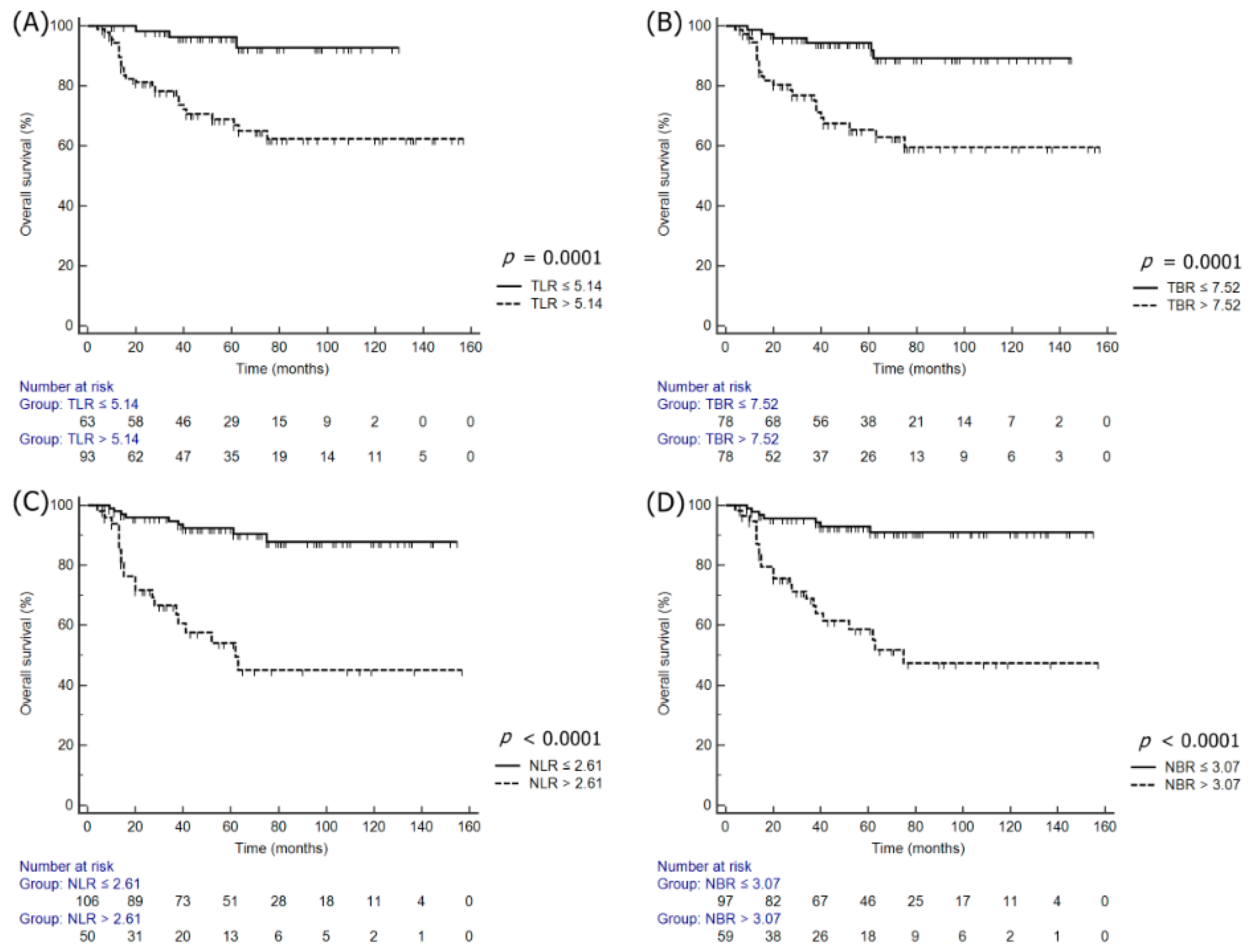
3.4. Comparison ROC for the Prediction of Tumor Recurrence and Death

To enhance the discrimination for the prediction of tumor recurrence and death, SUVmax was normalized to the liver and blood pool. TLR and TBR showed significant improvements in discrimination for tumor recurrence when compared with tSUVmax (tSUVmax vs. TLR; p = 0.0056, and tSUVmax vs. TBR; p = 0.0099) (Figure 3A). Furthermore, TLR and TBR demonstrated significant improvements in the accuracy of risk prediction for OS relative to tSUVmax (tSUVmax vs. TLR; p < 0.0001, and tSUVmax vs. TBR; p = 0.0001) (Figure 3C). However, the normalization of the nSUVmax did not improve the discrimination for tumor recurrence and death (Figure 3B,D).

4. Discussion

There are two major findings in this study. First, the normalization of tSUVmax to the liver or blood pool enhanced the level of discrimination for the prediction of tumor recurrence and death in locally advanced cervical cancer treated with CCRT. Second, TLR, TBR, NLR, and NBR are independent prognostic factors for predicting survival outcomes.
Several studies have demonstrated that pretreatment tSUVmax can predict survival outcomes. The largest retrospective study of 287 patients with FIGO stages IA2 to IVB cervical cancer established the following three prognostic groups based on tSUVmax with five-year OS rates: 95% for a tSUVmax of 5.2 or less, 70% for a tSUVmax between 5.2 and 13.3, and 44% for a tSUVmax greater than 13.3 [6]. Moreover, in a recent multicenter retrospective study that enrolled 270 patients with locally advanced cervical cancer treated with CCRT, a tSUVmax of 12 or greater was an independent prognostic biomarker for predicting tumor recurrence (HR: 2.14; 95% CI: 1.32–3.47; p = 0.002) and death (HR: 3.06; 95% CI: 1.46–6.44; p = 0.003) [16].
However, other studies have shown that tSUVmax is not an independent prognostic metabolic parameter. A study of 53 patients with FIGO stages IB1 to IVA determined the optimal tSUVmax cutoff to be 16.0; however, this did not significantly predict the progression or DFS [8]. SUVmax has several limiting factors, such as it representing the highest metabolic voxel but not the metabolic activity of the entire tumor. Meanwhile, research suggests volume-based metabolic parameters such as MTV and TLG are of prognostic value in cervical cancer [17]. However, MTV and TLG might include inflammation around the primary tumor associated with weakness [18]. SUVmax exhibits a high degree of reproducibility and is a less time-consuming method to apply. Despite these advantages, SUVmax may not be an adequate surrogate marker for representing the metabolic rate of the tumor, so other metabolic parameters that can predict prognosis should be further explored. The mediastinum vessel and normal liver tissue are the most frequent candidates among normal tissues [19].
Among various metabolic parameters, the tSUVmax normalized to liver uptake after neoadjuvant chemotherapy was the best predictor of a pathologic complete response in locally advanced rectal cancer [13]. Moreover, the standardized uptake TBR was an independent predictor of recurrence in non-small-cell lung cancer [14]. In our study, tSUVmax had no significant impact in predicting tumor recurrence or death. After the normalization of tSUVmax to the liver or blood pool, however, tSUVmax demonstrated a significant prognostic performance. We confirmed the improvement in discrimination for the prediction of tumor recurrence and death using the normalization of tSUVmax to the liver or blood pool by comparing ROC curves among tSUVmax, TLR, and TBR. Moreover, the normalization of tSUVmax to the liver or blood pool showed a significant prognostic value for predicting tumor recurrence or death in univariate and multivariate analyses. These enhancements of the prognostic value may make it possible to personalize treatment in a way that may involve higher doses of radiation boosts, consolidation chemotherapy, or adjuvant hysterectomy.
The lymph node status on 18F-FDG PET/CT images has also been shown to predict tumor recurrence and survival outcomes in patients with cervical cancer. A prospective cohort study of 560 patients who underwent pretreatment 18F-FDG PET/CT imaging demonstrated that patients with PET-positive lymph nodes had significantly worse DFS as compared with those with PET-negative nodes [20]. Furthermore, it has also been documented that the level of nSUVmax correlates with survival outcomes [7,21]. Only a limited number of studies to date have evaluated the prognostic roles of NLR or NBR. Furthermore, only one study has evaluated the normalization of nSUVmax to the liver. Higher NLR on pretreatment 18F-FDG PET/CT images was shown to be an independent prognostic factor for worse distant metastasis-free survival in locally advanced nasopharyngeal carcinoma [22]. Similarly to our previous research [9,16], we found in the current study that nSUVmax was a powerful biomarker for predicting tumor recurrence and death and that NLR and LBR were significant prognostic factors. However, normalized nSUVmax to the liver or blood pool did not demonstrate superiority over conventional nSUVmax for predicting tumor recurrence or survival.
Our study has some noteworthy limitations that must be considered. First, it was a retrospective study including a limited number of patients. Second, a histopathological verification of the lymph nodes was not performed. Finally, some variables tended to have a wider 95% CI than p-values, especially when considering the OS. The wider 95% CI width resulted in a relatively small sample and event size in some variables. A larger patient cohort study involving multi-institutional cooperative groups is needed to improve the statistical results.
Despite these limitations, our study offers some unique and significant findings and differs from previous studies in several aspects. To the best of our knowledge, this is the first study to evaluate how the normalization of tSUVmax and nSUVmax to the liver or blood pool affects prognostic values in cervical cancer. Metabolic parameters that did not demonstrate a prognostic role had a significant prognostic value for predicting tumor recurrence and death when normalized to the liver or blood pool. Moreover, this is the first research to demonstrate the prognostic value of TLR, TBR, NLR, and NBR in locally advanced cervical cancer treated with CCRT. TLR, TBR, NLR, and NBR were found to be independent prognostic factors for both DFS and OS.

5. Conclusions

Normalized tSUVmax and nSUVmax to the liver or blood pool showed a significant prognostic value in patients with locally advanced cervical cancer treated with CCRT. Moreover, the normalization of metabolic parameters improved the level of discrimination for the prediction of tumor recurrence and death, especially in the case of tSUVmax. The normalization of metabolic parameters to the liver or blood pool may enable the standardization of metabolic parameters for multicenter studies.

Supplementary Materials

The following are available online at https://www.mdpi.com/2077-0383/9/6/1878/s1, Table S1. Comparison of receiver operating characteristic curve analyses.

Author Contributions

Conceptualization, G.O.C. and S.Y.J.; methodology, S.Y.J., S.W.L., D.G.H., H.J.L. and Y.H.L.; software, G.O.C.; validation, S.Y.J., S.-W.L., H.J.L. and D.G.H.; formal analysis, G.O.C., S.-H.P. and S.Y.L.; investigation, Y.H.L. and H.J.L.; resources, Y.H.L. and H.J.L.; data curation, G.O.C., S.-H.P. and S.Y.L.; writing—original draft preparation, G.O.C., S.-H.P. and Y.H.L.; writing—review and editing, S.Y.J., S.-W.L., D.G.H. and Y.S.L.; visualization, H.J.L. and Y.H.L.; supervision, S.-W.L., D.G.H. and Y.S.L.; project administration, S.-W.L., S.-H.P., G.O.C. and S.Y.J.; funding acquisition, S.W.L. All authors have read and agreed to the published version of the manuscript.

Funding

This work was supported by a grant from the Korea Health Technology R&D Project through the Korea Health Industry Development Institute (KHIDI), funded by the Ministry of Health & Welfare, Republic of Korea (grant no.s HI16C1501).

Conflicts of Interest

The authors declare no conflict of interest.

Abbreviations

CCRTconcurrent chemoradiotherapy
CIconfidence interval
DFSdisease-free survival
18F-FDG PET/CTF-18 fluorodeoxyglucose positron emission tomography/computed tomography
FIGOAdvanced International Federation of Gynecology and Obstetrics
HFheterogeneity factor
HRhazard ratio
SUVmaxmaximum standardized uptake value
MTVmetabolic tumor volume
ROCreceiver operating characteristic
TLGtotal lesion glycolysis
WBwhole body

References

  1. Green, J.A.; Kirwan, J.M.; Tierney, J.F.; Symonds, P.; Fresco, L.; Collingwood, M.; Williams, C.J. Survival and recurrence after concomitant chemotherapy and radiotherapy for cancer of the uterine cervix: A systematic review and meta-analysis. Lancet 2001, 358, 781–786. [Google Scholar] [CrossRef]
  2. Whitney, C.W.; Sause, W.; Bundy, B.N.; Malfetano, J.H.; Hannigan, E.V.; Fowler, W.C., Jr.; Clarke-Pearson, D.L.; Liao, S.Y. Randomized comparison of fluorouracil plus cisplatin versus hydroxyurea as an adjunct to radiation therapy in stage IIB-IVA carcinoma of the cervix with negative para-aortic lymph nodes: A Gynecologic Oncology Group and Southwest Oncology Group study. J. Clin Oncol. 1991, 17, 1339–1348. [Google Scholar] [CrossRef] [PubMed] [Green Version]
  3. Collaboration Chemoradiotherapy for Cervical Cancer Meta-Analysis Collaboration. Reducing uncertainties about the effects of chemoradiotherapy for cervical cancer: A systematic review and metaanalysis of individual patient data from 18 randomized trials. J. Clin. Oncol. 2008, 26, 5802–5812. [Google Scholar] [CrossRef] [Green Version]
  4. Lee, D.W.; Kim, Y.T.; Kim, J.H.; Kim, S.; Kim, S.W.; Nam, E.J.; Kim, J.W. Clinical significance of tumor volume and lymph node involvement assessed by MRI in stage IIB cervical cancer patients treated with concurrent chemoradiation therapy. J. Gynecol. Oncol. 2010, 21, 18–23. [Google Scholar] [CrossRef] [PubMed]
  5. Endo, D.; Todo, Y.; Okamoto, K.; Ninobe, S.; Kato, H.; Nishiyama, N. Prognostic factors for patients with cervical cancer treated with concurrent chemoradiotherapy: A retrospective analysis in a Japanese cohort. J. Gynecol. Oncol. 2015, 26, 12–18. [Google Scholar] [CrossRef] [PubMed] [Green Version]
  6. Kidd, E.A.; Siegel, B.A.; Dehdashti, F.; Grigsby, P.W. Pelvic lymph node F-18 fluorodeoxyglucose uptake as a prognostic biomarker in newly diagnosed patients with locally advanced cervical cancer. Cancer 2010, 116, 1469–1475. [Google Scholar] [CrossRef]
  7. Onal, C.; Guler, O.C.; Reyhan, M.; Yapar, A.F. Prognostic value of 18F-fluorodeoxyglucose uptake in pelvic lymph nodes in patients with cervical cancer treated with definitive chemoradiotherapy. Gynecol. Oncol. 2015, 137, 40–46. [Google Scholar] [CrossRef]
  8. Bollineni, V.R.; Kramer, G.; Liu, Y.; Melidis, C.; deSouza, N.M. A literature review of the association between diffusion-weighted MRI derived apparent diffusion coefficient and tumour aggressiveness in pelvic cancer. Cancer Treat. Rev. 2015, 41, 496–502. [Google Scholar] [CrossRef]
  9. Chong, G.O.; Jeong, S.Y.; Park, S.H.; Lee, Y.H.; Lee, S.W.; Hong, D.G.; Kim, J.C.; Lee, Y.S.; Cho, Y.L. Comparison of the Prognostic Value of F-18 Pet Metabolic Parameters of Primary Tumors and Regional Lymph Nodes in Patients with Locally Advanced Cervical Cancer Who Are Treated with Concurrent Chemoradiotherapy. PLoS ONE 2015, 10, e0137743. [Google Scholar] [CrossRef]
  10. Kostakoglu, L.; Chauvie, S. Metabolic Tumor Volume Metrics in Lymphoma. Metabolic Tumor Volume Metrics in Lymphoma. Semin. Nucl. Med. 2018, 48, 50–66. [Google Scholar] [CrossRef] [PubMed]
  11. Schulte, M.; Brecht-Krauss, D.; Werner, M.; Hartwig, E.; Sarkar, M.R.; Keppler, P.; Kotzerke, J.; Guhlmann, A.; Delling, G.; Reske, S.N. Evaluation of neoadjuvant therapy response of osteogenic sarcoma using FDG PET. J. Nucl. Med. 1999, 40, 1637–1643. [Google Scholar] [PubMed]
  12. Chiaravalloti, A.; Danieli, R.; Abbatiello, P.; Di Pietro, B.; Travascio, L.; Cantonetti, M.; Guazzaroni, M.; Orlacchio, A.; Simonetti, G.; Schillaci, O. Factors affecting intrapatient liver and mediastinal blood pool ¹⁸F-FDG standardized uptake value changes during ABVD chemotherapy in Hodgkin’s lymphoma. Eur. J. Nucl. Med. Mol. Imaging 2014, 41, 1123–1132. [Google Scholar] [CrossRef] [PubMed]
  13. Park, J.; Chang, K.J.; Seo, Y.S.; Byun, B.H.; Choi, J.H.; Moon, H.; Lim, I.; Kim, B.I.; Choi, C.W.; Lim, S.M. Tumor SUVmax Normalized to Liver Uptake on (18)F-FDG PET/CT Predicts the Pathologic Complete Response After Neoadjuvant Chemoradiotherapy in Locally Advanced Rectal Cancer. Nucl. Med. Mol. Imaging 2014, 48, 295–302. [Google Scholar] [CrossRef] [PubMed] [Green Version]
  14. Shin, S.; Pak, K.; Kim, I.J.; Kim, B.S.; Kim, S.J. Prognostic Value of Tumor-to-Blood Standardized Uptake Ratio in Patients with Resectable Non-Small-Cell Lung Cancer. Nucl. Med. Mol. Imaging 2017, 51, 233–239. [Google Scholar] [CrossRef] [PubMed]
  15. Pecorelli, S. Revised FIGO staging for carcinoma of the vulva, cervix, and endometrium. Int. J. Gynaecol. Obstet. 2009, 105, 103–104. [Google Scholar] [CrossRef]
  16. Lee, W.K.; Chong, G.O.; Jeong, S.Y.; Lee, H.J.; Park, S.; Ryu, J.M.; Choi, Y.S.; Kang, S.; Koo, Y.; Lee, D.H.; et al. Prognosis-Predicting Model Based on [18F]fluorodeoxyglucose PET Metabolic Parameters in Locally Advanced Cervical Cancer Patients Treated with Concurrent Chemoradiotherapy: Multi-Center Retrospective Study. J. Clin. Med. 2020, 9, 427. [Google Scholar] [CrossRef] [Green Version]
  17. Han, S.; Kim, H.; Kim, Y.J.; Suh, C.H.; Woo, S. Prognostic Value of Volume-Based Metabolic Parameters of 18F-FDG PET/CT in Uterine Cervical Cancer: A Systematic Review and Meta-Analysis. AJR Am. J. Roentgenol. 2018, 211, 1112–1121. [Google Scholar] [CrossRef]
  18. Chu, K.P.; Murphy, J.D.; La, T.H.; Krakow, T.E.; Iagaru, A.; Graves, E.E.; Hsu, A.; Maxim, P.G.; Loo, B.; Chang, D.T.; et al. Prognostic value of metabolic tumor volume and velocity in predicting head-and-neck cancer outcomes. Int. J. Radiat. Oncol. Biol. Phys. 2012, 83, 1521–1527. [Google Scholar] [CrossRef] [Green Version]
  19. Lee, J.W.; Kim, S.K.; Lee, S.M.; Moon, S.H.; Kim, T. Detection of hepatic metastases using dual-time-point FDG PET/CT scans in patients with colorectal cancer. Mol. Imaging Biol. 2011, 13, 565–572. [Google Scholar] [CrossRef]
  20. Kidd, E.A.; Siegel, B.A.; Dehdashti, F.; Rader, J.S.; Mutch, D.S.; Powell, M.A.; Grigsby, P.W. Lymph node staging by positron emission tomography in cervical cancer: Relationship to prognosis. J. Clin. Oncol. 2010, 28, 2108–2113. [Google Scholar] [CrossRef]
  21. Ramlov, A.; Kroon, P.S.; Jürgenliemk-Schulz, I.M.; De Leeuw, A.C.; Gormsen, L.C.; Fokdal, L.U.; Tanderup, K.; Lindegaard, J.C. Impact of radiation dose and standardized uptake value of (18) FDG PET on nodal control in locally advanced cervical cancer. Acta Oncol. 2015, 54, 1567–1573. [Google Scholar] [CrossRef] [PubMed] [Green Version]
  22. Cho, H.; Kim, S.H.; Kim, H.; Koh, Y.W.; Kim, S.; Choi, E.C.; Yun, M. Lymph Node With the Highest FDG Uptake Predicts Distant Metastasis-Free Survival in Patients With Locally Advanced Nasopharyngeal Carcinoma. Clin. Nucl. Med. 2018, 43, e220–e225. [Google Scholar] [CrossRef] [PubMed]
Figure 1. Kaplan–Meier survival plots of DFS according to the normalized tSUVmax and nSUVmax to the liver or blood: (A) TLR, (B) TBR, (C) NLR, and (D) NBR.
Jcm 09 01878 g001
Figure 2. Kaplan–Meier survival plots of OS according to the normalized tSUVmax and nSUVmax to the liver or blood: (A) TLR, (B) TBR, (C) NLR, and (D) NBR.
Jcm 09 01878 g002
Figure 3. Comparison of ROC curve analyses. (A) tSUVmax versus TLR and TBR for DFS. (B) nSUVmax versus NLR and NBR for DFS. (C) tSUVmax versus TLR and TBR for OS. (D) nSUVmax versus NLR and NBR for OS.
Jcm 09 01878 g003
Table 1. Clinicopathologic characteristics and PET metabolic parameters of patients with and without recurrence.
VariablesAll
(n = 156)
No Recurrence
(n = 101)
Recurrence
(n = 55)
p-Value
Age (years)55.1 ± 12.955.5 ± 12.854.3 ± 13.00.5965
Histology (n, %) 0.1244
SCC141 (90.4)94 (93.1)47 (85.5)
AC/ASC15 (9.6)7 (6.9)8 (14.5)
FIGO stage (n, %) 0.0006
IIB120 (76.9)87 (86.1)33 (60.0
IIIA14 (9.0)8 (7.9)6 (10.9)
IIIB16 (10.3)5 (5.0)11 (20.0)
IVA6 (3.8)1 (1.0)5 (9.1)
Tumor size (cm)4.6 ± 1.54.4 ± 1.45.1 ± 1.60.0080
LN metastasis (n, %)102 (65.4)55 (54.5)47 (85.5)0.0001
Paraaortic LN metastasis (n, %)25 (16.0)18 (17.8)7 (12.7)0.4088
SCC antigen (ng/mL)20.8 ± 36.016.8 ± 35.628.1 ± 35.90.0715
tSUVmax6.0 ± 2.56.0 ± 2.76.0 ± 2.10.9542
TLR5.9 ± 4.46.8 ± 4.17.5 ± 4.80.3356
TBR9.5 ± 6.09.2 ± 6.010.1 ± 6.10.3717
nSUVmax3.3 ± 7.33.5 ± 5.17.9 ± 9.50.0003
NLR2.3 ± 4.01.5 ± 2.53.8 ± 5.60.0008
NBR3.1 ± 5.02.1 ± 3.45.0 ± 6.70.0005
AC = adenocarcinoma; ASC = adenosquamous cell carcinoma; FIGO = International Federation of Gynecology and Obstetrics; LN = lymph node; NBR = node-to-blood ratio; NLR = node-to-liver ratio; nSUVmax = nodal maximum standardized uptake value; SCC = squamous cell carcinoma; SUVmax = maximum standardized uptake value; TBR = tumor-to-blood ratio; TLR = tumor-to-liver ratio; tSUVmax = tumor maximum standardized uptake value.
Table 2. Univariate analyses of clinical variable and quantitative metabolic parameters for DFS and OS.
Variables Disease-Free Survival Overall Survival
LevelHR95% CIp-ValueLevelHR95% CIp-Value
Age (years)>491.00-->491.00--
≤491.250.70–2.230.4584≤492.241.06–4.770.0355
HistologySCC1.00--SCC1.00--
AC/ASC2.040.78–4.980.1531AC/ASC1.570.46–5.350.4699
FIGO stageIIB1.00--IIB1.00--
>IIB4.642.31–9.32<0.0001>IIB7.302.85–18.69<0.0001
Tumor size<4 cm1.00--<4.2 cm1.00--
≥4 cm2.481.41–4.340.0016≥4.2 cm3.091.50–6.380.0023
LN metastasesNegative1.00--Negative1.00--
Positive3.091.79–5.32<0.0001Positive2.961.41–6.180.0039
Paraaortic LN metastasesNegative1.000.71–2.850.3245Negative1.001.10–6.990.0305
Positive1.42Positive2.78
Serum SCC<19.31.00--<19.31.00--
≥19.33.711.94–7.120.0001≥19.33.751.55–9.080.0035
tSUVmax≤4.811.00--≤6.431.00--
>4.811.390.26–7.300.6975>6.432.050.60–7.030.2528
TLR≤4.991.00--≤5.141.00--
>4.992.261.37–3.870.0031>5.144.232.05–8.700.0001
TBR≤6.961.00--≤7.521.00-
>6.962.231.31–3.810.0033>7.524.262.07–8.790.0001
nSUVmax≤4.631.00--≤4.911.00--
>4.636.863.81–12.36< 0.0001>4.917.723.53–16.88< 0.0001
NLR≤2.021.00--≤2.61--
>2.026.263.51–11.14< 0.0001>2.6110.274.51–23.39<0.0001
NBR≤1.971.00--≤3.071.00--
>1.974.742.76–8.15< 0.0001>3.078.023.71–17.36<0.0001
AC = adenocarcinoma; ASC = adenosquamous cell carcinoma; CI = confidence interval; FIGO = International.
Table 3. Multivariate analyses of PET parameters in relation to DFS and OS after adjusting for clinicopathologic factors.
VariablesDisease-Free SurvivalOverall Survival
LevelHR95% CIp-ValueLevelHR95% CIp-Value
tSUVmax≤4.811.00--≤6.431.00--
>4.810.290.04–2.380.2478>6.430.660.08–5.850.7117
TLR≤4.98851.00--≤5.13681.00--
>4.98851.480.76–2.870.2509>5.13684.161.19–14.500.0252
TBR≤6.96121.00--≤7.52351.00--
>1.96121.420.75–2.700.2864>7.52353.011.04–8.700.0415
nSUVmax≤4.631.00--≤4.911.00--
>4.634.061.74–9.440.0012>4.916.991.56–31.250.0109
LNR≤2.00211.00--≤2.60981.00--
>2.00213.541.53–8.190.0032>2.60984.841.58–14.810.0057
NBR≤1.96811.00--≤3.07051.00--
>1.96813.381.02–11.190.0457>3.07056.871.55–30.540.0113
CI = confidence interval; FIGO = International Federation of Gynecology and Obstetrics; HR = hazard ratio; NBR = node-to-blood ratio; NLR = node-to-liver ratio; nSUVmax = nodal maximum standardized uptake value; SUVmax = maximum standardized uptake value; TBR = tumor-to-blood ratio; TLR = tumor-to-liver ratio; tSUVmax = tumor maximum standardized uptake value.

