Article

Comparison of Ilizarov Bifocal, Acute Shortening and Relengthening with Bone Transport in the Treatment of Infected, Segmental Defects of the Tibia

1
The Bone Infection Unit, Nuffield Orthopaedic Centre, Oxford University Hospitals, Foundation NHS Trust, Windmill Rd, Headington, Oxford OX3 7HE, UK
2
Department of Orthopaedics and Trauma Surgery, Medical University of Vienna, Spitalgasse 23, Vienna 1090, Austria
3
Department of Trauma Surgery, University of Utrecht, University Medical Centre Utrecht (UMCU), 3512 Utrecht, The Netherlands
*
Author to whom correspondence should be addressed.
J. Clin. Med. 2020, 9(2), 279; https://doi.org/10.3390/jcm9020279
Submission received: 20 December 2019 / Revised: 20 January 2020 / Accepted: 22 January 2020 / Published: 28 January 2020
(This article belongs to the Section Orthopedics)

Abstract

:
This prospective study compared bifocal acute shortening and relengthening (ASR) with bone transport (BT) in a consecutive series of complex tibial infected non-unions and osteomyelitis, for the reconstruction of segmental defects created at the surgical resection of the infection. Patients with an infected tibial segmental defect (>2 cm) were eligible for inclusion. Patients were allocated to ASR or BT, using a standardized protocol, depending on defect size, the condition of soft tissues and the state of the fibula (intact or divided). We recorded the Weber–Cech classification, previous operations, external fixation time, external fixation index (EFI), follow-up duration, time to union, ASAMI bone and functional scores and complications. A total of 47 patients (ASR: 20 patients, BT: 27 patients) with a median follow-up of 37.9 months (range 16–128) were included. In the ASR group, the mean bone defect size measured 4.0 cm, and the mean frame time was 8.8 months. In the BT group, the mean bone defect size measured 5.9cm, and the mean frame time was 10.3 months. There was no statistically significant difference in the EFI between ASR and BT (2.0 and 1.8 months/cm, respectively) (p = 0.223). A total of 3/20 patients of the ASR and 15/27 of the BT group needed further unplanned surgery during Ilizarov treatment (p = 0.006). Docking site surgery was significantly more frequent in BT; 66.7%, versus ASL; 5.0% (p < 0.0001). The infection eradication rate was 100% in both groups at final follow-up. Final ASAMI functional rating scores and bone scores were similar in both groups. Segmental resection with the Ilizarov method is effective and safe for reconstruction of infected tibial defects, allowing the eradication of infection and high union rates. However, BT demonstrated a higher rate of unplanned surgeries, especially docking site revisions. Acute shortening and relengthening does not reduce the fixator index. Both techniques deliver good functional outcome after completion of treatment.

1. Introduction

Infected segmental defects are common after tibial injury [1,2,3]. They are problematic for patients, due to the prolonged treatment time, permanent functional deficits and high reinfection and non-union rates [4]. Although there has been an improvement in the treatment of open fractures since the introduction of management guidelines, advanced implant designs and less traumatic surgical techniques, the infection rate is still high, ranging from 9% to 18% [5,6,7,8,9,10]. Furthermore, recurrence of infection is common, affecting 10% to 20% of patients [11,12,13,14]. A structured approach, with adequate debridement, excision of necrotic tissue, antimicrobial therapy and reconstruction of the bone and soft tissues, is of immense importance to eradicate infection and restore function [15,16,17]. To manage the resulting bone defects, distraction osteogenesis techniques such as Ilizarov acute shortening with bifocal relengthening (ASR) and bone transport (BT) have been shown to be effective treatment options to achieve union and infection clearance [1,12,15,16,18]. This technique permits not only the gradual restoration of the bone, but also deformity correction, correction of joint contractures, and rehabilitation, with early weight bearing to avoid muscle wasting and disuse osteopenia.
Generally, each surgeon decides on the method of distraction histogenesis (ASR vs. BT), based on clinical features and personal experience [19,20,21,22]. Small bone defects are commonly treated by acute shortening, with or without relengthening, while in large bone defects a BT is often preferred. However, no standardized or validated protocol has been described in the literature and it is often impossible to determine why a particular method has been chosen.
In this study, we compared acute shortening and relengthening with bone transport for the reconstruction of segmental defects in a consecutive, prospectively analysed series of complex infected tibias, by using a standardized protocol. We evaluated the treatment duration, outcome and complications during and after the procedures.

2. Materials and Methods

2.1. Study Design

This prospective study was conducted in a tertiary healthcare centre, providing advanced specialty care (orthopaedic surgeons, plastic surgeons, specialist Ilizarov nurses and physiotherapists and infectious disease physicians specialized in musculoskeletal infections) to patients with musculoskeletal infections. The study was performed in accordance with the Declaration of Helsinki and approved by the hospital governance board (ID 5778).

2.2. Study Population

Consecutive patients over 18 years old, who had a segmental bone defect after surgery for infected non-union or osteomyelitis of the tibia were included. Information on demographics, comorbidities, associated deformities, imaging, histological and microbiological results from intraoperative sampling, surgical treatment procedures, and complications were recorded.
Preoperatively, each patient was reviewed by a multidisciplinary team (orthopaedic and plastic surgeons, infectious disease physicians, and a specialist Ilizarov nurse practitioner) who agreed the treatment plan. The classification of Weber and Cech [23], was determined at operation, based on the non-union viability as defined according to the vascularity of the bone.
The following outcome parameters were evaluated after surgery: recurrent infection, total frame time, external fixator index in months/cm (EFI), complications, the Association for the Advancement of Methods of Ilizarov (ASAMI) bone and functional classification scores, and further unplanned surgery (defined as any additional surgery undertaken after frame removal).

2.3. Infection Definition

An infected segmental tibial bone defect was diagnosed according to the International Consensus criteria for fracture-related infections (FRI) [24,25]. Infection was present when at least one of the following criteria applied: (i) a draining sinus, (ii) an abscess or intraoperative purulence, (iii) significant growth of a microorganism from intraoperative tissue sampling (at least two or more positive sterile site cultures with indistinguishable organisms), and (iv) positive histopathology supportive of deep active infection.

2.4. Tissue Sampling and Surgical Procedures

All patients were treated with a single operation comprising an excision of the infected dead bone segment, application of the Ilizarov fixator, corticotomy and skin closure (including free tissue transfer, if needed). Antimicrobial treatment was stopped for at least 14 days before surgery. A tourniquet was used in all patients. In the first step, the collection of specimens was performed as described previously [26].
For histopathological analysis, at least two deep tissue specimens were collected. If a sinus was present for more than five years, it was excised and sent for histological review to exclude squamous carcinoma.
In the second step, the infected area was fully exposed to allow adequate debridement. Protecting the neurovascular structures, a segmental resection was performed to eradicate the non-viable zone. A careful resection of indurated subcutaneous tissue, infected and necrotic soft tissue, and necrotic bone was performed in all cases. Thereafter, the wound was manually washed with generous volumes of 0.05% aqueous chlorhexidine at low pressure. Without releasing the tourniquet, the pattern of bone bleeding was observed. If punctate bleeding was absent, a further resection was undertaken.

2.5. Standardized Treatment Protocol

The choice of Ilizarov method was made depending on the criteria outlined below. An Ilizarov ring fixator was used in all cases (Smith and Nephew, Memphis, TN, USA).
Acute shortening and relengthening (Figure 1) was performed in patients with bone defects of up to 5 cm, where it could be performed safely without soft tissue or neurovascular compromise. Segmental bone resection was performed to allow good bone contact with good alignment at the docking site. A corticotomy was performed at a separate metaphyseal location outside the infected zone. After 6–7 days latent period, length was gradually restored by distraction through the corticotomy site at 1 mm per day. Lengthening was continued until equal leg length was restored. The docking site was compressed at 0.5 mm per week for 2–3 weeks.
Bone transport (Figure 2) was selected in all patients with bone defects larger than 5 cm or when the fibula was intact and well aligned. In some patients, with defects <5 cm, the soft tissues were heavily scarred and indurated due to chronic infection. If attempted, acute shortening caused vascular compromise in the foot, thus bone transport was elected as the reconstruction method. Corticotomy, latent period and distraction rate were the same as in the ASR group. When docking was achieved in the BT group, no elective surgery on the docking site was planned.
Skin closure was performed in all cases. If a direct wound closure was possible, early mobilization was commenced, and full-weight-bearing encouraged on day two. If a local or free microvascular muscle flap was used to restore a healthy soft tissue envelope, full weight bearing was allowed after flap healing based on the plastic surgeon’s discretion (usually after seven days).
Empiric antibiotics (Vancomycin and Meropenem) were administered intraoperatively, after sampling. According to the susceptibility of the isolated pathogen(s), the antimicrobial treatment was adjusted by the infectious disease specialist team and continued for at least six weeks.

2.6. Statistical Analysis

Continuous variables are expressed as mean and range; categorical variables are described as absolute and relative frequencies (percentage). Student’s t-test and Fisher’s exact test were performed to compare metric and binary variables between the distributions of both groups. A chi-squared test was used to compare the overall complication rate between both groups. All estimated parameters are reported with 95% confidence intervals. The significance level for all tests was 5% (p < 0.05). The software package XLSTATPM (version 2017; XLSTAT; Addinsoft, New York, NY, USA) was used for statistical analysis.

3. Results

3.1. Demographics and Infection Characteristics
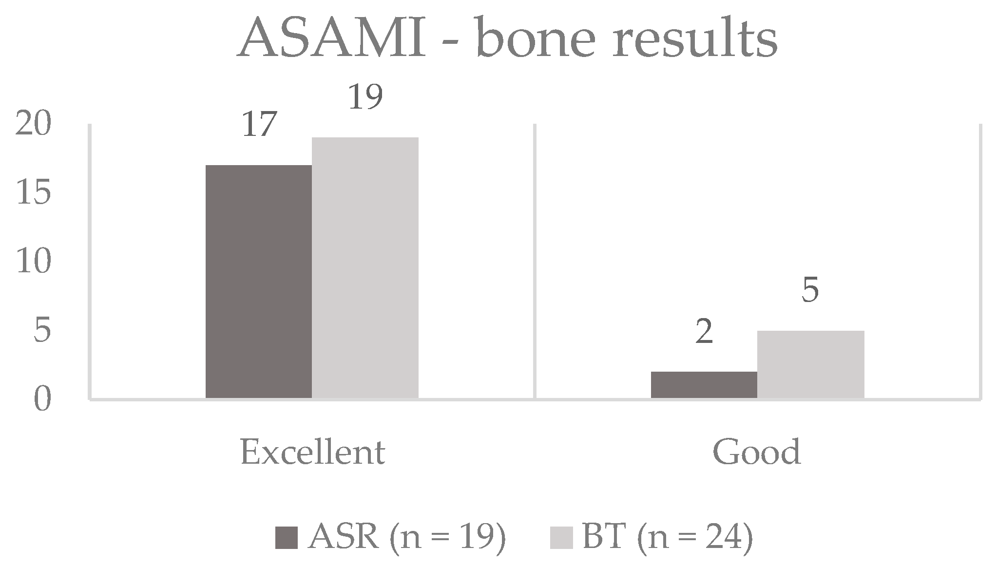
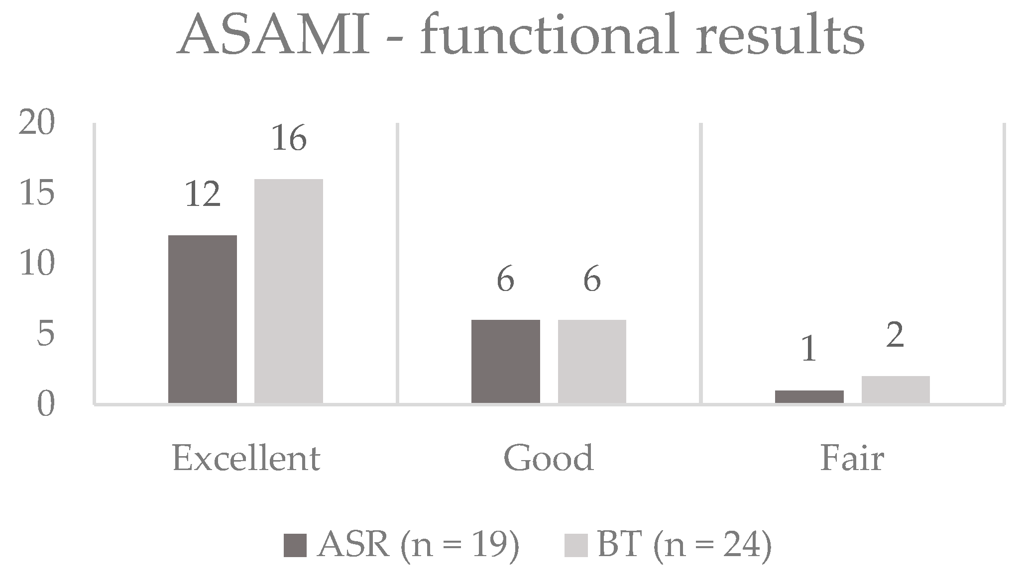
Forty-seven patients with an infected tibial segmental defect were included. Of these, 20 (43%) patients were treated with ASR and 27 (57%) with BT. In eight cases with bone defects under 5 cm, ASR was not possible because of scarred soft tissue or neurovascular compromise after attempted shortening. Demographic data and surgical features of the 47 analysed cases are shown in Table 1. Median follow-up was 37.9 months (range 16–128).
Six (13%) patients presented with muscle flaps in place, which were “reused” (ASR [n = 3], BT [n = 3]). In a further 11 (23%) patients, a new muscle flap was required to allow soft tissue closure. Of these, eight (73%) had a free gracilis muscle flap (ASR [n = 4], BT [n = 4]), two (18%) a free latissimus dorsi muscle flap (BT group), and one (9%) a local gastrocnemius muscle flap (BT group). There was no difference in the need for new free flap reconstruction between the groups (ASR, 4/17; BT, 7/24 p = 1.000).
The most common causative microorganism was Staphylococcus aureus, followed by coagulase negative staphylococci and Pseudomonas aeruginosa.

3.2. Outcome

No patient suffered a recurrent FRI during follow-up (Table 2).
The initial union rate was 18/20 (90%) for ASR and 17/27 (63%) for BT. This difference was statistically significant (p = 0.047). All non-union cases were infection free and healed after further fixation (see below). Therefore, the final union rate was 100% in both groups.
At the end of treatment, no patient had a significant leg length discrepancy (>1 cm) or angular or rotational deformity (>5 degrees).
Figure 3 illustrates the final ASAMI bone classification results for each group. The ASAMI bone scores showed no significant difference between both groups at the end of treatment (p = 0.682).
Figure 4 shows the final ASAMI functional classification results. There was no significant difference between both groups regarding ASAMI functional score (p = 0.705).

3.3. Unplanned Surgeries during Fixator Time

Eighteen (38%) unplanned surgeries (including docking site procedures) had to be performed in both groups. Of these, 15 (n = 15/27; 56%) were needed in the BT group with only three (n = 3/20; 15%) surgeries in the ASR group. There was a statistically significant difference between groups (p = 0.006). Thirteen of the 15 (87%) patients with an unplanned procedure in the BT group had an operation at the docking site. All surgeries are summarized in Table 3.
If the docking site procedures were excluded, no statistically significant difference was found between the ASR and BT group (p = 0.638).

3.4. Unplanned Surgeries after Fixator Removal

After the removal of the Ilizarov frame, thirteen (n = 13/47; 28%) unplanned reoperations were needed in both groups. In the ASR group only three (n = 3/20; 15%) further revision surgeries were required with ten (n = 10/27; 37%) in the BT group. However, no significant difference was found (p = 0.114). Overall, docking site surgery was significantly more frequent in BT; 66.7%, versus ASL; 5.0% (p = 0.0001). A detailed list of revision surgeries after the removal of the Ilizarov frame is shown in Table 3.
In total, 31 unplanned surgeries were needed during the whole observation period, six (30%) in the ASR group and 25 (93%) in the BT group. This difference was statistically significant (p = 0.013).

3.5. Other Complications

During treatment, 22 patients (47%) suffered at least one superficial pin infection requiring treatment with oral antibiotics. These were equally common after ASR or BT. One patient had the removal of a pin due to persistent pin site infection, which was resolved after removal. One patient had a release of painful tethered skin around a pin and two bone transport patients needed repeat corticotomy due to premature fusion of the lengthening site. There were no other significant complications observed.

4. Discussion

This series confirms that segmental excision is an effective method of eradicating infection, with no patient suffering a recurrent infection at follow-up. Resection of an entire segment of bone allows the complete removal of all dead bone and biofilm, and so will give a better chance of eradication compared to an intralesional excision. Outcomes may also be improved by the multidisciplinary approach from the specialist bone infection team, with careful attention to diagnostics, surgical management, soft tissue reconstruction and antimicrobial therapy.
Few studies have compared the outcome of acute shortening and relengthening with bone transport by using Ilizarov techniques for an infected tibial non-union with a segmental bone defect [19,20,22] (Table 4). To the best of our knowledge, this series represents the largest single-centre prospective study with a standardized treatment protocol. In previous studies, the surgical treatment was chosen based on clinical parameters according to the surgeon’s preference, and the reason for each decision is usually not described. This makes it very difficult to give any recommendation on which cases would be best treated by any particular technique.
In this study, we had a clear protocol for the choice of Ilizarov method, allowing a better comparison of the techniques in cases where they may be more appropriately applied. Also, we limited the variability between groups by using the same fixator type (Ilizarov rings), same sampling method and diagnostic criteria, same antibiotic regime and same latent period before distraction. It has been shown that different fixator constructs may affect the outcome of the Ilizarov method [27].
Acute shortening and relengthening using an Ilizarov ring fixator had a higher initial union rate (90% vs. 63%) and a much lower rate of unplanned surgery during treatment (30% vs. 93%) compared to bone transport. The majority of unplanned operations were docking site procedures. Our rate of 67% docking site surgery in the bone transport group is similar to that reported by Tetsworth, et al. [22] (67%) and Eralp, et al. [19] (62%), but our interventions in the ASR group were much less frequent, at 5%. Tetsworth, et al. [22] operated on 38% of ASR cases and suggested that there may be a place for primary bone grafting of acutely shortened docking sites. We do not agree with this. In his series, Tetsworth, et al. included patients with defects larger than 5 cm in the ASR group. These patients could not be acutely shortened and so had a partial shortening followed by a gradual shortening to bone contact. This is analogous to a short bone transport and may account for the higher rate of docking site surgery. We agree that ASR is a technically more demanding procedure than bone transport. It is often difficult to achieve acute shortening due to swelling in the soft tissues around the defect. Displacement of the tissues may prevent accurate apposition of the bone ends with good alignment. Because of these concerns, we recommend that ASR should be reserved for defects under 5 cm, to allow patients to receive the benefits of the technique.
It has been suggested that the routine freshening and bone grafting of docking sites should be performed [28,29,30]. However, in our series and others, this would result in the overtreatment of one third of cases. We therefore do not recommend routine docking site surgery, but the decision to operate should not be delayed for a prolonged period. If there is no clear sign of bridging callous across the docking site within eight weeks of docking, we advocate surgery. In this series, we usually freshened the docking site and provided bone grafting if there was a defect present. We only advocate internal fixation if the stability of the fixator has been lost.
Unfortunately, ASR did not reduce the total time in treatment compared to BT. The duration of treatment is determined by three phases. The latent period (from corticotomy to the onset of distraction) was the same in both groups. The lengthening phase and the consolidation phase (from completion of relengthening or docking to frame removal) are mainly determined by the length of the segment to be reconstructed, and therefore are unlikely to be affected by the choice of technique. In most cases, docking site healing occurred (with or without adjunctive surgery) before consolidation of the regenerate, and so did not delay treatment time in the bone transport group.
We were also concerned that acute shortening may defunction muscles crossing the ankle joint, making early mobilisation more difficult, with a potentially reduced functional outcome. This was not observed. Patients in both groups mobilised full-weight-bearing, beginning as soon as the soft tissues allowed. In our study, the majority of patients showed excellent or good bony and functional results after treatment. This is in line with the reported data in the literature [19,20,22] (Table 4). Clearly, both techniques allow good functional recovery, but this is only achieved with careful attention to rehabilitation with a guided physiotherapy program, supervised by therapists who are familiar with Ilizarov methods.
Numerous methods have been described for the restoration of bone in segmental tibial defects, including cancellous bone grafting, the implantation of bone substitutes, Papineau technique, non-vascularised and vascularised bone grafts and the induced membrane technique [31,32,33,34,35]. All have reported successful results, but treatment times are long and reoperation rates are high. In recent literature reviews comparing defect management methods, there was no clear advantage of one technique over another, either in outcome or treatment time [36,37]. Ilizarov methods do have an advantage as they can address concomitant deformity and leg length difference more easily.
At our institution, the soft tissue closure is always performed in the same operation as the Ilizarov procedure as a single stage operation. This requires long operative times but reduces the number of visits to theatre and the overall time in treatment. We have shown that it is successful in this series and in cases of chronic osteomyelitis [38]. In the tibia, with chronic infection, up to one third of patients may require tissue transfer (local or free flap) to achieve closure. This requires meticulous preoperative planning between the orthopaedic and plastic surgeon [17]. Often, frame application may need to be modified to allow access to microvascular anastomosis for free flaps. However, we have shown that it is safe to transfer free flaps and reuse existing flaps in combination with distraction osteogenesis [39]. In this study, no patient treated with a flap had a wound-healing disorder or failure of the flap.
A limitation of the study is the small sample size in both groups. Nevertheless, it represents the largest prospective single-centre study in adults comparing ASR and BT, and is sufficiently capable of comparing the techniques [22]. Another concern is the difference in the segment length of the two groups. This was dictated as part of the protocol and was due to the pragmatic difficulty of acute shortening of long defects with scarred and indurated soft tissues. It may be appropriate to consider a randomised trial of ASR versus BT for defects up to 5 cm where shortening is possible, to address this. However, the sample size would need to be large, as the differences in outcome and treatment parameters are likely to be small.

5. Conclusions

Bone transport using the Ilizarov technique is a less demanding procedure, but showed a higher rate of unplanned surgeries, especially docking site revisions. Almost all patients having a bone transport required an unplanned procedure. While acute shortening and relengthening is technically more difficult, (fibula osteotomy, soft tissue dissection, increased risk of neurovascular injuries, etc.), less revision surgeries are needed. ASR is limited by neurovascular compromise, while BT is also applicable in large bone defects, with no need to expose the fibula. Nevertheless, both methods are safe and effective distraction osteogenesis techniques for the treatment of infected tibial non-unions with similar functional and bony outcomes.

Author Contributions

I.K.S. collected data, wrote the paper, performed surgery and analysed the results. M.A.M. collected the data, wrote the paper, designed the study, performed surgery and analysed the results. J.F. collected the data, performed surgery, edited the paper and analysed the results. G.A.M.G. edited the paper, performed surgery and analysed the results. D.S. edited the paper and performed surgery. All authors have read and agreed to the published version of the manuscript.

Funding

We acknowledge support from Oxford Bone Infection Consultancy and the Nuffield Orthopaedic Centre Trust.

Acknowledgments

Part of this study was presented at the European Bone and Joint Infection Society conference (EBJIS 2018) in Helsinki, Finland.

Conflicts of Interest

The authors declare no conflict of interest.

References

  1. Cattaneo, R.; Catagni, M.; Johnson, E.E. The treatment of infected nonunions and segmental defects of the tibia by the methods of Ilizarov. Clin. Orthop. Relat. Res. 1992, 280, 143–152. [Google Scholar] [CrossRef]
  2. Dendrinos, G.K.; Kontos, S.; Lyritsis, E. Use of the Ilizarov technique for treatment of non-union of the tibia associated with infection. J. Bone Jt. Surg. Am. Vol. 1995, 77, 835–846. [Google Scholar] [CrossRef]
  3. Kocaoglu, M.; Eralp, L.; Rashid, H.U.; Sen, C.; Bilsel, K. Reconstruction of segmental bone defects due to chronic osteomyelitis with use of an external fixator and an intramedullary nail. J. Bone Jt. Surg. Am. Vol. 2006, 88, 2137–2145. [Google Scholar] [CrossRef]
  4. Metsemakers, W.J.; Kuehl, R.; Moriarty, T.F.; Richards, R.G.; Verhofstad, M.H.J.; Borens, O.; Kates, S.; Morgenstern, M. Infection after fracture fixation: Current surgical and microbiological concepts. Injury 2018, 49, 511–522. [Google Scholar] [CrossRef] [PubMed] [Green Version]
  5. Antonova, E.; Le, T.K.; Burge, R.; Mershon, J. Tibia shaft fractures: Costly burden of nonunions. Bmc Musculoskelet. Disord. 2013, 14, 42. [Google Scholar] [CrossRef] [PubMed] [Green Version]
  6. Audige, L.; Griffin, D.; Bhandari, M.; Kellam, J.; Ruedi, T.P. Path analysis of factors for delayed healing and nonunion in 416 operatively treated tibial shaft fractures. Clin. Orthop. Relat. Res. 2005, 438, 221–232. [Google Scholar] [CrossRef]
  7. British Orthopaedic Association & British Association of Plastic, R.A.S. Open Fractures. Available online: https://www.boa.ac.uk/resources/boast-4-pdf.html (accessed on 1 December 2017).
  8. Harley, B.J.; Beaupre, L.A.; Jones, C.A.; Dulai, S.K.; Weber, D.W. The effect of time to definitive treatment on the rate of nonunion and infection in open fractures. J. Orthop. Trauma 2002, 16, 484–490. [Google Scholar] [CrossRef]
  9. Malik, M.H.; Harwood, P.; Diggle, P.; Khan, S.A. Factors affecting rates of infection and nonunion in intramedullary nailing. J. Bone Jt. Surg. Br. Vol. 2004, 86, 556–560. [Google Scholar] [CrossRef] [Green Version]
  10. Nice, National Institute for Health and Care Excellence. Infection Prevention and Control. 2014. Available online: https://www.nice.org.uk/guidance/qs61/resources/infection-prevention-and-control-pdf-2098782603205 (accessed on 17 April 2014).
  11. Atesalp, A.S.; Komurcu, M.; Basbozkurt, M.; Kurklu, M. The treatment of infected tibial nonunion with aggressive debridement and internal bone transport. Mil. Med. 2002, 167, 978–981. [Google Scholar]
  12. Maini, L.; Chadha, M.; Vishwanath, J.; Kapoor, S.; Mehtani, A.; Dhaon, B.K. The Ilizarov method in infected nonunion of fractures. Injury 2000, 31, 509–517. [Google Scholar] [CrossRef]
  13. McHale, K.A.; Ross, A.E. Treatment of infected tibial nonunions with debridement, antibiotic beads, and the Ilizarov method. Mil. Med. 2004, 169, 728–734. [Google Scholar] [CrossRef] [PubMed] [Green Version]
  14. Ring, D.; Jupiter, J.B.; Gan, B.S.; Israeli, R.; Yaremchuk, M.J. Infected nonunion of the tibia. Clin. Orthop. Relat. Res. 1999, 302–311. [Google Scholar] [CrossRef] [PubMed]
  15. McNally, M.; Ferguson, J.; Kugan, R.; Stubbs, D. Ilizarov Treatment Protocols in the Management of Infected Nonunion of the Tibia. J. Orthop. Trauma 2017, 31 (Suppl. S5), S47–S54. [Google Scholar] [CrossRef]
  16. Yin, P.; Ji, Q.; Li, T.; Li, J.; Li, Z.; Liu, J.; Wang, G.; Wang, S.; Zhang, L.; Mao, Z.; et al. A Systematic Review and Meta-Analysis of Ilizarov Methods in the Treatment of Infected Nonunion of Tibia and Femur. PLoS ONE 2015, 10, e0141973. [Google Scholar] [CrossRef] [PubMed]
  17. Chan, J.K.K.; Ferguson, J.Y.; Scarborough, M.; McNally, M.A.; Ramsden, A.J. Management of Post-Traumatic Osteomyelitis in the Lower Limb: Current State of the Art. Indian J. Plast. Surg. Off. Publ. Assoc. Plast. Surg. India 2019, 52, 62–72. [Google Scholar] [CrossRef] [PubMed] [Green Version]
  18. Pearson, R.L.; Perry, C.R. The Ilizarov technique in the treatment of infected tibial nonunions. Orthop. Rev. 1989, 18, 609–613. [Google Scholar]
  19. Eralp, L.; Kocaoglu, M.; Celiktas, M.; Gulsen, M. Is acute compression and distraction superior to segmental bone transport techniques in chronic tibial osteomyelitis? Comparison of Distraction Osteogenesis Techniques. Acta Orthop. Belg. 2016, 82, 599–609. [Google Scholar]
  20. Khan, M.S.; Rashid, H.; Umer, M.; Qadir, I.; Hafeez, K.; Iqbal, A. Salvage of infected non-union of the tibia with an Ilizarov ring fixator. J. Orthop. Surg. (Hong Kong) 2015, 23, 52–55. [Google Scholar] [CrossRef]
  21. Paley, D.; Catagni, M.A.; Argnani, F.; Villa, A.; Benedetti, G.B.; Cattaneo, R. Ilizarov treatment of tibial nonunions with bone loss. Clin. Orthop. Relat. Res. 1989, 241, 146–165. [Google Scholar] [CrossRef]
  22. Tetsworth, K.; Paley, D.; Sen, C.; Jaffe, M.; Maar, D.C.; Glatt, V.; Hohmann, E.; Herzenberg, J.E. Bone transport versus acute shortening for the management of infected tibial non-unions with bone defects. Injury 2017, 48, 2276–2284. [Google Scholar] [CrossRef] [Green Version]
  23. Weber, B.G.; Cech, O. Pseudarthrosis: Pathophysiology, Biomechanics, Therapy, Results; Grune & Stratton: New York, NY, USA, 1976. [Google Scholar]
  24. Morgenstern, M.; Athanasou, N.A.; Ferguson, J.Y.; Metsemakers, W.J.; Atkins, B.L.; McNally, M.A. The value of quantitative histology in the diagnosis of fracture-related infection. Bone Jt. J. 2018, 100-b, 966–972. [Google Scholar] [CrossRef] [PubMed]
  25. Metsemakers, W.J.; Morgenstern, M.; McNally, M.A.; Moriarty, T.F.; McFadyen, I.; Scarborough, M.; Athanasou, N.A.; Ochsner, P.E.; Kuehl, R.; Raschke, M.; et al. Fracture-related infection: A consensus on definition from an international expert group. Injury 2018, 49, 505–510. [Google Scholar] [CrossRef] [PubMed] [Green Version]
  26. Sigmund, I.K.; McNally, M.C. Diagnosis of bone and joint infections. Orthop. Trauma 2019, 33, 144–152. [Google Scholar] [CrossRef]
  27. Zhang, Z.; Swanson, W.B.; Wang, Y.H.; Lin, W.; Wang, G. Infection-free rates and Sequelae predict factors in bone transportation for infected tibia: A systematic review and meta-analysis. BMC Musculoskelet. Disord. 2018, 19, 442. [Google Scholar] [CrossRef] [Green Version]
  28. Hatzokos, I.; Stavridis, S.I.; Iosifidou, E.; Karataglis, D.; Christodoulou, A. Autologous bone marrow grafting combined with demineralized bone matrix improves consolidation of docking site after distraction osteogenesis. J. Bone Jt. Surg. Am. Vol. 2011, 93, 671–678. [Google Scholar] [CrossRef]
  29. Lovisetti, G.; Sala, F. Clinical strategies at the docking site of distraction osteogenesis: Are open procedures superior to the simple compression of Ilizarov? Injury 2013, 44 (Suppl. S1), S58–S62. [Google Scholar] [CrossRef]
  30. Tetsworth, K.D.; Dlaska, C.E. The art of tibial bone transport using the Ilizarov fixator: The suspension wire technique. Tech. Orthop. 2015, 30, 142–155. [Google Scholar] [CrossRef]
  31. Ferguson, J.; Diefenbeck, M.; McNally, M. Ceramic Biocomposites as Biodegradable Antibiotic Carriers in the Treatment of Bone Infections. J. Bone Jt. Infect. 2017, 2, 38–51. [Google Scholar] [CrossRef] [Green Version]
  32. Gordon, L.; Chiu, E.J. Treatment of infected non-unions and segmental defects of the tibia with staged microvascular muscle transplantation and bone-grafting. J. Bone Jt. Surg. Am. Vol. 1988, 70, 377–386. [Google Scholar] [CrossRef]
  33. Green, S.A. Skeletal defects. A comparison of bone grafting and bone transport for segmental skeletal defects. Clin. Orthop. Relat. Res. 1994, 301, 111–117. [Google Scholar] [CrossRef]
  34. Le Thua, T.H.; Pham, D.N.; Boeckx, W.; De Mey, A. Vascularized fibular transfer in longstanding and infected large bone defects. Acta Orthop. Belg. 2014, 80, 50–55. [Google Scholar] [PubMed]
  35. Papineau, L.J.; Alfageme, A.; Dalcourt, J.P.; Pilon, L. Chronic osteomyelitis: Open excision and grafting after saucerization (author’s transl). Int. Orthop. 1979, 3, 165–176. [Google Scholar] [PubMed]
  36. Bezstarosti, H.; Van Lieshout, E.M.M.; Voskamp, L.W.; Kortram, K.; Obremskey, W.; McNally, M.A.; Metsemakers, W.J.; Verhofstad, M.H.J. Insights into treatment and outcome of fracture-related infection: A systematic literature review. Arch. Orthop. Trauma. Surg. 2019, 139, 61–72. [Google Scholar] [CrossRef] [PubMed] [Green Version]
  37. Tong, K.; Zhong, Z.; Peng, Y.; Lin, C.; Cao, S.; Yang, Y.; Wang, G. Masquelet technique versus Ilizarov bone transport for reconstruction of lower extremity bone defects following posttraumatic osteomyelitis. Injury 2017, 48, 1616–1622. [Google Scholar] [CrossRef] [PubMed]
  38. McNally, M.A.; Ferguson, J.Y.; Lau, A.C.; Diefenbeck, M.; Scarborough, M.; Ramsden, A.J.; Atkins, B.L. Single-stage treatment of chronic osteomyelitis with a new absorbable, gentamicin-loaded, calcium sulphate/hydroxyapatite biocomposite: A prospective series of 100 cases. Bone Jt. J. 2016, 98-b, 1289–1296. [Google Scholar] [CrossRef] [PubMed] [Green Version]
  39. Mifsud, M.; Ferguson, J.; Stubbs, D.; Ramsden, A.; McNally, M.A. Outcomes of simultaneous Ilizarov frame reconstruction and free muscle flaps in the management of complex limb infection. Orthop. Proc. 2018, 100-B (Suppl. S8). [Google Scholar] [CrossRef]
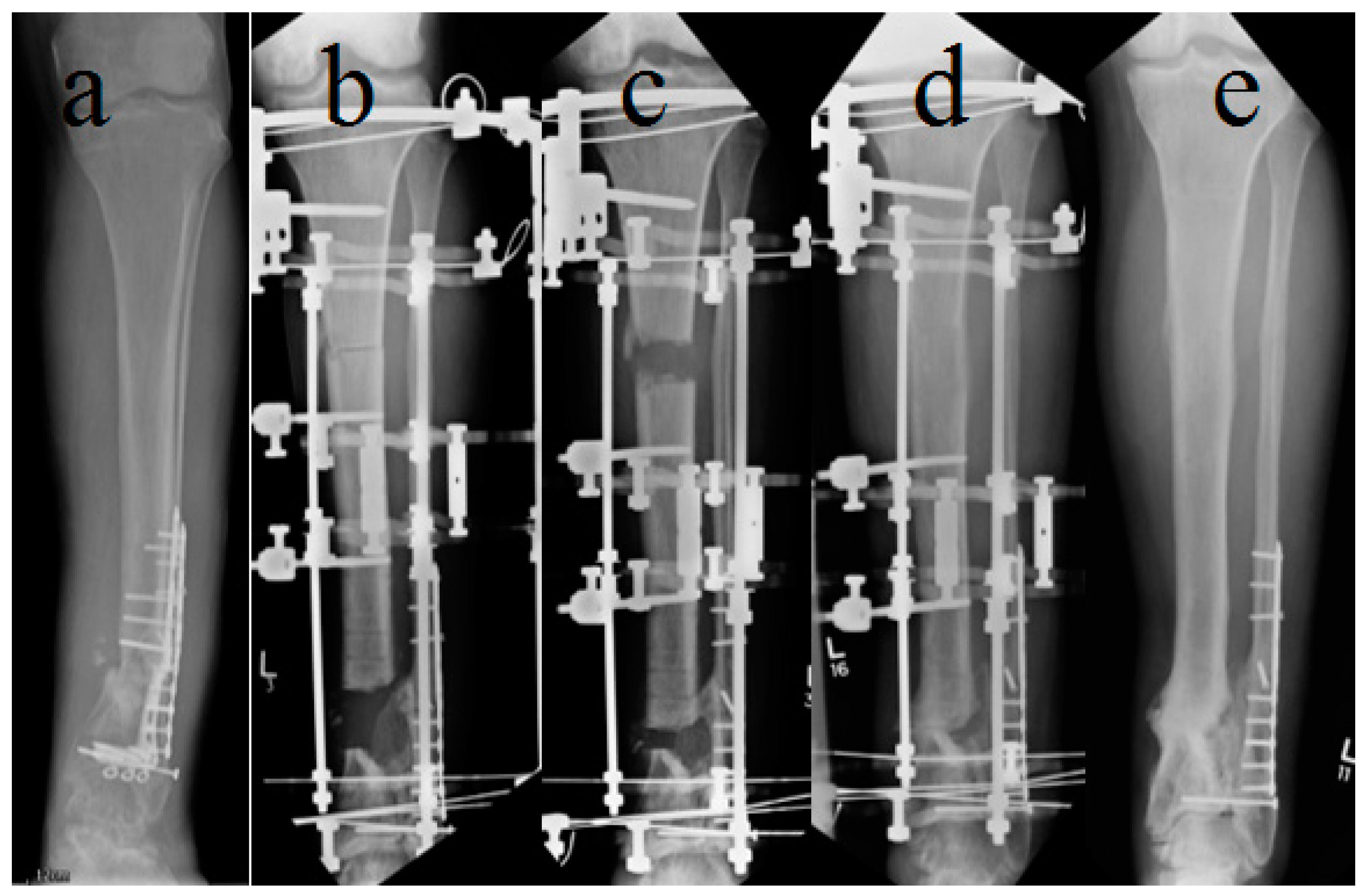
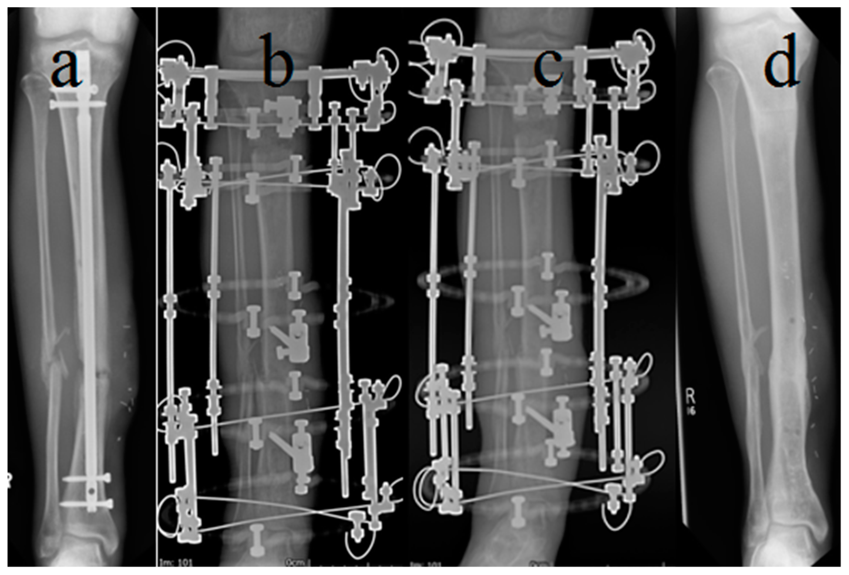
Figure 1. Acute shortening with relengthening. (a) Open fracture of tibia treated with early debridement, IM nailing and flap cover. Presented at three months, with a discharging sinus and exposed medial tibial bone. (b) A 2.5 cm resection was required. The tibia was acutely shortened to good bone contact and the flap reused to cover the defect. (c) Distraction was performed at 1 mm per day through the proximal corticotomy (b) and continued until equal leg length was achieved (at four weeks after surgery). (d) Eight months after surgery, the tibia was solidly healed with good alignment and no evidence of infection.
Jcm 09 00279 g001
Figure 2. Bone Transport. (a) Previous open fracture of distal tibia, treated with internal fixation. The wound broke down and he was referred with a mobile infected non-union. Preoperative AP x-ray shows that the lateral tibial plate has broken at the non-union. (b) Four centimetre resection of the non-union after removal of the infected tibial plate. The fibula was intact and well aligned, so a bone transport was preferred. (c) Twenty-six days after surgery. The proximal corticotomy is distracted at 1 mm per day. (d) Six weeks after docking. No surgery was required to secure union at the docking site. (e) Four months after frame removal with well consolidated docking site, regenerate and good alignment.
Jcm 09 00279 g002
Figure 3. Results of the Association for the Advancement of Methods of Ilizarov (ASAMI) bone classification in the acute shortening/relengthening (ASR) and bone transport (BT) group. There was no statistically significant difference between the groups (p = 0.682). The y-axis gives the absolute numbers.
Jcm 09 00279 g003
Figure 4. Results of the ASAMI functional classification in the acute shortening/relengthening (ASR) and bone transport (BT) group. There was no statistically significant difference between the groups (p = 0.705). The y-axis gives the absolute numbers.
Jcm 09 00279 g004
Table 1. Demographic data and surgical features of the study cohort stratified in groups with acute shortening and relengthening (ASR) and bone transport (BT). Values in bold are statistically significant.
Demographic DataASR (n = 20)BT (n = 27)p-Value
Patient age (mean)48.9 (21–78)50.9 (29–75)0.738
Bone Defect (cm)4.0 (2–5)5.9 (3–10)0.007
Prior Surgeries per patient2.9 (1–5)2.7 (1–6)0.858
Sinus tract890.761
Muscle flap7101.000
EF time (months)8.8 (5–16)10.3 (7–17)0.064
EFI (months/cm)2.0 (1.3–2.8)1.8 (0.9–2.7)0.223
Weber–Cech type (n[%])
Type C1 (5)0 (0)
Type D4 (20)4 (15)
Type E3 (15)5 (19)
Type F12 (60)18 (67)
A statistically significant difference between both study groups was demonstrated for the magnitude of bone defect (p = 0.007) with larger defects in the BT group. The absolute external fixator time was shorter in the ASR group, but it was not statistically significant (p = 0.064).
Table 2. Outcome of the treatment of infected tibial segmental defect in the acute shortening/relengthening (ASR) group and the bone transport (BT) group.
Total
(n = 47)
ASR
(n = 20)
BT
(n = 27)
p-Value
Infection-free47 (100)20 (100)27 (100)1.000
Union without further surgery35 (74)18 (90)17 (63)0.047
Unplanned Reoperation during EF treatment including docking site18 (38)3 (15)15 (56)0.006
Unplanned Reoperation during EF treatment excluding docking site5 (11)3 (15)2 (7)0.638
Unplanned Reoperation after EF removal13 (28)3 (15)10 (37)0.114
Refracture after EF removal3 (7)2 (11)1 (4)0.575
Final Infection-free union47 (100)20 (100)27 (100)1.000
Table 3. Surgeries during and after external fixator (EF) treatment in the acute shortening/relengthening (ASR) group and the bone transport (BT) group.
TotalASRBTp-Value
During EF treatment
Fibular Division1 (2)1 (5)0 (0)0.426
Insertion of further pins1 (2)1 (5)0 (0)0.426
Tethered pin site release1 (2)1 (5)0 (0)0.426
Bone grafting only4 (9)0 (0)4 (15)0.126
Freshening of docking site3 (6)0 (0)3 (11)0.251
Docking Site Realignment4 (9)0 (0)4 (15)0.126
Re-Corticotomy2 (4)0 (0)2 (7)0.500
BMP only2 (4)0 (0)2 (7)0.500
After EF removal
Plate only4 (9)0 (0)4 (15)0.126
Plating and bone grafting1 (2)1 (5)0 (0)0.426
Plating and BMP1 (2)0 (0)1 (4)1.000
EF reapplication1 (2)1 (5)0 (0)0.426
Intramedullary Nail4 (9)1 (5)3 (11)0.626
Ankle Fusion2 (4)0 (0)2 (7)0.500
Total316250.013
NOTE. The values given are the number of cases, with percentage in parentheses. Values in bold are statistically significant. EF = external fixator, BMP = Bone Morphogenic Protein.
Table 4. Results of comparative trials in the literature.
Khan et al. [20] Eralp et al. [19] Testworth et al. [22]Present Study
Study designprospective retrospective retrospective prospective
Number of patients24 74 42 47
ProcedureASR
(n = 16)
BT
(n = 8)
ASR
(n = 45)
BT
(n = 29)
ASR
(n = 21)
BT
(n = 21)
ASR
(n = 20)
BT
(n = 27)
Mean bone defect (cm)3.3 (in all patients)5.95.35.87.04.05.9
Mean EF time (months)8 91010138.810.3
Mean EFI (m/cm)4.2 1.62.11.71.82.01.8
ASAMI functional7 excellent
8 good
1 fair
1 excellent
4 good
1 fair
1 failure
1 died (advanced liver disease)
34 excellent
8 good
2 fair1 poor
22 excellent
3 good
4 poor
14 excellent
6 good
1 fair
14 excellent
6 good
1 fair
12 excellent
7 good
1 fair
19 excellent
6 good
2 fair
ASAMI bone6 excellent
9 good
1 fair
5 good
2 poor
1 died
35 excellent
8 good
2 fair
21 excellent
4 good
4 poor
19 excellent
2 good
15 excellent
5 good
1 fair
18 excellent
2 good
22 excellent
5 good

