Digital Pathology: Advantages, Limitations and Emerging Perspectives


Abstract
1. Introduction
2. From Telepathology to Whole Slide Imaging (WSI)
3. Regulatory Requirements for WSI for Patient Diagnostics in Europe and the US
4. Concordance of Digital Pathology (DP) with Glass Slides
5. Critical Quality Parameters in WSI for Diagnostic Imaging
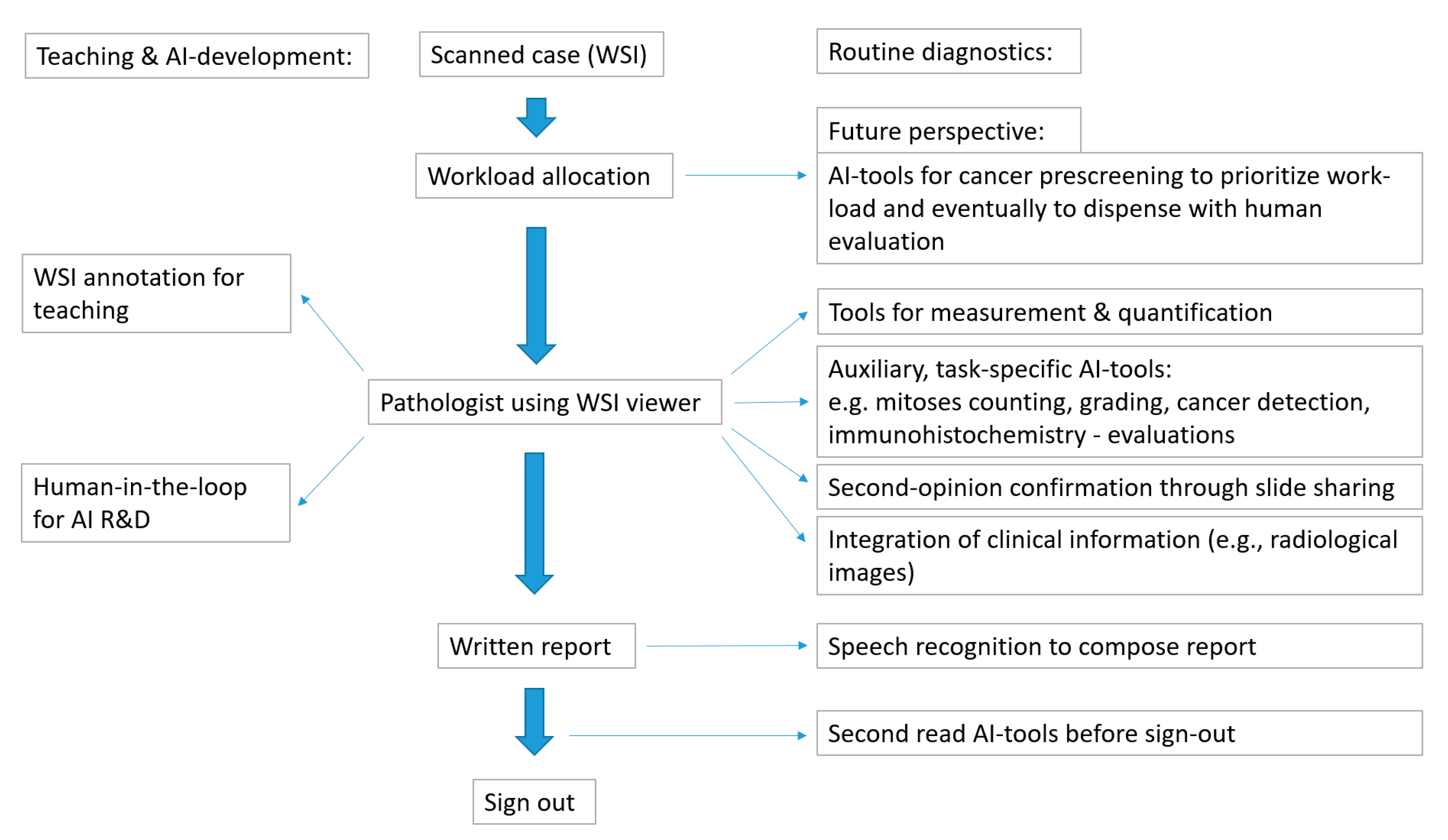
6. The Integrated DP Work-Flow
7. Experience from DP Implementations in Routine Diagnostics Using WSI
8. Medical Education and the Consultation Setting—Advantages and Challenges
9. Computational Pathology (CPATH)
10. Cost-Effectiveness Considerations
11. Digital Pathology and Occupational Health—Computer Vision Syndrome (CVS)
12. Digital Pathology and the Pathologist’s Profession
13. Important Open Challenges and How They Could Be Addressed
14. Conclusions
Author Contributions
Funding
Conflicts of Interest
References
- Hanna, M.G.; Reuter, V.E.; Samboy, J.; England, C.; Corsale, L.; Fine, S.W.; Agaram, N.P.; Stamelos, E.; Yagi, Y.; Hameed, M.; et al. Implementation of digital pathology offers clinical and operational increase in efficiency and cost savings. Arch. Pathol. Lab. Med. 2019, 143, 1545–1555. [Google Scholar] [CrossRef] [PubMed]
- García-Rojo, M. International clinical guidelines for the adoption of digital pathology: A review of technical aspects. Pathobiol. J. Immunopathol. Mol. Cell. Biol. 2016, 83, 99–109. [Google Scholar] [CrossRef] [PubMed]
- Abels, E.; Pantanowitz, L. Current state of the regulatory trajectory for whole slide imaging devices in the USA. J. Pathol. Inform. 2017, 8, 23. [Google Scholar] [CrossRef] [PubMed]
- Mukhopadhyay, S.; Feldman, M.D.; Abels, E.; Ashfaq, R.; Beltaifa, S.; Cacciabeve, N.G.; Cathro, H.P.; Cheng, L.; Cooper, K.; Dickey, G.E.; et al. Whole slide imaging versus microscopy for primary diagnosis in surgical pathology: A multicenter blinded randomized noninferiority study of 1992 cases (pivotal study). Am. J. Surg. Pathol. 2018, 42, 39–52. [Google Scholar] [CrossRef] [PubMed]
- Goacher, E.; Randell, R.; Williams, B.; Treanor, D. The diagnostic concordance of whole slide imaging and light microscopy: A systematic review. Arch. Pathol. Lab. Med. 2017, 141, 151–161. [Google Scholar] [CrossRef] [PubMed]
- Snead, D.R.; Tsang, Y.W.; Meskiri, A.; Kimani, P.K.; Crossman, R.; Rajpoot, N.M.; Blessing, E.; Chen, K.; Gopalakrishnan, K.; Matthews, P.; et al. Validation of digital pathology imaging for primary histopathological diagnosis. Histopathology 2016, 68, 1063–1072. [Google Scholar] [CrossRef]
- Azam, A.S.; Miligy, I.M.; Kimani, P.K.; Maqbool, H.; Hewitt, K.; Rajpoot, N.M.; Snead, D.R.J. Diagnostic concordance and discordance in digital pathology: A systematic review and meta-analysis. J. Clin. Pathol. 2020. [Google Scholar] [CrossRef]
- Pantanowitz, L.; Sinard, J.H.; Henricks, W.H.; Fatheree, L.A.; Carter, A.B.; Contis, L.; Beckwith, B.A.; Evans, A.J.; Lal, A.; Parwani, A.V. Validating whole slide imaging for diagnostic purposes in pathology: Guideline from the college of american pathologists pathology and laboratory quality center. Arch. Pathol. Lab. Med. 2013, 137, 1710–1722. [Google Scholar] [CrossRef]
- Abels, E.; Pantanowitz, L.; Aeffner, F.; Zarella, M.D.; van der Laak, J.; Bui, M.M.; Vemuri, V.N.; Parwani, A.V.; Gibbs, J.; Agosto-Arroyo, E.; et al. Computational pathology definitions, best practices, and recommendations for regulatory guidance: A white paper from the digital pathology association. J. Pathol. 2019, 249, 286–294. [Google Scholar] [CrossRef]
- Garcia-Rojo, M.; De Mena, D.; Muriel-Cueto, P.; Atienza-Cuevas, L.; Dominguez-Gomez, M.; Bueno, G. New european union regulations related to whole slide image scanners and image analysis software. J. Pathol. Inform. 2019, 10, 2. [Google Scholar] [CrossRef]
- Bernard, C.; Chandrakanth, S.A.; Cornell, I.S.; Dalton, J.; Evans, A.; Garcia, B.M.; Godin, C.; Godlewski, M.; Jansen, G.H.; Kabani, A.; et al. Guidelines from the canadian association of pathologists for establishing a telepathology service for anatomic pathology using whole-slide imaging. J. Pathol. Inform. 2014, 5, 15. [Google Scholar] [PubMed]
- Royal College of Pathologists. Best Practice Recommendations for Digital Pathology. Available online: https://www.rcpath.org/uploads/assets/f465d1b3-797b-4297-b7fedc00b4d77e51/Best-practice-recommendations-for-implementing-digital-pathology.pdf (accessed on 3 October 2020).
- Williams, B.J.; Brettle, D.; Aslam, M.; Barrett, P.; Bryson, G.; Cross, S.; Snead, D.; Verrill, C.; Clarke, E.; Wright, A.; et al. Guidance for remote reporting of digital pathology slides during periods of exceptional service pressure: An emergency response from the uk royal college of pathologists. J. Pathol. Inform. 2020, 11, 12. [Google Scholar] [CrossRef] [PubMed]
- Haroske, G.; Zwönitzer, R.; Hufnagl, P. “Digital Pathology in Diagnostics-Reporting on Digital Images” guideline of the professional association of german pathologists. Der Pathologe 2018, 39, 250–252. [Google Scholar] [CrossRef] [PubMed]
- Williams, B.J.; Knowles, C.; Treanor, D. Maintaining quality diagnosis with digital pathology: A practical guide to iso 15189 accreditation. J. Clin. Pathol. 2019, 72, 663–668. [Google Scholar] [CrossRef] [PubMed]
- Krupinski, E.A.; Silverstein, L.D.; Hashmi, S.F.; Graham, A.R.; Weinstein, R.S.; Roehrig, H. Observer performance using virtual pathology slides: Impact of lcd color reproduction accuracy. J. Digit. Imaging 2012, 25, 738–743. [Google Scholar] [CrossRef] [PubMed]
- Norgan, A.P.; Suman, V.J.; Brown, C.L.; Flotte, T.J.; Mounajjed, T. Comparison of a medical-grade monitor vs commercial off-the-shelf display for mitotic figure enumeration and small object (Hellicobacter pylori) detection. Am. J. Clin. Pathol. 2018, 149, 181–185. [Google Scholar] [CrossRef]
- Randell, R.; Ambepitiya, T.; Mello-Thoms, C.; Ruddle, R.A.; Brettle, D.; Thomas, R.G.; Treanor, D. Effect of display resolution on time to diagnosis with virtual pathology slides in a systematic search task. J. Digit. Imaging 2015, 28, 68–76. [Google Scholar] [CrossRef]
- Mills, A.M.; Gradecki, S.E.; Horton, B.J.; Blackwell, R.; Moskaluk, C.A.; Mandell, J.W.; Mills, S.E.; Cathro, H.P. Diagnostic efficiency in digital pathology: A comparison of optical versus digital assessment in 510 surgical pathology cases. Am. J. Surg. Pathol. 2018, 42, 53–59. [Google Scholar] [CrossRef]
- Abel, J.T.; Ouillette, P.; Williams, C.L.; Blau, J.; Cheng, J.; Yao, K.; Lee, W.Y.; Cornish, T.C.; Balis, U.G.J.; McClintock, D.S. Display characteristics and their impact on digital pathology: A current review of pathologists’ future “microscope”. J. Pathol. Inform. 2020, 11, 23. [Google Scholar]
- Stathonikos, N.; Nguyen, T.Q.; Spoto, C.P.; Verdaasdonk, M.A.M.; van Diest, P.J. Being fully digital: Perspective of a dutch academic pathology laboratory. Histopathology 2019, 75, 621–635. [Google Scholar] [CrossRef]
- Steiner, D.F.; MacDonald, R.; Liu, Y.; Truszkowski, P.; Hipp, J.D.; Gammage, C.; Thng, F.; Peng, L.; Stumpe, M.C. Impact of deep learning assistance on the histopathologic review of lymph nodes for metastatic breast cancer. Am. J. Surg. Pathol. 2018, 42, 1636–1646. [Google Scholar] [CrossRef] [PubMed]
- Stathonikos, N.; Nguyen, T.Q.; van Diest, P.J. Rocky road to digital diagnostics: Implementation issues and exhilarating experiences. J. Clin. Pathol. 2020. [Google Scholar] [CrossRef] [PubMed]
- HalioDx. Immunoscore® IC in Non-Small Cell Lung Cancer. Available online: https://www.haliodx.com/diagnostic/immunoscorer-ic-in-lung-cancer/ (accessed on 26 October 2020).
- HalioDx. Haliodx and Philips Team up to Offer Immunoscore® Colon ivd on Philips Intellisite Pathology Solution. Available online: https://www.haliodx.com/about-us/news/detail/News/haliodx-and-philips-team-up-to-offer-immunoscorer-colon-ivd-on-philips-intellisite-pathology-soluti/ (accessed on 26 October 2020).
- Akbar, S.; Peikari, M.; Salama, S.; Panah, A.Y.; Nofech-Mozes, S.; Martel, A.L. Automated and manual quantification of tumour cellularity in digital slides for tumour burden assessment. Sci. Rep. 2019, 9, 14099. [Google Scholar] [CrossRef] [PubMed]
- Ström, P.; Kartasalo, K.; Olsson, H.; Solorzano, L.; Delahunt, B.; Berney, D.M.; Bostwick, D.G.; Evans, A.J.; Grignon, D.J.; Humphrey, P.A.; et al. Artificial intelligence for diagnosis and grading of prostate cancer in biopsies: A population-based, diagnostic study. Lancet Oncol. 2020, 21, 222–232. [Google Scholar] [CrossRef]
- Pantanowitz, L.; Quiroga-Garza, G.M.; Bien, L.; Heled, R.; Laifenfeld, D.; Linhart, C.; Sandbank, J.; Albrecht Shach, A.; Shalev, V.; Vecsler, M.; et al. An artificial intelligence algorithm for prostate cancer diagnosis in whole slide images of core needle biopsies: A blinded clinical validation and deployment study. Lancet Digit. Health 2020, 2, e407–e416. [Google Scholar] [CrossRef]
- Bulten, W.; Pinckaers, H.; van Boven, H.; Vink, R.; de Bel, T.; van Ginneken, B.; van der Laak, J.; Hulsbergen-van de Kaa, C.; Litjens, G. Automated deep-learning system for gleason grading of prostate cancer using biopsies: A diagnostic study. Lancet Oncol. 2020, 21, 233–241. [Google Scholar] [CrossRef]
- Campanella, G.; Hanna, M.G.; Geneslaw, L.; Miraflor, A.; Werneck Krauss Silva, V.; Busam, K.J.; Brogi, E.; Reuter, V.E.; Klimstra, D.S.; Fuchs, T.J. Clinical-grade computational pathology using weakly supervised deep learning on whole slide images. Nat. Med. 2019, 25, 1301–1309. [Google Scholar] [CrossRef]
- Holzinger, A. Interactive machine learning for health informatics: When do we need the human-in-the-loop? Brain Inform. 2016, 3, 119–131. [Google Scholar] [CrossRef]
- Lutnick, B.; Ginley, B.; Govind, D.; McGarry, S.D.; LaViolette, P.S.; Yacoub, R.; Jain, S.; Tomaszewski, J.E.; Jen, K.Y.; Sarder, P. An integrated iterative annotation technique for easing neural network training in medical image analysis. Nat. Mach. Intell. 2019, 1, 112–119. [Google Scholar] [CrossRef]
- Alami, H.; Fortin, J.P.; Gagnon, M.P.; Pollender, H.; Têtu, B.; Tanguay, F. The challenges of a complex and innovative telehealth project: A qualitative evaluation of the eastern quebec telepathology network. Int. J. Health Policy Manag. 2018, 7, 421–432. [Google Scholar] [CrossRef]
- Têtu, B.; Perron, É.; Louahlia, S.; Paré, G.; Trudel, M.C.; Meyer, J. The eastern québec telepathology network: A three-year experience of clinical diagnostic services. Diagn. Pathol. 2014, 9 (Suppl. 1), S1. [Google Scholar]
- Pare, G.; Meyer, J.; Trudel, M.C.; Tetu, B. Impacts of a large decentralized telepathology network in canada. Telemed. J. E-Health Off. J. Am. Telemed. Assoc. 2016, 22, 246–250. [Google Scholar] [CrossRef] [PubMed]
- Têtu, B.; Boulanger, J.; Houde, C.; Fortin, J.P.; Gagnon, M.P.; Roch, G.; Paré, G.; Trudel, M.C.; Sicotte, C. The eastern quebec telepathology network: A real collective project. Med. Sci. M/S 2012, 28, 993–999. [Google Scholar]
- Thorstenson, S.; Molin, J.; Lundström, C. Implementation of large-scale routine diagnostics using whole slide imaging in sweden: Digital pathology experiences 2006-2013. J. Pathol. Inform. 2014, 5, 14. [Google Scholar] [PubMed]
- Asa, S.L.; Bodén, A.C.; Treanor, D.; Jarkman, S.; Lundström, C.; Pantanowitz, L. 2020 vision of digital pathology in action. J. Pathol. Inform. 2019, 10, 27. [Google Scholar]
- Evans, A.J.; Salama, M.E.; Henricks, W.H.; Pantanowitz, L. Implementation of whole slide imaging for clinical purposes: Issues to consider from the perspective of early adopters. Arch. Pathol. Lab. Med. 2017, 141, 944–959. [Google Scholar] [CrossRef]
- Williams, B.J.; Treanor, D. Practical guide to training and validation for primary diagnosis with digital pathology. J. Clin. Pathol. 2020, 73, 418–422. [Google Scholar] [CrossRef]
- Turnquist, C.; Roberts-Gant, S.; Hemsworth, H.; White, K.; Browning, L.; Rees, G.; Roskell, D.; Verrill, C. On the edge of a digital pathology transformation: Views from a cellular pathology laboratory focus group. J. Pathol. Inform. 2019, 10, 37. [Google Scholar] [CrossRef]
- Leica Biosystems. Top Considerations When Buying a Digital Pathology Scanner. Available online: https://www.leicabiosystems.com/de/resources/top-considerations-when-buying-a-digital-pathology-scanner/ (accessed on 25 October 2020).
- Griffin, J.; Treanor, D. Digital pathology in clinical use: Where are we now and what is holding us back? Histopathology 2017, 70, 134–145. [Google Scholar] [CrossRef]
- Hanna, M.G.; Reuter, V.E.; Ardon, O.; Kim, D.; Sirintrapun, S.J.; Schuffler, P.J.; Busam, K.J.; Sauter, J.L.; Brogi, E.; Tan, L.K.; et al. Validation of a digital pathology system including remote review during the covid-19 pandemic. Mod. Pathol. 2020, 33, 2115–2127. [Google Scholar] [CrossRef]
- Boyce, B.F. Whole slide imaging: Uses and limitations for surgical pathology and teaching. Biotech. Histochem. Off. Publ. Biol. Stain Comm. 2015, 90, 321–330. [Google Scholar] [CrossRef] [PubMed]
- van Diest, P.J.; Huisman, A.; van Ekris, J.; Meijer, J.; Willems, S.; Hofhuis, H.; Verbeek, X.; van der Wel, M.; Vos, S.; Leguit, R.; et al. Pathology image exchange: The dutch digital pathology platform for exchange of whole-slide images for efficient teleconsultation, telerevision, and virtual expert panels. JCO Clin. Cancer Inform. 2019, 3, 1–7. [Google Scholar] [CrossRef] [PubMed]
- Al-Janabi, S.; van Slooten, H.J.; Visser, M.; van der Ploeg, T.; van Diest, P.J.; Jiwa, M. Evaluation of mitotic activity index in breast cancer using whole slide digital images. PLoS ONE 2013, 8, e82576. [Google Scholar] [CrossRef] [PubMed]
- Saha, M.; Chakraborty, C.; Arun, I.; Ahmed, R.; Chatterjee, S. An advanced deep learning approach for ki-67 stained hotspot detection and proliferation rate scoring for prognostic evaluation of breast cancer. Sci. Rep. 2017, 7, 3213. [Google Scholar] [CrossRef] [PubMed]
- Dennis, J.; Parsa, R.; Chau, D.; Koduru, P.; Peng, Y.; Fang, Y.; Sarode, V.R. Quantification of human epidermal growth factor receptor 2 immunohistochemistry using the ventana image analysis system: Correlation with gene amplification by fluorescence in situ hybridization: The importance of instrument validation for achieving high (>95%) concordance rate. Am. J. Surg. Pathol. 2015, 39, 624–631. [Google Scholar]
- Pagès, F.; Mlecnik, B.; Marliot, F.; Bindea, G.; Ou, F.S.; Bifulco, C.; Lugli, A.; Zlobec, I.; Rau, T.T.; Berger, M.D.; et al. International validation of the consensus immunoscore for the classification of colon cancer: A prognostic and accuracy study. Lancet 2018, 391, 2128–2139. [Google Scholar] [CrossRef]
- Jiang, Y.; Yang, M.; Wang, S.; Li, X.; Sun, Y. Emerging role of deep learning-based artificial intelligence in tumor pathology. Cancer Commun. 2020, 40, 154–166. [Google Scholar] [CrossRef]
- Samek, W.; Binder, A.; Montavon, G.; Lapuschkin, S.; Müller, K. Evaluating the visualization of what a deep neural network has learned. IEEE Trans. Neural Netw. Learn. Syst. 2017, 28, 2660–2673. [Google Scholar] [CrossRef]
- Holzinger, A.; Plass, M.; Holzinger, K.; Crisan, G.C.; Pintea, C.-M.; Palade, V. A glass-box interactive machine learning approach for solving np-hard problems with the human-in-the-loop. arXiv 2017, arXiv:1708.01104. [Google Scholar]
- Holzinger, A. From machine learning to explainable ai. In Proceedings of the 2018 World Symposium on Digital Intelligence for Systems and Machines (DISA), Kosice, Slovakia, 23–25 August 2018; pp. 55–66. [Google Scholar]
- Pohn, B.; Kargl, M.; Reihs, R.; Holzinger, A.; Zatloukal, K.; Müller, H. Towards a deeper understanding of how a pathologist makes a diagnosis: Visualization of the diagnostic process in histopathology. In Proceedings of the 2019 IEEE Symposium on Computers and Communications (ISCC), Barcelona, Spain, 29 June–3 July 2019; pp. 1081–1086. [Google Scholar]
- Benjamens, S.; Dhunnoo, P.; Meskó, B. The state of artificial intelligence-based FDA-approved medical devices and algorithms: An online database. NPJ Digit. Med. 2020, 3, 118. [Google Scholar] [CrossRef]
- Coudray, N.; Ocampo, P.S.; Sakellaropoulos, T.; Narula, N.; Snuderl, M.; Fenyö, D.; Moreira, A.L.; Razavian, N.; Tsirigos, A. Classification and mutation prediction from non-small cell lung cancer histopathology images using deep learning. Nat. Med. 2018, 24, 1559–1567. [Google Scholar] [CrossRef] [PubMed]
- Kim, R.H.; Nomikou, S.; Dawood, Z.; Jour, G.; Donnelly, D.; Moran, U.; Weber, J.S.; Razavian, N.; Snuderl, M.; Shapiro, R.; et al. A deep learning approach for rapid mutational screening in melanoma. bioRxiv 2019, 610311. [Google Scholar] [CrossRef]
- Schaumberg, A.J.; Rubin, M.A.; Fuchs, T.J. H&E-stained whole slide image deep learning predicts spop mutation state in prostate cancer. bioRxiv 2018. [Google Scholar] [CrossRef]
- Wulczyn, E.; Steiner, D.F.; Moran, M.; Plass, M.; Reihs, R.; Mueller, H.; Sadhwani, A.; Cai, Y.; Flament, I.; Chen, P.-H.C.; et al. Abstract 2096: A deep learning system to predict disease-specific survival in stage ii and stage iii colorectal cancer. Cancer Res. 2020, 80, 2096. [Google Scholar]
- Skrede, O.J.; De Raedt, S.; Kleppe, A.; Hveem, T.S.; Liestøl, K.; Maddison, J.; Askautrud, H.A.; Pradhan, M.; Nesheim, J.A.; Albregtsen, F.; et al. Deep learning for prediction of colorectal cancer outcome: A discovery and validation study. Lancet 2020, 395, 350–360. [Google Scholar] [CrossRef]
- FDAnews. Fda Hands Paige. AL Breakthrough Designation for Cancer Diagnosis Tool. Available online: https://www.fdanews.com/articles/190525-fda-hands-paigeai-breakthrough-designation-for-cancer-diagnosis-tool (accessed on 26 October 2020).
- Komura, D.; Ishikawa, S. Machine learning methods for histopathological image analysis. Comput. Struct. Biotechnol. J. 2018, 16, 34–42. [Google Scholar] [CrossRef]
- Zhang, Y.; Zhang, B.; Coenen, F.; Xiao, J.; Lu, W. One-class kernel subspace ensemble for medical image classification. EURASIP J. Adv. Signal Process. 2014, 2014, 17. [Google Scholar] [CrossRef]
- Aresta, G.; Araújo, T.; Kwok, S.; Chennamsetty, S.S.; Safwan, M.; Alex, V.; Marami, B.; Prastawa, M.; Chan, M.; Donovan, M.; et al. Bach: Grand challenge on breast cancer histology images. Med. Image Anal. 2019, 56, 122–139. [Google Scholar] [CrossRef]
- Ehteshami Bejnordi, B.; Veta, M.; Johannes van Diest, P.; van Ginneken, B.; Karssemeijer, N.; Litjens, G.; van der Laak, J.; Hermsen, M.; Manson, Q.F.; Balkenhol, M.; et al. Diagnostic assessment of deep learning algorithms for detection of lymph node metastases in women with breast cancer. JAMA 2017, 318, 2199–2210. [Google Scholar] [CrossRef]
- Cireşan, D.C.; Giusti, A.; Gambardella, L.M.; Schmidhuber, J. Mitosis detection in breast cancer histology images with deep neural networks. Medical image computing and computer-assisted intervention: MICCAI. In Proceedings of the International Conference on Medical Image Computing and Computer-Assisted Intervention, Nagoya, Japan, 22–26 September 2013; pp. 411–418. [Google Scholar]
- Ho, J.; Ahlers, S.M.; Stratman, C.; Aridor, O.; Pantanowitz, L.; Fine, J.L.; Kuzmishin, J.A.; Montalto, M.C.; Parwani, A.V. Can digital pathology result in cost savings? A financial projection for digital pathology implementation at a large integrated health care organization. J. Pathol. Inform. 2014, 5, 33. [Google Scholar] [CrossRef]
- Rosenfield, M. Computer vision syndrome: A review of ocular causes and potential treatments. Ophthalmic & physiological optics. Ophthal Physl Opt 2011, 31, 502–515. [Google Scholar]
- Rossignol, A.M.; Morse, E.P.; Summers, V.M.; Pagnotto, L.D. Video display terminal use and reported health symptoms among massachusetts clerical workers. J. Occup. Med. Off. Publ. Ind. Med Assoc. 1987, 29, 112–118. [Google Scholar]
- Daum, K.M.; Clore, K.A.; Simms, S.S.; Vesely, J.W.; Wilczek, D.D.; Spittle, B.M.; Good, G.W. Productivity associated with visual status of computer users. Optometry 2004, 75, 33–47. [Google Scholar] [CrossRef]
- Blehm, C.; Vishnu, S.; Khattak, A.; Mitra, S.; Yee, R.W. Computer vision syndrome: A review. Surv. Ophthalmol. 2005, 50, 253–262. [Google Scholar] [CrossRef]
- Bohr, P.C. Efficacy of office ergonomics education. J. Occup. Rehabil. 2000, 10, 12. [Google Scholar] [CrossRef]
- Chui, M.; Manyika, J.; Miremadi, M. Where machines could replace humans—And where they can’t (yet). McKinsey Q. 2016, 30, 1–9. [Google Scholar]
- Vertinsky, T.; Forster, B. Prevalence of eye strain among radiologists: Influence of viewing variables on symptoms. AJR. Am. J. Roentgenol. 2005, 184, 681–686. [Google Scholar] [CrossRef]


| Digital Pathology (DP) Feature | Possible Advantages | Possible Disadvantages |
|---|---|---|
| In-house telepathology | ○ Quick second opinion ○ Social distancing (COVID-19 pandemic) | ○ Second opinion overuse (interrupted work-flows) ○ Decreased interpersonal (face-to-face) communication |
| Extramural telepathology | ○ Service for remote/understaffed areas ○ Specialization through DP in low volume labs ○ Home-office use ○ Healthcare cost reduction through global histopathology market | ○ Social isolation in remote telepathology ○ Loss of routine on-site expertise through home office ○ Wage competition through global histopathology market |
| Consultation telepathology | ○ Quick access possible ○ No physical slide transfer ○ Lower threshold for consultation due to shorter turnaround time | ○ No tissue block available for additional stains/molecular assays ○ Consulted pathologist unaccustomed to work-up (stains/scanner calibration) at the primary center ○ Compatibility issues due to diverse proprietary DP formats ○ Possible medico-legal implications due to restricted work-up |
| WSI-general | ○ No physical slide distribution ○ No fading of stored slides ○ No irretrievable/lost slides ○ Shorter sign-out time ○ Reduced misidentification of slides due to barcoded slides automatically allocated to the case ○ Easy dynamic workload allocation (e.g., management of backlogged work, redistribution in case of sick leave) | ○ Time to evaluable-ready slide increased due to additional scan time ○ Integration into a laboratory information system (LIS) for full efficiency gains needed → possible costs for LIS update ○ Regular calibration required (scanners/displays) ○ Small particles omitted by scan → manual checking for rescan ○ Artifacts (out-of-focus areas, digital stitching artifacts) ○ Increased IT-dependence (IT-downtime) compared to optical microscopy |
| WSI-reporting/user experience | ○ Parallel (side-by-side) viewing, digital slide superposition ○ Shorter sign-out time ○ Quick access to prior slides → less immunohistochemistry ○ Facilitates slide presentation at multidisciplinary tumor board ○ Easy image sharing in clinical communication ○ Computational pathology possible (see below) ○ Occupational health: less neck strain, more flexible posture | ○ Slower evaluation compared to optical microscopes ○ Mostly only single focus plane in routine DP → difficulties with interpretation ○ Some structures harder to recognize on WSI → glass slide needed ○ Polarization not possible on DP → glass slide needed ○ Extra training for safe practice required (perceived insecurity on digital sign-out) if not DP from career start ○ Easy availability of prior digital slides might shift medico-legal onus towards more extensive re-examination → increased workload ○ Dual infrastructure generally necessary (glass and digital) ○ Occupational health: Computer Vision Syndrome (CVS) |
| WSI-Image Analysis, ML/AI | ○ Faster/efficient and more accurate measurements/quantifications ○ Exact quantification of tumor cell content for molecular analyses ○ Digital enhancement of image features ○ AI for second-read safety net ○ Direct link morphology to clinical parameters “novel biomarker” beyond human recognition ○ Inspection/correction of suggestions from AI-apps in development on WSI-viewer: “human-in-the-loop” interaction | ○ Benefit of more accurate quantification not necessarily clinically relevant ○ Applications beyond human evaluation not yet approved/used for clinical management ○ AI intransparent (“black box”) ○ Regulatory oversight challenges with self-modifying (adaptive) AI as algorithm/performance not constant over time |
| WSI-teaching | ○ Digital images for presentation and exams readily available ○ Remote teaching and self-study ○ Increased student motivation, modern appeal | ○ None |
| Costs and efficiency gains | ○ Work time saved through faster turnaround times ○ Decreased auxiliary techniques (less immunohistochemistry) ○ Decreased physical slide-transfer costs | ○ DP implementation and maintenance and storage costs add to current fixed costs if productivity gains remain unrealized (fixed work contracts) ○ Dual infrastructure costs (workstations and microscopes if kept) ○ Glass and digital storage still generally deemed necessary ○ Technical expert knowledge for hardware acquisitions needed |
Publisher’s Note: MDPI stays neutral with regard to jurisdictional claims in published maps and institutional affiliations. |
© 2020 by the authors. Licensee MDPI, Basel, Switzerland. This article is an open access article distributed under the terms and conditions of the Creative Commons Attribution (CC BY) license (http://creativecommons.org/licenses/by/4.0/).
Share and Cite
Jahn, S.W.; Plass, M.; Moinfar, F. Digital Pathology: Advantages, Limitations and Emerging Perspectives. J. Clin. Med. 2020, 9, 3697. https://doi.org/10.3390/jcm9113697
Jahn SW, Plass M, Moinfar F. Digital Pathology: Advantages, Limitations and Emerging Perspectives. Journal of Clinical Medicine. 2020; 9(11):3697. https://doi.org/10.3390/jcm9113697
Chicago/Turabian StyleJahn, Stephan W., Markus Plass, and Farid Moinfar. 2020. "Digital Pathology: Advantages, Limitations and Emerging Perspectives" Journal of Clinical Medicine 9, no. 11: 3697. https://doi.org/10.3390/jcm9113697
APA StyleJahn, S. W., Plass, M., & Moinfar, F. (2020). Digital Pathology: Advantages, Limitations and Emerging Perspectives. Journal of Clinical Medicine, 9(11), 3697. https://doi.org/10.3390/jcm9113697