Article

Genetic Variants of lncRNA MALAT1 Exert Diverse Impacts on the Risk and Clinicopathologic Characteristics of Patients with Hepatocellular Carcinoma

1
Division of Hepatology and Gastroenterology, Department of Internal Medicine, Yuan’s General Hospital, Kaohsiung 80249, Taiwan
2
Department of Internal Medicine, School of Medicine, College of Medicine, Taipei Medical University, Taipei 11031, Taiwan
3
Pulmonary Research Center, Wan Fang Hospital, Taipei Medical University, Taipei 11696, Taiwan
4
School of Medicine, Chung Shan Medical University, Taichung 40201, Taiwan
5
Department of Surgery, Chung Shan Medical University Hospital, Taichung 40201, Taiwan
6
Graduate Institute of Clinical Medicine, College of Medicine, Taipei Medical University, Taipei 11031, Taiwan
7
Whole-Genome Research Core Laboratory of Human Diseases, Chang Gung Memorial Hospital, Keelung 20401, Taiwan
8
Department of Pharmacy, FooYin University Hospital, Pingtung 92847, Taiwan
9
Institute of Medicine, Chung Shan Medical University, Taichung 40201, Taiwan
10
Department of Medical Research, Chung Shan Medical University Hospital, Taichung 40201, Taiwan
11
TMU Research Center of Cancer Translational Medicine, Taipei Medical University, Taipei 11031, Taiwan
*
Authors to whom correspondence should be addressed.
Lan-Ting Yuan and Jer-Hwa Chang made an equal contribution to this work.
J. Clin. Med. 2019, 8(9), 1406; https://doi.org/10.3390/jcm8091406
Submission received: 25 July 2019 / Revised: 26 August 2019 / Accepted: 3 September 2019 / Published: 6 September 2019
(This article belongs to the Special Issue Molecular Mechanisms of Cancer Chemoprevention)

Abstract

:
The long noncoding (lnc)RNA, metastasis-associated lung adenocarcinoma transcript 1 (MALAT1), plays a crucial role in the development of hepatocellular carcinoma (HCC). However, potential genetic variants (single nucleotide polymorphisms, SNPs) in MALAT1 that affect the susceptibility and progression of HCC have rarely been explored. Three tagging SNPs, viz., rs3200401 C > T, rs619586 A > G, and rs1194338 C > A, in MALAT1 were genotyped by a TaqMan allelic discrimination assay in 394 HCC patients and 1199 healthy controls. A stratified analysis showed that younger patients (<55 years) with the MALAT1 rs619586 G allele had a decreased risk of HCC under a codominant model (AOR = 0.289, 95% CI: 0.108–0.773, p = 0.013) and dominant model (AOR = 0.286, 95% CI: 0.107–0.765, p = 0.013). Female patients and patients with a smoking habit who carried the CA + AA genotype of rs1194338 had a lower risk of developing vascular invasion (p = 0.049) and a high Child–Pugh grade (B or C) (p = 0.036), respectively. Under the dominant model, smokers with the MALAT1 rs3200401 CT + TT genotype had a higher frequency of hepatitis B virus (HBV) infection (p = 0.034). Moreover, the aspartate aminotransferase was higher in patients with the rs3200401 CT + TT genotype. Furthermore, analyses of clinical datasets revealed that MALAT1 expression level was gradually unregulated during HCC development from normal liver, cirrhotic liver, dysplastic liver to HCC and correlated with poor survival rates in HCC patients, especially in the hepatitis virus-infected population.

1. Introduction

Liver cancer is the sixth most common malignant tumor with more than 840,000 new cases reported per year worldwide, and this disease is the fourth leading cause of cancer-related deaths [1]. Hepatocellular carcinoma (HCC) is the major type of primary liver cancer, comprising 75–85% of cases. Early-stage HCC can be effectively treated by liver transplantation or curative surgery. However, therapeutic strategies are still limited for advanced disease [2]. The incurable property of HCC is due to its extensive heterogeneity of clinical manifestations, and these heterogeneities are manifested through diverse genetic and epigenetic features, as well as ethnic differences in patients [3,4]. For example, HCC causes higher mortality in developing countries, such as Asian and African countries, because of chronic infection with hepatitis B virus (HBV) or hepatitis C virus (HCV) [1]. In addition, a variety of genetic and epigenetic variations were also reported to be correlated with HCC [5]. Intensive investigations over the last few decades have focused on the roles of protein-coding genes in the pathogenesis of HCC. Recently, research hotspots regarding genetic and epigenetic variations have focused on noncoding (nc)RNAs.
Long noncoding (lnc)RNAs are a type of ncRNA with a length of more than 200 bp, which were identified as oncogenes or tumor suppressors and participate in most processes of tumorigenesis, including uncontrollable proliferation, evasion of cell death, and metastasis [6]. The highly conserved metastasis-associated lung adenocarcinoma transcript 1 (MALAT1) lncRNA with >8000 bp was first identified as an oncogene in non-small-cell lung cancer (NSCLC) because of its functional role in promoting metastasis via induction of the epithelial–mesenchymal transition (EMT) and its clinical significance in predicting metastasis of early-stage NSCLC [7,8]. Subsequently, overexpression of MALAT1 was shown to exhibit marked effects on tumor cell proliferation, migration, invasion, and apoptosis in numerous types of cancer. Moreover, accumulating evidence indicated that expression levels of MALAT1 in tumor tissues and/or body fluids could serve as a biomarker for tumor diagnosis and prognosis [9,10]. For instance, aberrant expression of MALAT1 in colorectal, pancreatic, papillary thyroid, and renal cancers was identified as an unfavorable predictor for their clinical progression and prognosis [11,12,13,14].
Regarding the role of MALAT1 in HCC, overexpression of MALAT1 in tumor tissues or sera may correlate with advanced tumor stages and reduced overall survival of HCC patients and indicate a higher risk of tumor recurrence following liver transplantation [15,16]. Moreover, upregulation of MALAT1 in HCC was reported to be associated with chemoresistance to multiple agents, including 5-fluorouracil, mitomycin C, and Adriamycin via a hypoxia-inducible factor (HIF)-1α-MALAT1-microRNA (mir)-216b pathway [17]. Also, Malakar et al. demonstrated that MALAT1 enhances HCC progression by inducing serine/arginine-rich splicing factor 1 (SRSF1) upregulation and mammalian target of rapamycin (mTOR) activation [18]. Undoubtedly, MALAT1 is recognized as an oncogene in HCC.
Results from a whole-genome mutational analysis of an Asian population showed that several mutational signatures were related to liver carcinogenesis including MALAT1 lncRNA, but their clinical significance remains unclear [19]. Genetic variations such as single-nucleotide polymorphisms (SNPs) can alter expressions of coding genes and lncRNAs. Actually, MALAT1 SNPs were reported to be associated with the risk or progression of several cancer types and expression of MALAT1. For example, the AG genotype or G allele of MALAT1 rs619586 SNP was reported to be associated with decreased risks of breast cancer, colorectal cancer (CRC), and papillary thyroid cancer (PTC) and a decreased expression level of MALAT1 [20,21,22]. Moreover, an A allele of rs1194338 in the promoter region of MALAT1 was also reported to be correlated with a decreased CRC risk [21,23]. Furthermore, the polymorphism rs3200401 C > T in the MALAT1 gene was recently reported to be associated with an increased risk of esophageal squamous cell carcinoma (ESCC) in a Chinese population [24]. To date, whether polymorphisms of MALAT1 play any role in the risk and progression of HCC remains less investigated, especially in Asian populations. Accordingly, we performed a case-control study in a Taiwanese population to identify the roles of MALAT1 SNPs in the risk and clinical characteristics of HCC.

2. Materials and Methods

2.1. Study Subjects, Ethics, and Consent

HCC patients of this case-control study were recruited from 2007 to 2015 at Chung Shan Medical University Hospital (Taichung, Taiwan), and were composed of 273 men and 121 women. In total, 1199 age- and gender-matched cancer-free controls were randomly selected from the Taiwan Biobank Project, and all study participants were of a Han Chinese population from the same geographic area. In addition to HCC, any patients with a history of other cancer types were excluded. HCC patients were clinically staged at the time of diagnosis according to the tumor, node, metastasis (TNM) staging system of the American Joint Committee on Cancer (AJCC). Demographic data and related risk factors including the smoking and drinking status were obtained through a structured questionnaire. Tobacco consumption was categorized in “never-user” and “ever-user” (current and former user) at the time of diagnosis. Medical information of our recruited HCC patients such as alpha-fetoprotein (AFP), aspartate aminotransferase (AST), alanine aminotransferase (ALT), the TNM status, Child–Pugh grade, vascular invasion, hepatitis virus infection, and liver cirrhosis was obtained from their medical records. Peripheral blood was collected from each participant after informed consent. The research design was approved by the Institutional Review Board of Chung Shan Medical University Hospital.

2.2. Genomic DNA Extraction

Whole-blood samples collected from our recruited subjects were placed in tubes containing ethylenediaminetetraacetic acid (EDTA) and further centrifuged for 10 min at 3000 rpm. Buffy coats of DNA were extracted from whole-blood specimens using a QIAamp DNA Blood Mini Kit (Qiagen, Valencia, CA, USA) according to the manufacturer’s instructions. Extracted DNA was dissolved in TE buffer (10 mM Tris and 1 mM EDTA; pH 7.8) and the quality of DNA was evaluated based on a corresponding analysis on a Nanodrop-2000 spectrophotometer (Thermo Scientific, Waltham, MA, USA). The final preparation was stored at −20 °C and was used as templates for the polymerase chain reaction (PCR).

2.3. Selection of MALAT1 SNPs

In this study, we selected two tagSNPs, rs3200401 and rs619586, from the International HapMap Project data, because these SNPs fit the following criteria: (i) SNPs located in MALAT1; (ii) minor allele frequency (MAF) of each SNP of >5% in a Han Chinese population (rs3200401 C/T: 0.87/0.13; rs619586 A/G: 0.87/0.13; rs1194338 C/A: 0.76/0.24); (iii) linkage disequilibrium value of r2 < 0.8 for the candidate SNP; and (iv) functional SNPs with a known biological mechanism based on published literature [25,26]. We also included another novel SNP, viz., rs1194338, located in the promoter region of MALAT1, which was recently reported to be associated with susceptibility to colorectal carcinoma (CRC) in a Han Chinese population [21].

2.4. Genotyping of MALAT1 SNPs

Genotyping of MALAT1 SNPs, including rs3200401 (assay ID: C_3246069_10), rs619586 (assay ID: C_1060479_10), and rs1194338 (assay ID: C_11661801_10), was done using the TaqMan SNP Genotyping Assay with an ABI StepOnePlus™ Real-Time PCR System and further evaluated with SDS version 3.0 software (Applied Biosystems, Foster City, CA, USA).

2.5. Bioinformatics Analysis

The Genome-Tissue Expression (GTEx) database (https://www.gtexportal.org/home/) [27] was used to identify the regulatory potential of candidate functional MALAT1 variants. GTEx data were used to identify correlations between SNPs and tissue-specific gene expression levels. Transcriptomic data from samples of a normal liver, cirrhotic liver, dysplastic liver, and HCC were analyzed using the microarray datasets with accession number GSE6764 obtained from the Gene Expression Omnibus (GEO) database (https://www.ncbi.nlm.nih.gov/geo/) to investigate whether MALAT1 expression (probe ID: 224568_x_at) was associated with HCC tumor formation. RNA sequencing analysis, the visualization platform Gene Expression Profiling Interactive Analysis (GEPIA) (http://gepia.cancer-pku.cn/) [28], and the Kaplan–Meier (KM) plotter (http://kmplot.com/analysis/) [29] were used to determine whether MALAT1 expression was correlated with the prognosis of HCC patients. The correlation of MALAT1 and prognosis were calculated by median cutoff in GEPIA and auto selected best cutoff in KM-plotter analysis. The GEPIA performs data mining based on The Cancer Genome Atlas (TCGA) data.

2.6. Statistical Analysis

To compare age, gender, demographic characteristic distributions and categorical variables between HCC patients and control groups, the Mann–Whitney U-test and Chi-squared test was applied for statistical analysis. Multiple logistic regression models were used to estimate the adjusted odds ratio (AOR) and 95% confidence interval (CI) of the association of genotype frequencies with HCC risk; p < 0.05 was considered statistically significant. Experimental data were analyzed using SAS statistical software (vers. 9.1, 2005; SAS Institute, Cary, NC, USA).

3. Results

3.1. Demographic Characteristics of Recruited Subjects

Distributions of demographic and clinical characteristics of recruited subjects are shown in Table 1. No significant differences were found between HCC patients and the controls with regard to the distribution of age <55 and ≥55 years (p = 0.451), gender (p = 0.797), or smoking status (p = 0.913). In contrast, significantly higher frequencies of HCC patients compared to controls had a drinking habit (14.1% vs. 35.3%; p < 0.001) and were positive for hepatitis B surface antigen (HBsAg) (12.2% vs. 42.4%; p < 0.001) and anti-HCV antibodies (4.4% vs. 45.2%; p < 0.001). Among HCC patients, higher proportions were diagnosed as being at the early clinical (I + II) and T (T1 + T2) stages, and with no lymph node (N0) or distal (M0) metastasis. Most patients had slight liver cirrhosis (Child–Pugh grade 0 or A).

3.2. Associations between MALAT1 Candidate SNPs and HCC Susceptibility

To examine possible associations of MALAT1 SNPs with the risk of developing HCC, the genotype frequencies of three SNPs (rs3200401, rs619586, and rs1194338) were first investigated in the entire population we recruited. Alleles with the highest distribution frequencies for rs3200401, rs619586, and rs1194338 SNPs of our recruited subjects were homozygous C/C, A/A, and C/C, respectively. Genotypic distributions of MALAT1 rs3200401, rs619586, and rs1194338 in the control group conformed to Hardy–Weinberg equilibrium (p = 0.112, χ2 value: 2.521; p = 0.423, χ2 value: 0.641 and p = 0.566, χ2 value: 0.330, respectively). After adjusting for age, gender, cigarette smoking, and alcohol consumption, no significant differences were observed between HCC patients with these candidate SNPs (rs3200401 CT/TT, rs619586 AG/GG, rs1194338 CA/AA) and those with the wild-type (WT) gene (Table 2). We further divided the tested population by age, and found that younger patients (<55 years) with the AG/GG genotypes of MALAT1 rs619586 had a decreased risk of HCC under the codominant model (AG vs. AA: AOR, 0.289; 95% CI, 0.108–0.773; p = 0.013) and dominant model (AG + GG vs. AA: AOR, 0.286; 95% CI, 0.107–0.765; p = 0.013) (Table 3), while no associations were observed in elderly HCC patients (≥55 years) (data not shown).

3.3. Relationships between Clinicopathological Features of HCC and MALAT1 SNPs in Different Genders

Next, to clarify the effects of MALAT1 genetic polymorphisms on the HCC clinicopathological status, including clinical stage, primary tumor size, lymph node and distant metastasis, vascular invasion, Child–Pugh grade, hepatitis virus infection, and liver cirrhosis. Among all HCC patients, the rs3200401, rs619586, and rs1194338 SNPs showed no significant association with the clinicopathologic status (Table S1). We further divided the HCC population by sex, and found that only the female HCC patients who carried at least one minor allele (CA and AA) of rs1194338 had a significantly (p = 0.049) 0.377-fold lower risk (95% CI, 0.143–0.994) of developing vascular invasion compared to their corresponding WT homozygotes (Table 4 and Table S2).

3.4. Relationships between Clinicopathological Features and MALAT1 SNPs in HCC Patients Who Smoked Tobacco

We further divided HCC patients into smoking and nonsmoking subgroups and investigated differences between MALAT1 SNPs and the HCC clinicopathological status in these two groups. No association between MALAT1 SNPs (rs3200401, rs619586, and rs1194338) and HCC clinical features was observed in nonsmoking subgroup (Table S3). Among 156 HCC smokers, we observed that patients with at least one polymorphic A allele at the rs1194338 SNP (CA + AA genotype) had lower risks of developing moderate to severe liver failure (Child–Pugh B or C grade; p = 0.036) (Table 5 and Table S3). Moreover, patients with at least one polymorphic T allele at the rs3200401 SNP (CT + TT genotype) had a higher risk of being infected with HBV (p = 0.034) than did WT patients (Table 6 and Table S3).

3.5. Serum Alpha-Fetoprotein (AFP), Aspartate Aminotransferase (AST), and Alanine Aminotransferase (ALT) Levels among Different MALAT1 SNP Carriers in HCC Patients

Serum AFP is a common clinical pathological marker of HCC. Serum ALT and AST levels are most often used to evaluate liver function [30]. Table S4 shows associations of MALAT1 genotypic frequencies with the HCC laboratory status. After adjusting for age, gender, cigarette smoking, and alcohol consumption, we observed that AST levels were significantly higher in HCC patients harboring the rs3200401 CT + TT genotype compared to those with CC homozygotes. Otherwise, no significant associations between levels of these HCC clinical pathologic markers and genotypes of any other MALAT1 SNPs (rs619586 and rs1194338) were noted.

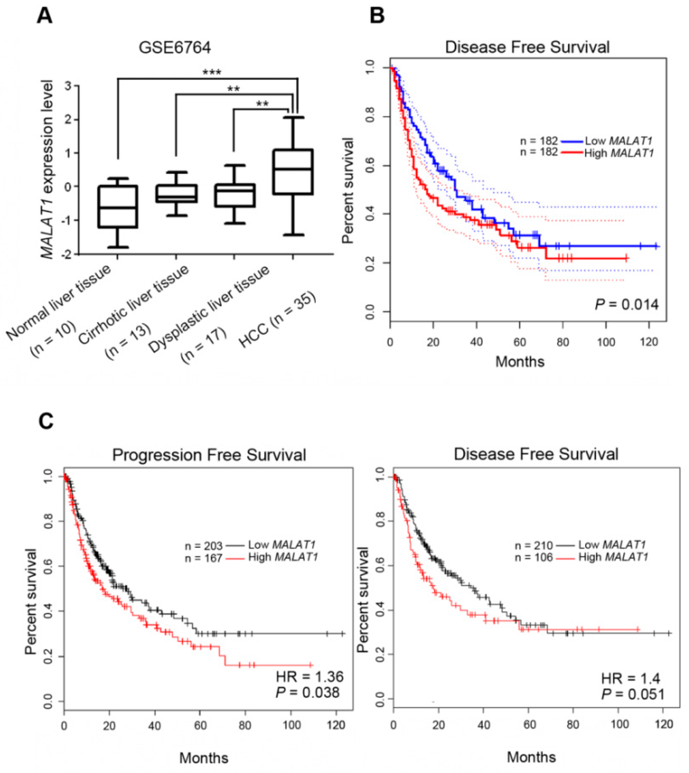
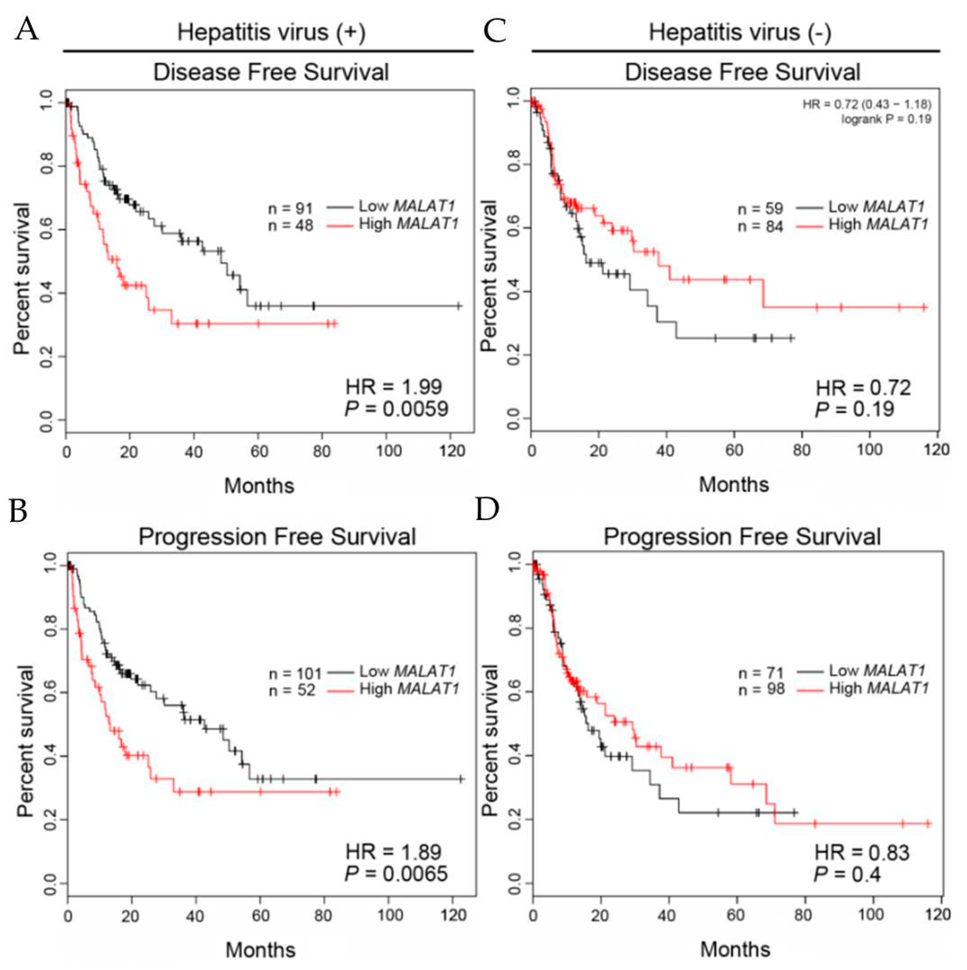
3.6. Clinical Relevance of MALAT1 Expression Levels in HCC Patients with or without Hepatitis Infection

Considering the potential effects of MALAT1 polymorphic genotypes on MALAT1 expression levels, we further clarified the clinical significance of MALAT1 in HCC formation and progression. Tissues of different stages of HCC formation including normal liver, cirrhotic liver, dysplastic liver, and HCC were first analyzed from the GEO database (GSE6764), and we found significantly higher MALAT1 transcripts in HCC tissues than in normal liver, cirrhotic liver, and dysplastic liver tissues (Figure 1A). MALAT1 expression in the cirrhotic liver or dysplastic liver also showed higher trends than in the normal liver, suggesting that MALAT1 might play a role in the process of HCC development. Moreover, MALAT1 expression data in TCGA database were analyzed using the online tool, GEPIA [28], and we found that HCC patients with MALAT1high tumors had shorter disease free survival (DFS) times compared to those with MALAT1low tumors (p = 0.014; Figure 1B). Furthermore, correlations between MALAT1 expression and DFS or progression-free survival (PFS) in HCC patients were analyzed using the KM plotter [29], and we found that MALAT1 expression was negatively correlated with DFS (p = 0.051) and the PFS time (p = 0.038; Figure 1C). All tested HCC patients from the KM plotter were divided by the presence or absence of hepatitis virus infection, and we found that the negative correlation of MALAT1 levels with DSF (p = 0.0059) or PFS (p = 0.0065) time was only observed in the hepatitis virus-infected population, and this correlation was more striking in the virus-infected population than in the overall population (Figure 1C and Figure 2). Taken together, the above clinical data suggest that MALAT1 expression may modulate the formation and progression of HCC. Hepatitis virus infection may play a critical role in MALAT1-modulated HCC progression.

4. Discussion

HCC is one of the most prevalent forms of cancer, and its incidence is increasing at a very high rate. To date, the pathophysiology and underlying causes of HCC are less well understood. Recently, accumulating studies have focused on the causative ncRNAs involved in HCC development, which may assist us in understanding the still unclear molecular processes involved in HCC [31,32]. lncRNAs are a type of ncRNA which were reported to exhibit tissue-specific expression patterns and have high utility as prognostic biomarkers for various cancer types [33,34]. Moreover, lncRNAs are expressed at low copy numbers [35] and show high diversity in transcripts in different tissues. Actually, numerous lncRNAs function as oncogenes or tumor-suppressor genes, which were reported to be involved in metastasis and prognosis of HCC, such as MEG3, GAS5, HOTAIR, HULC, H19, and MALAT1 [36]. The oncogenic roles of MALAT1 in HCC were previously extensively studied, such as promotion of motility, proliferation, and metastasis of HCC through inducing latent transforming growth factor β-binding protein 3 (LTBP3) expression and obliterating anticancer effects of miR146-5p and mir-195 [37,38,39]. However, the epidemiological focus on HCC susceptibility and clinicopathologic characteristics conferred by genetic variants on loci of MALAT1 have rarely been investigated.
The present study demonstrated that individuals younger than 55 years with the mutant base G of rs619586 had a significantly lower risk of HCC, regardless of the codominant model (OR: 0.684, 95%CI: 0.478–0.979) or dominant model (OR: 0.675, 95%CI: 0.479–0.951). Previous studies conducted in Chinese populations also showed that the AG, GG genotype or G allele of MALAT1 rs619586 polymorphism was significantly associated with a decreased risk of breast cancer, CRC, ESCC, and PTC [20,21,22,24]. Genetic variants of lncRNA were reported to influence its expression or function due to structural changes and further contribute to cancer development [40]. Indeed, miR-214 was reported to directly target MALAT1 with the rs619586 G allele and further induce downregulation of MALAT1 [25]. Actually, messenger (m)RNA expression levels of MALAT1 were significantly lower in PTC patients or cancer-free subjects who harbored the AG + GG genotype than those with the AA genotype [20,22]. Those results suggested that genetic mutations of MALAT1 rs619586 might affect the corresponding expression of mRNA through miR-214 binding and further affect the development of HCC. Relationships between MALAT1 rs619586 SNPs and miR-214 to regulate MALAT1 expression in HCC should be further addressed in the future.
HCC is characterized by its propensity for vascular invasion, and MALAT1 was reported to promote invasion by HCC cells. In non-small cell lung cancer (NSCLC), higher MALAT1 expression was significantly related to female sex, TMN stage, vessel invasion, and recurrence [41]. In the current study, a novel genetic variant, viz., rs1194338, in the promoter region of MALAT1 was evaluated, and we found that female HCC patients with the MALAT1 rs1194338 A allele were significantly associated with a decreased risk of developing vascular invasion compared to those with the C allele. We also observed that a higher MALAT1 level was correlated with poor DFS and PFS in patients with HCC based on a bioinformatics analysis. It was reported that genetic variants in the promoter region can affect the expression, stability, and subcellular localization of the transcriptome, resulting in functional changes and disease occurrence [42]. However, Li et al. indicated that the rs1194338 polymorphism (C > A) did not affect expression levels of MALAT1 in CRC tissues [23]. In contrast to colon tissues, data from the GTEx database [27] showed that a significant decrease of MALAT1 expression with the rs1194338 SNP (CA + AA) compared to the WT genotype (CC, p = 0.000095) was observed in 491 skeletal muscle tissue samples. We hypothesized that the functional relevance of rs1194338 on MALAT1 expression might be tissue specific, and further studies are needed to investigate the correlation between the rs1194338 SNP and MALAT1 expression and the precise mechanism underlying the function of rs1194338 SNP in HCC. In addition, sex hormones have been considered to play a role in HCC development. Estrogen is the main female sex hormone. Recently, estrogen was reported to attenuate HCC progression by regulating cell proliferation and invasion [43]. Moreover, high estrogen was shown to suppress invasion of breast cancer cells through decreasing MALAT1 level [44]. However, the correlations among genetic variant rs1194338, estrogen concentration, and vascular invasion of female HCC patients also need to be further investigated.
Tobacco smoking is reported to be associated with HBV-related HCC recurrence and patient survival after curative surgery. More HCC patients with liver function of Child–Pugh class B were identified in current smokers than nonsmokers [45]. MALAT1 was reported to be involved in hepatitis B virus X protein (HBx)-mediated HCC formation and to be induced by HBx in HCC cells [37]. Actually, we only observed that MALAT1 expression was a poor prognostic factor in hepatitis virus-infected HCC patients. Moreover, cigarette smoke extract was also reported to induce upregulation of MALAT1 in human bronchial epithelial cells [46]. In the current study, HCC patients were further divided into smoking and nonsmoking groups, and we further investigated differences between MALAT1 genetic variants and the HCC clinicopathological status. We found that smokers with the MALAT1 rs1194338 polymorphism (CA + AA) had a lower risk of developing moderate to severe liver failure (Child–Pugh B or C grade). In addition, smokers with the MALAT1 rs3200401 polymorphism (CT + TT) had a higher frequency of HBV infection. We speculated that tobacco carcinogens might alter MALAT1 expression dependent upon the presence of the rs1194338 and rs3200401 polymorphisms and further influence the severity of liver failure and frequency of HBV infection in HCC patients. Furthermore, it was reported that interactions of lncRNAs and other molecules were probably correlated with their structures, which are influenced by genetic variations of lncRNAs [47]. The C > T mutation of rs3200401 may alter the structural features of MALAT1 and result in loss of MALAT1-SRSF2 binding, downregulation of SRSF2 phosphorylation, changes in the alternative splicing of pre-mRNAs, and finally changes in expression levels of cancer-associated genes [26]. Whether rs3200401 polymorphism-related MALAT1-SRSF2 binding loss might affect HBV infection in HCC patients needs to be further investigated.
A recent study from Xiaohui Ji et al. investigated the associations between MALAT1 SNPs and HCC and indicated that three intronic SNPs, rs11227209, rs619586, and rs3200401, at MALAT1 were not associated with HCC susceptibility and clinicopathological features in a southern Chinese population [48]. They also showed no significant association between MALAT1 rs619586 and HCC risk in HBV-positive subgroup. Similar to Ji’s study, we also observed that SNPs rs619586 and rs3200401 were not significantly associated with HCC susceptibility and clinicopathological features in a Taiwanese population. HBV and HCV are independent factor of HCC. No matter in HBV- or HCV-infected HCC patients, no significant associations were observed between MALAT1 rs619586 and rs3200401 and clinicopathologic status in our study (Tables S5 and S6). In addition to intronic SNPs, a novel genetic variant, viz., rs1194338, in the promoter region of MALAT1 was first evaluated in HCC in our study. Although MALAT1 rs1194338 SNPs did not correlate with HCC susceptibility either, we found that female HCC patients with the MALAT1 rs1194338 A allele were significantly associated with a decreased risk of developing vascular invasion compared to those with the C allele. In hepatitis virus-infected HCC patients, we observed that MALAT1 rs1194338 SNPs did not associate with clinicopathologic status. Combined effects of environmental factors and MALAT1 gene SNPs on the risk of HCC were also analyzed by Ji’s study and showed no significant association between MALAT1 rs11227209, rs619586, rs3200401 and HCC susceptibility in HCC patients with different environmental factors exposure. In contrast, our study revealed that HCC smokers with the MALAT1 rs1194338 polymorphism (CA + AA) had a lower risk of developing moderate to severe liver failure. In addition, smokers with the MALAT1 rs3200401 polymorphism (CT + TT) had a higher frequency of HBV infection. The differences between these association results from Ji’s and our studies might be due to dissimilarities in the genetic background of participants and the tissue-specific effect of lncRNA.
Our study is not without limitations. First, the sample size of the current study was still not large enough and might lead to a limited statistical impact on the accuracy and precision of the results. For example, in some statistical analyses of selected subgroups (e.g., vascular invasive status of female population), the rather small number of patients might mean that the statistical significance was practically irrelevant (p = 0.049). Moreover, although the clinical data from Figure 2 suggested that hepatitis virus infection may influence MALAT1-modulated HCC progression. The rather small number of hepatitis virus-infected patients might cause the insignificant associations between MALAT1 SNPs and clinicopathologic status in hepatitis virus-infected HCC patients. Therefore, larger sample sizes from other medical centers are needed to further confirm the role of MALAT1 SNPs in HCC susceptibility and development, especially in HBV- or HCV-infected populations. Moreover, this study is restricted to the Taiwanese population; the effect of MALAT1 SNPs on HCC risk in other ethnic populations is necessary to compare with and double-check our current results. Furthermore, in our assessment of MALAT1 association with clinical parameters of HCC, we have performed a multivariate analysis to control potential confounding factors. However, another weakness is that we did not adjust for multiple testing to avoid type I error. The association detected in the present study, therefore, might be limited unless additional replication experiments are conducted. Finally, the functional relevance of MALAT1 SNPs in HCC is still unclear, the mRNA and DNA should be collected simultaneously from the same samples to further validate the effect of MALAT1 SNPs on MALAT1 expression in future studies.

5. Conclusions

Despite our best efforts, a significant proportion of HCC patients will eventually develop advanced disease, and we do not currently have reliable tools to predict who those patients are. In this study, we identified the diverse allelic effects of MALAT1 genetic variants (rs3200401, rs619586, and rs1194338) which contribute to the susceptibility and clinicopathologic development of HCC in a Taiwanese population. These results can lead to a better understanding of risk and early detection of HCC.

Supplementary Materials

The following are available online at https://www.mdpi.com/2077-0383/8/9/1406/s1, Table S1: Odds ratio (OR) and 95% confidence interval (CI) of clinical status and MALAT1 genotypic frequencies in 394 hepatocellular carcinoma patients, Table S2: Distribution frequency of the clinical status and for MALAT1 SNPs frequencies in hepatocellular carcinoma patients with stratified by gender, Table S3: Distribution frequency of the clinical status and for MALAT1 SNPs frequencies in hepatocellular carcinoma patients with stratified by smoking status, Table S4: Association of MALAT1 genotypic frequencies with hepatocellular carcinoma laboratory findings, Table S5: Odds ratio (OR) and 95% confidence interval (CI) of clinical status and MALAT1 genotypic frequencies in hepatocellular carcinoma patients with hepatitis B virus infection (n = 167), Table S6: Odds ratio (OR) and 95% confidence interval (CI) of clinical status and MALAT1 genotypic frequencies in hepatocellular carcinoma patients with hepatitis C virus infection (n = 178).

Author Contributions

L.T.Y., M.H.C. and S.F.Y. contributed to the conception and design, and critically revised the manuscript; M.H.C. and J.H.C. contributed to the conception, and drafted the manuscript; H.L.L., Y.C.Y. and S.F.Y. contributed to data acquisition and interpretation. Y.C.Y., S.C.S. and C.L.L. contributed to analyzing the clinical data from the database.

Funding

This study was supported by grant no. 108YGH-TMU-05 from Yuan’s General Hospital, Kaohsiung, Taiwan (to. L.-T. Yuan and M.-H. Chien). This study was also supported by the TMU Research Center of Cancer Translational Medicine from The Featured Areas Research Center Program within the framework of the Higher Education Sprout Project by the Ministry of Education (MOE) in Taiwan (to M.-H. Chien).

Acknowledgments

We thank the Tissue Bank from Chung Shan Medical University Hospital (Taichung, Taiwan) for sample preparation.

Conflicts of Interest

The authors declare that no conflict of interest exists.

References

  1. Bray, F.; Ferlay, J.; Soerjomataram, I.; Siegel, R.L.; Torre, L.A.; Jemal, A. Global cancer statistics 2018: GLOBOCAN estimates of incidence and mortality worldwide for 36 cancers in 185 countries. CA Cancer J. Clin. 2018, 68, 394–424. [Google Scholar] [CrossRef] [Green Version]
  2. Llovet, J.M. Liver cancer: Time to evolve trial design after everolimus failure. Nat. Rev. Clin. Oncol. 2014, 11, 506–507. [Google Scholar] [CrossRef]
  3. Roessler, S.; Budhu, A.; Wang, X.W. Deciphering cancer heterogeneity: The biological space. Front. Cell. Dev. Biol. 2014, 2, 12. [Google Scholar] [CrossRef]
  4. Shen, H.; Laird, P.W. Interplay between the cancer genome and epigenome. Cell 2013, 153, 38–55. [Google Scholar] [CrossRef]
  5. Lee, S.M.; Kim-Ha, J.; Choi, W.Y.; Lee, J.; Kim, D.; Lee, J.; Choi, E.; Kim, Y.J. Interplay of genetic and epigenetic alterations in hepatocellular carcinoma. Epigenomics 2016, 8, 993–1005. [Google Scholar] [CrossRef] [Green Version]
  6. Arun, G.; Diermeier, S.D.; Spector, D.L. Therapeutic Targeting of Long Noncoding RNAs in Cancer. Trends Mol. Med. 2018, 24, 257–277. [Google Scholar] [CrossRef]
  7. Shen, L.; Chen, L.; Wang, Y.; Jiang, X.; Xia, H.; Zhuang, Z. Long noncoding RNA MALAT1 promotes brain metastasis by inducing epithelial–mesenchymal transition in lung cancer. J. Neurooncol. 2015, 121, 101–108. [Google Scholar] [CrossRef]
  8. Ji, P.; Diederichs, S.; Wang, W.; Böing, S.; Metzger, R.; Schneider, P.M.; Tidow, N.; Brandt, B.; Buerger, H.; Bulk, E.; et al. MALAT-1, a novel noncoding RNA, and thymosin beta4 predict metastasis and survival in early-stage non-small cell lung cancer. Oncogene 2003, 22, 8031–8041. [Google Scholar] [CrossRef]
  9. Zhao, M.; Wang, S.; Li, Q.; Ji, Q.; Guo, P.; Liu, X. MALAT1: A long noncoding RNA highly associated with human cancers. Oncol. Lett. 2018, 16, 19–26. [Google Scholar]
  10. Amodio, N.; Raimondi, L.; Juli, G.; Stamato, M.A.; Caracciolo, D.; Tagliaferri, P.; Tassone, P. MALAT1: A druggable long noncoding RNA for targeted anti-cancer approaches. J. Hematol. Oncol. 2018, 11, 63. [Google Scholar] [CrossRef]
  11. Zheng, H.T.; Shi, D.B.; Wang, Y.W.; Li, X.X.; Xu, Y.; Tripathi, P.; Gu, W.L.; Cai, G.X.; Cai, S.J. High expression of lncRNA MALAT1 suggests a biomarker of poor prognosis in colorectal cancer. Int. J. Clin. Exp. Pathol. 2014, 7, 3174–3181. [Google Scholar]
  12. Pang, E.J.; Yang, R.; Fu, X.B.; Liu, Y.F. Overexpression of long noncoding RNA MALAT1 is correlated with clinical progression and unfavorable prognosis in pancreatic cancer. Tumor Biol. 2015, 36, 2403–2407. [Google Scholar] [CrossRef]
  13. Zhang, H.M.; Yang, F.Q.; Chen, S.J.; Che, J.; Zheng, J.H. Upregulation of long noncoding RNA MALAT1 correlates with tumor progression and poor prognosis in clear cell renal cell carcinoma. Tumor Biol. 2015, 36, 2947–2955. [Google Scholar] [CrossRef]
  14. Liu, J.; Dong, H.; Yang, Y.; Qian, Y.; Liu, J.; Li, Z.; Guan, H.; Chen, Z.; Li, C.; Zhang, K.; et al. Upregulation of long noncoding RNA MALAT1 in papillary thyroid cancer and its diagnostic value. Future Oncol. (Lond. Engl.) 2018, 14, 3015–3022. [Google Scholar] [CrossRef]
  15. Lai, M.C.; Yang, Z.; Zhou, L.; Zhu, Q.Q.; Xie, H.Y.; Zhang, F.; Wu, L.M.; Chen, L.M.; Zheng, S.S. Long noncoding RNA MALAT-1 overexpression predicts tumor recurrence of hepatocellular carcinoma after liver transplantation. Med. Oncol. (Northwood Lond. Engl.) 2012, 29, 1810–1816. [Google Scholar] [CrossRef]
  16. Luo, F.; Sun, B.; Li, H.; Xu, Y.; Liu, Y.; Liu, X.; Lu, L.; Li, J.; Wang, Q.; Wei, S.; et al. A MALAT1/HIF-2alpha feedback loop contributes to arsenite carcinogenesis. Oncotarget 2016, 7, 5769–5787. [Google Scholar]
  17. Yuan, P.; Cao, W.; Zang, Q.; Li, G.; Guo, X.; Fan, J. The HIF-2alpha-MALAT1-miR-216b axis regulates multi-drug resistance of hepatocellular carcinoma cells via modulating autophagy. Biochem. Biophys. Res. Commun. 2016, 478, 1067–1073. [Google Scholar] [CrossRef]
  18. Malakar, P.; Shilo, A.; Mogilevsky, A.; Stein, I.; Pikarsky, E.; Nevo, Y.; Benyamini, H.; Elgavish, S.; Zong, X.; Prasanth, K.V.; et al. Long Noncoding RNA MALAT1 Promotes Hepatocellular Carcinoma Development by SRSF1 Upregulation and mTOR Activation. Cancer Res. 2017, 77, 1155–1167. [Google Scholar] [CrossRef]
  19. Fujimoto, A.; Furuta, M.; Totoki, Y.; Tsunoda, T.; Kato, M.; Shiraishi, Y.; Tanaka, H.; Taniguchi, H.; Kawakami, Y.; Ueno, M.; et al. Whole-genome mutational landscape and characterization of noncoding and structural mutations in liver cancer. Nat. Genet. 2016, 48, 500–509. [Google Scholar] [CrossRef]
  20. Peng, R.; Luo, C.; Guo, Q.; Cao, J.; Yang, Q.; Dong, K.; Wang, S.; Wang, K.; Song, C. Association analyses of genetic variants in long noncoding RNA MALAT1 with breast cancer susceptibility and mRNA expression of MALAT1 in Chinese Han population. Gene 2018, 642, 241–248. [Google Scholar] [CrossRef]
  21. Zhao, K.; Jin, S.; Wei, B.; Cao, S.; Xiong, Z. Association study of genetic variation of lncRNA MALAT1 with carcinogenesis of colorectal cancer. Cancer Manag. Res. 2018, 10, 6257–6261. [Google Scholar] [CrossRef]
  22. Zhu, H.; Lv, Z.; An, C.; Shi, M.; Pan, W.; Zhou, L.; Yang, W.; Yang, M. Onco-lncRNA HOTAIR and its functional genetic variants in papillary thyroid carcinoma. Sci. Rep. 2016, 6, 31969. [Google Scholar] [CrossRef] [Green Version]
  23. Li, Y.; Bao, C.; Gu, S.; Ye, D.; Jing, F.; Fan, C.; Jin, M.; Chen, K. Associations between novel genetic variants in the promoter region of MALAT1 and risk of colorectal cancer. Oncotarget 2017, 8, 92604–92614. [Google Scholar] [CrossRef] [Green Version]
  24. Qu, Y.; Shao, N.; Yang, W.; Wang, J.; Cheng, Y. Association of polymorphisms in MALAT1 with the risk of esophageal squamous cell carcinoma in a Chinese population. Onco. Targets Ther. 2019, 12, 2495–2503. [Google Scholar] [CrossRef]
  25. Zhuo, Y.; Zeng, Q.; Zhang, P.; Li, G.; Xie, Q.; Cheng, Y. Functional polymorphism of lncRNA MALAT1 contributes to pulmonary arterial hypertension susceptibility in Chinese people. Clin. Chem. Lab. Med. 2017, 55, 38–46. [Google Scholar] [CrossRef]
  26. Wang, J.Z.; Xiang, J.J.; Wu, L.G.; Bai, Y.S.; Chen, Z.W.; Yin, X.Q.; Wang, Q.; Guo, W.H.; Peng, Y.; Guo, H.; et al. A genetic variant in long noncoding RNA MALAT1 associated with survival outcome among patients with advanced lung adenocarcinoma: A survival cohort analysis. BMC Cancer 2017, 17, 167. [Google Scholar]
  27. Human genomics. The Genotype-Tissue Expression (GTEx) pilot analysis: Multitissue gene regulation in humans. Science 2015, 348, 648–660. [CrossRef]
  28. Tang, Z.; Li, C.; Kang, B.; Gao, G.; Li, C.; Zhang, Z. GEPIA: A web server for cancer and normal gene expression profiling and interactive analyses. Nucleic Acids Res. 2017, 45, W98–W102. [Google Scholar] [CrossRef]
  29. Menyhart, O.; Nagy, A.; Gyorffy, B. Determining consistent prognostic biomarkers of overall survival and vascular invasion in hepatocellular carcinoma. R. Soc. Open Sci. 2018, 5, 181006. [Google Scholar] [CrossRef] [Green Version]
  30. Gowda, S.; Desai, P.B.; Hull, V.V.; Math, A.A.K.; Vernekar, S.N.; Kulkarni, S.S. A review on laboratory liver function tests. Pan Afr. Med. J. 2009, 3, 17. [Google Scholar]
  31. Klingenberg, M.; Matsuda, A.; Diederichs, S.; Patel, T. Noncoding RNA in hepatocellular carcinoma: Mechanisms, biomarkers and therapeutic targets. J. Hematol. 2017, 67, 603–618. [Google Scholar]
  32. Mehra, M.; Chauhan, R. Long Noncoding RNAs as a Key Player in Hepatocellular Carcinoma. Biomark. Cancer 2017, 9, 1179299X17737301. [Google Scholar] [CrossRef]
  33. Wu, W.; Wagner, E.K.; Hao, Y.; Rao, X.; Dai, H.; Han, J.; Chen, J.; Storniolo, A.M.V.; Liu, Y.; He, C. Tissue-specific Co-expression of Long Noncoding and Coding RNAs Associated with Breast Cancer. Sci. Rep. 2016, 6, 32731. [Google Scholar] [CrossRef]
  34. Foroughi, K.; Amini, M.; Atashi, A.; Mahmoodzadeh, H.; Hamann, U.; Manoochehri, M. Tissue-Specific Down-Regulation of the Long Noncoding RNAs PCAT18 and LINC01133 in Gastric Cancer Development. Int. J. Mol. Sci. 2018, 19, 3881. [Google Scholar] [CrossRef]
  35. Fok, E.T.; Scholefield, J.; Fanucchi, S.; Mhlanga, M.M. The emerging molecular biology toolbox for the study of long noncoding RNA biology. Epigenomics 2017, 9, 1317–1327. [Google Scholar] [CrossRef]
  36. Abbastabar, M.; Sarfi, M.; Golestani, A.; Khalili, E. lncRNA involvement in hepatocellular carcinoma metastasis and prognosis. EXCLI J. 2018, 17, 900–913. [Google Scholar]
  37. Hou, Z.; Xu, X.; Fu, X.; Tao, S.; Zhou, J.; Liu, S.; Tan, D. HBx-related long noncoding RNA MALAT1 promotes cell metastasis via up-regulating LTBP3 in hepatocellular carcinoma. Am. J. Cancer Res. 2017, 7, 845–856. [Google Scholar]
  38. Li, C.; Miao, R.; Liu, S.; Wan, Y.; Zhang, S.; Deng, Y.; Bi, J.; Qu, K.; Zhang, J.; Liu, C. Down-regulation of miR-146b-5p by long noncoding RNA MALAT1 in hepatocellular carcinoma promotes cancer growth and metastasis. Oncotarget 2017, 8, 28683–28695. [Google Scholar] [CrossRef]
  39. Liu, D.; Zhu, Y.; Pang, J.; Weng, X.; Feng, X.; Guo, Y. Knockdown of long noncoding RNA MALAT1 inhibits growth and motility of human hepatoma cells via modulation of miR-195. J. Cell. Biochem. 2018, 119, 1368–1380. [Google Scholar] [CrossRef]
  40. Schmitt, A.M.; Chang, H.Y. Long Noncoding RNAs in Cancer Pathways. Cancer Cell 2016, 29, 452–463. [Google Scholar] [CrossRef] [Green Version]
  41. Lin, L.; Li, H.; Zhu, Y.; He, S.; Ge, H. Expression of metastasis-associated lung adenocarcinoma transcript 1 long noncoding RNA in vitro and in patients with non-small cell lung cancer. Oncol. Lett. 2018, 15, 9443–9449. [Google Scholar]
  42. Deng, N.; Zhou, H.; Fan, H.; Yuan, Y. Single nucleotide polymorphisms and cancer susceptibility. Oncotarget 2017, 8, 110635–110649. [Google Scholar] [CrossRef]
  43. Xu, H.; Wei, Y.; Zhang, Y.; Xu, Y.; Li, F.; Liu, J.; Zhang, W.; Han, X.; Tan, R.; Shen, P. Oestrogen attenuates tumour progression in hepatocellular carcinoma. J. Pathol. 2012, 228, 216–229. [Google Scholar] [CrossRef]
  44. Zhao, Z.; Chen, C.; Liu, Y.; Wu, C. 17beta-Estradiol treatment inhibits breast cell proliferation, migration and invasion by decreasing MALAT-1 RNA level. Biochem. Biophys. Res. Commun. 2014, 445, 388–393. [Google Scholar] [CrossRef]
  45. Zhang, X.F.; Wei, T.; Liu, X.M.; Liu, C.; Lv, Y. Impact of cigarette smoking on outcome of hepatocellular carcinoma after surgery in patients with hepatitis B. PLoS ONE 2014, 9, e85077. [Google Scholar] [CrossRef]
  46. Lu, L.; Luo, F.; Liu, Y.; Liu, X.; Shi, L.; Lu, X.; Liu, Q. Posttranscriptional silencing of the lncRNA MALAT1 by miR-217 inhibits the epithelial–mesenchymal transition via enhancer of zeste homolog 2 in the malignant transformation of HBE cells induced by cigarette smoke extract. Toxicol. Appl. Pharmacol. 2015, 289, 276–285. [Google Scholar] [CrossRef]
  47. Hrdlickova, B.; de Almeida, R.C.; Borek, Z.; Withoff, S. Genetic variation in the noncoding genome: Involvement of micro-RNAs and long noncoding RNAs in disease. Biochim. Biophys. Acta 2014, 1842, 1910–1922. [Google Scholar] [CrossRef]
  48. Ji, X.; Zhang, J.; Liu, L.; Lin, Z.; Pi, L.; Lin, Z.; Tian, N.; Lin, X.; Chen, S.; Yu, X.; et al. Association of tagSNPs at lncRNA MALAT-1 with HCC Susceptibility in a Southern Chinese Population. Sci. Rep. 2019, 9, 10895. [Google Scholar] [CrossRef]
Figure 1. Clinical relevance of MALAT1 expression in the development and prognosis of hepatocellular carcinoma (HCC). (A) MALAT1 gene expression levels in normal liver, cirrhotic liver, dysplastic liver, and HCC tissues compared according to data from GEO datasets (GSE6764). (B,C) Kaplan–Meier plots of disease-free and progression-free survival of HCC patients stratified by MALAT1 expression, which were analyzed by GEPIA (B) and KM plotter (C), respectively. A log-rank test was used to examine between-group differences. HR, hazard ratio.
Jcm 08 01406 g001
Figure 2. Prognostic significance of MALAT1 in hepatocellular carcinoma (HCC) patients with (A,B) or without (C,D) hepatitis virus infection. Kaplan–Meier plots of disease-free survival (DFS) and progression-free survival (PFS) of HCC patients with or without hepatitis virus infection which were analyzed by the KM plotter. Overall DFS and PFS predicted by low versus high MALAT1 expression. HR, hazard ratio.
Jcm 08 01406 g002
Table 1. Distributions of demographic characteristics in 1199 controls and 394 patients with hepatocellular carcinoma.
VariableControls (N = 1199)
n (%)
Patients (N = 394)
n (%)
p Value
Age (years)
≥55912 (76.1%)307 (77.9%)0.451
Gender
Male839 (70%)273 (69.3%)0.797
Cigarette smoking
Yes471 (39.3%)156 (39.6%)0.913
Alcohol consumption
Yes169 (14.1%)139 (35.3%)<0.001 *
HBsAg
Positive146 (12.2%)167 (42.4%)<0.001 *
Anti-HCV
Positive53 (4.4%)178 (45.2%)<0.001 *
AFP (ng/mL) a 20.83
AST (IU/L) a 48
ALT (IU/L) a 43
Stage
III + IV 117 (29.7%)
Tumor T status
T3 + T4 112 (28.4%)
Lymph node status
N1 + N2 + N3 12 (3%)
Metastasis
M1 19 (4.8%)
Child–Pugh grade
B or C 74 (18.8%)
Liver cirrhosis
Positive 326 (82.7%)
Mann–Whitney U-test or Chi-squared test was used between healthy controls and patients with HCC. * p value < 0.05 as statistically significant. a Median level. HBsAg, hepatitis B surface antigen; HCV, hepatitis C virus; AFP, alpha-fetoprotein; AST, aspartate aminotransferase; ALT, alanine aminotransferase.
Table 2. Genotyping and allele frequency of MALAT1 single nucleotide polymorphisms (SNPs) in hepatocellular carcinoma and normal controls.
VariableControls (N = 1199)
n (%)
Patients (N = 394)
n (%)
OR (95% CI)AOR (95% CI) a
rs3200401
CC802 (66.9%)263 (66.8%)1.000 (reference)1.000 (reference)
CT347 (28.9%)117 (29.7%)1.028 (0.799–1.322)1.051 (0.805–1.372)
TT50 (4.2%)14 (3.6%)0.854 (0.465–1.570)0.881 (0.459–1.689)
CT + TT397 (33.1%)131 (33.2%)1.006 (0.790–1.281)1.030 (0.797–1.331)
rs619586
AA1014 (84.6%)330 (83.8%)1.000 (reference)1.000 (reference)
AG175 (14.6%)61 (15.5%)1.071 (0.780–1.470)0.982 (0.701–1.376)
GG10 (0.8%)3 (0.8%)0.922 (0.252–3.370)0.563 (0.141–2.250)
AG + GG185 (15.4%)64 (16.2%)1.063 (0.780–1.450)0.954 (0.686–1.327)
rs1194338
CC510 (42.5%)172 (43.7%)1.000 (reference)1.000 (reference)
CA 537 (44.8%)175 (44.4%)0.966 (0.758–1.232)0.980 (0.759–1.266)
AA 152 (12.7%)47 (11.9%)0.917 (0.633–1.327)0.995 (0.672–1.474)
CA + AA689 (57.5%)222 (56.3%)0.955 (0.759–1.202)0.983 (0.772–1.253)
The odds ratios (ORs) with their 95% confidence intervals (CIs) were estimated by logistic regression models. AOR, adjusted odds ratio. a Adjusted for the effects of age, gender, cigarette smoking and alcohol consumption.
Table 3. Genotyping and allele frequency of MALAT1 single nucleotide polymorphisms (SNPs) in hepatocellular carcinoma and normal controls among those aged <55 years.
VariableControls (N = 287)
n (%)
Patients (N = 87)
n (%)
OR (95% CI)AOR (95% CI) a
rs3200401
CC193 (67.2%)55 (63.2%)1.000 (reference)1.000 (reference)
CT82 (28.6%)30 (34.5%)1.284 (0.768–2.147)1.452 (0.837–2.516)
TT12 (4.2%)2 (2.3%)0.585 (0.127–2.692)0.725 (0.148–3.548)
CT + TT94 (32.8%)32 (36.8%)1.195 (0.724–1.971)1.364 (0.799–2.330)
rs619586
AA239 (83.3%)82 (94.3%)1.000 (reference)1.000 (reference)
AG47 (16.4%)5 (5.7%)0.310 (0.119–0.806)
p = 0.016 *
0.289 (0.108–0.773)
p = 0.013 *
GG1 (0.3%)0 (0%)NANA
AG + GG48 (16.7%)5 (5.7%)0.304 (0.117–0.789)
p = 0.014 *
0.286 (0.107–0.765)
p = 0.013 *
rs1194338
CC124 (43.2%)36 (41.4%)1.000 (reference)1.000 (reference)
CA126 (43.9%)42 (48.3%)1.148 (0.690–1.911)1.313 (0.760–2.266)
AA37 (12.9%)9 (10.3%)0.838 (0.370–1.898)1.330 (0.548–3.232)
CA + AA163 (56.8%)51 (58.6%)1.078 (0.663–1.753)1.316 (0.778–2.224)
The odds ratios (ORs) with their 95% confidence intervals (CIs) were estimated by logistic regression models. a Adjusted for the effects of gender, cigarette smoking, and alcohol consumption. * p value < 0.05 as statistically significant. NA, not applicable; AOR, adjusted odds ratio.
Table 4. Odds ratio (OR) and 95% confidence interval (CI) of clinical status and MALAT1 rs1194338 genotypic frequencies in 121 female hepatocellular carcinoma patients.
VariableGenotypic Frequencies
CC (%)
(n = 51)
CA + AA (%)
(n = 70)
OR (95% CI)p Value
Clinical Stage
Stage III/IV12 (23.5%)21 (30%)1.393 (0.611–3.177)0.431
Tumor size
>T29 (17.6%)20 (28.6%)1.867 (0.769–4.533)0.168
Lymph node metastasis
Yes2 (3.9%)0 (0%)NANA
Distant metastasis
Yes3 (5.9%)2 (2.9%)0.471 (0.076–2.925)0.419
Vascular invasion
Yes13 (25.5%)8 (11.4%)0.377 (0.143–0.994)0.049 *
Child–Pugh grade
B or C12 (23.5%)13 (18.6%)0.741 (0.306–1.794)0.507
HBsAg
Positive16 (31.4%)22 (31.4%)1.003 (0.461–2.182)0.995
Anti-HCV
Positive28 (54.9%)39 (55.7%)1.033 (0.500–2.135)0.929
Liver cirrhosis
Positive45 (88.2%)58 (82.9%)0.644 (0.225–1.850)0.414
The ORs with their 95% CIs were estimated by logistic regression models. >T2: multiple tumors of >5 cm or tumors involving a major branch of the portal or hepatic vein(s). * p value < 0.05 as statistically significant. NA, not applicable; HBsAg, hepatitis B surface antigen; HCV, hepatitis C virus.
Table 5. Odds ratio (OR) and 95% confidence interval (CI) of the clinical status and MALAT1 rs1194338 genotypic frequencies in 156 hepatocellular carcinoma patients among smokers.
VariableGenotypic Frequencies
CC (%)
(n = 64)
CA + AA (%)
(n = 92)
OR (95% CI)p Value
Clinical stage
Stage III/IV23 (35.9%)26 (28.3%)0.702 (0.355–1.39)0.311
Tumor size
>T223 (35.9%)26 (28.3%)0.702 (0.355–1.39)0.311
Lymph node metastasis
Yes2 (3.1%)3 (3.3%)1.045 (0.170–6.439)0.962
Distant metastasis
Yes2 (3.1%)5 (5.4%)1.782 (0.335–9.482)0.498
Vascular invasion
Yes14 (21.9%)11 (12%)0.485 (0.204–1.152)0.101
Child–Pugh grade
B or C17 (26.6%)12 (13%)0.415 (0.182–0.944)0.036 *
HBsAg
Positive28 (43.8%)38 (41.3%)0.905 (0.475–1.725)0.761
Anti-HCV
Positive31 (48.4%)40 (43.5%)0.819 (0.432–1.554)0.541
Liver cirrhosis
Positive53 (82.8%)77 (83.7%)1.065 (0.454–2.500)0.884
The ORs with their 95% CIs were estimated by logistic regression models. >T2: multiple tumors of >5 cm or tumors involving a major branch of the portal or hepatic vein(s). * p value < 0.05 as statistically significant. HBsAg, hepatitis B surface antigen; HCV, hepatitis C virus.
Table 6. Odds ratio (OR) and 95% confidence interval (CI) of clinical status and MALAT1 rs3200401 genotypic frequencies in hepatocellular carcinoma patients among smokers.
VariableGenotypic Frequencies
CC (%)
(n = 100)
CT + TT (%)
(n = 56)
OR (95% CI)p Value
Clinical stage
Stage III/IV34 (34%)15 (26.8%)0.710 (0.345–1.462)0.353
Tumor size
>T234 (34%)15 (26.8%)0.710 (0.345–1.462)0.353
Lymph node metastasis
Yes5 (5%)0 (0%)NANA
Distant metastasis
Yes6 (6%)1 (1.8%)0.285 (0.033–2.428)0.251
Vascular invasion
Yes18 (18%)7 (12.5%)0.651 (0.254–1.669)0.371
Child–Pugh grade
B or C19 (19%)10 (17.9%)0.927 (0.397–2.162)0.860
HBsAg
Positive36 (36%)30 (53.6%)2.051 (1.055–3.990)0.034 *
Anti-HCV
Positive48 (48%)23 (41.1%)0.755 (0.390–1.463)0.405
Liver cirrhosis
Positive80 (80%)50 (89.3%)2.083 (0.783–5.542)0.141
The ORs with their 95% CIs were estimated by logistic regression models. >T2: multiple tumors of >5 cm or tumors involving a major branch of the portal or hepatic vein(s). * p value < 0.05 as statistically significant. NA, not applicable; HBsAg, hepatitis B surface antigen; HCV, hepatitis C virus.

