Article

Effects of Repetitive Transcranial Magnetic Stimulation over Prefrontal Cortex on Attention in Psychiatric Disorders: A Systematic Review

1
Department of Psychiatry, Psychotherapy and Psychosomatic medicine, Christian Doppler Medical Center, 5020 Salzburg, Austria
2
Department of Neurology, Christian Doppler Klinik, Paracelsus Medical University, 5020 Salzburg, Austria
3
Department of Neurology, Franz Tappeiner Hospital, 39012 Merano, Italy
4
Department of Neurosciences, Biomedicine and Movement Sciences, University of Verona, 37100 Verona, Italy
5
Centre for Cognitive Neurosciences Salzburg, 5020 Salzburg, Austria
6
University for Medical Informatics and Health Technology, UMIT, 6060 Hall in Tirol, Austria
7
Department of Neurorehabilitation, Hospital of Vipiteno, 39049 Vipiteno, Italy
8
Department of Neurology, Hochzirl Hospital, 6170 Zirl, Austria
9
Department of Psychology, University of Akureyri, 6000 Akureyri, Iceland
*
Author to whom correspondence should be addressed.
J. Clin. Med. 2019, 8(4), 416; https://doi.org/10.3390/jcm8040416
Submission received: 22 January 2019 / Revised: 10 March 2019 / Accepted: 19 March 2019 / Published: 27 March 2019
(This article belongs to the Section Mental Health)

Abstract

:
Repetitive transcranial magnetic stimulation (rTMS) may be effective for enhancing cognitive functioning. In this review, we aimed to systematically evaluate the effects of rTMS on attention in psychiatric diseases. In particular, we searched PubMed and Embase to examine the effectiveness of rTMS administered to the dorsolateral prefrontal cortex (DLPFC) on this specific cognitive domain. The search identified 24 articles, 21 of which met inclusion and exclusion criteria. Among them, nine were conducted in patients with depression, four in patients with schizophrenia, three in patients with autism spectrum disorder (ASD), two in patients with attention deficit hyperactivity disorder, one each in patients with Alzheimer’s disease and in patients with alcohol or methamphetamine addiction. No evidence for cognitive adverse effects was found in all the included rTMS studies. Several studies showed a significant improvement of attentional function in patients with depression and schizophrenia. The beneficial effects on attention and other executive functions suggest that rTMS has the potential to target core features of ASD. rTMS may influence the attentional networks in alcohol-dependent and other addicted patients. We also reviewed and discussed the studies assessing the effects of rTMS on attention in the healthy population. This review suggests that prefrontal rTMS could exert procognitive effects on attention in patients with many psychiatric disorders.

1. Introduction

Attention is a cognitive and behavioral process that selectively focuses on individual aspects of subjective or objective information, allowing through voluntary top-down and automatic bottom-up mechanisms to selectively process or inhibit contents from the multiplicity of sensory inputs over different domains [1,2,3]. Attention facilitates or impairs other cognitive functions, such as memory, language, problem solving, and reflects complex interactions of multiple independent systems distributed within the brain [4,5].
Psychiatric disorders can also lead to attention deficits. Dysfunctions in attentional processes and selective set-shifting have been reported in depressed individuals [6]. Schizophrenia presents with positive clinical features but also with negative clinical features, such as attentional deficits [7]. In adult patients with attention deficit hyperactivity disorder (ADHD), cognitive disturbances are more pronounced than in the pediatric population [8] and are most evident as deficiencies of executive functions and attention [9,10]. In autism, the selective attention has been shown to be impaired even in situations where behavior is normal; especially a deficit in rapid attention shifting has been observed in behavioral tasks shifting between sensory modalities, spatial locations, and object features [11,12,13].
Attention does not localize anatomically [14] and is therefore difficult to study. However, frontal regions are particularly active during tasks of alerting attention [5]. Indeed, neuroimaging studies have demonstrated the engagement of the left dorsolateral prefrontal cortex (DLPFC) in executive functioning, and more specifically during selective attention. In particular, a functional magnetic resonance imaging (fMRI) study indicates the posterior DLPFC was active during a bimodal divided attention condition [15]. The posterior DLPFC may support the increased working memory load associated with divided, compared to selective attention.
If delivered repetitively, transcranial magnetic stimulation (TMS) can influence brain function and induce changes in neuroplasticity, also in brain regions recruited by attentional processes. Indeed, repetitive TMS (rTMS) can modulate cortical excitability, inducing lasting effects [16]. Therefore, rTMS has evolved into a powerful neuroscientific tool allowing to interfere transiently with specific brain functions.
A number of rTMS studies which targeted the DLPFC have shown significant improvements in cognitive function scores using both short- and long-term stimulation paradigms [17,18,19,20,21]. It might be of interest to explore whether rTMS could serve as an intervention in disorders with attention deficits. A number of studies has specifically targeted attention, while many others assessed broader effects.
The aim of this review was to summarize the most specific studies assessing the effects of rTMS over DLPFC on attentional processes in subjects with psychiatric disorders.

2. Transcranial Magnetic Stimulation

rTMS is a noninvasive and safe brain stimulation technique that uses brief, intense pulses of electric current delivered to a coil placed on the subject’s head in order to generate an electric field in the brain via electromagnetic induction. rTMS has been proven to influence cortical excitability and the metabolic activity of neurons. Indeed, the induced electrical field modulates the neural transmembrane potentials and, thereby, neural activity. These effects depend on the intensity, frequency, and number of pulses applied, the duration of the course, the coil location and the type of coil used. RTMS can be applied as continuous trains of low-frequency (LF, 1 Hz) or bursts of higher frequency (HF, ≥5 Hz) rTMS. In general, LF rTMS is thought to reduce, and HF rTMS is thought to enhance excitability in the targeted cortical region [22,23,24]. The physiological impact of rTMS and other neuromodulatory techniques involves synaptic plasticity, specifically long-term potentiation and long-term depression.
However, standard coils used in research and the clinic for rTMS are not capable of directly stimulating deep brain regions. The Heased coil (H-coil) is likely to have the ability of deep brain stimulation without the need of increasing the intensity to extreme levels [25]. Deep TMS (dTMS) thus enables deeper noninvasive cortical stimulation at an effective depth of approximately 3 cm depending on the coil’s design and the stimulation intensity.
There is a sufficient body of evidence to accept with level of recommendation A (definite efficacy, Evidence Based Health Care) the analgesic effect of HF rTMS applied over the primary motor cortex contralateral to pain and the antidepressant effect of HF rTMS applied over the DLPFC [24]. Overall, rTMS techniques have been shown to have potential therapeutic efficacy in cognitive neuroscience [26]. In turn, these techniques have attracted worldwide attention as possible therapeutic tools for various neurological and psychiatric conditions [24,27].

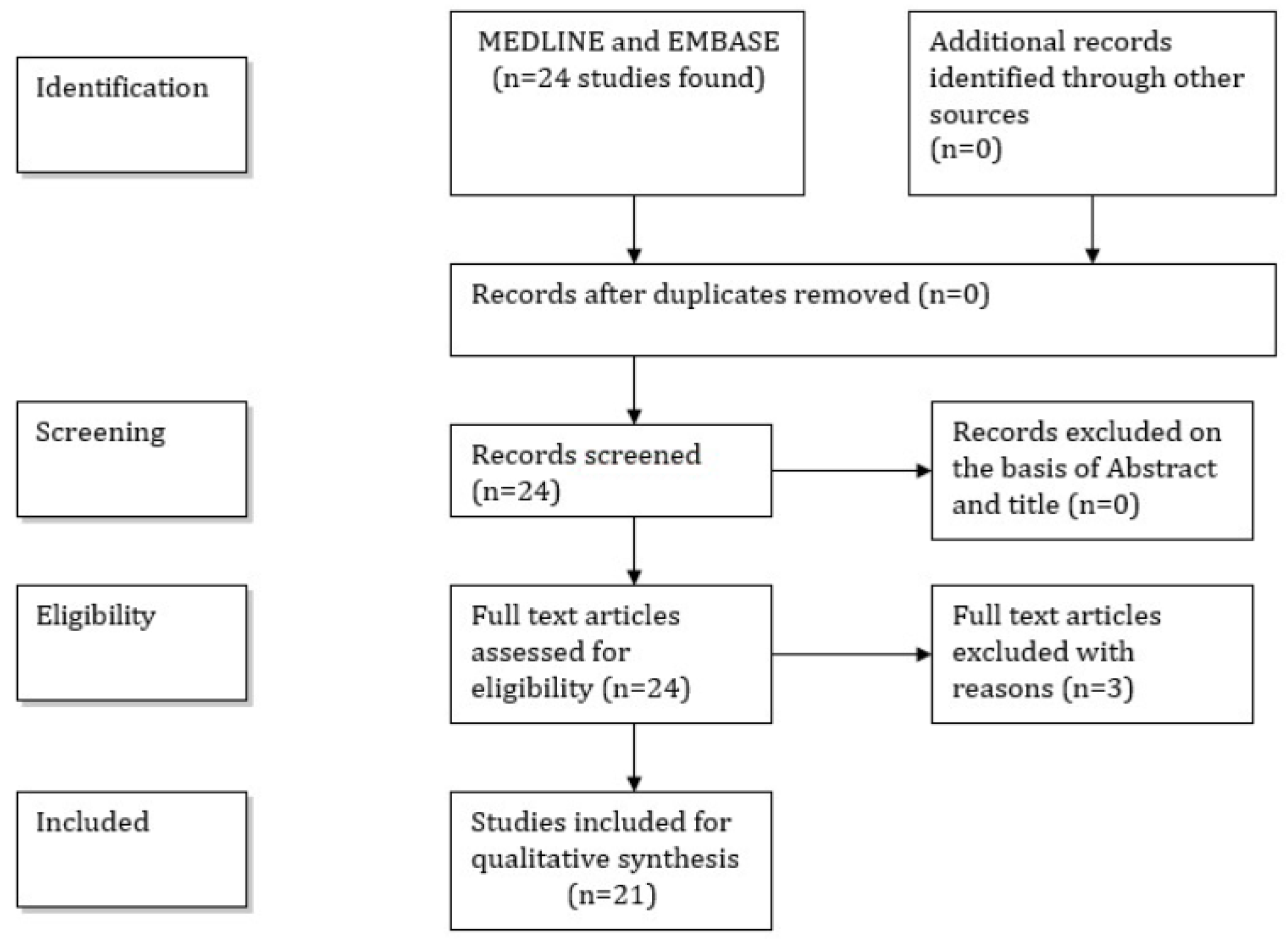
3. Material and Methods

In order to identify relevant articles for this review, we searched the MEDLINE, accessed by PubMed (1966–August 2018) and EMBASE (1980–August 2018) electronic databases were searched using the medical subject headings (MeSH) and free terms: “repetitive transcranial magnetic stimulation” OR “rTMS” AND “attention” OR “attentional” OR “attentive” AND “dorsolateral prefrontal cortex” OR “DLPFC”. Only original research articles were considered eligible for inclusion. Review articles or single case reports were excluded. The search was limited to studies written in English. Studies that met the following criteria were included: rTMS was conducted to patients with psychiatric diseases or neurological disorders with behavioral symptoms; administration site of rTMS was the DLPFC; the effect of rTMS on the cognitive domain attention was examined. In contrast, rTMS studies with animals as well as studies in which rTMS stimulation was administered on sites other than the DLPFC were excluded. Moreover, we included only studies that focused exclusively on attention, while studies with a broader scope within the umbrella concept of executive functions were excluded.
Full-text articles were retrieved for the selected titles, and reference lists of the retrieved articles were searched for additional publications. When data was missing or incomplete, principal investigators of included trials were contacted and additional information was requested. The titles and abstracts of the initially identified studies were screened by two authors to determine whether they satisfied the selection criteria. The methodological quality of each study and risk of bias were independently assessed, focusing on blinding, and any disagreement was solved through discussion. This search strategy yielded 24 results, three of which were excluded after reading the full paper, thereby leaving 21 studies which contributed to this review.
A flow-chart (Figure 1) shows the selection/inclusion process.

4. Results

The demographic characteristics of the patients in all included articles are shown in Table 1.
The description of the rTMS interventions in the reviewed articles is shown in Table 2.

Healthy Individuals

The breakdown of specific brain areas or neurotransmitter systems leads to selective disruptions of attentional networks in both healthy aging and pathological conditions [28]. The neural mechanisms underlying the ability to divide attention between multiple sensory modalities are still poorly understood [29].
The reviewed studies contribute to the understanding of the relationship between the DLPFC and attentional control, and suggest possible therapeutic applications for HF or LF rTMS.
These findings are consistent with those from several experimental studies in healthy humans.
Both single tasks demanding focused attention and dual task conditions requiring divided attention activate a widespread, mainly right-sided network including dorsolateral and ventrolateral prefrontal structures, superior and inferior parietal cortex, and anterior cingulate gyrus [30]. Vohn et al. performed fMRI in healthy subjects who underwent two within-modality (auditory/auditory, visual/visual) and one cross-modality (auditory/visual) divided attention task, as well as related selective attention control conditions [34]. The authors reported a significant activation in a predominantly right hemisphere network involving the PFC, the inferior parietal cortex, and the claustrum. Healthy subjects recognized fewer items after TMS over the left DLPFC than over the right DLPFC during encoding under full attention, while they produced fewer items after TMS over the right DLPFC in encoding under divided attention compared to a sham condition [31]. Taken together, these results favor the view that the right DLPFC is of special importance for attention, except for the last study which would point to a higher relevance of the left, compared to the right DLPFC.
It should be considered that selective and divided bimodal attention are concepts based on distinct neural processes. In fact, selective attention involves modulation of activity in the sensory cortices, while divided attention is achieved for most individuals via recruitment of the DLPFC [49].
TMS over PFC induced a significant reduction of performance time for both the verbal and visuo-spatial tests, thus suggesting the importance of this area in performing tasks requiring a high level of controlled attention [32].
Furthermore, 5 Hz rTMS over right DLPFC exerts remote effects on the activity of areas that functionally interact with this area during attentional processes [33].
HF rTMS over the right DLPFC was suggested to have an effect on top-down attentional processes by modulating the attentional set [35]. This is of interest, since top-down modulation mediated by the prefrontal cortex is a causal link between early attentional processes and subsequent memory performance [36].
Divided attention performance was significantly impaired about 30–60 min after a single rTMS session was applied over the left DLPFC, compared to a sham condition one week apart [26].
Daily HF-rTMS can improve attentional control in normally aging individuals [37]. Subjects who received five daily stimulation sessions of 10 Hz HF rTMS over the left DLPFC showed improved performance in reaction time during incongruent trials (i.e., those with distracting information) after HF-rTMS treatment compared with pretreatment assessment.

5. Results

5.1. Depression

Several studies assessed cognitive performance effects in patients with depression receiving rTMS. No major changes in the Continous Performace Task assessing attention and in other cognitive tests were observed in the first study of Speer and colleagues after LF or HF rTMS administered over the DLPFC [40]. Later studies assessed attention using psychometric tests, such as the d2 test, the sustained attention in the Cambridge Neuropsychological Test Automated Battery (CANTAB), the Test of Attentional Performance, and failed to find any significant effects of either HL or LF rTMS applied over the DLPFC [38,39,44]. Only one study using H-coils demonstrated that unilateral prefrontal left stimulation with H1/HIL-coils significantly improved the score on the rapid visual processing test as measured with the CANTAB [45].
A systematic review and meta-analysis of outcomes on individual neuropsychological tasks from sham-controlled RCTs where rTMS was administered to the DLPFC in depressed patients has recently been published [46]. No significant effect size for improvements with active compared to sham rTMS treatment was found.
For the purpose of this review, it is of interest that some studies used more specific tests to assess attentional processes. In a double-blind, placebo-controlled, crossover, within subjects design study, sixteen depressed patients performed a modified task switching paradigm, before and after receiving HF rTMS versus placebo rTMS over the left DLPFC [41]. One session of HF rTMS over the left DLPFC had a specific beneficial effect on task-switching performance, whereas mood remained stable. The same research group also found that after 2 weeks of HF rTMS over the left DLPFC, depressive symptoms improved in more than half of a therapy-resistant population [42]. After a single session, mood did not improve but attentional control was increased solely within the group of treatment responders. Of course, it needs to be considered that depression has very broad negative effects on cognitive function, so that a relieving of depressive symptoms might in turn have overall positive effects on cognition.
A more recent study examined whether acute and long-term HF deep rTMS to the DLPFC can attenuate attentional deficits associated with Major Depressive Disorder [47]. Twenty-one patients and 26 matched control subjects were characterized with the Beck Depression Inventory and the Sustained Attention to Response Task (SART) at baseline. Patients were retested following a single session and after 4 weeks of HF (20 Hz) deep rTMS applied to the DLPFC. To control for the practice effect, the controls were reassessed with the SART two further times. The patients exhibited deficits in sustained attention and cognitive inhibition. Both acute and long-term HF frontal repetitive dTMS ameliorated sustained attention deficits in the patient group. Improvement after acute dTMS was related to attentional recovery after long-term dTMS. It should be noted that longer-term improvement in sustained attention was not related to antidepressant effects of dTMS treatment.
Kavanaugh et al. examined recently the neurocognitive results of a randomized, double-blind, sham-controlled trial with an investigational 2-coil rTMS device [47]. The authors included patients with antidepressant treatment or treatment-intolerant major depressive disorder. A significant effect of active rTMS was observed for the quality of episodic memory, while there were no effects for continuity and power of attention as well as for working memory.

5.2. Schizophrenia

In patients with chronic schizophrenia, no significant change of cognitive performances, including the d2 attention task to assess attentional capacity, was observed as the result of a HF rTMS treatment [43]. Wölwer and coworkers also failed to find any significant cognitive effects in patients with schizophrenia who received HF rTMS [50].
In another study with schizophrenia patients, excitatory rTMS applied to the DLPFC was found to improve, among other cognitive functions, the selective and divided attention, as assessed by means of the Tübinger Aufmerksamkeitsprüfung [51].
Active rTMS can lead to a statistically significant reduction on the Scale for the Assessment of Negative Symptoms total score and of all domains of negative symptoms of schizophrenia, including impaired attention [52].

5.3. Autism Spectrum Disorder

Event-related brain potentials (ERPs) provide high temporal resolution measures of neuronal activity associated with several perceptual and cognitive processes. Sokhadze et al. assessed post-TMS differences in 13 subjects with autism [53]. The authors examined amplitude and latency of early and late attention-orienting frontal ERP components, indicating improved attentional processing. After rTMS, the parieto-occipital P50 amplitude decreased to novel distractors but not to targets; also, the amplitude and latency to targets increased for the frontal P50 while decreasing to nontarget stimuli.
Twenty-five subjects with autism spectrum disorder (ASD) were assessed in order to characterize selective attention using illusory figures before and after 12 sessions of rTMS applied bilaterally to the DLPFC [54]. This study was conducted in a controlled design where a waiting-list of 20 children with autism spectrum disorder was examined with the same time-interval, but with no rTMS intervention. A significant increase in amplitude of both N200 and P300 components as well as a significant reduction in response errors as a result of rTMS were detected.
The same research group also found, in 124 high functioning ASD children, that 18 sessions of rTMS applied over the DLPFC facilitates cognitive control, attention, and target stimuli recognition by improving discrimination between task-relevant and task-irrelevant illusory figures in an oddball test [55].

5.4. Attention Deficit Hyperactivity Disorder

In a crossover double-blind randomized, sham-controlled pilot study, patients with ADHD received either a single session of HF rTMS directed to the right DLPFC (real rTMS) or a single session of sham rTMS [56]. The post-real rTMS attention score improved significantly compared to the prereal rTMS attention score. rTMS had no effect on measures of mood and anxiety, and sham rTMS showed no effects.
In a more recent study, twenty daily sessions were conducted in patients diagnosed as having ADHD, using the bilateral HF dTMS coil in order to stimulate the PFC. The Conners’ Adult ADHD Rating Scale questionnaire and a computerized continuous performance test, the Test of Variables of Attention, were used for the assessment of cognitive functions. No differences in clinical outcomes were observed between groups receiving real dTMS or sham TMS [57].

5.5. Addiction

HF (10 Hz) rTMS of the left DLPFC was found to improve emotional attention of 31 methamphetamine addicts [58]. The attention bias effect to negative information persisted in the active rTMS group over two weeks.
An fMRI study in 26 recently detoxified alcohol-dependent patients documented effects of accelerated HF rTMS applied to the right DLPFC [59]. The findings suggest that the intervention did not manifestly affect the craving neurocircuit during an alcohol-related cue-exposure, but instead it may have influenced the attentional network. In fact, brain activation changes after one and 15 HF rTMS sessions were observed in regions associated with the extended reward system and the default mode network, respectively, during the presentation of event-related alcohol cue-reactivity paradigms.

5.6. Alzheimer’s Disease

A single study has examined the effects of HF rTMS, applied over the DLPFC on behavioral and psychological symptoms of dementia as well as on cognitive function in 52 patients with Alzheimer’s disease (AD) [60].
The intervention group, which was treated with 20 Hz rTMS five days a week for four weeks, showed significantly lower scores (i.e., greater improvement) than the control group on the Alzheimer’s Disease Assessment Scale-Cognitive (ADAS-Cog) total score, as well as on all four ADAS-Cog factor scores (memory, language, constructional praxis, and attention).

6. Discussion

This review highlights that rTMS applied over the DLPFC can positively influence the attentional function in subjects with several psychiatric disorders. The outcome measures were not uniform but mostly dealt with attentional performance.
Some studies revealed that prefrontal rTMS could exert procognitive effects on executive function and attention in patients with depression [3]. Antidepressant effects of rTMS could be related to the same neurochemical mechanisms that underlie cognitive functioning, or just facilitate the normal cognitive function that was repressed because of the severe effects depression has on overall physical and cognitive functioning. It has been hypothesized that the extent of antidepressant effects could be considered as second-order long-term effects possibly related to primary alternations in cognitive functioning. Concurrence of depression and cognitive dysfunctions is well known in a wide range of clinical populations [61]. In particular, impaired cognition is closely related to depressive symptoms in AD [62,63], thus possibly potentiating the devastating effects of the disease itself or being an early sign of neural dysfunction [64].
In patients with schizophrenia, imaging studies have demonstrated abnormalities in the left globus pallidus, which lead to widespread hypometabolism affecting the frontal lobes, especially the DLPFC and the anterior cingulate gyrus [65]. Furthermore, abnormalities of visually orienting the frontal lobes/executive attentional network could interact with the parietal lobes/orienting network to affect the initiation of attentional shift, thus leading to abnormalities of visual orienting [66]. It is therefore of interest that rTMS to the DLPFC could improve attentional functioning in this patient population [38]. However, the findings were contradictory, as other studies could not identify any beneficial effects. A more systematic investigation comparing the different parameters of TMS to each other may shed more light on the mechanisms of action.
The results of some studies support the use of LF rTMS as a modulatory tool to alter the disrupted balance between cortical excitation and inhibition in autism. LF rTMS application to DLPFC would result in an alteration of the abnormal excitatory/inhibitory ratio through the activation of inhibitory GABAergic double bouquet interneurons.
Similarly, in patients suffering from ADHD initial findings suggest the possibility that attentional difficulties can be improved by using HF rTMS applied to the right DLPFC, and have encouraged future research [41]. However, the evidence from a more recent study does not support the effectiveness of bilateral prefrontal stimulation to treat adult ADHD [42]. Due to the small sample size, these preliminary results should be interpreted with caution.
rTMS can significantly improve, among other cognitive functions, attentional impairment that often accompanies AD. Impairments in visual attention and visual information processing have been identified as part of the neuropsychological features of AD, even in its earliest stages, and dissociations in visual attention deficits have been detected also in mild cognitive impairment (MCI) using a measure that assesses simple, divided, and selective attention [67]. It is unclear whether the memory impairment in patients with amnestic MCI (aMCI) and AD is associated with attentional deficits. An fMRI study revealed that there are changes in the functional network subserving divided attention in patients with aMCI, as reflected in the attenuated activation of PFC [68]. Interestingly, depressive symptoms in AD patients increase the deficits of cognitive flexibility and divided attention [69].
This review has some limitations. First of all, there is considerable variability between studies in patients with different neuropsychiatric diseases. Very few trials have used exactly the same study design. The stimulation protocols, with respect to frequency, intensity, orientation of the coil, pattern, number of pulses by train, total number of pulses, duration of stimulation, frequency and intensity of stimulation, number of sessions delivered, are highly heterogeneous. Therefore, estimating the real effectiveness and reproducibility is very difficult. Systematic investigation of the effects of the various stimulation protocols are highly warranted, because the border between effectiveness and ineffectiveness may be very small and occurs somewhere in the dimensions spanned by the abovementioned parameters.
Furthermore, we have included in this review only studies employing specific cognitive tests/tasks focusing on attention, even if working memory and other executive functions are strongly correlated with this cognitive domain. Indeed, the role of the right DLPFC and of the right posterior parietal cortex (PPC) in controlling the interaction between working memory and attention during a visual search has been explored using rTMS in a recent study [70]. Both the rDLPFC and the right PPC were found to be critical for controlling working memory biases in human visual attention. However, the broader scope of including executive functions should be addressed in another systematic summarizing work; possibly a meta-analysis could be conducted given that the study protocols were more comparable.
It should be considered that most therapeutic attempts are based on rTMS techniques aiming at enhancing cortical excitability, in particular HF rTMS. However, the underlying pathophysiologic mechanisms differ among the various neurological and psychiatric diseases which can be treated with this noninvasive brain stimulation technique. Therefore, appropriate testing of cortical physiology before and after therapeutic interventions is needed.

7. Conclusions

In conclusion, a better understanding of attention networks could allow targeting the most suitable area of the brain according to the specific attention domain affected. Moreover, a detailed examination of the best stimulation frequency, surface or deep stimulation, duration and intensity of the intervention, among other important core features of TMS-protocols, should be done when moving closer to clinical application of TMS to treat attentional deficits.
Despite the above-mentioned limitations, this review indicates that neuromodulatory techniques such as rTMS are promising approaches to be used as attentional enhancers in people with neuropsychiatric conditions where impaired attention is a prominent feature.

Author Contributions

Study conception and design: L.H., J.S., Y.H., R.N.; analysis and interpretation of the data: F.B., V.V.; article draft and revision L.S. (Luca Sebastianelli), L.S. (Leopold Saltuari), E.T., R.N.

Conflicts of Interest

The authors declare no conflict of interest.

References

  1. Treisman, A.M.; Gelade, G. A feature-integration theory of attention. Cogn. Psychol. 1980, 12, 97–136. [Google Scholar] [CrossRef]
  2. Treisman, A. Feature binding, attention and object perception. Philos. Trans. R Soc. Lond. B Biol. Sci. 1998, 353, 1295–1306. [Google Scholar] [CrossRef]
  3. Iimori, T.; Nakajima, S.; Miyazaki, T.; Tarumi, R.; Ogyu, K.; Wada, M.; Tsugawa, S.; Masuda, F.; Daskalakis, Z.J.; Blumberger, D.M.; et al. Effectiveness of the prefrontal repetitive transcranial magnetic stimulation on cognitive profiles in depression, schizophrenia, and Alzheimer’s disease: A systematic review. Prog. Neuropsychopharmacol. Biol. Psychiatry 2019, 88, 31–40. [Google Scholar] [CrossRef]
  4. Pessoa, L.; Kastner, S.; Ungerleider, L.G. Neuroimaging studies of attention: From modulation of sensory processing to top-down control. J. Neurosci. 2003, 23, 3990–3998. [Google Scholar] [CrossRef] [PubMed]
  5. Fan, J.; McCandliss, B.D.; Fossella, J.; Flombaum, J.I.; Posner, M.I. The activation of attentional networks. Neuroimage 2005, 26, 471–479. [Google Scholar] [CrossRef] [Green Version]
  6. Hartlage, S.; Alloy, L.B.; Vazquez, C.; Dykman, B. Automatic and effortful processing in depression. Psychol. Bull. 1993, 113, 247–278. [Google Scholar] [CrossRef]
  7. Ochoa, E.L.; Lasalde-Dominicci, J. Cognitive deficits in schizophrenia: Focus on neuronal nicotinic acetylcholine receptors and smoking. Cell. Mol. Neurobiol. 2007, 27, 609–639. [Google Scholar] [CrossRef]
  8. Woods, S.P.; Lovejoy, D.W.; Ball, J.D. Neuropsychological characteristics of adults with ADHD: A comprehensive review of initial studies. Clin. Neuropsychol. 2002, 16, 12–34. [Google Scholar] [CrossRef] [PubMed]
  9. Tucha, O.; Mecklinger, L.; Laufkotter, R.; Klein, H.E.; Walitza, S.; Lange, K.W. Methylphenidate-induced improvements of various measures of attention in adults with attention deficit hyperactivity disorder. J. Neural. Transm. (Vienna) 2006, 113, 1575–1592. [Google Scholar] [CrossRef]
  10. Tucha, L.; Tucha, O.; Walitza, S.; Sontag, T.A.; Laufkotter, R.; Linder, M.; Lange, K.W. Vigilance and sustained attention in children and adults with ADHD. J. Atten. Disord. 2009, 12, 410–421. [Google Scholar] [CrossRef]
  11. Burack, J.A. Selective attention deficits in persons with autism: Preliminary evidence of an inefficient attentional lens. J. Abnorm. Psychol. 1994, 103, 535–543. [Google Scholar] [CrossRef]
  12. Belmonte, M.K.; Yurgelun-Todd, D.A. Anatomic dissociation of selective and suppressive processes in visual attention. Neuroimage 2003, 19, 180–189. [Google Scholar] [CrossRef] [Green Version]
  13. Belmonte, M.K.; Yurgelun-Todd, D.A. Functional anatomy of impaired selective attention and compensatory processing in autism. Cogn. Brain Res. 2003, 17, 651–664. [Google Scholar] [CrossRef]
  14. Alvarez, J.A.; Emory, E. Executive function and the frontal lobes: A meta-analytic review. Neuropsychol. Rev. 2006, 16, 17–42. [Google Scholar] [CrossRef]
  15. Johnson, J.A.; Strafella, A.P.; Zatorre, R.J. The role of the dorsolateral prefrontal cortex in bimodal divided attention: Two transcranial magnetic stimulation studies. J. Cogn. Neurosci. 2007, 19, 907–920. [Google Scholar] [CrossRef] [PubMed]
  16. Fitzgerald, P.B.; Fountain, S.; Daskalakis, Z.J. A comprehensive review of the effects of rTMS on motor cortical excitability and inhibition. Clin. Neurophysiol. 2006, 117, 2584–2596. [Google Scholar] [CrossRef]
  17. Cotelli, M.; Calabria, M.; Manenti, R.; Rosini, S.; Zanetti, O.; Cappa, S.F.; Miniussi, C. Improved language performance in Alzheimer disease following brain stimulation. J. Neurol. Neurosurg. Psychiatry 2011, 82, 794–797. [Google Scholar] [CrossRef] [PubMed]
  18. Cotelli, M.; Manenti, R.; Cappa, S.F.; Geroldi, C.; Zanetti, O.; Rossini, P.M.; Miniussi, C. Effect of transcranial magnetic stimulation on action naming in patients with Alzheimer disease. Arch. Neurol. 2006, 63, 1602–1604. [Google Scholar] [CrossRef]
  19. Ahmed, M.A.; Darwish, E.S.; Khedr, E.M.; El Serogy, Y.M.; Ali, A.M. Effects of low versus high frequencies of repetitive transcranial magnetic stimulation on cognitive function and cortical excitability in Alzheimer’s dementia. J. Neurol. 2012, 259, 83–92. [Google Scholar] [CrossRef]
  20. Devi, G.; Voss, H.U.; Levine, D.; Abrassart, D.; Heier, L.; Halper, J.; Martin, L.; Lowe, S. Open-label, short-term, repetitive transcranial magnetic stimulation in patients with Alzheimer’s disease with functional imaging correlates and literature review. Am. J. Alzheimers Dis. Other Dement. 2014, 29, 248–255. [Google Scholar] [CrossRef] [PubMed]
  21. Padala, P.R.; Padala, K.P.; Lensing, S.Y.; Jackson, A.N.; Hunter, C.R.; Parkes, C.M.; Dennis, R.A.; Bopp, M.M.; Caceda, R.; Mennemeier, M.S.; et al. Repetitive transcranial magnetic stimulation for apathy in mild cognitive impairment: A double-blind, randomized, sham-controlled, cross-over pilot study. Psychiatry Res. 2018, 261, 312–318. [Google Scholar] [CrossRef]
  22. Pascual-Leone, A.; Tormos, J.M.; Keenan, J.; Tarazona, F.; Canete, C.; Catala, M.D. Study and modulation of human cortical excitability with transcranial magnetic stimulation. J. Clin. Neurophysiol. 1998, 15, 333–343. [Google Scholar] [CrossRef]
  23. Rossi, S.; Hallett, M.; Rossini, P.M.; Pascual-Leone, A. Safety, ethical considerations, and application guidelines for the use of transcranial magnetic stimulation in clinical practice and research. Clin. Neurophysiol. 2009, 120, 2008–2039. [Google Scholar] [CrossRef] [Green Version]
  24. Lefaucheur, J.P.; Andre-Obadia, N.; Antal, A.; Ayache, S.S.; Baeken, C.; Benninger, D.H.; Cantello, R.M.; Cincotta, M.; de Carvalho, M.; De Ridder, D.; et al. Evidence-based guidelines on the therapeutic use of repetitive transcranial magnetic stimulation (rTMS). Clin. Neurophysiol. 2014, 125, 2150–2206. [Google Scholar] [CrossRef]
  25. Zangen, A.; Roth, Y.; Voller, B.; Hallett, M. Transcranial magnetic stimulation of deep brain regions: Evidence for efficacy of the H-coil. Clin. Neurophysiol. 2005, 116, 775–779. [Google Scholar] [CrossRef]
  26. Wagner, M.; Rihs, T.A.; Mosimann, U.P.; Fisch, H.U.; Schlaepfer, T.E. Repetitive transcranial magnetic stimulation of the dorsolateral prefrontal cortex affects divided attention immediately after cessation of stimulation. J. Psychiatr. Res. 2006, 40, 315–321. [Google Scholar] [CrossRef]
  27. Bersani, F.S.; Minichino, A.; Enticott, P.G.; Mazzarini, L.; Khan, N.; Antonacci, G.; Raccah, R.N.; Salviati, M.; Delle Chiaie, R.; Bersani, G.; et al. Deep transcranial magnetic stimulation as a treatment for psychiatric disorders: A comprehensive review. Eur. Psychiatry 2013, 28, 30–39. [Google Scholar] [CrossRef]
  28. Coulthard, E.; Singh-Curry, V.; Husain, M. Treatment of attention deficits in neurological disorders. Curr. Opin. Neurol. 2006, 19, 613–618. [Google Scholar] [CrossRef]
  29. Sharma, K.; Davis, T.; Coulthard, E. Enhancing attention in neurodegenerative diseases: Current therapies and future directions. Transl. Neurosci. 2016, 7, 98–109. [Google Scholar] [CrossRef]
  30. Nebel, K.; Wiese, H.; Stude, P.; de Greiff, A.; Diener, H.C.; Keidel, M. On the neural basis of focused and divided attention. Cogn. Brain Res. 2005, 25, 760–776. [Google Scholar] [CrossRef]
  31. Blanchet, S.; Gagnon, G.; Schneider, C. The contribution of the dorsolateral prefrontal cortex in full and divided encoding: A paired-pulse transcranial magnetic stimulation study. Behav. Neurol. 2010, 23, 107–115. [Google Scholar] [CrossRef]
  32. Sabatino, M.; Di Nuovo, S.; Sardo, P.; Abbate, C.S.; La Grutta, V. Neuropsychology of selective attention and magnetic cortical stimulation. Int. J. Psychophysiol. 1996, 21, 83–89. [Google Scholar] [CrossRef]
  33. Rounis, E.; Stephan, K.E.; Lee, L.; Siebner, H.R.; Pesenti, A.; Friston, K.J.; Rothwell, J.C.; Frackowiak, R.S. Acute changes in frontoparietal activity after repetitive transcranial magnetic stimulation over the dorsolateral prefrontal cortex in a cued reaction time task. J. Neurosci. 2006, 26, 9629–9638. [Google Scholar] [CrossRef]
  34. Vohn, R.; Fimm, B.; Weber, J.; Schnitker, R.; Thron, A.; Spijkers, W.; Willmes, K.; Sturm, W. Management of attentional resources in within-modal and cross-modal divided attention tasks: An fMRI study. Hum. Brain Mapp. 2007, 28, 1267–1275. [Google Scholar] [CrossRef]
  35. Vanderhasselt, M.A.; De Raedt, R.; Baeken, C.; Leyman, L.; Clerinx, P.; D’Haenen, H. The influence of rTMS over the right dorsolateral prefrontal cortex on top-down attentional processes. Brain Res. 2007, 1137, 111–116. [Google Scholar] [CrossRef]
  36. Zanto, T.P.; Rubens, M.T.; Thangavel, A.; Gazzaley, A. Causal role of the prefrontal cortex in top-down modulation of visual processing and working memory. Nat. Neurosci. 2011, 14, 656–661. [Google Scholar] [CrossRef] [PubMed] [Green Version]
  37. Kim, S.H.; Han, H.J.; Ahn, H.M.; Kim, S.A.; Kim, S.E. Effects of five daily high-frequency rTMS on Stroop task performance in aging individuals. Neurosci. Res. 2012, 74, 256–260. [Google Scholar] [CrossRef]
  38. Hoppner, J.; Schulz, M.; Irmisch, G.; Mau, R.; Schlafke, D.; Richter, J. Antidepressant efficacy of two different rTMS procedures. High frequency over left versus low frequency over right prefrontal cortex compared with sham stimulation. Eur. Arch. Psychiatry Clin. Neurosci. 2003, 253, 103–109. [Google Scholar] [CrossRef]
  39. Januel, D.; Dumortier, G.; Verdon, C.M.; Stamatiadis, L.; Saba, G.; Cabaret, W.; Benadhira, R.; Rocamora, J.F.; Braha, S.; Kalalou, K.; et al. A double-blind sham controlled study of right prefrontal repetitive transcranial magnetic stimulation (rTMS): Therapeutic and cognitive effect in medication free unipolar depression during 4 weeks. Prog. Neuropsychopharmacol. Biol. Psychiatry 2006, 30, 126–130. [Google Scholar] [CrossRef]
  40. Speer, A.M.; Repella, J.D.; Figueras, S.; Demian, N.K.; Kimbrell, T.A.; Wasserman, E.M.; Post, R.M. Lack of adverse cognitive effects of 1 Hz and 20 Hz repetitive transcranial magnetic stimulation at 100% of motor threshold over left prefrontal cortex in depression. J. ECT 2001, 17, 259–263. [Google Scholar] [CrossRef]
  41. Vanderhasselt, M.A.; De Raedt, R.; Leyman, L.; Baeken, C. Acute effects of repetitive transcranial magnetic stimulation on attentional control are related to antidepressant outcomes. J. Psychiatry Neurosci. 2009, 34, 119–126. [Google Scholar]
  42. Vanderhasselt, M.A.; De Raedt, R.; Baeken, C.; Leyman, L.; D’Haenen, H. A single session of rTMS over the left dorsolateral prefrontal cortex influences attentional control in depressed patients. World J. Biol. Psychiatry 2009, 10, 34–42. [Google Scholar] [CrossRef] [PubMed]
  43. Mittrach, M.; Thunker, J.; Winterer, G.; Agelink, M.W.; Regenbrecht, G.; Arends, M.; Mobascher, A.; Kim, S.J.; Wolwer, W.; Brinkmeyer, J.; et al. The tolerability of rTMS treatment in schizophrenia with respect to cognitive function. Pharmacopsychiatry 2010, 43, 110–117. [Google Scholar] [CrossRef]
  44. Ullrich, H.; Kranaster, L.; Sigges, E.; Andrich, J.; Sartorius, A. Ultra-high-frequency left prefrontal transcranial magnetic stimulation as augmentation in severely ill patients with depression: A naturalistic sham-controlled, double-blind, randomized trial. Neuropsychobiology 2012, 66, 141–148. [Google Scholar] [CrossRef]
  45. Levkovitz, Y.; Harel, E.V.; Roth, Y.; Braw, Y.; Most, D.; Katz, L.N.; Sheer, A.; Gersner, R.; Zangen, A. Deep transcranial magnetic stimulation over the prefrontal cortex: Evaluation of antidepressant and cognitive effects in depressive patients. Brain Stimul. 2009, 2, 188–200. [Google Scholar] [CrossRef]
  46. Martin, D.M.; McClintock, S.M.; Forster, J.J.; Lo, T.Y.; Loo, C.K. Cognitive enhancing effects of rTMS administered to the prefrontal cortex in patients with depression: A systematic review and meta-analysis of individual task effects. Depress Anxiety 2017, 34, 1029–1039. [Google Scholar] [CrossRef]
  47. Kavanaugh, B.C.; Aaronson, S.T.; Clarke, G.N.; Holtzheimer, P.E.; Johnson, C.W.; McDonald, W.M.; Schneider, M.B.; Carpenter, L.L. Neurocognitive effects of repetitive transcranial magnetic stimulation with a 2-coil device in treatment-resistant major depressive disorder. J. ECT 2018, 34, 258–265. [Google Scholar] [CrossRef]
  48. Naim-Feil, J.; Bradshaw, J.L.; Sheppard, D.M.; Rosenberg, O.; Levkovitz, Y.; Dannon, P.; Fitzgerald, P.B.; Isserles, M.; Zangen, A. Neuromodulation of attentional control in major depression: A pilot deep TMS study. Neural Plast. 2016, 2016, 5760141. [Google Scholar] [CrossRef] [PubMed]
  49. Johnson, J.A.; Zatorre, R.J. Neural substrates for dividing and focusing attention between simultaneous auditory and visual events. Neuroimage 2006, 31, 1673–1681. [Google Scholar] [CrossRef]
  50. Wolwer, W.; Lowe, A.; Brinkmeyer, J.; Streit, M.; Habakuck, M.; Agelink, M.W.; Mobascher, A.; Gaebel, W.; Cordes, J. Repetitive transcranial magnetic stimulation (rTMS) improves facial affect recognition in schizophrenia. Brain Stimul. 2014, 7, 559–563. [Google Scholar] [CrossRef]
  51. Guse, B.; Falkai, P.; Gruber, O.; Whalley, H.; Gibson, L.; Hasan, A.; Obst, K.; Dechent, P.; McIntosh, A.; Suchan, B.; et al. The effect of long-term high frequency repetitive transcranial magnetic stimulation on working memory in schizophrenia and healthy controls--a randomized placebo-controlled, double-blind fMRI study. Behav. Brain Res. 2013, 237, 300–307. [Google Scholar] [CrossRef]
  52. Prikryl, R.; Ustohal, L.; Prikrylova Kucerova, H.; Kasparek, T.; Venclikova, S.; Vrzalova, M.; Ceskova, E. A detailed analysis of the effect of repetitive transcranial magnetic stimulation on negative symptoms of schizophrenia: A double-blind trial. Schizophr. Res. 2013, 149, 167–173. [Google Scholar] [CrossRef]
  53. Sokhadze, E.; Baruth, J.; Tasman, A.; Mansoor, M.; Ramaswamy, R.; Sears, L.; Mathai, G.; El-Baz, A.; Casanova, M.F. Low-frequency repetitive transcranial magnetic stimulation (rTMS) affects event-related potential measures of novelty processing in autism. Appl. Psychophysiol. Biofeedback 2010, 35, 147–161. [Google Scholar] [CrossRef]
  54. Casanova, M.F.; Baruth, J.M.; El-Baz, A.; Tasman, A.; Sears, L.; Sokhadze, E. Repetitive Transcranial Magnetic Stimulation (rTMS) Modulates event-related potential (ERP) indices of attention in autism. Transl. Neurosci. 2012, 3, 170–180. [Google Scholar] [CrossRef]
  55. Sokhadze, E.M.; Lamina, E.V.; Casanova, E.L.; Kelly, D.P.; Opris, I.; Tasman, A.; Casanova, M.F. Exploratory study of rTMS neuromodulation effects on electrocortical functional measures of performance in an oddball test and behavioral symptoms in autism. Front. Syst. Neurosci. 2018, 12, 20. [Google Scholar] [CrossRef]
  56. Bloch, Y.; Harel, E.V.; Aviram, S.; Govezensky, J.; Ratzoni, G.; Levkovitz, Y. Positive effects of repetitive transcranial magnetic stimulation on attention in ADHD Subjects: A randomized controlled pilot study. World J. Biol. Psychiatry 2010, 11, 755–758. [Google Scholar] [CrossRef]
  57. Paz, Y.; Friedwald, K.; Levkovitz, Y.; Zangen, A.; Alyagon, U.; Nitzan, U.; Segev, A.; Maoz, H.; Koubi, M.; Bloch, Y. Randomised sham-controlled study of high-frequency bilateral deep transcranial magnetic stimulation (dTMS) to treat adult attention hyperactive disorder (ADHD): Negative results. World J. Biol. Psychiatry 2018, 19, 561–566. [Google Scholar] [CrossRef]
  58. Zhang, L.; Cao, X.; Liang, Q.; Li, X.; Yang, J.; Yuan, J. High-frequency repetitive transcranial magnetic stimulation of the left dorsolateral prefrontal cortex restores attention bias to negative information in methamphetamine addicts. Psychiatry Res. 2018, 265, 151–160. [Google Scholar] [CrossRef]
  59. Herremans, S.C.; Van Schuerbeek, P.; De Raedt, R.; Matthys, F.; Buyl, R.; De Mey, J.; Baeken, C. The impact of accelerated right prefrontal high-frequency repetitive transcranial magnetic stimulation (rTMS) on cue-reactivity: An fMRI study on craving in recently detoxified alcohol-dependent patients. PLoS ONE 2015, 10, e0136182. [Google Scholar] [CrossRef]
  60. Wu, Y.; Xu, W.; Liu, X.; Xu, Q.; Tang, L.; Wu, S. Adjunctive treatment with high frequency repetitive transcranial magnetic stimulation for the behavioral and psychological symptoms of patients with Alzheimer’s disease: A randomized, double-blind, sham-controlled study. Shanghai Arch. Psychiatry 2015, 27, 280–288. [Google Scholar] [CrossRef]
  61. Marazziti, D.; Consoli, G.; Picchetti, M.; Carlini, M.; Faravelli, L. Cognitive impairment in major depression. Eur. J. Pharmacol. 2010, 626, 83–86. [Google Scholar] [CrossRef] [PubMed]
  62. Lyketsos, C.G.; DelCampo, L.; Steinberg, M.; Miles, Q.; Steele, C.D.; Munro, C.; Baker, A.S.; Sheppard, J.M.; Frangakis, C.; Brandt, J.; et al. Treating depression in Alzheimer disease: Efficacy and safety of sertraline therapy, and the benefits of depression reduction: The DIADS. Arch. Gen. Psychiatry 2003, 60, 737–746. [Google Scholar] [CrossRef] [PubMed]
  63. Rutherford, G.; Gole, R.; Moussavi, Z. rTMS as a treatment of Alzheimer’s disease with and without comorbidity of depression: A review. Neurosci. J. 2013, 2013, 679389. [Google Scholar] [CrossRef]
  64. Stogmann, E.; Moser, D.; Klug, S.; Gleiss, A.; Auff, E.; Dal-Bianco, P.; Pusswald, G.; Lehrner, J. Activities of Daily Living and Depressive Symptoms in Patients with Subjective Cognitive Decline, Mild Cognitive Impairment, and Alzheimer’s Disease. J. Alzheimers Dis. 2016, 49, 1043–1050. [Google Scholar] [CrossRef]
  65. Early, T.S.; Posner, M.I.; Reiman, E.M.; Raichle, M.E. Hyperactivity of the left striato-pallidal projection. Part I: Lower level theory. Psychiatr. Dev. 1989, 7, 85–108. [Google Scholar] [PubMed]
  66. Fernandez-Duque, D.; Posner, M.I. Brain imaging of attentional networks in normal and pathological states. J. Clin. Exp. Neuropsychol. 2001, 23, 74–93. [Google Scholar] [CrossRef]
  67. Okonkwo, O.C.; Wadley, V.G.; Ball, K.; Vance, D.E.; Crowe, M. Dissociations in visual attention deficits among persons with mild cognitive impairment. Neuropsychol. Dev. Cogn. B Aging Neuropsychol. Cogn. 2008, 15, 492–505. [Google Scholar] [CrossRef]
  68. Dannhauser, T.M.; Walker, Z.; Stevens, T.; Lee, L.; Seal, M.; Shergill, S.S. The functional anatomy of divided attention in amnestic mild cognitive impairment. Brain 2005, 128, 1418–1427. [Google Scholar] [CrossRef] [PubMed] [Green Version]
  69. Nakaaki, S.; Murata, Y.; Sato, J.; Shinagawa, Y.; Tatsumi, H.; Hirono, N.; Furukawa, T.A. Greater impairment of ability in the divided attention task is seen in Alzheimer’s disease patients with depression than in those without depression. Dement. Geriatr. Cogn. Disord. 2007, 23, 231–240. [Google Scholar] [CrossRef] [PubMed]
  70. Wang, M.; Yang, P.; Wan, C.; Jin, Z.; Zhang, J.; Li, L. Evaluating the role of the dorsolateral prefrontal cortex and posterior parietal cortex in memory-guided attention with repetitive transcranial magnetic stimulation. Front. Hum. Neurosci. 2018, 12, 236. [Google Scholar] [CrossRef]
Figure 1. Flow-chart showing the selection/inclusion process.
Jcm 08 00416 g001
Table 1. Demographic characteristics of the patients in the included studies.
StudiesNoGenderMean AgeDisease DurationEducation
M/F(y)(y)(y)
Depression
Speer et al., 2001 [28]18-45 ± 7--
Höppner et al., 2003 [29]308/2256.4 ± 11.1--
Januel et al., 2006 [30]276/2137.78 ± 11.2777.77 ± 90.82 mo-
Levkovitz et al., 2009 [31]23 H112/1145.57 ± 13.3413.96 ± 2.96-
22 H211/1145.77 ± 11.9913.00 ± 2.12-
11 HIL 110%3/544.27 ± 11.3615.45 ± 2.02-
8 HIL 120%10/1049.88 ± 9.5213.13 ± 2.81-
Vanderhasselt et al., 2009a [32]166/1042 ± 11.2
Vanderhasselt et al., 2009b [33]156/945.6 ± 5.87--
Ullrich et al., 2012 [34]Active 2231.8/68.2%56.9/10.2%--
Sham 2142.9/57.1%54.1/7.8%
Naim-Feil et al., 2016 [35]2110/1144 ± 915 ± 3-
Kavanaugh et al., 2018 [36]Active 4310/3145.84 ± 11.8717.94 ± 3.7-
Sham 4112/3147.95 ± 12.7815.59 ± 9.17
Schizophrenia
Mittrach et al., 2010 [37]Active 1814/434.5 ± 0.55.7 ± 5.2-
Sham 1411/334.4 ± 10.55.6 ± 8.7
Guse et al., 2013 [38]Active 1310/337 (22-58)15.5-
Sham 129/336 (20-5112.6
Prikryl et al., 2013 [39]Active 2323/031.6 ± 8.044.91 ± 5.09 y12.43 ± 2.06 y
Control 1717/033.94 ± 9.985.89 ± 7.91 y12.44 ± 1.97
Wölwer et al., 2014 [40]Active 1814/434.3 ± 5.75.7 ± 5.2-
Sham 1411/334.4 ± 5.65.6 ± 8.7
Attention deficit hyperactivity disorder
Bloch et al., 2010 [41]137/6---
Active 96/332 ± 11
Paz. et al., 2017 [42]Sham 138/530.85 ± 6.82--
Alzheimer disease
Wu et al., 2015 [43]Active 2610/1671.4 ± 4.95.1 ± 1.511.4 ± 2.7 y
Control 2611/1571.9 ± 4.85.1 ± 1.511.5 ± 2.1 y
Autism
Sokhadze et al., 2010 [44]1312/115.6 ± 5.8--
Casanova et al., 2012 [45]4539/613 ± 2.7--
Sokhadze et al., 2018 [46]11293/1913.1 ± 1.78--
Addiction
Herremans et al, 2015 [47]2617/945.2 ± 9.3--
Zang et al., 2018 [48]3131/043 ± 9.1513 ± 7.45-
no. = number of patients; M = male; F = female; y = years; mo. = months, “-“ not reported.
Table 2. Description of the repetitive transcranial magnetic stimulation (rTMS) interventions in the included studies.
StudiesStimulation ParametersOutcome MeasuresPrincipal Findings
PositionIntensitityFrequencyTotal Pulses Per SessionNo. Sessions
Depression
Speer et al., 2001 [28]L DLPFC100% MT20 Hz
1 Hz
160010Continuous Performance TaskNo significant changes
Hoeppner et al., 2003 [29]L DLPFC
R DLPFC
80% MT20 Hz
1 Hz
?10d2 TestNo significant changes
Januel et al., 2006 [30]R DLPFC90% MT1 Hz?16Auditory and visual attention spanNo significant differences
Levkovitz et al., 2009 [31]H-Coil
DLPFC
120% MT20 Hz168920CANTAB, RVP↑ RVP performances
Vanderhasselt et al., 2009a [32]L DLPFC110% MT10 Hz156010VAS
Self-paced switching task
↑ Attentional processes
Vanderhasselt et al., 2009b [33]L DLPFC110% MT10 Hz156010Self-paced switching task↑ Attentional control
Ullrich et al.; 2012 [34]L DLPFC110% MT30 Hz
1 Hz
1800
990
15ZVT, SKT↑ Processing speed performance ↑
Naim-Feil et al., 2016 [35]H-Coil
L > R DLPFC
120% MT20 Hz16801 (n = 21)
20 (n = 13)
BDI, SART↓ Sustained attention deficits
Kavanaugh et al., 2018 [36]2-coil
L > R DLPF
120% MT10 Hz300020CDR System↑ Continuity and power of attention
Schizophrenia
Mittrach et al., 2010 [37]L DLPFC110% MT10 Hz100010d2 TestNo significant changes
Guse et al., 2013 [38]L DLPFC110% MT10 Hz100015TAPSignificant time-by-stimulation interaction in divided attention
Prikryl et al., 2013 [39]L DLPFC110% MT10 Hz200015SANS↓ SANS total score + all domains of negative symptoms
Woelwer et al., 2014 [40]L DLPFC110% MT10 Hz1000010d2 TestNo significant changes
Attention deficit hyperactivity disorder
Bloch et al., 2010 [41]R DLPFC100% MT20 Hz??PANAS, VAS attention/mood
CANTAB
↑ VAS for attention
Paz. et al., 2017 [42]H-Coil
L/R DLPFC
120% MT18 Hz198020TOVA, CAARSNo differences sham/active rTMS
Alzheimer disease
Wu et al., 2015 [43]L DLPFC80% RMT20 Hz120020BEHAVE-AD, ADAS-Cog scoresImprovement in all ADAS-Cog scores
Autism
Sokhadze et al., 2010 [44]L DLPFC90 % RMT0,5 Hz1506ABC, SCR, RBS Early and late ERP componentsImprovement of error percentage to targets P50 parieto-occipital↓, frontal ↑
Casanova et al., 2012 [45]L/R DLPFC90 % RMT≤ 1 Hz15012Selective attention illusory figures ERP indices of selective attention↓ in response errors
↑ N200 and P300 components
Sokhadze et al., 2018 [46]L/R DLPFC90 % RMT1 Hz18018Visual oddball with Kanizsa figures
Stimulus and response-locked ERP
↑ Motor responses accuracy
↑ Early and later-stage ERP indices
Addiction
Herremans et al, 2015 [47]R DLPFC110 % RMT20 Hz156015AUQ, OCDSCue-induced alcohol craving was not altered
Zang et al., 2018 [48]L DLPFC90 % RMT10 Hz200014Chinese Affective Picture SystemImprovement of emotional attention in meth addicts
Table legend: R = right; L = left; DLPFC = dorsolateral prefrontal cortex; MT = motor threshold; CANTAB = Cambridge Neuropsychological Test Automated Battery; RVP = Rapid Visual Processing; VAS = Visual Analogue Scale; ZVT = Zahlen-Verbindungs-Test; SKT = Syndrom-Kurztest; BDI = Beck depression Inventory; SART = Sustained Attention to response task; CDR System = Cognitive Drug Research Computerized Assessment System; TAP = Test of Attentional Performance; SANS = Scale for the Assessment of Negative Symptoms; PANAS = Positive and Negative Affect Schedule; TOVA = Test of Variables of Attention; CAARS = Conners’ Adult ADAH Rating Scale; BEHAVE-AD = Behavioral Pathology in Alzheimer’s Rating Scale; ADAS-Cog = Alzheimer’s Disease Assessment Scale-Cognitive; ABC = Aberrant Behavior Checklist; SCR = Social Responsiveness Scale; RBS = Repetitive Behavior Scale; AUQ = Alcohol Urge Questionnaire; OCDS = Obsessive Compulsive Drinking Scale; ERP = event-related potentials; ↑ = enhancement; ↓ = reduction.

