Article

Systematic Review of Capnography with Mask Ventilation during Cardiopulmonary Resuscitation Maneuvers

by
Francisco José Cereceda-Sánchez
1,* and
Jesús Molina-Mula
2
1
Emergency Care Management 061, Balearic Islands, C/. Illes Balears s/n., 07014 Palma de Mallorca, Spain
2
Physiotherapy Department at the University of Balearic Islands, Ctra. de Valldemossa, km 7.5, 07122 Palma de Mallorca, Balearic Islands, Spain
*
Author to whom correspondence should be addressed.
J. Clin. Med. 2019, 8(3), 358; https://doi.org/10.3390/jcm8030358
Submission received: 26 February 2019 / Revised: 8 March 2019 / Accepted: 9 March 2019 / Published: 13 March 2019
(This article belongs to the Special Issue Cardiac Rehabilitation)

Abstract

:
The latest guidelines identify capnography as an instrument used to assess bag-valve-mask ventilation during cardiopulmonary resuscitation (CPR). In this review, we analyzed the feasibility and reliability of capnography use with face mask ventilation during CPR maneuvers in adults and children. This systematic review was completed in December 2018; data for the study were obtained from the following databases: EBSCOhost, SCOPUS, PubMed, Índice Bibliográfico Español en Ciencias de la Salud (IBECS), TESEO, and Cochrane Library Plus. Two reviewers independently assessed the eligibility of the articles; we analyzed publications from different sources and identified studies that focused on the use of capnography with a face mask during CPR maneuvers in order to describe the capnometry value and its correlation with resuscitation outcomes and the assistance of professionals. A total of 888 papers were collected, and 17 papers were included that provided objective values for the use of capnography with a mask for ventilation. Four were randomized clinical trials (RCT) and the rest were observational studies. Four studies were completed in adults and 13 were completed in newborns. After the analysis of the papers, we recommended a capnographic level of C in adults and B in newborns. Despite the little evidence obtained, capnography has been demonstrated to facilitate the advanced clinical practice of mask ventilation in cardiopulmonary resuscitation, to be reliable in the early detection of heart rate increase in newborns, and to asses in-airway patency and lung aeration during newborn resuscitation.

1. Introduction

Myocardial infarction is the major cause of death worldwide in a certain age group according to the most current global disease-specific mortality data [1]. The initial airway management support provided by medical staff to patients with cardiac arrest is usually a bag-valve-mask (BVM) in cardiopulmonary resuscitation (CPR) [2,3]. There is still controversy about the best device to use for airway management during CPR [4]. Historically, orotracheal intubation (OTI) was the recommended golden standard [5,6]; the latest recommendations of the International Liaison Committee on Resuscitation (ILCOR) has doubted whether there are any superior advanced airway management devices that can be compared to BVM and that provide better results after a cardiac arrest, as supported by several observational studies [4].
Capnography is the non-invasive monitoring of ventilation via carbon dioxide (CO2) present in exhaled gas with a waveform representation, while capnometry is the value alone without waveform [7,8]; it complements the monitoring of a patients’ oxygenation by means of pulse-oximetry [9]. Capnography is usually expressed as the partial pressure (normal levels are 35 to 40 mmHg) of the maximum CO2 level obtained at the end of expiration (better known as end-tidal CO2 or EtCO2) [10]. The technological advances of the last decades have produced evolved instruments for monitoring this parameter with increasingly precise and compacted instruments. Now capnographs are even incorporated in defibrillator monitors used in emergency services [9] as well as in respiratory function monitors in delivery rooms [11]. They are compact devices with a portable size.
Different technologies are used for monitoring EtCO2: (1) Colorimetric detectors, which are simple devices that react chemically with CO2, providing semi-quantitative values in a three-color range according to the existing CO2 concentration in the exhaled gas sample; (2) capnometers represent the measurement as a numerical value of exhaled CO2, expressed in pressure units: Torr (mm Hg) or % CO2; (3) capnographs determine CO2 levels and provide a graphical, waveform representation of the detected values (known as a capnogram) [12], which can be represented by its relation to CO2 concentration based on volume (volumetric capnography) or time (temporal capnography). The latter is most commonly used [13].
Scientific societies have been recommending capnography during CPR maneuvers for correct OTI monitoring because it is used for effective and/or quality cardiac massage, and for early detection of the return of spontaneous circulation (ROSC) [4]. The validity of this parameter as a prognostic indicator of the result of cardiac arrest has been tested, correlating the low survival probability of newly intubated patients as those who show values <10 mmHg or the same level after 20 min of reanimation [14].
In the last European Resuscitation Council recommendations, the prominence of capnography in physiological monitoring during cardiac arrest increased. The authors indicated the potential of using this parameter together with complementary ventilation systems (Supraglottic Airway Devices or BVM) for patients experiencing cardiac arrest [15].
The clinical question was: In patients under resuscitation maneuvers (P), (1) is capnographic monitoring during ventilation with a mask (I) feasible? (2) Is it reliable to assess the quality of ventilation techniques and ROSC? (O). The secondary aim of this work was to describe the kinds of instruments used to determine capnometry or capnography and to identify professionals who performed CPR maneuvers with mask ventilation and capnographic monitoring.

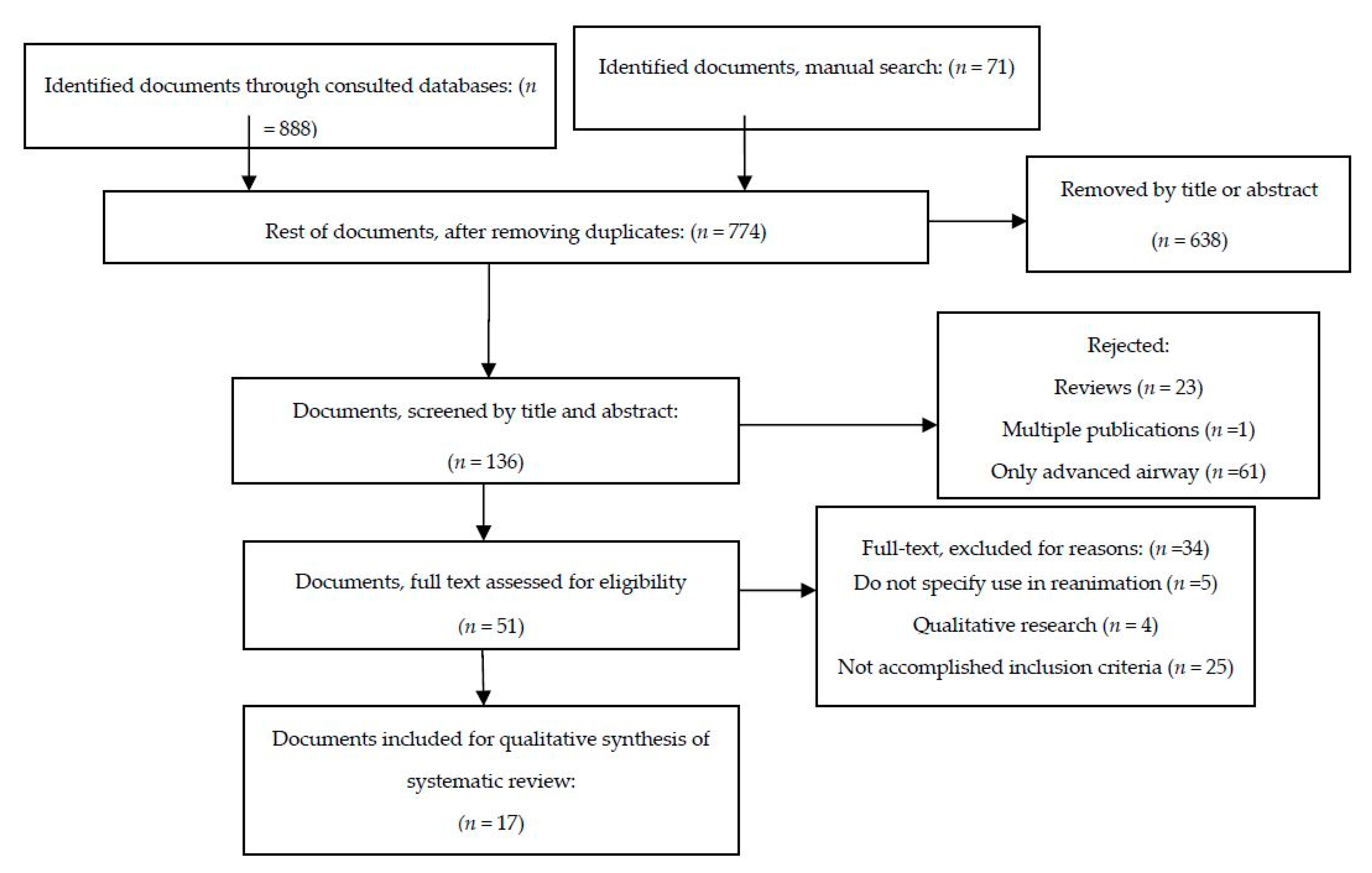
2. Materials and Methods

This systematic review was completed without an earlier cut-off limit up to 31 December 2018. We followed the steps of the Preferred Reporting Items for Systematic Review and Meta-Analysis protocols (PRISMA) [16], using Health Sciences Descriptors (DeCS/MeSH): Capnography and Cardiopulmonary Resuscitation, and the free terms: End-tidal, EtCO2, CPR, and resuscitation. The following combination was established with Boolean operators: (“Capnography” (Mesh) OR “end tidal” OR “etco2”) AND (“Cardiopulmonary Resuscitation” (Mesh) OR “CPR” OR “resuscitation”).
The primary sources of this review were the following databases: EBSCOhost, SCOPUS, PubMed, Índice Bibliográfico Español en Ciencias de la Salud (IBECS), TESEO (Spanish Theses repository), and Cochrane Library Plus. We also looked through the CPR guidelines published since 1998 by ILCOR, the European Resuscitation Council, and the American Heart Association, for papers that referenced the contents of this review.
The inclusion criteria used for the review were: (1) Observational and experimental studies published in English and Spanish, (2) studies should describe the implementation of capnography in CPR maneuvers for patients of any age; (3) studies should assess the effectiveness of ventilation using the mask only or in comparison with other devices for advanced airway management in cardiac arrest patients; and (4) studies should assess the correlation of values obtained from capnography and results of resuscitation (ROSC, survival rates, etc.).
Exclusion criteria included: (1) Studies performed on training mannequins, anesthetized patients, corpses, or animals; (2) clinical cases, letters to the editor, or other editorial texts; and (3) studies only focusing on the use of capnography in intubated patients.
The review was completed in pairs; the final decision as to whether to include or exclude a particular study was agreed upon by the research team, firstly by screening the titles and abstracts and then by a full-text reading. We used a template for the structured assessment of each consulted paper that included the following: Introduction, justification, objectives, kind of study, year of study, sample size, methodology, significant results, discussion, limitations, conclusions, remarks, and recommended bibliography. Then, we evaluated the degree to which each study was adequate on a 4-point Likert scale in terms of the criteria and methodological quality of the results shown. The Likert scale values were as follows: Paper with little relevance to the purpose of our review was assigned 1 point; 2 points when the study was relevant to the theoretical framework of justification of the review, but of low methodological quality; 3 points when the research methodology was relevant, but with results of low interest to the review; and 4 points when the methodology, results, conclusions, and theoretical framework of the work were relevant to the review. For a critical reading of the selected papers, we used the Critical Appraisal Skills Programme (CASP), which is used to complete systematic reviews, available on the web-based application FLC 3.0 [17].
After completing the literature research, the strategy was replicated by an expert in Documentation Sciences, using descriptors and their Boolean combination and the language used by databases. The expert obtained the same results. As such, the reliability of our review was ensured.
Both reviewers independently extracted the following data from each paper included in the systematic review and then compared the results obtained by means of a pre-designed chart: Sample size, kind of CPR (cardiac origin, traumatic, adult or pediatric patients, and intra-hospital or out-of-hospital), staff performing CPR, type of capnograph or capnometer used with BVM, physiological or survival values compared by ETCO2 (ROSC, survival at hospital admission and discharge, heart rate correlation, and time of measuring), evidence level according to CASP, results of the structured template, and Likert scale.
The methodological quality was evaluated by means of CASP. Based on the results, it was agreed to approximate to the grade recommendations based on the U.S. Preventive Services Task Force (USPSTF) evidence levels from “A” (substantial benefit) to “I” (insufficient evidence) levels.

3. Results

We reviewed the full text of 136 papers, and 17 were selected after applying the inclusion and exclusion criteria for critical reading (Figure 1). Distribution according to the study design was: eight descriptive prospective (47%), five retrospective (29%), and four RCT (23.5%). 13 were focused on neonatal patients (76.4%) and the other four on adults (23.6%). The mean number of patients in studies focusing on adults was 62, and the mean in newborn studies was 66. All studies on newborns were completed in the delivery room. On adults, three were completed in out-of-hospital cardiac arrest and one intra-hospital cardiac arrest. All the RCT focused on newborns; two of which focused on the viability and reliability of capnography monitoring during mask ventilation. We did not find any study about the use of capnography in children out of the neonatal period.
In adults, the articles relating the feasibility of capnography monitoring during CPR were all observational, and only Nakatani et al. [18] presented data and the correlation between registered values and outcomes from resuscitation, like ROSC or surveillance. In the cluster that used BVM, the ROSC proportion was 15% of the patient groups that showed EtCO2 values < 0.5% (<4 mmHg), 14% of patients from the group that showed EtCO2 values between 0.5% and 2% (between 4 and 15 mmHg), and 43% of patients from the group with values > 2% (>15 mmHg). The features of studies in adults are summarized in Table 1. Based on these results, the overall recommendation for capnometry use during mask ventilation in adults was level C according to USPSTF.
In newborns, there were more examples of feasibility and reliability capnography monitoring during CPR. From the 13 studies in these patients (four RCT, nine observational), the vast majority considered capnometric values as a parameter for assessing the quality of ventilation (Table 2), airway patency, mask leaks, and showed a good correlation between capnography and heart rates [19,20,21,22,23,24,25,26,27,28,29,30,31]. The two RCTs that emphasized the validity of the technique were: (1) Hawkes et al., who compared the accuracy of colorimetric detector versus capnography to determine normocarbia, but they did not find a difference in the incidence of normocarbia between the two detectors. (2) Kong et al. compared the utility of continuous capnography monitoring versus no capnography monitoring in the delivery room; they did not find a reduction in the occurrence cases in the targeted range (40–60 mmHg) of CO2 in the initial blood gas analysis at admission at neither of the two study branches. Although, both studies recommended the use of capnometry as a useful instrument because it provided useful physiologic information about the ventilatory status of the patient. Four articles correlated the use of capnography with assessing the increase in infant heart rate: Palme-Kilander and Tunell [31] appeared to be the first authors to correlate the upper values of exhaled CO2 with heart rate increase in newborns. During periods of assisted ventilation with CO2 of less than 2 mL/kg/min, they observed a mean of 87 beats/min; with records between 2 and 6 mL/kg/min, they observed an immediate increase in heart rate to 132–159 (mean 145) bpm. Hooper et al. [28] found EtCO2 levels increased to >10 mmHg in 28 s (median) before the heart rate increased up to 100 bpm. Mizumoto et al. [25] found an early detection of expired CO2 (>15 mmHg) between 8 and 73 s (median 15 s) before heart rate improvement. Blank et al. [26] indicated that, when the EtCO2 value was above 14 mmHg (2% in colorimetric detector), the detector showed an accuracy of 95%, detecting an increase in EtCO2 10 to 20 s before the increase in heart rate appeared on the monitor. They commented that the electrocardiogram did not provide a reliable measurement of heart rate in 12% (5/41) of neonates.
Considering these studies, we recommended the B level or the use of capnometry detectors during facemask ventilation during newborn resuscitation.
The staff that assist during resuscitations in pre-hospital environmental studies include paramedics and emergency medical technicians (EMTs), whereas in a hospital environment, medical, midwife, or nursing staff assist.
The limitations of the analyzed studies include the designs used, especially the retrospective design of five primary research papers, due to possible bias during data collection with small-sized samples. Otherwise, in newborn studies, the two RCT with larger samples focused on the values of EtCO2 as another variable observed for assessing the quality of ventilation in two different types of BVM [26] and compared the usual positive pressure ventilation through mask and T-piece device with a sustained ventilation technique, where a correlation was found between high EtCO2 values and larger volume tidal [25]. The other two RCT focused more on the reliability of capnometry during newborn resuscitation, and reported no improvement or correlation between capnometric values registered and blood PCO2 levels. Kong et al. [27] only used the value of capnometry from the last five breaths before disconnection of the positive pressure ventilation and compared this value with PCO2 levels with the average from the 40 minutes after EtCO2 measurement. Neither aspect of the study produced a reduction in the occurrence of hypocapnia and hypercapnia through capnography monitoring with mask ventilation. Hawkes et al. [20] recognized, as a limitation, a probable alteration in the CO2 blood test one hour after registration of the last EtCO2 value due to other clinical interventions. Although, both of them indicated that the use of capnography was feasible and appeared to be safe. The vast majority of studies in neonates did not use capnography monitoring as a unique variable or target in the study; the authors used it as a value for assessing the quality of ventilation. Another limitation was the high heterogeneity regarding the patients (four adults and 13 neonate studies), and the kind of cardiac arrest in adults. In the adult studies, one paper included only the cardiac arrest of a non-traumatic origin, another included all kinds of cardiac arrests, and the rest included only cardiac arrest of electromechanical dissociation and/or asystole. Leturiondo et al. [32]. did not describe if the patients were synchronized by chest compressions and ventilation, which obviously would artefact the capnogram. In adults we can say that there was too much variability in the measurement times utilized during the capnography monitoring between the studies.
In general, the instruments for monitoring CO2 were different in the studies, providing different ranges, volumetric, time capnography, or colorimetric values. Colorimetric devices were used in five studies; this kind of detector was unable to differentiate inadequate tidal volumes in comparison with the use of respiratory function monitors, which could incorporate CO2 monitoring and are used more in the delivery room.

4. Discussion

Within the newborn studies, there was more evidence about the use of capnometry detectors within mask ventilation. A previous review about the use of capnography in the delivery room [11] suggested a level of B for facemask ventilation according to the American Heart Association levels of evidence, which are recommendations that agree with our findings. We did not find any publications that reflected the correlation of quantitative values of capnography during adult CPR maneuvers with resuscitation outcome. Some evidence does exist for the feasibility of the technique in these patients. For example, Pearce et al. [14] assessed the correlation of capnographic values with ROSC as a predictor of hospital discharge. They stated that the capnography placed in OTI or BVM did not provide any more data about values recorded by BVM and capnography. Leturiondo et al. [32] studied the chest compression artifact on capnogram-based ventilation detection and found no distortion in the capnogram of seven patients ventilated with BVM; the rest of the patients were ventilated through OTI or supraglottic airways. They did not provide more capnographic values of resuscitations performed using BVM. Davis et al. [33] showed that capnography was performed immediately after starting ventilation with BVM and later on after OTI. The authors suggested that this approach established an appropriate capnography functioning and allowed for the recording of an EtCO2 reference value before intubation. Nakatani et al. [18], in their multicenter study, correlated the values of the CO2 detector together with BVM (40% of the cases), laryngeal mask (LMA) (58% of the cases), or esophageal tube (2% of the cases) with variables such as ROSC, survival at hospital admission, and initial electrocardiographic rhythm. They found a significant difference in both the rate of ROSC between patients with EtCO2 values of less than 0.5% and those with values greater than 2%.
We think that the initial parameterization using capnography with BVM for ventilation is crucial in order to provide more objective comparisons with the values obtained by different ventilation devices in CPR and clinical results during reanimation of adults. Although apparently obvious, Kodali and Urman [13] showed that providing ventilation with BVM could result in rather unreliable values due to probable air leaks through the facemask. Ventilation leaks may increase due to inexperience in sealing the interface, through inadequate airway opening, and an increased volume of blown air into the esophagus and subsequently into the stomach. These parameters would be easier to control by incorporating spirometers into the monitors [34], or with volumetric capnography, as has been performed with new-generation monitors in the delivery room and implemented in various newborn studies in order to determine ventilation pressures and volumes, mask leaks, and successful aeration through capnography. As such, capnography monitoring during mask ventilation is feasible and seems reliable in infants without congenital cardiopulmonary abnormalities. Pahuja et al. [19] demonstrated that avoiding high tidal volumes and hypocarbia by means of EtCO2 in the delivery room could reduce intraventricular hemorrhage. Finer et al. [30] showed that, with a CO2 detector, providers could recognize and attempt to correct airway obstruction. As demonstrated in the literature, newborn CPR used to be enough with mask ventilation and only during the first minutes after transmission from intrauterine to extrauterine life. At this moment, newborns must increase pulmonary blood flow as well as expand fluid-filled alveoli to facilitate gas exchange, and more assistance is required for preterms [11]. This is a physiological situation, which encompasses low flow in minor circulation and asphyxia treatment. Neonate arrest differs from adult arrest, where only artificial blood flow is achieved via compressions once ROSC is achieved. As infant capnography showed reliability in the detection of increase in heart rate, we hypothesized that this quality should be similar for detecting ROSC in adults.
The limitations of our review include the impossibility of performing translations of studies published in languages other than English and Spanish, which reduced the number of papers reviewed. We would like to emphasize the heterogeneity of measurement systems, like the variability in the moment of measurement. Future studies in adults should consider an adequate recording of the times of measurement and including the results obtained, if possible, for up to 20 min, after initiating the maneuvers. Besides the wide variety of professionals who assisted these patients based on the setting (in-hospital or out-of-hospital), country, and respective legislation, the maneuvers can be completed by various kinds of professionals. Trying to control for these variables when comparing of in future studies may help to reduce potential bias. With this many different variables, the meta-analysis of even two papers is unsuitable.

5. Conclusions

Despite having limited and low-level evidence in adults and moderate evidence in newborns (Table 1 and Table 2), according to the evidence levels of the USPSTF and results of the recorded critical reading sheets (FLC 3.0), we suggest that capnography with mask ventilation is feasible and seems to have the potential to advise rescuers about patients in cardiac arrest (adults and neonates). The reliability of the technique has been demonstrated with a correlation observed between capnometric values obtained from a ventilation using mask in those patients, as well as by the usefulness of this parameter for early detection of heart rate increase, sometimes more than 10 s in advance of rate increase, and to help maintain airway patency and lung aeration, which was mainly recorded in neonatal patients.

6. Recommendations for Practice

According to the USPSTF criteria, we recommend a C level for the implementation of capnography in adult mask ventilation during CPR, as evidence quality is low to moderate and net benefit would be small. In newborn resuscitation, we suggest a B level. Capnography can facilitate airway management as an indicator of gas exchange, lung aeration, early identification of obstruction, early recognition of ROSC or heart rate increase, and probably the quality of the administered chest compressions. The obtained results help establish an advanced clinical practice of facemask ventilation based on quantifiable evidence, which is often performed by staff who are not used to and/or not qualified to use more advanced devices; they may be nursing staff, EMTs, or even physicians of primary health care or other specialties not related to emergency and critical care in the pre-hospital setting.

Author Contributions

Formal analysis, J.M.-M.; Investigation, F.J.C.-S. and J.M.-M.; Methodology, F.J.C.-S. and J.M.-M.; Resources, F.J.C.-S.; Supervision, J.M.-M.; Writing-original draft, F.J.C.-S.; Writing-review and editing, J.M.-M.

Acknowledgments

We would like to thank the SATSE Research Cabinet of the Balearic Islands for their support with reviewing the bibliographical search.

Conflicts of Interest

The authors declare no conflicts of interest.

References

  1. Monsieurs, K.G.; Nolan, J.P.; Bossaert, L.L.; Greif, R.; Maconochie, I.K.; Nikolaou, N.I.; Perkins, G.D.; Soar, J.; Truhlář, A.; Wyllie, J.; et al. European Resuscitation Council Guidelines for Resuscitation 2015. Section 1. Executive summary. Resuscitation 2015, 95, 1–80. [Google Scholar] [CrossRef] [PubMed]
  2. Baskett, P.J.F.; Bossaert, L.; Carli, P.; Chamberlain, D.; Dick, W.; Nolan, J.P.; Parr, M.J.A. Guidelines for the advanced management of the airway and ventilation during resuscitation. A statement by the Airway and Ventilation Management of the Working Group of the European Resuscitation Council. Resuscitation 1996, 31, 201–230. [Google Scholar] [CrossRef]
  3. Deakin, C.D.; Nolan, J.P.; Soar, J.; Sunde, K.; Koster, R.W.; Smith, G.B.; Perkins, G.D. European Resuscitation Council Guidelines for Resuscitation 2010 Section 4. Adult advanced life support. Resuscitation 2010, 81, 1305–1352. [Google Scholar] [CrossRef] [PubMed]
  4. Callaway, C.W.; Soar, J.; Aibiki, M.; Bottiger, B.W.; Brooks, S.C.; Deakin, C.D.; Donnino, M.W.; Drajer, S.; Kloeck, W.; Morley, P.T.; et al. Part 4: Advanced Life Support: 2015 International Consensus on Cardiopulmonary Resuscitation and Emergency Cardiovascular Care Science With Treatment Recommendations. Circulation 2015, 132, S84–S145. [Google Scholar] [CrossRef] [PubMed]
  5. Baskett, P.J.F.; Bossaert, L.; Carli, P.; Chamberlain, D.; Dick, W.; Nolan, P.; Parr, M.J.A. Guidelines for the basic management of the airway and ventilation during resuscitation. Resuscitation 1996, 31, 187–200. [Google Scholar] [CrossRef]
  6. ILCOR, I.L.C. on R. Part 4: Advanced life support. Resuscitation 2005, 67, 213–247. [Google Scholar]
  7. Nicolás Arriaza, H.; María Mercedes Aguirre, C. Monitorización de la calidad de la reanimación cardiopulmonar. Rev. Chil. Anest. 2012, 41, 42–45. [Google Scholar]
  8. Davis, D.P. Quantitative capnometry as a critical resuscitation tool. J. Trauma Nurs. Off. J. Soc. Trauma Nurses 2005, 12, 40–42. [Google Scholar] [CrossRef]
  9. Díez-Picazo, L.D.; Barrado-Muñoz, L.; Blanco-Hermo, P.; Barroso-Matilla, S.; Espinosa Ramírez, S. La capnografía en los servicios de emergencia médica. Semer. Med. Fam. 2009, 35, 138–143. [Google Scholar] [CrossRef]
  10. Soleimanpour, H.; Gholipouri, C.; Golzari, S.E.; Rahmani, F.; Sabahi, M. Capnography in the Emergency Department. Available online: http://www.omicsgroup.org/journals/capnography-in-the-emergency-department -2165-7548.1000e123.pdf (accessed on 14 February 2016).
  11. Hawkes, G.A.; Kelleher, J.; Ryan, C.A.; Dempsey, E.M. A review of carbon dioxide monitoring in preterm newborns in the delivery room. Resuscitation 2014, 85, 1315–1319. [Google Scholar] [CrossRef]
  12. Bhende, M.S.; LaCovey, D.C. End-tidal carbon dioxide monitoring in the prehospital setting. Prehospital Emerg. Care Off. J. Natl. Assoc. EMS Physicians Natl. Assoc. State EMS Dir. 2001, 5, 208–213. [Google Scholar]
  13. Kodali, B.S.; Urman, R.D. Capnography during cardiopulmonary resuscitation: Current evidence and future directions. J. Emerg. Trauma. Shock 2014, 7, 332–340. [Google Scholar]
  14. Pearce, A.K.; Davis, D.P.; Minokadeh, A.; Sell, R.E. Initial end-tidal carbon dioxide as a prognostic indicator for inpatient PEA arrest. Resuscitation 2015, 92, 77–81. [Google Scholar] [CrossRef] [PubMed]
  15. Soar, J.; Nolan, J.P.; Böttiger, B.W.; Perkins, G.D.; Lott, C.; Carli, P.; Pellis, T.; Sandroni, C.; Skrifvars, M.B.; Smith, G.B.; et al. European Resuscitation Council Guidelines for Resuscitation 2015: Section 3. Adult advanced life support. Resuscitation 2015, 95, 100–147. [Google Scholar] [CrossRef] [PubMed]
  16. Moher, D.; Shamseer, L.; Clarke, M.; Ghersi, D.; Liberati, A.; Petticrew, M.; Shekelle, P.; Stewart, L.A.; PRISMA-P Group, P.-P. Preferred reporting items for systematic review and meta-analysis protocols (PRISMA-P) 2015 statement. Syst. Rev. 2015, 4, 1. [Google Scholar] [CrossRef] [PubMed] [Green Version]
  17. López de Argumedo, M.; Reviriego, E.; Andrío, E.; Rico, R.; Sobradillo, N.; Hurtado de Saracho, I. Revisión externa y validación de instrumentos metodológicos para la Lectura Crítica y la síntesis de la evidencia científica. Madrid: Plan Nacional para el SNS del MSC. Servicio de Evaluación de Tecnologías Sanitarias del País Vasco (Osteba); 2006. Inform. Available online: http://www.lecturacritica.com/es/acerca.php (accessed on 9 April 2017).
  18. Nakatani, K.; Yukioka, H.; Fujimori, M.; Maeda, C.; Noguchi, H.; Ishihara, S.; Yamanaka, I.; Tase, C. Utility of colorimetric end-tidal carbon dioxide detector for monitoring during prehospital cardiopulmonary resuscitation. Am. J. Emerg. Med. 1999, 17, 203–206. [Google Scholar] [CrossRef]
  19. Pahuja, A.; Hunt, K.; Murthy, V.; Bhat, P.; Bhat, R.; Milner, A.D.; Greenough, A. Relationship of resuscitation, respiratory function monitoring data and outcomes in preterm infants. Eur. J. Pediatr. 2018, 177, 1617–1624. [Google Scholar] [CrossRef] [PubMed]
  20. Hawkes, G.A.; Finn, D.; Kenosi, M.; Livingstone, V.; O’Toole, J.M.; Boylan, G.B.; O’Halloran, K.D.; Ryan, A.C.; Dempsey, E.M. A Randomized Controlled Trial of End-Tidal Carbon Dioxide Detection of Preterm Infants in the Delivery Room. J. Pediatr. 2017, 182, 74–78. [Google Scholar] [CrossRef]
  21. Hawkes, G.A.; Kenosi, M.; Finn, D.; O’Toole, J.M.; O’Halloran, K.D.; Boylan, G.B.; Ryan, A.C.; Dempsey, E.M. Delivery room end tidal CO2 monitoring in preterm infants <32 weeks. Arch. Dis. Child. Fetal Neonatal Ed. 2016, 101, F62–F65. [Google Scholar]
  22. Ngan, A.Y.; Cheung, P.-Y.; Hudson-Mason, A.; O’Reilly, M.; van Os, S.; Kumar, M.; Aziz, K.; Schmolzer, G.M. Using exhaled CO2 to guide initial respiratory support at birth: A randomised controlled trial. Arch. Dis. Child. 2017, 102, F525–F531. [Google Scholar] [CrossRef]
  23. Thallinger, M.; Ersdal, H.L.; Francis, F.; Yeconia, A.; Mduma, E.; Kidanto, H.; Linde, J.E.; Eilevstjønn, J.; Gunnes, N.; Størdal, K. Born not breathing: A randomised trial comparing two self-inflating bag-masks during newborn resuscitation in Tanzania. Resuscitation 2017, 116, 66–72. [Google Scholar] [CrossRef] [PubMed]
  24. Murthy, V.; D’Costa, W.; Shah, R.; Fox, G.F.; Campbell, M.E.; Milner, A.D.; Greenough, A. Prematurely born infants’ response to resuscitation via an endotracheal tube or a face mask. Early Hum. Dev. 2015, 91, 235–238. [Google Scholar] [CrossRef]
  25. Mizumoto, H.; Iki, Y.; Yamashita, S.; Hata, D. Expiratory CO2 as the first sign of successful ventilation during neonatal resuscitation. Pediatr. Int. 2015, 57, 186–188. [Google Scholar] [CrossRef]
  26. Blank, D.; Rich, W.; Leone, T.; Garey, D.; Finer, N. Pedi-cap color change precedes a significant increase in heart rate during neonatal resuscitation. Resuscitation 2014, 85, 1568–1572. [Google Scholar] [CrossRef] [PubMed]
  27. Kong, J.Y.; Rich, W.; Finer, N.N.; Leone, T.A. Quantitative end-tidal carbon dioxide monitoring in the delivery room: A randomized controlled trial. J. Pediatr. 2013, 163, 104–108. [Google Scholar] [CrossRef] [PubMed]
  28. Hooper, S.B.; Fouras, A.; Siew, M.L.; Wallace, M.J.; Kitchen, M.J.; Pas, A.B. t.; Klingenberg, C.; Lewis, R.A.; Davis, P.G.; Morley, C.J.; et al. Expired CO2 Levels Indicate Degree of Lung Aeration at Birth. PLoS ONE 2013, 8, e70895. [Google Scholar] [CrossRef]
  29. Murthy, V.; O’Rourke-Potocki, A.; Dattani, N.; Fox, G.F.; Campbell, M.E.; Milner, A.D.; Greenough, A. End tidal carbon dioxide levels during the resuscitation of prematurely born infants. Early Hum. Dev. 2012, 88, 783–787. [Google Scholar] [CrossRef] [PubMed]
  30. Finer, N.N.; Rich, W.; Wang, C.; Leone, T. Airway Obstruction During Mask Ventilation of Very Low Birth Weight Infants During Neonatal Resuscitation. Pediatrics 2009, 123, 865–869. [Google Scholar] [CrossRef] [PubMed]
  31. Palme-Kilander, C.; Tunell, R. Pulmonary gas exchange during facemask ventilation immediately after birth. Arch. Dis. Child. 1993, 68, 11–16. [Google Scholar] [CrossRef]
  32. Leturiondo, M.; Ruiz de Gauna, S.; Ruiz, J.M.; Julio Gutiérrez, J.; Leturiondo, L.A.; González-Otero, D.M.; Russell, J.K.; Zive, D.; Daya, M. Influence of chest compression artefact on capnogram-based ventilation detection during out-of-hospital cardiopulmonary resuscitation. Resuscitation 2018, 124, 63–68. [Google Scholar] [CrossRef] [PubMed]
  33. Davis, D.P.; Sell, R.E.; Wilkes, N.; Sarno, R.; Husa, R.D.; Castillo, E.M.; Lawrence, B.; Fisher, R.; Brainard, C.; Dunford, J.V. Electrical and mechanical recovery of cardiac function following out-of-hospital cardiac arrest. Resuscitation 2013, 84, 25–30. [Google Scholar] [CrossRef] [PubMed]
  34. Hess, D.; Eitel, D. Monitoring during resuscitation. Respir. Care 1992, 37, 739–768. [Google Scholar] [PubMed]
Figure 1. Flow-chart of information according to the review stages.
Jcm 08 00358 g001
Table 1. Relationship of variables analyzed in the adult studies with evidence levels, and evidence level according to USPSTF.
Author/yearStudy DesignTotal N/BVM Ventilated N Location/Cause of Cardiac ArrestProfessional Assisting Type of CapnographyOutcomeTime of End-tidal CO2 MeasurementEvidence Level USPSTF
Leturiondo et al. 2017Observational retrospective232/7Out-of-hospital cardiac arrestParamedicsMicrostreamDistortion of capnogram by chest compression 20 minIII
Pearce et al. 2015Observational retrospective50/NKIntra-hospital cardiac arrest by PEA/asystoleA multidisciplinary intra-hospital cardiac arrest teamMainstreamROSC and survival at discharge from hospital Continuous during the first 10 min of cardiac arrestNot classifiable *
Davis et al. 2013Observational prospective145/NKOut-of-hospital cardiac arrestParamedicsDid not specify ROSC and correlation with heart rate valuesBefore, during, and after pauses to check pulse: every 2 minNot classifiable *
Nakatani et al. 1999Observational prospective multicentric121/48Non-traumatic out-of-hospital cardiac arrestEmergency techniciansColorimetric ROSC, survival at admission7 to 15 minII-3
BVM: bag-valve-mask; NK, Not known which are ventilated through the mask; PEA, pulseless electrical activity; ROSC, return of spontaneous circulation; EtCO2, end-tidal CO2; USPSTF, U.S. Preventive Services Task Force. * according to our clinical question.
Table 2. Relationship of variables analyzed in the newborn studies, together with evidence levels and evidence level according to USPSTF.
Author/yearStudy DesignTotal N/Mask Ventilated Gestational AgeProfessional AssistingType of CapnographyOutcomeTime of End-tidal CO2 MeasurementEvidence Level USPSTF
Pahuja et al. 2018Observational retrospective70PretermCliniciansMainstreamIncidence of intraventricular hemorrhage and bronchopulmonary dysplasiaContinuous during resuscitationIII
Hawkes et al. 2017Observational prospective59PretermPhysiciansColorimetric and MicrostreamNormocarbia within the first hour of life.Continuous during delivery room ventilationI
Ngan et al. 2017Randomized Clinical Trial162PretermMultidisciplinary delivery teamMainstreamDuring sustained inflation or positive pressure ventilationContinuous, during first 60 sI
Thallinger et al. 2017Randomized Clinical Trial328TermMidwives and nurse anesthetistsMicrostreamTidal volumes and mask leak. Airway pressures and tidal volume comparing 2 devices10 minI
Hawkeset al. 2016Observational prospective35/29PretermUnspecifiedMicrostreamFeasibility of EtCO2 monitoring, normocapnia on admission neonatal intensive care unitContinuous, during first 10 minIII
Murthy et al. 2015Observational prospective35/15PretermUnspecifiedMainstreamEffectiveness active inflation, tidal volume and pressures.During 5 first inflationsIII
Mizumoto et al. 2015Observational prospective15/7PretermPediatricians and nursesMainstreamIncrease of heart rate and quality of ventilationUp to 3 min from delivery.III
Blank et al. 2014Observational retrospective4178% pretermMultidisciplinary delivery teamColorimetricIncrease of heart rateWhen detector turns to yellow (EtCO2 > 15 mmHg)III
Kong et al. 2013Randomized Clinical Trial48PretermMultidisciplinary delivery teamMainstream and colorimetricCorrelation PCO2 levels in blood gasAverage from last 5 ventilationsI
Hooperet al. 2013Observational prospective10PretermMultidisciplinary delivery teamMainstreamIncrease of heart rate and quality of ventilation, relationship with tidal volumeContinuous, not specified durationIII
Murthy et al. 2012Observational prospective40PretermUnspecifiedMainstreamFirst respiratory effort and tidal volumeSince first inspiratory effortIII
Fineret al. 2009Observational retrospective24PretermMultidisciplinary delivery teamColorimetricDetermining if airway was patentContinuous, not specified durationIII
Palme-Kilander and Turner. 1993Observational prospective30/28TermUnspecifiedBeckman Liston Becker II (volumetric)Increase of heart rate and quality of ventilationEvery 15 s. Up to 5 minIII
NK, Not known which are ventilated through the mask; PEA, pulseless electrical activity; ROSC, return of spontaneous circulation; EtCO2, end-tidal CO2; USPSTF, U.S. Preventive Services Task Force.

