Splenic Artery Aneurysms, a Rare Complication of Type 1 Gaucher Disease: Report of Five Cases

 ,
,
Abstract
:1. Introduction
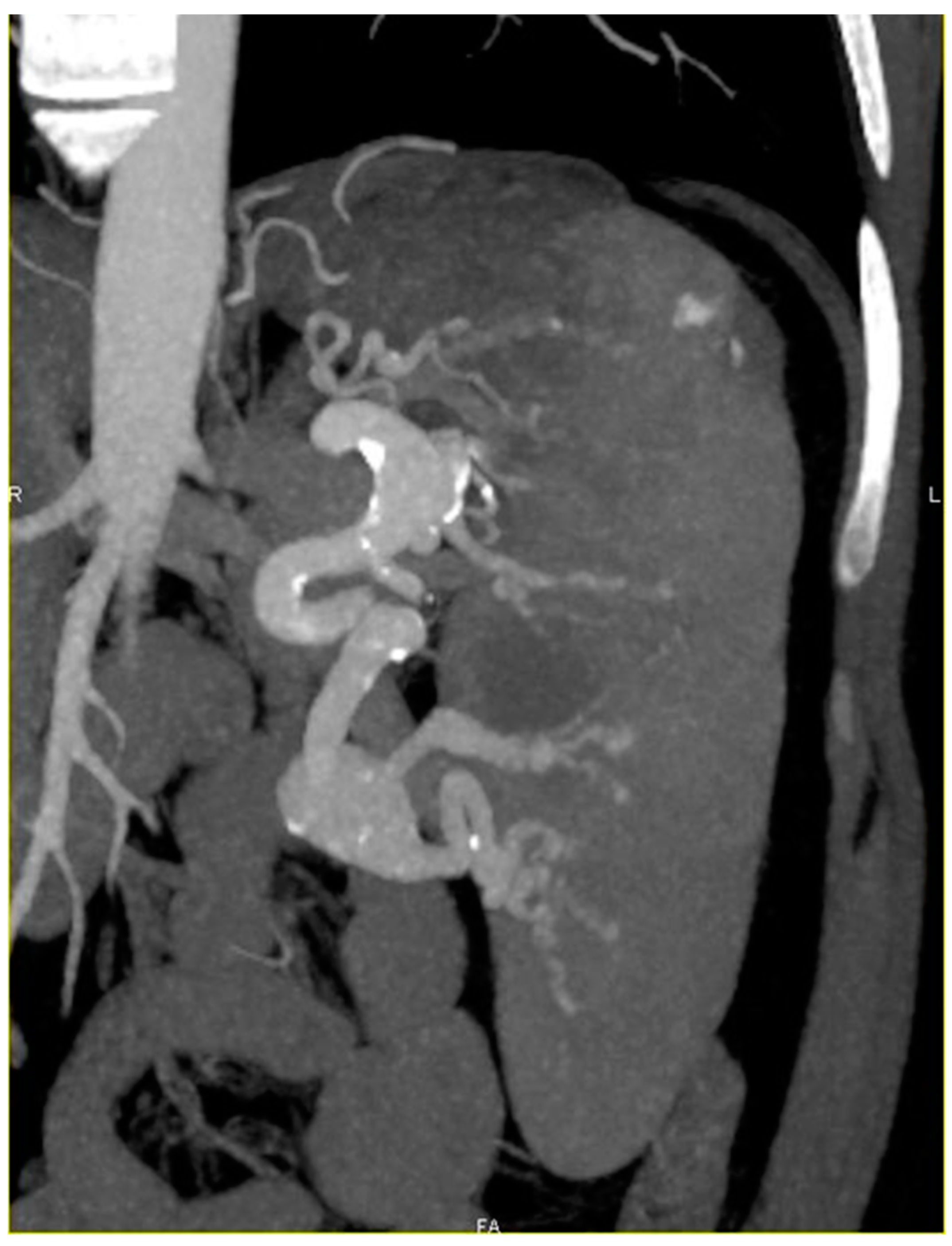
2. Discussion
3. Conclusions
Author Contributions
Conflicts of Interest
References
- Brady, R.O.; Kanfer, J.N.; Bradley, R.M.; Shapiro, D. Demonstration of a deficiency of glucocerebroside-cleaving enzyme in Gaucher’s disease. J. Clin. Investig. 1966, 45, 1112–1115. [Google Scholar] [CrossRef] [PubMed]
- Schnabel, D.; Schroder, M.; Sandhoff, K. Mutation in the sphingolipid activator protein 2 in a patient with a variant of Gaucher disease. FEBS Lett. 1991, 284, 57–59. [Google Scholar] [CrossRef]
- Stirnemann, J.; Vigan, M.; Hamroun, D.; Heraoui, D.; Rossi-Semerano, L.; Berger, M.G.; Rose, C.; Camou, F.; de Roux-Serratrice, C.; Grosbois, B.; et al. The french Gaucher’s disease registry: Clinical characteristics, complications and treatment of 562 patients. Orphanet. J. Rare Dis. 2012, 7, 77. [Google Scholar] [CrossRef] [PubMed]
- Weinreb, N.J.; Andersson, H.C.; Banikazemi, M.; Barranger, J.; Beutler, E.; Charrow, J.; Grabowski, G.A.; Hollak, C.E.; Kaplan, P.; Mankin, H.; et al. Prevalence of type 1 Gaucher disease in the united states. Arch. Intern. Med. 2008, 168, 326–327. [Google Scholar] [CrossRef] [PubMed]
- Raghavan, S.S.; Topol, J.; Kolodny, E.H. Leukocyte beta-glucosidase in homozygotes and heterozygotes for Gaucher disease. Am. J. Hum. Genet. 1980, 32, 158–173. [Google Scholar] [PubMed]
- Hruska, K.S.; LaMarca, M.E.; Scott, C.R.; Sidransky, E. Gaucher disease: Mutation and polymorphism spectrum in the glucocerebrosidase gene (GBA). Hum. Mutat. 2008, 29, 567–583. [Google Scholar] [CrossRef] [PubMed]
- Stirnemann, J.; Belmatoug, N. Adult Gaucher disease. Rev. Med. Interne 2001, 22 (Suppl. 3), 374–383. [Google Scholar]
- Mignot, C.; Doummar, D.; Maire, I.; De Villemeur, T.B. Type 2 Gaucher disease: 15 new cases and review of the literature. Brain Dev. 2006, 28, 39–48. [Google Scholar] [CrossRef] [PubMed]
- Sidransky, E. Gaucher disease: Insights from a rare mendelian disorder. Discov. Med. 2012, 14, 273–281. [Google Scholar] [PubMed]
- Akbulut, S.; Otan, E. Management of giant splenic artery aneurysm: Comprehensive literature review. Medicine (Baltimore) 2015, 94, 1016. [Google Scholar] [CrossRef] [PubMed]
- Sherlock, S.P.V.; Learmonth, J.R. Aneurysm of the splenic artery with an account of an example complicating Gaucher’s disease. Br. J. Surg. 1942, 151–160. [Google Scholar] [CrossRef]
- Van Rijn, M.J.; Ten Raa, S.; Hendriks, J.M.; Verhagen, H.J. Visceral aneurysms: Old paradigms, new insights? Best Pract. Res. Clin. Gastroenterol. 2017, 31, 97–104. [Google Scholar] [CrossRef] [PubMed]
- Bedford, P.D.; Lodge, B. Aneurysm of the splenic artery. Gut 1960, 1, 312–320. [Google Scholar] [CrossRef] [PubMed]
- Abbas, M.A.; Stone, W.M.; Fowl, R.J.; Gloviczki, P.; Oldenburg, W.A.; Pairolero, P.C.; Hallett, J.W.; Bower, T.C.; Panneton, J.M.; Cherry, K.J. Splenic artery aneurysms: Two decades experience at mayo clinic. Ann. Vasc. Surg. 2002, 16, 442–449. [Google Scholar] [CrossRef] [PubMed]
- Reynolds, M.R.; Heiferman, D.M.; Boucher, A.B.; Howard, B.M.; Barrow, D.L.; Dion, J.E. Multiple intracranial aneurysms in a patient with type I Gaucher disease: A case report and literature review. Br. J. Neurosurg. 2018, 1–3. [Google Scholar] [CrossRef] [PubMed]
- Nasu, M.; Fujiwara, H.; Sono, J.; Okada, Y.; Miyamoto, S.; Nishiuchi, S.; Tatemichi, K.; Shomura, T. Annulo-aortic ectasia with debakey type II dissecting aneurysm in Gaucher’s disease. J. Cardiovasc. Surg. (Torino) 1990, 31, 809–811. [Google Scholar]
- Agrawal, G.A.; Johnson, P.T.; Fishman, E.K. Splenic artery aneurysms and pseudoaneurysms: Clinical distinctions and ct appearances. AJR Am. J. Roentgenol. 2007, 188, 992–999. [Google Scholar] [CrossRef] [PubMed]
- Roberts, W.C.; Fredrickson, D.S. Gaucher’s disease of the lung causing severe pulmonary hypertension with associated acute recurrent pericarditis. Circulation 1967, 35, 783–789. [Google Scholar] [CrossRef] [PubMed]

| Patient | Sex/Age | Age at Diagnosis of GD | Genotype | Treatment | Age at Diagnosis of SAA | Evolution |
|---|---|---|---|---|---|---|
| 1 | F, NA | 30 | N370S/N370S | Intermittent ERT 10 U/kg eow from 44 to 52-year-old | 63 | Sudden collapse and death aged 63 years |
| 2 | F, 59 | 19 | N370S/84GG | ERT 16 U/kg eow | 49 | Complex set of four SAA |
| 3 | M, 50 | 10 | N370S/N370S | ERT 60 U/kg eow at 40-year-old | 40 | Aneurism size increase |
| 4 | M, 62 | 38 | N370S/D218A | ERT 75 U/kg every three weeks from 45 to 48-year-old | 58 | Embolization two years later |
| 5 | M, 31 | 29 | N370S/K196E | ERT 60 U/kg/eow at 29-year-old then eliglustat at 30-year-old | 29 | Aneurism size stabilization |
© 2019 by the authors. Licensee MDPI, Basel, Switzerland. This article is an open access article distributed under the terms and conditions of the Creative Commons Attribution (CC BY) license (http://creativecommons.org/licenses/by/4.0/).
Share and Cite
Serratrice, C.; Cox, T.M.; Leguy-Seguin, V.; Morris, E.; Yousfi, K.; Monnet, O.; Sibert, A.; Allaham, W.; Belmatoug, N. Splenic Artery Aneurysms, a Rare Complication of Type 1 Gaucher Disease: Report of Five Cases. J. Clin. Med. 2019, 8, 219. https://doi.org/10.3390/jcm8020219
Serratrice C, Cox TM, Leguy-Seguin V, Morris E, Yousfi K, Monnet O, Sibert A, Allaham W, Belmatoug N. Splenic Artery Aneurysms, a Rare Complication of Type 1 Gaucher Disease: Report of Five Cases. Journal of Clinical Medicine. 2019; 8(2):219. https://doi.org/10.3390/jcm8020219
Chicago/Turabian StyleSerratrice, Christine, Timothy M. Cox, Vanessa Leguy-Seguin, Elizabeth Morris, Karima Yousfi, Olivier Monnet, Annie Sibert, Wassim Allaham, and Nadia Belmatoug. 2019. "Splenic Artery Aneurysms, a Rare Complication of Type 1 Gaucher Disease: Report of Five Cases" Journal of Clinical Medicine 8, no. 2: 219. https://doi.org/10.3390/jcm8020219
APA StyleSerratrice, C., Cox, T. M., Leguy-Seguin, V., Morris, E., Yousfi, K., Monnet, O., Sibert, A., Allaham, W., & Belmatoug, N. (2019). Splenic Artery Aneurysms, a Rare Complication of Type 1 Gaucher Disease: Report of Five Cases. Journal of Clinical Medicine, 8(2), 219. https://doi.org/10.3390/jcm8020219