Article

Pre-, Intra-, and Post-Operative Evaluation of Extraocular Muscle Insertions Using Optical Coherence Tomography: A Comparison of Four Devices

Department of Ophthalmology, UPMC Children’s Hospital of Pittsburgh, University of Pittsburgh, Pittsburgh, PA 15224, USA
*
Author to whom correspondence should be addressed.
J. Clin. Med. 2019, 8(10), 1732; https://doi.org/10.3390/jcm8101732
Submission received: 30 August 2019 / Revised: 12 October 2019 / Accepted: 17 October 2019 / Published: 19 October 2019

Abstract

:
OCT (optical coherence tomography) is widely used in ophthalmology and pediatric ophthalmology, but limited research has been done on the use of OCT in strabismus. This study investigates the use of different OCT machines to image rectus muscle insertions pre-, intra-, and post-operatively in pediatric strabismus patients. The OCT machines used in the study were a Bioptigen (Leica Microsystems Inc., Buffalo Grove, IL, USA), Spectralis HRA+OCT with Anterior Segment Module (Heidelberg Engineering, Heidelberg, Germany), Visante (Carl Zeiss, Oberkochen, Germany), and Zeiss Rescan 700 (Carl Zeiss, Oberkochen, Germany). Measurements from the machines were compared with the caliper distance measured during the strabismus surgery before disinsertion or after reattachment. The OCT machines had moderate (Bioptigen: 0.62) to good intraclass correlation coefficients (Rescan: 0.83, Spectralis: 0.85, Visante: 0.88) with intra-operative measurements. To our knowledge, this is the first study to use an operating microscope with integrated intra-operative OCT to image rectus muscle insertions. OCT is a useful tool in strabismus surgical patients in the pre-, intra-, and post-operative settings, particularly in patients who have had previous surgery, when the muscle insertion is unknown. The ability to accurately image rectus muscle insertions has significant implications for the management of strabismus patients.

1. Introduction

Strabismus surgery on patients with previous strabismus, orbital, or other ophthalmic surgeries can be challenging, and the response to surgery less predictable than in patients without prior ocular surgery. The need for multiple strabismus surgeries is not uncommon [1]. The subsequent surgery or surgeries can frequently be performed by a different surgeon, with few details of the original surgery that may have been performed years or decades prior. Even with the previous operative reports or a history from the patient, the location of the extraocular muscles (EOMs) can be different than expected. Slit lamp examination can give clues to the prior surgery, but not specifics. Knowing the location of the EOMs prior to surgery and what previous surgery was performed can be very beneficial to the surgeon. Several techniques have been used to image the EOM pre-, intra-, and post-operatively to help plan surgery and reduce surgical exploration and time, including computed tomography (CT), magnetic resonance imaging (MRI), B-Scan ultrasonography (US), and ultrasound biomicroscopy (UBM) [2,3,4,5,6,7,8,9,10,11]. More recent studies have used the optical coherence tomography (OCT) to image EOM insertion on un-operated-on and previously operated-on muscles, but to our knowledge, none have used an operating microscope with integrated intra-operative OCT [12,13,14,15,16,17,18,19,20,21,22,23,24,25,26,27,28,29].

2. Experimental Section

A prospective study of pediatric strabismus patients (≤18 years old at enrollment) scheduled for strabismus surgery was enrolled. All subjects or parents signed informed consent, depending on age, for inclusion before they participated in the study. The study was conducted in accordance with the Declaration of Helsinki, and the protocol was approved by the University of Pittsburgh Institutional Review Board. OCT was used to image the rectus muscle insertions on pediatric strabismus patients pre-operatively, intra-operatively, and post-operatively. The different OCT machines used were a Bioptigen, Inc. (Leica Microsystems Inc., Leica Microsystems Inc., Buffalo Grove, IL, USA), Spectralis HRA+OCT with Anterior Segment Module (Heidelberg Engineering, Heidelberg, Germany), Visante (Carl Zeiss, Oberkochen, Germany), and Zeiss Rescan 700 (Carl Zeiss, Oberkochen, Germany). The measurements were compared to the handheld caliper distance measured during the strabismus surgery prior to disinsertion and where reattached to the sclera after surgery was performed. The muscle(s) scheduled to be operated on were imaged pre-operatively if the patients were 3 years or older and cooperative for OCT imaging with the Spectralis and Visante, as well as at the post-operative 1 month and the 4–6 month visit. All patients of any age had OCT images taken intra-operatively before the start of the surgery with the Bioptigen and Rescan.
A fixation light or fixation target was used to have patients look in different gaze positions, in order to image the muscle insertions pre-operatively and post-operatively. For example, the lateral rectus (LR) insertion was imaged with the eye in adduction. Of the images acquired, the highest quality image, as determined by the interpreting pediatric ophthalmologist (MP), was selected to measure the distance from the limbus to the muscle insertion with digital calipers incorporated into each machine’s software.
The EOM insertion in this study was defined as the most anterior point of the muscle tendon, which is visible as a dark structure tapering toward the limbus between the sclera and conjunctiva/Tenon’s (See Figure 1). The limbus was defined on the Visante as the transition between the corneal epithelium and the conjunctival epithelium, as described by Rosetto et al. [27]. The limbus was marked on the external photo with the Spectralis. Measurements were taken using digital calipers integrated in the AS-OCT software by a pediatric ophthalmologist on the Visante and Spectralis. All images were obtained by a single, well-experienced ophthalmic photographer pre-operatively and post-operatively, and all intraoperative measurements by the operating pediatric ophthalmologist (MP).
The Bioptigen and Rescan (Figure 2) were used to capture intra-operative measurements prior to the start of the strabismus surgery. The 10 mm anterior probe was used to capture the images with the Bioptigen, which was either hand-held or placed in an adjustable custom mount. The Rescan was used in the anterior segment mode. Intra-operatively, the eye was brought the direction opposite of the desired muscle to be imaged, with forceps grasping the episclera near the limbus. The limbus was identified clinically, and handheld calipers were used to make a shadow on the intra-operative OCT image at the anterior insertion of the tendon, or to mark the insertion on the conjunctiva and then measure to the limbus.
The “intra-operative” measurements during the surgical procedure were obtained with handheld calipers from the limbus to the most anterior insertion of the muscle centrally after “cleaning” the muscle of Tenon’s, the surrounding connective tissue, and any scar. After the muscle was reattached to the sclera during surgery, the new insertion to limbus was measured with handheld calipers.
To compare the different measurements, Bland–Altman plots were performed, along with intraclass correlation coefficients (ICCs). The statistical analysis was done with Excel 2013 (Microsoft, Redmond, WA, USA).

3. Results

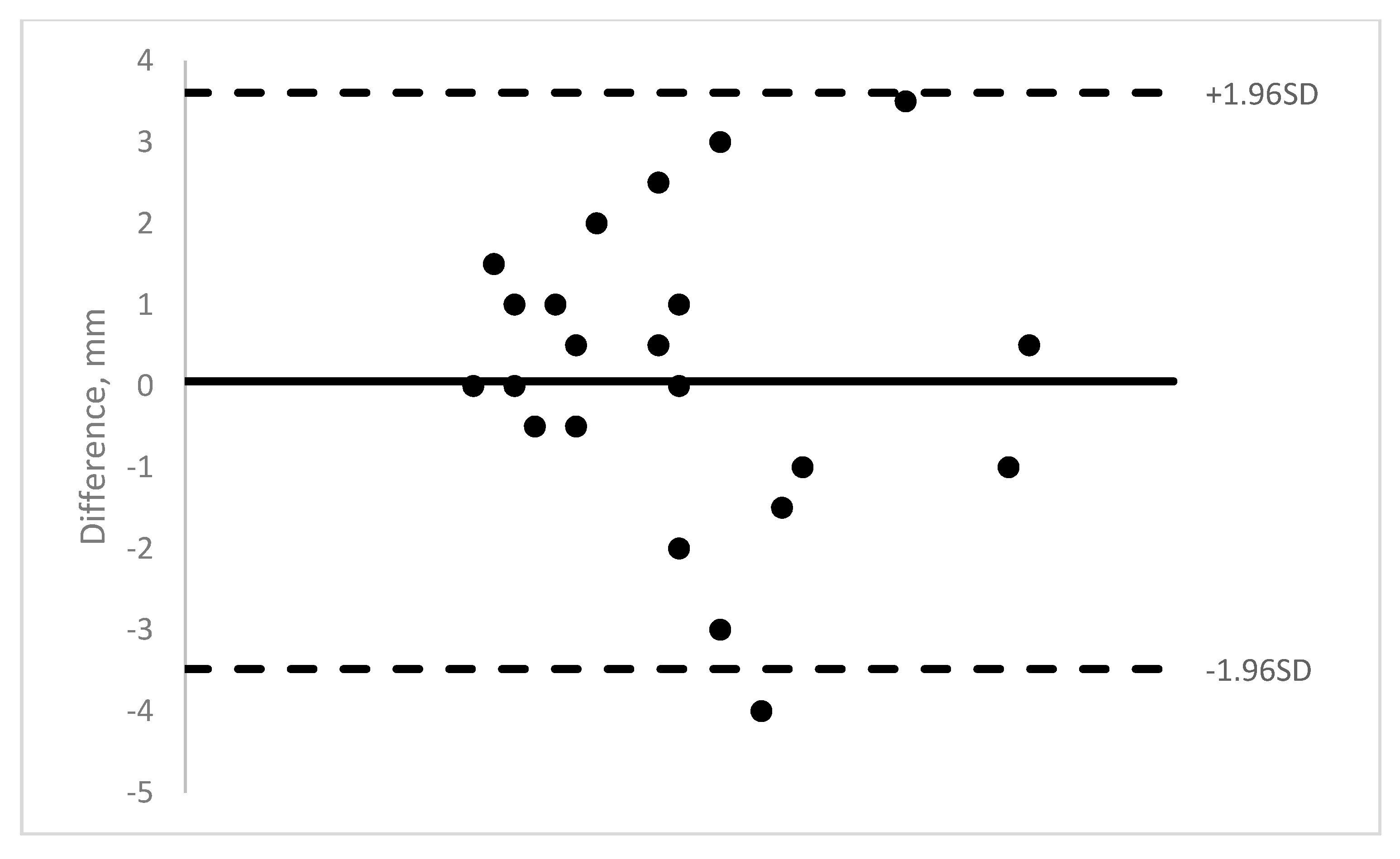
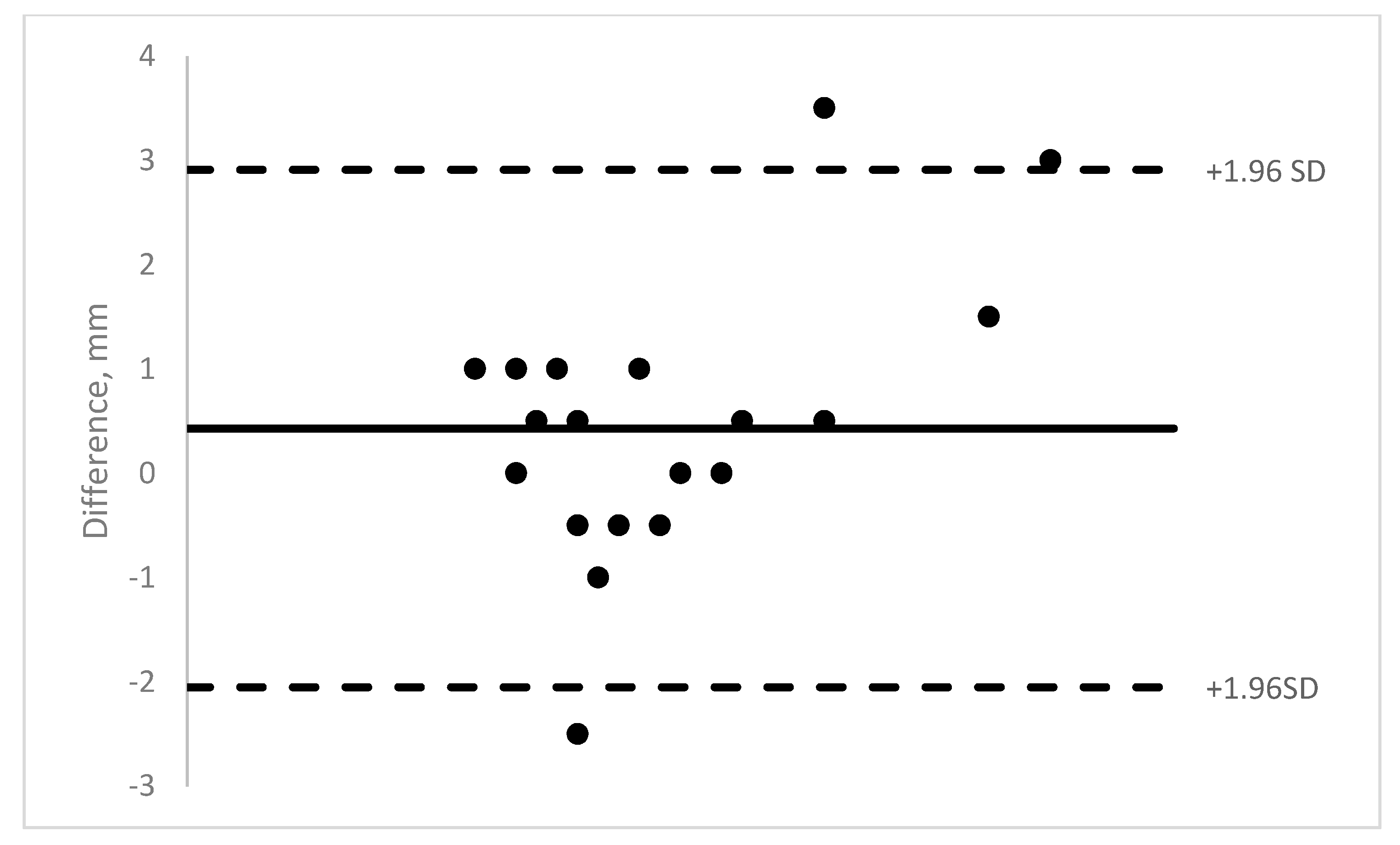
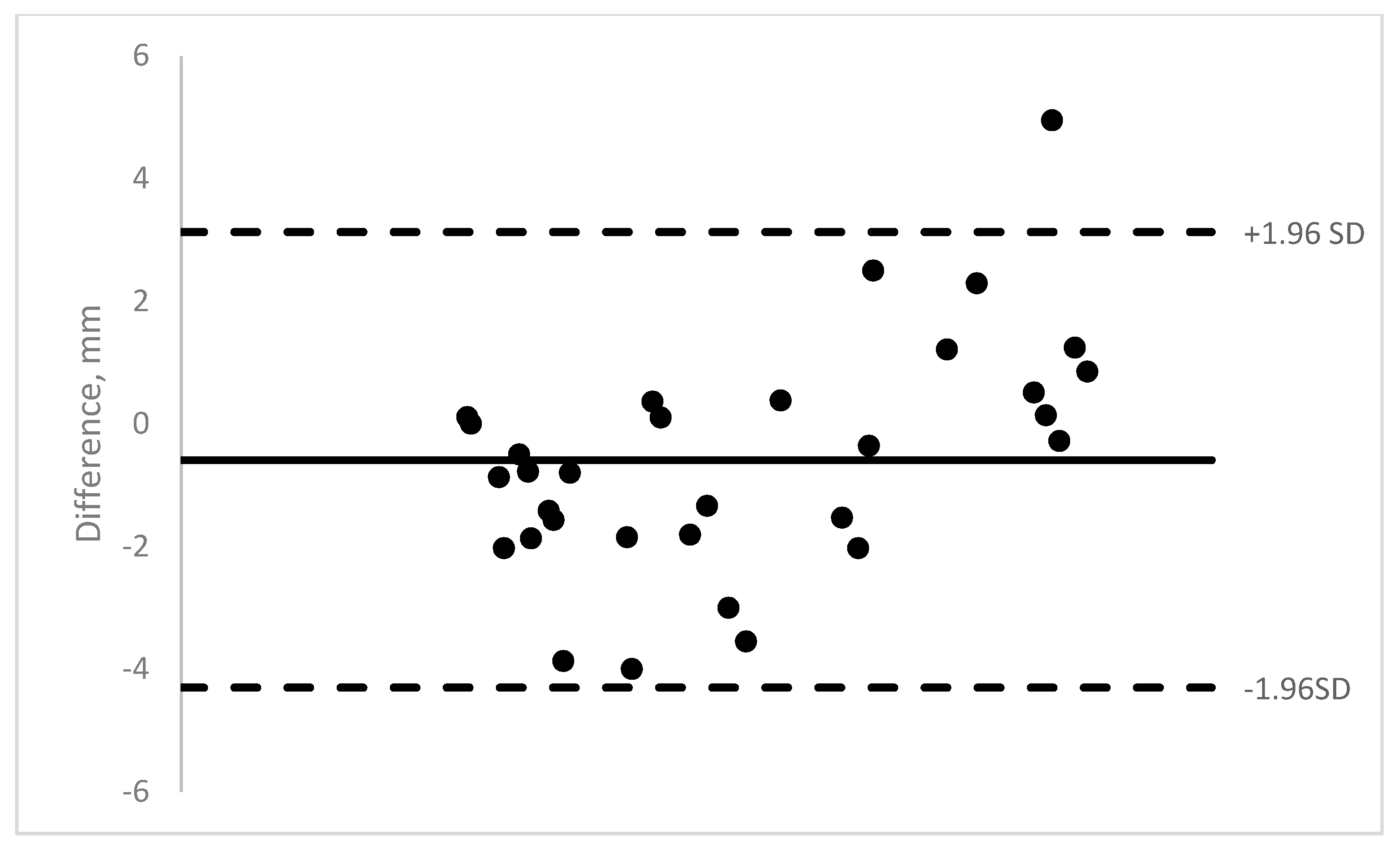
Nineteen pediatric patients were enrolled in the study. Ten were males and 9 were females. The age range was 11 months to 13 years (mean = 5.8 years). A total of 140 OCT images of 40 muscles were obtained (14 medial rectus, 22 lateral rectus, and 4 inferior rectus). The majority of the rectus muscle insertions were able to be imaged (58–89% depending on machine). Those results show moderate to good ICCs (0.62–0.88) (See Table 1). See Figure 3, Figure 4, Figure 5 and Figure 6 for Bland–Altman plots for each OCT machine. The percentage of muscles measuring ≤1 mm was higher pre-operatively than post-operatively (Spectralis: 89% vs. 54%; and Visante: 45% vs. 31%). See Table 2 for the list of patients able to be imaged with all four optical coherence tomography machines.

4. Discussion

OCT is a useful tool in strabismus surgical patients, particularly for previously operated patients where the muscle insertion may not be known. OCT can be useful pre-operatively, intra-operatively, and post-operatively. The ability to accurately image rectus muscle insertions has significant implications for the management of strabismus patients.
UBM, CT, MRI, and B-scan ultrasound can also be used to image the EOMs. These methods have advantages and disadvantages. The advantages of some include the ability to image the entire EOM, orbits, and pulley system; however, these techniques can be difficult to perform in certain patients due to claustrophobia, requirement for sedation or general anesthesia, and ionizing radiation. OCT can be obtained by a minimally invasive, quicker approach, and can be used pre-operatively and post-operatively in cooperative patients (3 years and older in this study), as well as intra-operatively while the patient is already under general anesthesia.
However, the OCT had limitations—rectus muscles are not always able to be imaged, so the accuracy was limited (42–84% of insertions were ≤1 mm of the OCT measurement, depending on the machine). In some cases, the muscle insertion had an ambiguous transition from muscle tendon to sclera or Tenon’s capsule on the OCT image, or could not be identified, which was not useful. In contrast, in other cases the insertion was very distinct and provided more definitive information with higher level of confidence to the surgeon. The limitation of ambiguous, unidentified, or misidentified insertion may be improved with newer, higher-resolution OCT technology that continues to develop. The limitations of OCT also include the inability to image the oblique muscles or posterior portions of the EOMs. Also, many previously recessed muscles were not able to be imaged posteriorly enough to capture the new muscle insertion (only 17/32 rectus muscles ≥10 mm from the limbus could be imaged), with less success post-operatively than pre-operatively. However, the inability to image the rectus insertion a certain distance from the limbus can provide useful information for future surgical planning.
Our results of 61% (62/102) of imaged muscles being at ≤1 mm difference is not as successful as previous studies by Ngo et al., at 89.9% for pediatric patients, or Rosetto et al. at 77% and Pihlblad et al. at 70% on an adult populations [13,21,27]. Our results add to the literature on this subject and add previously unreported results using the Biotogen and Rescan OCT machines.
The OCT machines each have their individual advantages and disadvantages, as well. The Spectralis and Visante can only be used in an outpatient setting, and the Rescan only in the operating room, but the Bioptogen can be used in both. Younger and uncooperative patients (seven patients under 3 years old in our study and one with developmental delay) were not able to be imaged as outpatients. Having the information before going to surgery is helpful for planning, but intraoperative information with the patient already under anesthesia for the planned surgery can also be helpful. The Rescan can be used while operating with real-time information, which can be very advantageous in some circumstances. The results also show that many different OCT machines that are available are capable of imaging rectus muscle insertions. Within the limitations discussed above, the ability to image the rectus muscle insertions pre-operatively, intra-operatively, and post-operatively in a non-invasive way gives valuable information to the strabismus surgeon to plan surgery, execute the surgery, and interpret the surgical outcomes.
Some limitations of the study include that measurements were all performed by the same pediatric ophthalmologist, who was not blinded to the patient or surgery. The study had a small number of subjects, and an even smaller amount that were successfully imaged by all four machines (Table 2). The study also assumed the rectus muscle insertion did not change post-operatively.
In conclusion, different OCT machines are capable of imaging rectus muscle insertions pre-operatively, intra-operatively, and post-operatively, which gives additional useful information to the strabismus surgeon. To our knowledge, this is the first study to use an operating microscope with integrated intra-operative OCT to image rectus muscle insertions. As technology continues to advance, we hope to be able to image muscle insertions with greater accuracy. Further studies will be needed to evaluate the different available and emerging techniques for imaging muscle insertions, in order to further refine our surgical approach.

5. Conclusions

OCT machines are capable of imaging the rectus muscle insertions pre-operatively, intra-operatively, and post-operatively. The muscle insertion information provided by the different OCT machines can be advantageous to the strabismus surgeon, and may help with planning surgery, performing surgery and interpreting surgical outcomes.

Author Contributions

Conceptualization, M.S.P.; methodology, M.S.P.; formal analysis, M.S.P.; investigation, P.R.S., A.T. and S.T.; writing—original draft preparation, M.S.P.; writing—review and editing, P.R.S., A.T. and S.T.; funding acquisition, M.S.P.

Funding

This research was funded by the Knights Templar Eye Foundation.

Acknowledgments

Ken K. Nischal contributed through the supervision and mentorship of this research project. The research results were presented at the 4th World Congress of Paediatric Ophthalmology and Strabismus, Hyderabad, India on 2 December 2017.

Conflicts of Interest

The author M.S.P. has received lecture honorariums sponsored by the World Society of Paediatric Ophthalmology and Strabismus related to some of the research presented above. The funders had no role in the design of the study; in the collection, analyses, or interpretation of data; in the writing of the manuscript; or in the decision to publish the results.

References

  1. Leffler, C.T.; Vaziri, K.; Schwartz, S.G.; Cavuoto, K.M.; McKeown, C.A.; Kishor, K.S.; Janot, A.C. Rates of Reoperation and Abnormal Binocularity Following Strabismus Surgery in Children. Am. J. Ophthalmol. 2016, 162, 159.e9–166.e9. [Google Scholar] [CrossRef] [PubMed]
  2. Demer, J.L.; Clark, R.A.; Kono, R.; Wright, W.; Velez, F.; Rosenbaum, A.L. A 12-year, prospective study of extraocular muscle imaging in complex strabismus. J. Aapos Off. Publ. Am. Assoc. Pediatric Ophthalmol. Strabismus 2002, 6, 337–347. [Google Scholar] [CrossRef]
  3. Ettl, A.; Kramer, J.; Daxer, A.; Koornneef, L. High-resolution magnetic resonance imaging of the normal extraocular musculature. Eye 1997, 11, 793–797. [Google Scholar] [CrossRef] [PubMed] [Green Version]
  4. Demer, J.L.; Ortube, M.C.; Engle, E.C.; Thacker, N. High-resolution magnetic resonance imaging demonstrates abnormalities of motor nerves and extraocular muscles in patients with neuropathic strabismus. J. Aapos Off. Publ. Am. Assoc. Pediatric Ophthalmol. Strabismus 2006, 10, 135–142. [Google Scholar] [CrossRef]
  5. Demer, J.L.; Dushyanth, A. T2-weighted fast spin-echo magnetic resonance imaging of extraocular muscles. J. Aapos Off. Publ. Am. Assoc. Pediatric Ophthalmol. Strabismus 2011, 15, 17–23. [Google Scholar] [CrossRef] [Green Version]
  6. Tamburrelli, C.; Salgarello, T.; Vaiano, A.S.; Scullica, L.; Palombi, M.; Bagolini, B. Ultrasound of the horizontal rectus muscle insertion sites: Implications in preoperative assessment of strabismus. Investig. Ophthalmol. Vis. Sci. 2003, 44, 618–622. [Google Scholar] [CrossRef]
  7. Sacca, S.; Polizzi, A.; Macri, A.; Patrone, G.; Rolando, M. Echographic study of extraocular muscle thickness in children and adults. Eye 2000, 14 Pt 5, 765–769. [Google Scholar] [CrossRef]
  8. Khan, H.A.; Smith, D.R.; Kraft, S.P. Localising rectus muscle insertions using high frequency wide-field ultrasound biomicroscopy. Br. J. Ophthalmol. 2012, 96, 683–687. [Google Scholar] [CrossRef]
  9. Watts, P.; Smith, D.; Mackeen, L.; Kraft, S.; Buncic, J.R.; Abdolell, M. Evaluation of the ultrasound biomicroscope in strabismus surgery. J. Aapos Off. Publ. Am. Assoc. Pediatric Ophthalmol. Strabismus 2002, 6, 187–190. [Google Scholar] [CrossRef]
  10. Dai, S.; Kraft, S.P.; Smith, D.R.; Buncic, J.R. Ultrasound biomicroscopy in strabismus reoperations. J. Aapos Off. Publ. Am. Assoc. Pediatric Ophthalmol. Strabismus 2006, 10, 202–205. [Google Scholar] [CrossRef]
  11. Solarte, C.E.; Smith, D.R.; Buncic, J.R.; Tehrani, N.N.; Kraft, S.P. Evaluation of vertical rectus muscles using ultrasound biomicroscopy. J. Aapos Off. Publ. Am. Assoc. Pediatric Ophthalmol. Strabismus 2008, 12, 128–131. [Google Scholar] [CrossRef]
  12. Takkar, B.; Sharma, P.; Singh, A.K.; Sahay, P. Anterior segment optical coherence tomography for identifying muscle status in strabismus surgery. Int. J. Ophthalmol. 2016, 9, 933–934. [Google Scholar] [CrossRef] [PubMed]
  13. Ngo, C.S.; Smith, D.; Kraft, S.P. The accuracy of anterior segment optical coherence tomography (AS-OCT) in localizing extraocular rectus muscles insertions. J. Aapos Off. Publ. Am. Assoc. Pediatric Ophthalmol. Strabismus 2015, 19, 233–236. [Google Scholar] [CrossRef] [PubMed]
  14. Pihlblad, M.S.; Erenler, F.; Sharma, A.; Manchandia, A.; Reynolds, J.D. Anterior Segment Optical Coherence Tomography of the Horizontal and Vertical Extraocular Muscles With Measurement of the Insertion to Limbus Distance. J. Pediatric Ophthalmol. Strabismus 2016, 53, 141–145. [Google Scholar] [CrossRef] [Green Version]
  15. Liu, X.; Wang, F.; Xiao, Y.; Ye, X.; Hou, L. Measurement of the limbus-insertion distance in adult strabismus patients with anterior segment optical coherence tomography. Investig. Ophthalmol. Vis. Sci. 2011, 52, 8370–8373. [Google Scholar] [CrossRef]
  16. Park, K.A.; Lee, J.Y.; Oh, S.Y. Reproducibility of horizontal extraocular muscle insertion distance in anterior segment optical coherence tomography and the effect of head position. J. Aapos Off. Publ. Am. Assoc. Pediatric Ophthalmol. Strabismus 2014, 18, 15–20. [Google Scholar] [CrossRef]
  17. Salcedo-Villanueva, G.; Paciuc-Beja, M.; Harasawa, M.; Velez-Montoya, R.; Olson, J.L.; Oliver, S.C.; Mandava, N.; Quiroz-Mercado, H. Identification and biometry of horizontal extraocular muscle tendons using optical coherence tomography. Graefe’s Arch. Clin. Exp. Ophthalmol. 2015, 253, 477–485. [Google Scholar] [CrossRef]
  18. Haner, N.U.; Dysli, M.; Abegg, M.; Zinkernagel, M.S. Enhanced-depth optical coherence tomography for imaging horizontal rectus muscles in Graves’ orbitopathy. Graefe’s Arch. Clin. Exp. Ophthalmol. 2015, 253, 1569–1573. [Google Scholar] [CrossRef]
  19. Radhakrishnan, S.; Rollins, A.M.; Roth, J.E.; Yazdanfar, S.; Westphal, V.; Bardenstein, D.S.; Izatt, J.A. Real-time optical coherence tomography of the anterior segment at 1310 nm. Arch. Ophthalmol. 2001, 119, 1179–1185. [Google Scholar] [CrossRef]
  20. De-Pablo-Gomez-de-Liano, L.; Fernandez-Vigo, J.I.; Ventura-Abreu, N.; Morales-Fernandez, L.; Fernandez-Perez, C.; Garcia-Feijoo, J.; Gomez-de-Liano, R. Spectral domain optical coherence tomography to assess the insertion of extraocular rectus muscles. J. Aapos Off. Publ. Am. Assoc. Pediatric Ophthalmol. Strabismus 2016, 20, 201–205. [Google Scholar] [CrossRef]
  21. Pihlblad, M.S.; Reynolds, J.D. Anterior Segment Optical Coherence Tomography of Previously Operated Extraocular Muscles. Am. Orthopt. J. 2017, 67, 61–66. [Google Scholar] [CrossRef] [PubMed]
  22. Ocak, O.B.; Inal, A.; Yilmaz, I.; Aygit, E.D.; Ocak, S.Y.; Celik, S.; Taskapili, M.; Gokyigit, B. Measurement of extraocular horizontal muscle insertion distance via anterior segment optical coherence tomography of healthy children and comparison with healthy adults. Int. Ophthalmol. 2019, 39, 1037–1042. [Google Scholar] [CrossRef] [PubMed]
  23. De-Pablo-Gomez-de-Liano, L.; Fernandez-Vigo, J.I.; Ventura-Abreu, N.; Troyano-Rivas, J.; Nino-Rueda, C.; Romo-Lopez, A.; Gomez-de-Liano, R. Optical Coherence Tomography Thickness Measurements of the Extraocular Rectus Muscle Tendons in Graves’ Ophthalmopathy. J. Pediatric Ophthalmol. Strabismus 2018, 55, 356–362. [Google Scholar] [CrossRef] [PubMed]
  24. Han, J.Y.; Lee, D.C.; Lee, S.Y. Horizontal Extraocular Muscle and Scleral Anatomy in Children: A Swept-Source Anterior Segment Optical Coherence Tomography Study. Korean J. Ophthalmol. 2018, 32, 83–88. [Google Scholar] [CrossRef] [Green Version]
  25. Venincasa, M.J.; Osigian, C.J.; Cavuoto, K.M.; Rossetto, J.D.; Capo, H. Combination of anterior segment optical coherence tomography modalities to improve accuracy of rectus muscle insertion location. J. Aapos Off. Publ. Am. Assoc. Pediatric Ophthalmol. Strabismus 2017, 21, 243–246. [Google Scholar] [CrossRef]
  26. Lee, J.Y.; Park, K.A.; Lyu, I.J.; Oh, S.Y. Postoperative change in lateral rectus muscle insertion measured by anterior segment optical coherence tomography. Eye 2017, 31, 1556–1561. [Google Scholar] [CrossRef] [Green Version]
  27. Rossetto, J.D.; Cavuoto, K.M.; Allemann, N.; McKeown, C.A.; Capo, H. Accuracy of Optical Coherence Tomography Measurements of Rectus Muscle Insertions in Adult Patients Undergoing Strabismus Surgery. Am. J. Ophthalmol. 2017, 176, 236–243. [Google Scholar] [CrossRef]
  28. De-Pablo-Gomez-de-Liano, L.; Fernandez-Vigo, J.I.; Ventura-Abreu, N.; Garcia-Feijoo, J.; Fernandez-Vigo, J.A.; Gomez-de-Liano, R. Agreement Between Three Optical Coherence Tomography Devices to Assess the Insertion Distance and Thickness of Horizontal Rectus Muscles. J. Pediatric Ophthalmol. Strabismus 2017, 54, 168–176. [Google Scholar] [CrossRef] [Green Version]
  29. De-Pablo-Gomez-de-Liano, L.; Fernandez-Vigo, J.I.; Ventura-Abreu, N.; Morales-Fernandez, L.; Garcia-Feijoo, J.; Gomez-de-Liano, R. Agreement between intraoperative measurements and optical coherence tomography of the limbus-insertion distance of the extraocular muscles. Arch. De La Soc. Esp. De Oftalmol. 2016, 91, 567–572. [Google Scholar] [CrossRef]
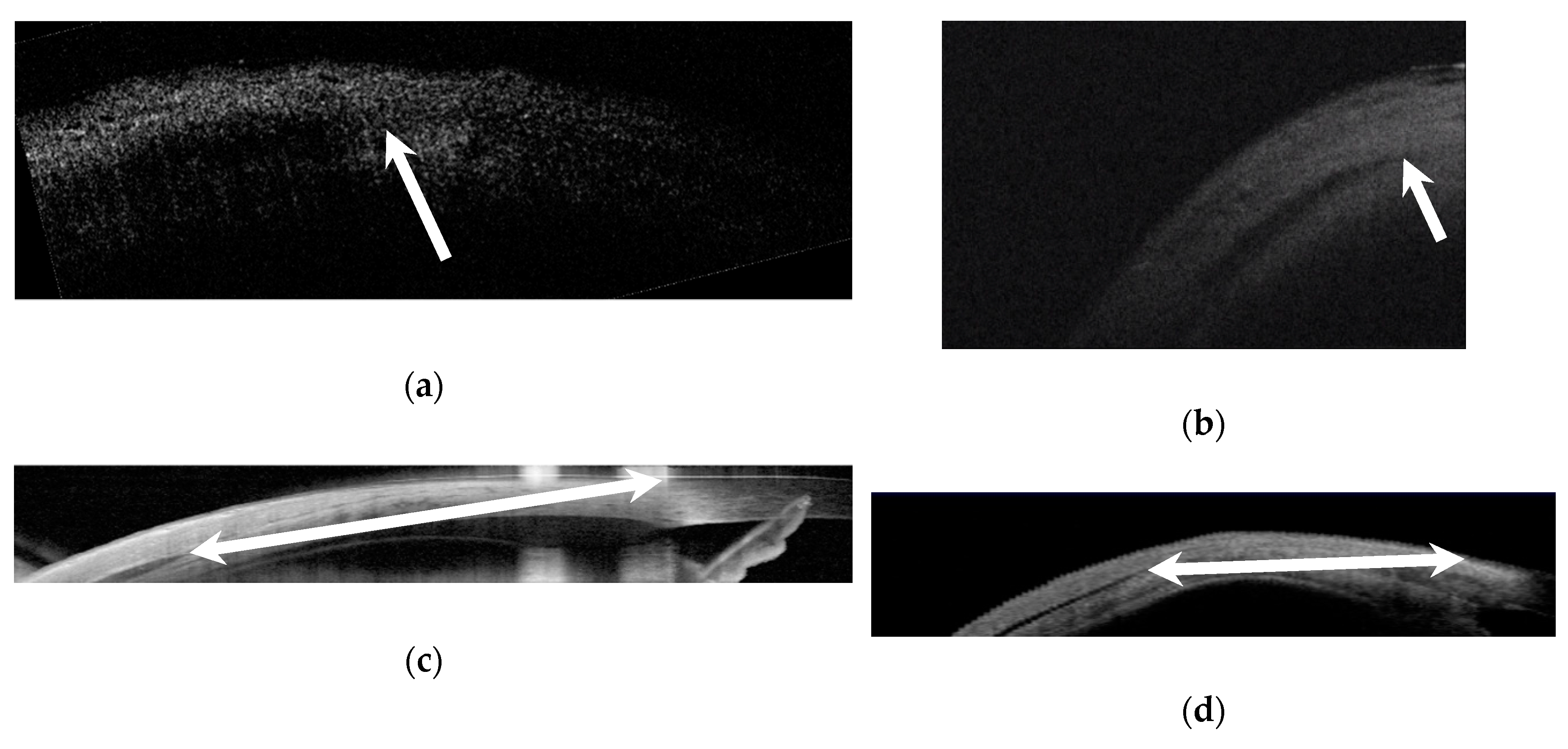
Figure 1. Anterior segment optical coherence tomography of a lateral rectus muscle insertion: (a) Bioptigen, (b) Rescan, (c) Spectralis, and (d) Visante. The rectus muscle insertion was located as a dark structure tapering toward the limbus between the sclera and conjunctiva/Tenon’s. Arrows mark the insertion in (a,b); the limbus was identified intra-operatively, and then measured to the insertion on the optical coherence tomography (OCT) with handheld calipers. Double arrows mark the insertion and limbus in (c,d), and calipers on the machine measured the distance.
Jcm 08 01732 g001
Figure 2. Anterior segment optical coherence tomography (integrated intraoperative microscope, Zeiss Rescan 700 (Carl Zeiss, Germany)) of a previously resected medial rectus muscle. (A): intraoperative view. (B): arrow marks muscle insertion. (C): cross section of muscle shown.
Jcm 08 01732 g002
Figure 3. Bioptigen Bland–Altman plot of intra-operative measurements versus OCT measurement.
Jcm 08 01732 g003
Figure 4. Rescan Bland–Altman plot of intra-operative measurements versus OCT measurement.
Jcm 08 01732 g004
Figure 5. Spectralis Bland–Altman plot of intra-operative measurements versus OCT measurement.
Jcm 08 01732 g005
Figure 6. Visante Bland–Altman plot of intra-operative measurements versus OCT measurement.
Jcm 08 01732 g006
Table 1. Anterior segment optical coherence tomography measurement results.
BioptigenRescanSpectralisVisante
Able to image muscle insertion24/34 (71%)25/28 (89%)22/38 (58%)31/40 (78%)
Mean difference between OCT measurement and caliper (SD)0.06 mm (1.81) range −4.0 to +3.50.43 mm (1.27) range −2.5 to +3.50.47 mm (1.54) range −3.7 to +4.3−0.56 mm (1.9) range −4.0 to +5.0
Intraclass correlation coefficient (ICC)0.620.830.850.88
p-value<0.01<0.001<0.001<0.001
≤1 mm13/24 (54%)21/25 (84%)15/22 (68%)13/31 (42%)
≤2 mm18/24 (75%)21/25 (84%)20/22 (91%)22/31 (71%)
Pre-op or before surgery on unoperated muscles: ≤1 mm10/29 (34%)19/24 (79%)8/9 (89%)5/11 (45%)
Post-op: ≤1 mmaa7/13 (54%)5/16 (31%)
Muscles imaged with all four machines (% ≤ 1 mm)37.5% (3/8)75% (6/8)62.5% (5/8)37.5% (3/8)
a Only had intra-operative measurements.
Table 2. Patients able to be imaged with all four optical coherence tomography machines.
PatientsIntra-Op MeasurmentBioptigenRescanSpectralisVisante
Patient 1: LR a12 mmUnable9.0 mmUnable9.5 mm
Patient 2: MR4.5 mmUnable3.5 mm5.2 mm6.4 mm
Patient 2: LR7.0 mm10.0 mm8.0 mm7.3 mm10.7 mm
Patient 3: MR a9.5 mm10.5 mm6.0 mmUnable9.1 mm
Patient 3: LR6.0 mm6.0 mm5.0 mm6.0 mm6.1 mm
Patient 4: MR5.0 mm4.5 mm4.5 mm4.1 mm6.6 mm
Patient 4: LR6.0 mmunable6.0 mm6.4 mm7.9 mm
Patient 4: IR5.0 mmunable4.0 mmUnable5.8 mm
a previously operated muscle, LR: lateral rectus, MR: medial rectus, IR: inferior rectus.