Article

Clinical and Rehabilitative Predictors of Peak Oxygen Uptake Following Cardiac Transplantation

by
Katelyn E. Uithoven
1,*,
Joshua R. Smith
2,
Jose R. Medina-Inojosa
2,
Ray W. Squires
2,
Erik H. Van Iterson
3 and
Thomas P. Olson
2
1
School of Kinesiology, University of Minnesota, Minneapolis, MN 55455, USA
2
Division of Preventive Cardiology, Department of Cardiovascular Medicine, Mayo Clinic, Rochester, MN 55905, USA
3
Section of Preventive Cardiology and Rehabilitation, Cleveland Clinic, Cleveland, OH 44195, USA
*
Author to whom correspondence should be addressed.
J. Clin. Med. 2019, 8(1), 119; https://doi.org/10.3390/jcm8010119
Submission received: 21 December 2018 / Revised: 14 January 2019 / Accepted: 16 January 2019 / Published: 19 January 2019
(This article belongs to the Special Issue Cardiac Rehabilitation)

Abstract

:
The measurement of peak oxygen uptake (VO2peak) is an important metric for evaluating cardiac transplantation (HTx) eligibility. However, it is unclear which factors (e.g., recipient demographics, clinical parameters, cardiac rehabilitation (CR) participation) influence VO2peak following HTx. Consecutive HTx patients with cardiopulmonary exercise testing (CPET) between 2007–2016 were included. VO2peak was measured from CPET standard protocol. Regression analyses determined predictors of the highest post-HTx VO2peak (i.e., quartile 4: VO2peak > 20.1 mL/kg/min). One hundred-forty HTx patients (women: n = 41 (29%), age: 52 ± 12 years, body mass index (BMI): 27 ± 5 kg/m2) were included. History of diabetes (Odds Ratio (OR): 0.17, 95% Confidence Interval (CI): 0.04–0.77, p = 0.021), history of dyslipidemia (OR: 0.42, 95% CI: 0.19–0.93, p = 0.032), BMI (OR: 0.90, 95% CI: 0.82–0.99, p = 0.022), hemoglobin (OR: 1.29, 95% CI: 1.04–1.61, p = 0.020), white blood cell count (OR: 0.81, 95% CI: 0.66–0.98, p = 0.033), CR exercise sessions (OR: 1.10, 95% CI: 1.04–1.15, p < 0.001), and pre-HTx VO2peak (OR: 1.17, 95% CI: 1.07–1.29, p = 0.001) were significant predictors. Multivariate analysis showed CR exercise sessions (OR: 1.10, 95% CI: 1.03–1.16, p = 0.002), and pre-HTx VO2peak (OR: 1.16, 95% CI: 1.04–1.30, p = 0.007) were independently predictive of higher post-HTx VO2peak. Pre-HTx VO2peak and CR exercise sessions are predictive of a greater VO2peak following HTx. These data highlight the importance of CR exercise session attendance and pre-HTx fitness in predicting VO2peak post-HTx.

1. Introduction

Peak oxygen uptake (VO2peak) is an important metric for cardiac transplantation (HTx) eligibility. The measurement of exercise capacity using VO2peak obtained from cardiopulmonary exercise testing (CPET) is one criteria used for determination of transplant eligibility; with a value of ≤14 mL/kg/min considered transplant eligible [1,2,3,4,5]. The association between higher VO2peak and reduced hospitalizations and mortality risk has been shown in both the heart failure (HF) and HTx population, with even modest improvements in VO2peak associated with improved outcomes [6,7,8,9]. Because of this, investigating demographic and clinical factors that are predictive of post-HTx VO2peak has significant implications not only for functional capacity but also long-term survival.
Previous studies have investigated demographic and clinical determinants of VO2peak in various populations [10,11,12,13,14,15,16,17,18]. For example, the predictive influence of age, sex, and body mass index (BMI) on VO2peak has been shown in healthy populations [16,17]. In HTx patients, these variables have also been shown to be prominent predictors of post-HTx VO2peak along with chronotropic reserve, donor age, and time from HTx [11,13,14,18]. However, a limitation of these previous studies is the small sample sizes used (n = 60–95) [11,13,14,18]. In addition, recent work has indicated that participation in cardiac rehabilitation (CR) in HTx postoperative care has been shown to be related to improvements in VO2peak [15,19,20]; however, it is unclear if CR participation is predictive of a greater VO2peak following HTx.
Therefore, the purpose of this study was to investigate whether pre-HTx clinical characteristics and/or postoperative CR exercise session attendance provide utility in predicting VO2peak following HTx. Based on previous studies on the relationship between CR involvement and VO2peak [15,19,20], we hypothesize that CR will surpass other predictive factors of post-HTx VO2peak in HTx patients.

2. Experimental Section

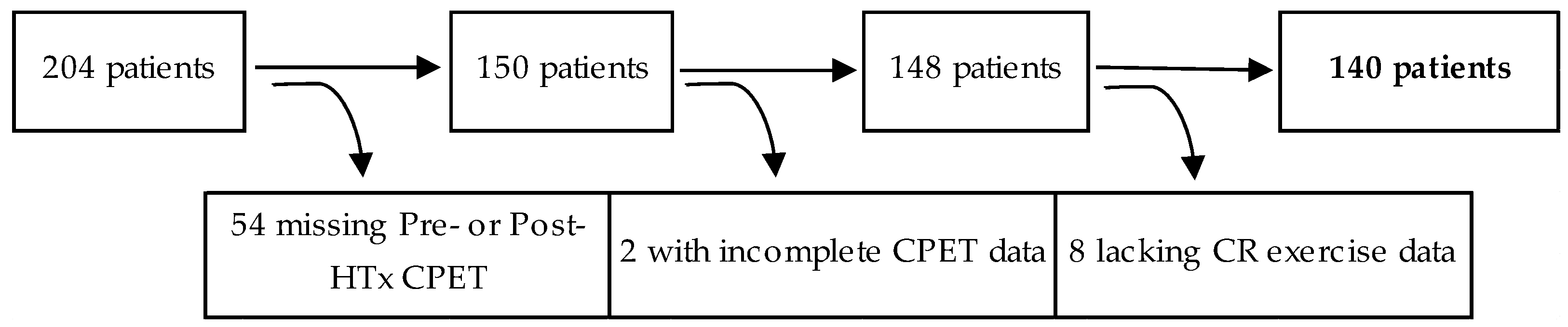
2.1. Participants and Study Design

A retrospective, single-center study cohort design evaluated consecutive adult HTx patients who performed symptom-limited CPET prior to HTx (pre-HTx) and following HTx (post-HTx) between the years of 2007–2016. Demographic and clinical characteristics were obtained from an institutional database. Inclusion criteria included completion of pre-HTx CPET within 24 months prior to procedural date and post-HTX CPET within 1-year of HTx. Patients were excluded if they lacked CR exercise session data or had incomplete CPET data. Of the 204 HTx patients, 140 were analyzed in this study (Figure 1). This study was approved by the Mayo Clinic Institutional Review Board (IRB #15-007965) and followed research authorization protocol for the use of medical records as required by the state of Minnesota [21].

2.2. Clinical Characteristics

Clinical baseline information from the time of HTx procedural date was obtained via medical record extraction. Demographic data along with previous disease history, previous left ventricular assist device (LVAD), current laboratory measurements (i.e., hemoglobin, hematocrit, white blood cell count, and creatinine), indication for HTx (i.e., restrictive cardiomyopathy, dilated cardiomyopathy, hypertrophic cardiomyopathy, ischemic cardiomyopathy, or other), and pre-HTx medication status for the following: angiotensin-converting enzyme (ACE) inhibitor, amiodarone, aspirin, beta blocker, calcium channel blocker, and diuretic were extracted from the procedural sedation assessment at the time of HTx. For the purpose of monitoring data correctness, two investigators independently reviewed a random sampling of medical record charts.

2.3. Cardiac Rehabilitation Participation

Patients included in this study were referred for CR participation and attended at least one documented session following HTx. Medical records were examined to determine CR attendance specifically relating to postoperative HTx care versus CR for any cardiac-related event. As the initial visit for CR typically involves orientation procedures with little to no exercise involvement, this was not assessed in this study. Only those CR sessions with documented exercise participation were included for analysis. All exercise sessions were supervised throughout activity by clinical exercise physiologists with cardiologist oversight. During the course of CR participation patients performed 20–45 min of aerobic activity in a monitored setting, with the usual addition of strength training components for 10–15 min. In addition to CR attendance patients were encouraged to partake in light to moderate physical activity for at least 30 min on all days of the week. Guidance regarding nutrition, medication management, stress management, and depressive symptom management were all components of the comprehensive approach of the CR program. Social support was also provided in this environment as patients all engaged in group education/learning activities throughout CR.

2.4. Cardiopulmonary Exercise Testing Procedures

Clinical exercise physiologists conducted the clinically-indicated CPET with cardiologist oversight. An institutionally designed protocol was performed on a motorized treadmill (GE Case, Milwaukee, WI, USA) with workload increasing every 2 min until volitional fatigue [22,23]. Heart rhythm and heart rate (HR) were continuously monitored via 12-lead electrocardiogram during exercise and recovery. Prior to the start of exercise and at the last 30 s of each stage systolic blood pressure (SBP) and diastolic blood pressure (DBP) were measured via manual sphygmomanometry. Flow and gas exchange variables were measured during exercise using indirect calorimetry (MGC Diagnostics, St. Paul, MN, USA). Peak values were obtained by averaging the last 30 s of the CPET data collection. Measured gas exchange variables included VO2peak, carbon dioxide production (VCO2), minute ventilation (VE), and respiratory exchange ratio (RER). The value of VE/VCO2 slope was determined from rest-peak values for VE and VCO2 and %predicted VO2peak [24] was calculated. Peak values were chosen for VE/VCO2 slope as they have been shown to have superior prognostic value as opposed to the pre-ventilatory threshold slope equation [25]. Patients were monitored closely for development of significant dysrhythmia, electrocardiographic abnormalities, and/or abnormal blood pressure responses (e.g., symptomatic hypotension). Cardiac medications were not withheld prior to CPET to maximize patient safety and generalizability of the data.

2.5. Statistical Analysis

Data are reported as mean ± standard deviation (SD) or frequency (percentage) where applicable. Statistical analysis was conducted using SPSS (version 22.0, Chicago, IL, USA) and JMP (JMP, Cary, NC, USA). The Student’s t test was used to compare pre-HTx CPET variables to post-HTx CPET variables. As previously described [26,27], univariate binary regression analysis was used to assess individual predictors for the highest post-HTx VO2peak (i.e., quartile 4) based on VO2peak data presented herein. Quartile 4 = VO2peak > 20.1 mL/kg/min, quartile 3 = VO2peak > 17.4 mL/kg/min, quartile 2 = VO2peak > 14.3 mL/kg/min, and quartile 1 = VO2peak ≤ 14.3 mL/kg/min (total range = 24.5 mL/kg/min). A multivariate regression analysis was then performed including all significant univariate variables. Three additional adjustment analyses were conducted to account for potential factors involved in exercise capacity. Model 1 was adjusted for demographic influencers of exercise capacity (i.e., age and sex), Model 2 was adjusted for demographic and clinical influencers of exercise capacity (i.e., age, sex, BMI, history of diabetes, and hemoglobin), and Model 3 included all significant univariate predictors for multivariate analysis adjusted for age and sex. The receiver operating characteristic (ROC) curve model was assessed to establish the area under the curve (AUC) for predicting quartile 4 (i.e., the highest VO2peak) and the cut-off for CR for maximal post-HTx VO2peak benefits. Least squares linear regression analysis was performed between pre-HTx VO2peak and post-HTx VO2peak, with reporting of the Pearson product-moment correlation coefficient (r) as an indicator of the strength of association between before and after measurements. For all analyses, statistical significance was set at an alpha level of p < 0.05.

3. Results

3.1. Patient Population and Clinical Characteristics

Demographic and clinical characteristics of the patients included are presented in Table 1. Of the 140 HTx patients analyzed in this study the mean age was 52 ± 12 years, mean BMI was 27 ± 5 kg/m2, and n = 41 (29%) were women. All data shown in Table 1 was obtained from each patient within 24 months prior to procedural data. The mean number of CR sessions attended was 18 ± 9, with a range of 2–36 sessions.

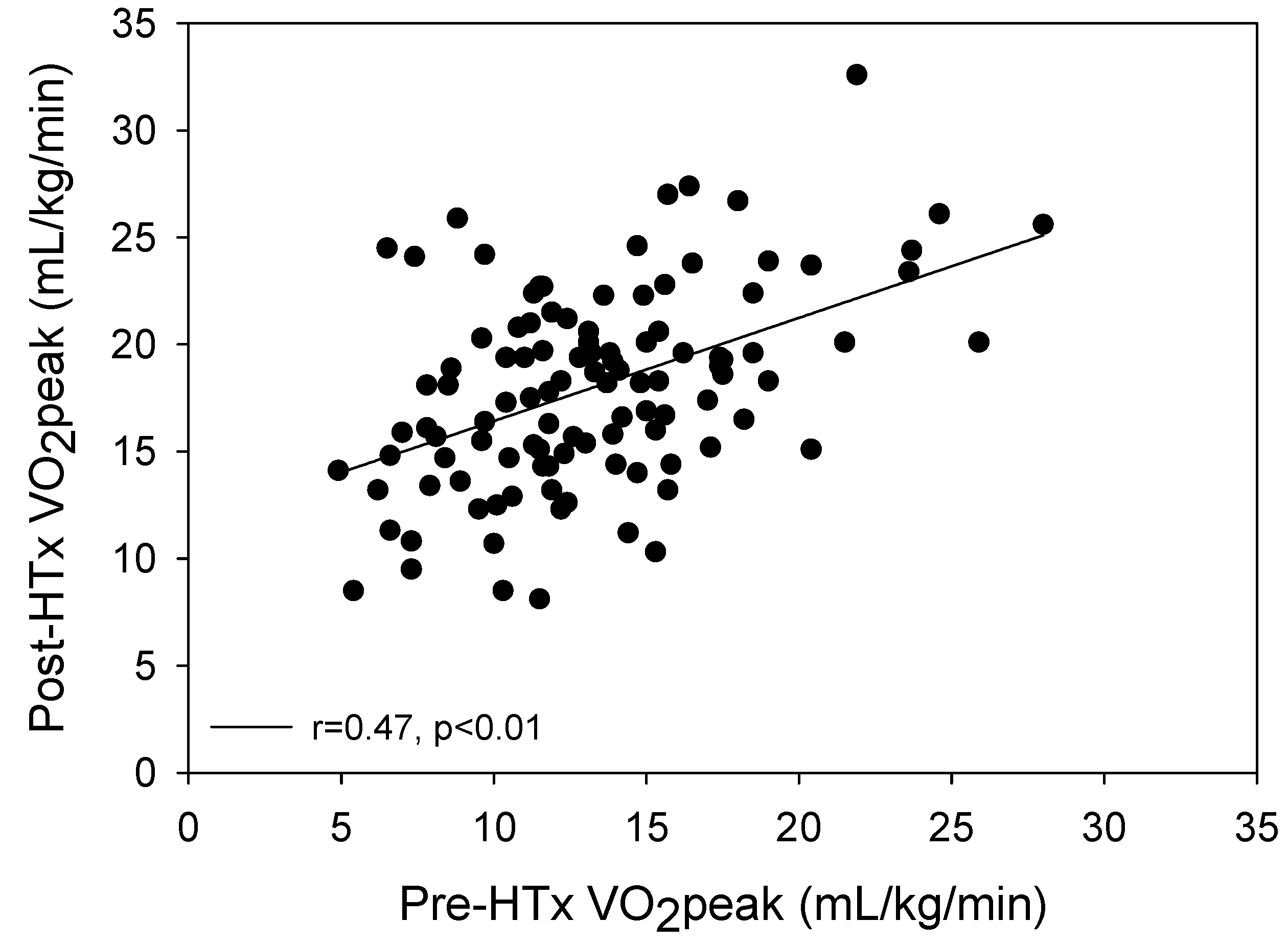
3.2. Exercise Testing Data Prior to and Following Transplant

Peak exercise testing data for CPET pre-HTx versus post-HTx are shown in Table 2. All variables included show significant improvements from pre-HTx to post-HTx, with VE/VCO2 slope decreasing while all other variables increased. Notably, relative VO2peak increased substantially from pre- to post-HTx (12.9 ± 4.4 vs. 17.5 ± 4.6 mL/kg/min, p < 0.001). A significant positive correlation was present between pre-HTx VO2peak and post-HTx VO2peak (Figure 2) (r = 0.47, p < 0.01). Additionally, this correlation between pre and post-HTx VO2peak was further analyzed with adjustment for the number of CR exercise sessions attended and remained significant (p < 0.001).

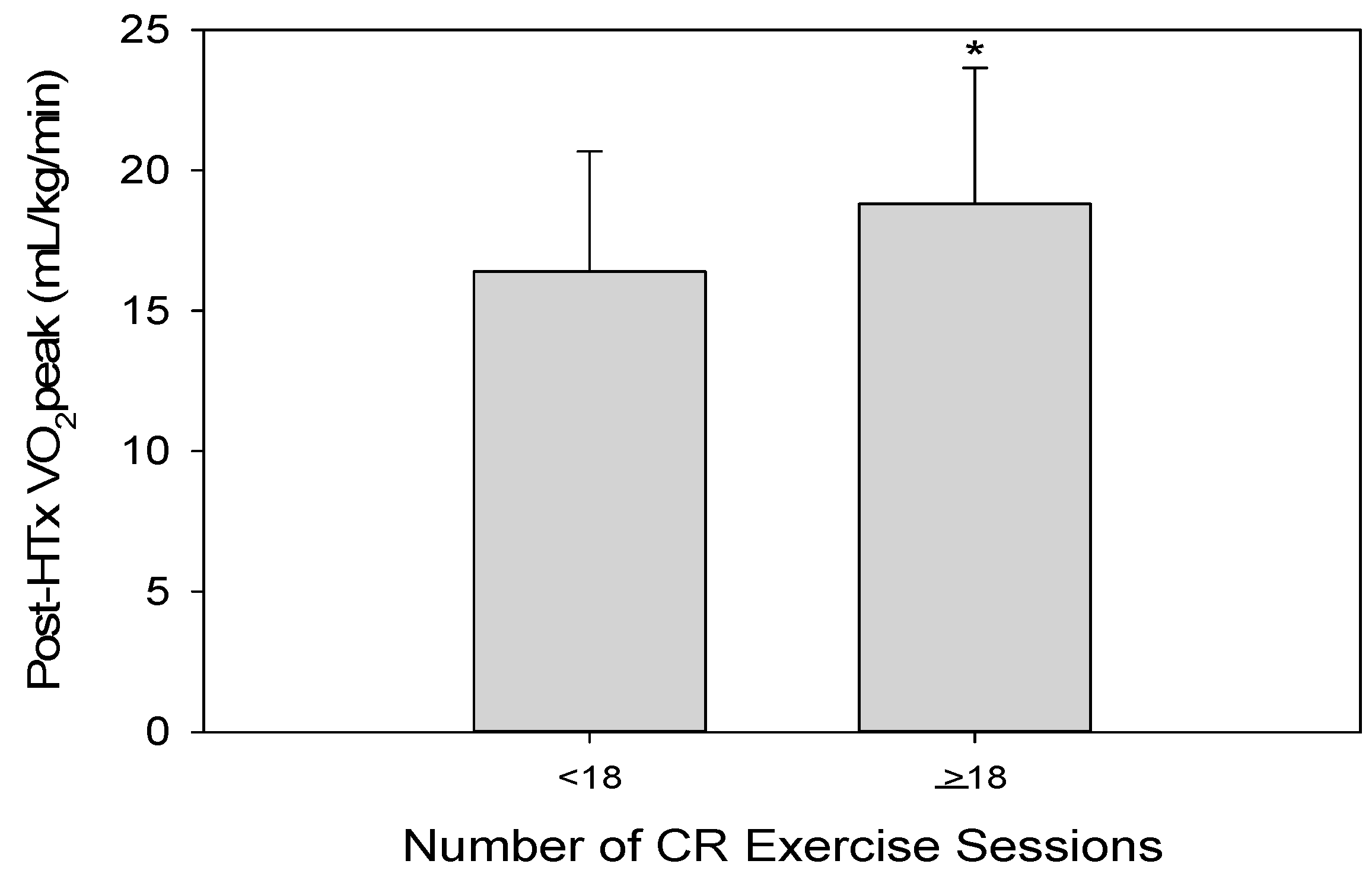
3.3. Predictors of VO2peak

Univariate regression analysis indicated that BMI, history of diabetes, history of dyslipidemia, hemoglobin, white blood cell count, CR exercise sessions, and pre-HTx VO2peak were significant predictors of higher VO2peak post-HTx (Table 3). The significance of pre-HTx VO2peak and CR exercise sessions on post-HTx VO2peak remained after adjustment for demographic influencers of exercise capacity (age and sex) as shown in Model 1, and adjustment for demographic and clinical influencers of exercise capacity (age, sex, BMI, history of diabetes, hemoglobin, peak HR, and HR recovery (HRR)) as shown in Model 2 (Table 4).
All significant univariate predictors were included into the multivariate regression analysis with adjustment for age and sex. This analysis (as shown in Model 3) indicated that CR exercise sessions and pre-HTx VO2peak were the independent predictors of higher post-HTx VO2peak (Table 4). When CR exercise session attendance was divided into two groups by the median attendance of 18 sessions, those who attended ≥18 sessions had significantly higher post-HTx VO2peak compared to those with <18 sessions attended (≥18 sessions: 18.8 ± 4.8 vs. <18 sessions: 16.4 ± 4.3, p < 0.01) (Figure 3). The value of 18 sessions was also determined to be an appropriate cutoff for CR attendance necessary to maximize benefits in post-HTx VO2peak (AUC: 0.692, specificity: 0.549, sensitivity: 0.684, p < 0.001).

4. Discussion

The purpose of this study was to determine whether pre-HTx clinical characteristics and/or postoperative participation in CR provide utility in predicting VO2peak following HTx. Pre-HTx VO2peak and the number of CR exercise sessions attended were the only variables predictive of greater VO2peak following HTx. These metrics independently predicted higher post-HTx VO2peak even after adjusting for other factors of exercise capacity, surpassing other significant univariate factors. Our data provide new evidence of the critical benefits of CR in improving functional capacity following HTx.
Previous work has evaluated potential predictors of VO2peak in various populations [10,11,12,13,14,17,18,28,29]. In healthy subjects the general characteristics of younger age, male sex, and training state have been shown to be associated with improved exercise capacity [17]. For those with cardiovascular disease (CVD), several studies measuring exercise capacity directly by VO2peak or indirectly using predicted METS have shown overall that age and baseline fitness are the strongest predictors of VO2peak [10,28,29]. The predictive quality of age also remains in the HTx population [11,13,14]. One of the first studies to evaluate predictors of VO2peak specifically for HTx patients found that chronotropic reserve, donor age, recipient age, and time from HTx were the most significant predictors of VO2peak post-HTx [11]. This study, however, collected CPET data in the broad range of 1–100 months following HTx, which limits the specificity of temporal cardiopulmonary adaptation post-HTx. Although not significant in multivariate analysis, clinical history of diabetes and dyslipidemia were significant univariate predictors in this study, emphasizing the importance of multimorbidity management [30,31,32,33]. More recent studies in HTx patients have shown that the demographic components of sex, BMI, and age are prominent predictors of VO2peak [13,14,18]. Table 5 provides information on four specific studies particularly relevant to the findings of the current study examining predictors of VO2peak in HTx patients.
Although the findings of these studies concur with our study regarding the predictive quality of BMI, CR exercise session attendance and baseline fitness as assessed by pre-HTx VO2peak were the only independent predictors of post-HTx VO2peak. Baseline fitness levels have previously been shown to predict VO2peak in CVD patients [28,29], yet this study is the first to show this relationship in HTx patients. A significant correlation was present between pre-HTx VO2peak and post-HTx VO2peak in our study, as those with the highest pre-HTx VO2peak were more likely to have higher VO2peak measurements following HTx. These results highlight the importance of maintaining or reaching a higher functional capacity leading up to HTx, as baseline values predict postoperative VO2peak levels.
The predictive impact of CR involvement following HTx on achieving higher VO2peak values was a unique component to this study. Our findings clearly demonstrate the importance of postoperative CR participation in improving functional capacity and implications of survival following HTx. Specifically following HTx, the benefits of CR involvement are widespread for this clinically unique population; including counteracting marked deconditioning and skeletal muscle weakness associated with end-stage HF, corticosteroid treatment, and surgical recovery [18,34,35,36]. Although the predictive quality of CR session attendance has not been previously reported, the increase in VO2peak following CR in HTx patients is well-documented [15,19,20,37,38]. The importance of CR involvement on VO2peak following HTx is critical, as it elicits significantly greater increases in VO2peak compared to at-home therapy [37]. One such study reported an increase of 3.6 mL/kg/min in VO2peak following a 12-week CR program after HTx [19]. Additionally, a recent review evaluating CR exercise in HTx patients that encompassed 10 randomized controlled trials with a total of 300 patients found that exercise capacity was, on average, 2.49 mL/kg/min higher for those who exercised [15]. Our study showed a mean increase of 4.6 mL/kg/min (equivalent to over 1.5 METS) from pre- to post-HTx, which has substantial meaning with regard to improved quality of life and survival. Further, those who attended >18 sessions demonstrated a post-HTx VO2peak of 2.4 mL/kg/min higher than those who attended <18 sessions (i.e., 15% greater). Studies in HF have found that an increased VO2peak is associated with better outcomes (i.e., primary endpoints of all-cause hospitalizations and/or mortality) [6,7,39,40]. One such study found a 5% lower risk of all-cause mortality or hospitalization for every 6% increase in VO2peak, thereby highlighting the importance of even seemingly small improvements in VO2peak on long-term clinical outcomes [7]. It should be noted, however, that despite significant functional improvements observed following HTx, VO2peak values still remain abnormal in HTx patients compared to age-matched control subjects [34,41,42]. Further understanding of the demographic, clinical, and rehabilitative components that predict post-HTx VO2peak values offers greater insight into the complex cardiopulmonary and peripheral adaptations occurring following HTx.
It is important to recognize the retrospective observational nature of the present study design. Additional research in this area is encouraged to confirm these results in a prospective fashion. The distribution of post-HTx medications could potentially influence CPET measurements, therefore this should be taken into account as a limitation to the application of these results. Further, data pertaining to donor information (e.g., donor age, donor sex) and surgical procedural notes (e.g., cold ischemic time) were unavailable for this study and may be important additional metrics to include in follow-up studies.

5. Conclusions

In summary, HTx patients demonstrated substantial improvements in VO2peak following HTx. The only factors that provided significant predictive value of higher post-HTx VO2peak values were CR exercise session attendance and pre-HTx VO2peak, even after adjustment for other univariate predictors of post-HTx exercise capacity. These data demonstrate the influential role of sustaining and/or improving cardiorespiratory fitness leading up to and following HTx, specifically in the CR setting.

Author Contributions

Conceptualization, K.E.U., J.R.W.S., J.R.M.-I., E.H.V.I., T.P.O.; Methodology, K.E.U., J.R.W.S., J.R.M.-I., E.H.V.I., R.W.S., T.P.O.; Software, K.E.U., J.R.W.S., J.R.M.-I.; Validation, K.E.U., J.R.W.S., J.R.M.-I.; Formal Analysis, K.E.U., J.R.W.S., J.R.M.-I., T.P.O.; Investigation, K.E.U., J.R.W.S., J.R.M.-I., E.H.V.I., R.W.S., T.P.O.; Resources, K.E.U., J.R.W.S., J.R.M.-I.., E.H.V.I., R.W.S., T.P.O.; Data Curation, K.E.U., J.R.W.S., J.R.M.-I., T.P.O.; Writing—Original Draft Preparation, K.E.U., J.R.W.S.; Writing—Review and Editing, J.R.W.S., J.R.M.-I., R.W., T.P.O.; Visualization, K.E.U., J.R.W.S., J.R.M.-I., R.W., T.P.O.; Supervision, J.R.W.S., T.P.O.; Project Administration, J.R.W.S., T.P.O.; Funding Acquisition, J.R.W.S., T.P.O.

Funding

This research was funded by the National Institutes of Health [HL-126638 to TPO] and American Heart Association [18POST3990251 to JRS].

Conflicts of Interest

The authors declare no conflict of interest.

References

  1. Mancini, D.M.; Eisen, H.; Kussmaul, W.; Mull, R.; Edmunds, L.H., Jr.; Wilson, J.R. Value of peak exercise oxygen consumption for optimal timing of cardiac transplantation in ambulatory patients with heart failure. Circulation 1991, 83, 778–786. [Google Scholar] [CrossRef] [PubMed]
  2. Elmariah, S.; Goldberg, L.R.; Allen, M.T.; Kao, A. Effects of gender on peak oxygen consumption and the timing of cardiac transplantation. J. Am. Coll. Cardiol. 2006, 47, 2237–2242. [Google Scholar] [CrossRef] [PubMed]
  3. Ponikowski, P.; Voors, A.A.; Anker, S.D.; Bueno, H.; Cleland, J.G.; Coats, A.J.; Falk, V.; Gonzalez-Juanatey, J.R.; Harjola, V.P.; Jankowska, E.A.; et al. 2016 ESC Guidelines for the diagnosis and treatment of acute and chronic heart failure. Kardiol. Pol. 2016, 74, 1037–1147. [Google Scholar] [CrossRef] [PubMed]
  4. Mehra, M.R.; Canter, C.E.; Hannan, M.M.; Semigran, M.J.; Uber, P.A.; Baran, D.A.; Danziger-Isakov, L.; Kirklin, J.K.; Kirk, R.; Kushwaha, S.S.; et al. The 2016 International Society for Heart Lung Transplantation listing criteria for heart transplantation: A 10-year update. J. Heart Lung Transplant. 2016, 35, 1–23. [Google Scholar] [CrossRef] [PubMed]
  5. Peterson, L.R.; Schechtman, K.B.; Ewald, G.A.; Geltman, E.M.; Meyer, T.; Krekeler, P.; Rogers, J.G. The effect of beta-adrenergic blockers on the prognostic value of peak exercise oxygen uptake in patients with heart failure. J. Heart Lung Transplant. 2003, 22, 70–77. [Google Scholar] [CrossRef]
  6. Keteyian, S.J.; Patel, M.; Kraus, W.E.; Brawner, C.A.; McConnell, T.R.; Pina, I.L.; Leifer, E.S.; Fleg, J.L.; Blackburn, G.; Fonarow, G.C.; et al. Variables measured during cardiopulmonary exercise testing as predictors of mortality in chronic systolic heart failure. J. Am. Coll. Cardiol. 2016, 67, 780–789. [Google Scholar] [CrossRef] [PubMed]
  7. Swank, A.M.; Horton, J.; Fleg, J.L.; Fonarow, G.C.; Keteyian, S.; Goldberg, L.; Wolfel, G.; Handberg, E.M.; Bensimhon, D.; Illiou, M.C.; et al. Modest increase in peak VO2 is related to better clinical outcomes in chronic heart failure patients: Results from heart failure and a controlled trial to investigate outcomes of exercise training. Circ. Heart Fail. 2012, 5, 579–585. [Google Scholar] [CrossRef] [PubMed]
  8. Kavanagh, T.; Mertens, D.J.; Shephard, R.J.; Beyene, J.; Kennedy, J.; Campbell, R.; Sawyer, P.; Yacoub, M. Long-term cardiorespiratory results of exercise training following cardiac transplantation. Am. J. Cardiol. 2003, 91, 190–194. [Google Scholar] [CrossRef]
  9. Yardley, M.; Havik, O.E.; Grov, I.; Relbo, A.; Gullestad, L.; Nytroen, K. Peak oxygen uptake and self-reported physical health are strong predictors of long-term survival after heart transplantation. Clin. Transplant. 2016, 30, 161–169. [Google Scholar] [CrossRef]
  10. Branco, C.F.; Viamonte, S.; Matos, C.; Magalhaes, S.; Cunha, I.; Barreira, A.; Fernandes, P.; Torres, S. Predictors of changes in functional capacity on a cardiac rehabilitation program. Rev. Port. Cardiol. 2016, 35, 215–224. [Google Scholar] [CrossRef]
  11. Douard, H.; Parrens, E.; Billes, M.A.; Labbe, L.; Baudet, E.; Broustet, J.P. Predictive factors of maximal aerobic capacity after cardiac transplantation. Eur. Heart J. 1997, 18, 1823–1828. [Google Scholar] [CrossRef] [PubMed] [Green Version]
  12. Hammond, H.K.; Kelly, T.L.; Froelicher, V.F.; Pewen, W. Use of clinical data in predicting improvement in exercise capacity after cardiac rehabilitation. J. Am. Coll. Cardiol. 1985, 6, 19–26. [Google Scholar] [CrossRef] [Green Version]
  13. Leung, T.C.; Ballman, K.V.; Allison, T.G.; Wagner, J.A.; Olson, L.J.; Frantz, R.P.; Edwards, B.S.; Dearani, J.A.; Daly, R.C.; McGregor, C.G.; et al. Clinical predictors of exercise capacity 1 year after cardiac transplantation. J. Heart Lung Transplant. 2003, 22, 16–27. [Google Scholar] [CrossRef]
  14. Oliveira Carvalho, V.; Guimaraes, G.V.; Vieira, M.L.; Catai, A.M.; Oliveira-Carvalho, V.; Ayub-Ferreira, S.M.; Bocchi, E.A. Determinants of peak VO2 in heart transplant recipients. Rev. Bras. Cir. Cardiovasc. 2015, 30, 9–15. [Google Scholar] [CrossRef]
  15. Anderson, L.; Nguyen, T.T.; Dall, C.H.; Burgess, L.; Bridges, C.; Taylor, R.S. Exercise-based cardiac rehabilitation in heart transplant recipients. Cochrane Database Syst. Rev. 2017, 4, CD012264. [Google Scholar] [CrossRef] [PubMed]
  16. Ogawa, T.; Spina, R.J.; Martin, W.H., 3rd; Kohrt, W.M.; Schechtman, K.B.; Holloszy, J.O.; Ehsani, A.A. Effects of aging, sex, and physical training on cardiovascular responses to exercise. Circulation 1992, 86, 494–503. [Google Scholar] [CrossRef] [PubMed]
  17. Woo, J.S.; Derleth, C.; Stratton, J.R.; Levy, W.C. The influence of age, gender, and training on exercise efficiency. J. Am. Coll. Cardiol. 2006, 47, 1049–1057. [Google Scholar] [CrossRef]
  18. Nytroen, K.; Rustad, L.A.; Gude, E.; Hallen, J.; Fiane, A.E.; Rolid, K.; Holm, I.; Aakhus, S.; Gullestad, L. Muscular exercise capacity and body fat predict VO(2peak) in heart transplant recipients. Eur. J. Prev. Cardiol. 2014, 21, 21–29. [Google Scholar] [CrossRef]
  19. Hsu, C.J.; Chen, S.Y.; Su, S.; Yang, M.C.; Lan, C.; Chou, N.K.; Hsu, R.B.; Lai, J.S.; Wang, S.S. The effect of early cardiac rehabilitation on health-related quality of life among heart transplant recipients and patients with coronary artery bypass graft surgery. Transplant. Proc. 2011, 43, 2714–2717. [Google Scholar] [CrossRef]
  20. Rosenbaum, A.N.; Kremers, W.K.; Schirger, J.A.; Thomas, R.J.; Squires, R.W.; Allison, T.G.; Daly, R.C.; Kushwaha, S.S.; Edwards, B.S. Association between early cardiac rehabilitation and long-term survival in cardiac transplant recipients. Mayo Clin. Proc. 2016, 91, 149–156. [Google Scholar] [CrossRef]
  21. Yawn, B.P.; Yawn, R.A.; Geier, G.R.; Xia, Z.; Jacobsen, S.J. The impact of requiring patient authorization for use of data in medical records research. J. Fam. Pract. 1998, 47, 361–365. [Google Scholar] [PubMed]
  22. Squires, R.W.; Allison, T.G.; Johnson, B.D.; Gau, G.T. Non-physician supervision of cardiopulmonary exercise testing in chronic heart failure: Safety and results of a preliminary investigation. J. Cardiopulm. Rehabil. 1999, 19, 249–253. [Google Scholar] [CrossRef] [PubMed]
  23. Larsen, C.M.; Ball, C.A.; Hebl, V.B.; Ong, K.C.; Siontis, K.C.; Olson, T.P.; Ackerman, M.J.; Ommen, S.R.; Allison, T.G.; Geske, J.B. Effect of body mass index on exercise capacity in patients with hypertrophic cardiomyopathy. Am. J. Cardiol. 2017. [Google Scholar] [CrossRef] [PubMed]
  24. Myers, J.; Kaminsky, L.A.; Lima, R.; Christle, J.W.; Ashley, E.; Arena, R. A reference equation for normal standards for VO2 Max: Analysis from the Fitness Registry and the Importance of Exercise National Database (FRIEND Registry). Prog. Cardiovasc. Dis. 2017, 60, 21–29. [Google Scholar] [CrossRef] [PubMed]
  25. Arena, R.; Humphrey, R.; Peberdy, M.A. Prognostic ability of VE/VCO2 slope calculations using different exercise test time intervals in subjects with heart failure. Eur. J. Cardiovasc. Prev. Rehabil. 2003, 10, 463–468. [Google Scholar] [CrossRef] [PubMed]
  26. Francis, D.P.; Shamim, W.; Davies, L.C.; Piepoli, M.F.; Ponikowski, P.; Anker, S.D.; Coats, A.J. Cardiopulmonary exercise testing for prognosis in chronic heart failure: Continuous and independent prognostic value from VE/VCO2 slope and peak VO2. Eur. Heart J. 2000, 21, 154–161. [Google Scholar] [CrossRef] [PubMed]
  27. Smith, J.R.; Medina-Inojosa, J.R.; Layrisse, V.; Ommen, S.R.; Olson, T.P. Predictors of exercise capacity in patients with hypertrophic obstructive cardiomyopathy. J. Clin. Med. 2018, 7. [Google Scholar] [CrossRef]
  28. Pierson, L.M.; Miller, L.E.; Herbert, W.G. Predicting exercise training outcome from cardiac rehabilitation. J. Cardiopulm. Rehabil. 2004, 24, 113–118. [Google Scholar] [CrossRef]
  29. McKee, G.; Kerins, M.; Fitzgerald, G.; Spain, M.; Morrison, K. Factors that influence obesity, functional capacity, anxiety and depression outcomes following a Phase III cardiac rehabilitation programme. J. Clin. Nurs. 2013, 22, 2758–2767. [Google Scholar] [CrossRef]
  30. Fang, Z.Y.; Sharman, J.; Prins, J.B.; Marwick, T.H. Determinants of exercise capacity in patients with type 2 diabetes. Diabetes Care 2005, 28, 1643–1648. [Google Scholar] [CrossRef]
  31. Higgins, J.; Pflugfelder, P.W.; Kostuk, W.J. Increased morbidity in diabetic cardiac transplant recipients. Can. J. Cardiol. 2009, 25, e125–e129. [Google Scholar] [CrossRef] [Green Version]
  32. Horwich, T.B.; Hamilton, M.A.; Maclellan, W.R.; Fonarow, G.C. Low serum total cholesterol is associated with marked increase in mortality in advanced heart failure. J. Card Fail. 2002, 8, 216–224. [Google Scholar] [CrossRef] [PubMed]
  33. Rauchhaus, M.; Clark, A.L.; Doehner, W.; Davos, C.; Bolger, A.; Sharma, R.; Coats, A.J.; Anker, S.D. The relationship between cholesterol and survival in patients with chronic heart failure. J. Am. Coll. Cardiol. 2003, 42, 1933–1940. [Google Scholar] [CrossRef] [PubMed] [Green Version]
  34. Tucker, W.J.; Beaudry, R.I.; Samuel, T.J.; Nelson, M.D.; Halle, M.; Baggish, A.L.; Haykowsky, M.J. Performance limitations in heart transplant recipients. Exerc. Sport Sci. Rev. 2018, 46, 144–151. [Google Scholar] [CrossRef]
  35. Georgiadou, P.; Adamopoulos, S. Skeletal muscle abnormalities in chronic heart failure. Curr. Heart Fail. Rep. 2012, 9, 128–132. [Google Scholar] [CrossRef] [PubMed]
  36. Lampert, E.; Mettauer, B.; Hoppeler, H.; Charloux, A.; Charpentier, A.; Lonsdorfer, J. Structure of skeletal muscle in heart transplant recipients. J. Am. Coll. Cardiol. 1996, 28, 980–984. [Google Scholar] [CrossRef]
  37. Kobashigawa, J.A.; Leaf, D.A.; Lee, N.; Gleeson, M.P.; Liu, H.; Hamilton, M.A.; Moriguchi, J.D.; Kawata, N.; Einhorn, K.; Herlihy, E.; et al. A controlled trial of exercise rehabilitation after heart transplantation. N. Engl. J. Med. 1999, 340, 272–277. [Google Scholar] [CrossRef]
  38. Daida, H.; Squires, R.W.; Allison, T.G.; Johnson, B.D.; Gau, G.T. Sequential assessment of exercise tolerance in heart transplantation compared with coronary artery bypass surgery after phase II cardiac rehabilitation. Am. J. Cardiol. 1996, 77, 696–700. [Google Scholar] [CrossRef]
  39. Pina, I.L.; Bittner, V.; Clare, R.M.; Swank, A.; Kao, A.; Safford, R.; Nigam, A.; Barnard, D.; Walsh, M.N.; Ellis, S.J.; et al. Effects of exercise training on outcomes in women with heart failure: Analysis of HF-ACTION (Heart Failure-A Controlled Trial Investigating Outcomes of Exercise TraiNing) by sex. JACC Heart Fail. 2014, 2, 180–186. [Google Scholar] [CrossRef]
  40. Sarullo, F.M.; Fazio, G.; Brusca, I.; Fasullo, S.; Paterna, S.; Licata, P.; Novo, G.; Novo, S.; Di Pasquale, P. Cardiopulmonary exercise testing in patients with chronic heart failure: Prognostic comparison from peak VO2 and VE/VCO2 Slope. Open Cardiovasc. Med. J. 2010, 4, 127–134. [Google Scholar] [CrossRef]
  41. Givertz, M.M.; Hartley, L.H.; Colucci, W.S. Long-term sequential changes in exercise capacity and chronotropic responsiveness after cardiac transplantation. Circulation 1997, 96, 232–237. [Google Scholar] [CrossRef] [PubMed]
  42. Kao, A.C.; Van Trigt, P., 3rd; Shaeffer-McCall, G.S.; Shaw, J.P.; Kuzil, B.B.; Page, R.D.; Higginbotham, M.B. Central and peripheral limitations to upright exercise in untrained cardiac transplant recipients. Circulation 1994, 89, 2605–2615. [Google Scholar] [CrossRef] [PubMed]
Figure 1. Flowchart for patient inclusion and exclusion. Of the initially identified 204 HTx patients, 54 patients lacked a pre-HTx or post-HTx CPET, 2 patients had incomplete CPET data, and 8 patients were lacking CR exercise session data, resulting in 140 patients for study analysis. HTx, cardiac transplantation; CPET, cardiopulmonary exercise testing.
Jcm 08 00119 g001
Figure 2. Relationship between measurements of relative VO2peak from pre-HTx to post-HTx. A significant positive correlation was present between pre-HTx VO2peak and post-HTx VO2peak (r = 0.47, p < 0.01). Post-HTx: following cardiac transplantation; Pre-HTx: prior to cardiac transplantation; VO2peak: peak oxygen uptake.
Jcm 08 00119 g002
Figure 3. Post-HTx VO2peak based on median CR exercise session attendance. Those HTx patients who attended ≥18 CR exercise sessions had significantly higher post-HTx VO2peak compared to those with <18 CR exercise sessions attended (≥18 sessions: 18.8 ± 4.8 vs. <18 sessions: 16.4 ± 4.3, p < 0.01) *, significantly higher than <18 CR sessions. CR: cardiac rehabilitation; Post-HTx: following cardiac transplantation; VO2peak: peak oxygen uptake.
Jcm 08 00119 g003
Table 1. Recipient demographics and clinical characteristics.
n140
Age (years)52 ± 12
Sex (Female)41 (29)
Height (cm)172 ± 14
Weight (kg)83 ± 19
BSA (m2)2.0 ± 0.2
BMI (kg/m2)27.1 ± 4.6
History of Diabetes31 (22.1)
History of Smoking52 (37.1)
History of Dyslipidemia74 (52.9)
History of Hypertension63 (45)
Previous LVAD27 (19.3)
Indication for Heart Transplant
Restrictive Cardiomyopathy29 (20.7)
Dilated Cardiomyopathy53 (37.9)
Hypertrophic Cardiomyopathy8 (5.6)
Ischemic Cardiomyopathy25 (17.9)
Other25 (17.9)
Labs
Hemoglobin (g/dL)12.0 ± 2.0
Hematocrit (%)36 ± 6
White Blood Cell Count (109/L)7.5 ± 3.0
Creatinine (mg/dL)1.4 ± 1.0
Medications *
ACE Inhibitor52 (37.1)
Amiodarone12 (8.6)
Aspirin65 (46.4)
Beta Blocker103 (73.6)
Calcium Channel Blocker3 (2.1)
Diuretic92 (65.7)
Number of CR Exercise Sessions18 ± 9
Note: BMI, body mass index; BSA, body surface area; CR, cardiac rehabilitation; LVAD, left ventricular assist device. All data are presented as mean ± standard deviation or frequency (percentage). *, medication distributions are prior to cardiac transplantation procedure; ACE, angiotensin converting enzyme.
Table 2. Peak exercise testing values prior to and following cardiac transplantation.
Pre-HTxPost-HTxp-Value
n140140
Exercise time (min)5.3 ± 1.76.7 ± 1.7<0.001
METS4.9 ± 1.86.4 ± 1.8<0.001
Absolute VO2peak (L/min)1.1 ± 0.41.4 ± 0.4<0.001
VO2peak % Predicted (%)42 ± 1458 ± 17<0.001
Relative VO2peak (mL/kg/min)12.9 ± 4.417.5 ± 4.7<0.001
VE/VCO2 slope42 ± 1237 ± 6<0.001
VCO2 (L/min)1.2 ± 0.51.7 ± 0.5<0.001
RER1.15 ± 0.131.21 ± 0.12<0.001
SBP (mmHg)105 ± 25145 ± 30<0.001
DBP (mmHg)60 ± 1067 ± 11<0.001
HR (bpm)110 ± 21125 ± 20<0.001
METS, metabolic equivalents; RER, respiratory exchange ratio; SBP, systolic blood pressure; DBP, diastolic blood pressure; HR, heart rate; VCO2, production of carbon dioxide; VE, minute ventilation; VE/VCO2 slope, ventilatory efficiency; VO2peak, peak oxygen uptake.
Table 3. Univariate regression analysis for predictors of VO2peak.
VariableUnivariate Analysis
OR95% CIp-Value
Age (years)0.9780.949–1.0090.170
Sex (female)0.4570.173–1.2090.115
BMI (kg/m2)0.8960.815–0.9850.022
History of Diabetes0.1740.039–0.7720.021
History of Smoking0.8050.354–1.8310.605
History of Hypertension0.7410.335–1.6410.460
History of Dyslipidemia0.4150.185–0.9290.032
History of LVAD1.1710.446–3.0760.748
Beta Blocker Medication0.6420.275–1.4990.306
Indication for HTx
Restrictive Cardiomyopathy0.8120.299–2.2010.682
Dilated Cardiomyopathy0.9200.409–2.0660.840
Hypertrophic Cardiomyopathy2.0400.461–9.0350.348
Ischemic Cardiomyopathy0.5650.179–1.7820.330
Other1.6940.655–4.3800.277
Labs
Hemoglobin (g/dL)1.2941.041–1.6080.020
Hematocrit (%)1.0630.985–1.1460.115
White Blood Cell Count (109/L)0.8060.662–0.9820.033
Creatinine (mg/dL)0.4020.155–1.0390.060
Pre-HTx CPET Data
Peak SBP (mmHg)0.9980.971–1.0050.158
Heart Rate Recovery (bpm)1.0210.987–1.0560.225
Relative VO2peak (mL/kg/min)1.1741.070–1.2890.001
CR Exercise Sessions1.0951.041–1.152<0.001
OR, odds ratio; CI, confidence interval; BMI, body mass index; CR, cardiac rehabilitation; HTx, cardiac transplantation; LVAD, left ventricular assist device; SBP, systolic blood pressure, VO2peak, peak oxygen uptake.
Table 4. Adjusted and multivariate regression analysis for predictors of Post-HTx VO2peak.
VariableModel 1Model 2Model 3
OR95% CIp-ValueOR95% CIp-ValueOR95% CIp-Value
Age (years)0.9680.932–1.0060.0940.9700.933–1.0090.1300.9620.922–1.0040.077
Sex (female)0.3950.128–1.2170.1060.2840.080–1.0100.0520.2920.087–0.9860.047
BMI (kg/m2)0.8890.791–0.9990.0490.0970.811–1.0180.097
History of Diabetes3.0500.602–15.4500.1782.7600.635–11.9950.176
History of Dyslipidemia0.8800.315–2.4650.807
Labs
Hemoglobin (g/dL)1.1720.930–1.4780.1781.0450.829–1.310.710
White Blood Cell Count (109/L)0.7960.620–1.0210.072
Pre-HTx CPET Data
Relative VO2peak (mL/kg/min)1.1451.037–1.2640.0071.1101.002–1.2310.0471.2061.068–1.3610.002
Peak HR (bpm)0.9790.951–1.0070.147
HRR (bpm)1.0080.959–1.0590.764
CR Exercise Sessions1.1021.037–1.264<0.0011.0951.037–1.1570.0011.1031.042–1.1670.001
OR, odds ratio; CI, confidence interval; BMI, body mass index; CPET, cardiopulmonary exercise testing; CR, cardiac rehabilitation; HR, heart rate; HRR, heart rate recovery; LVAD, left ventricular assist device; SBP, systolic blood pressure; VO2peak, peak oxygen uptake. Model 1: Adjustment for age and sex. Model 2: Adjustment for age, sex, and influencers of exercise capacity (body mass index, history of diabetes, hemoglobin, peak HR, and HRR). Model 3: Significant univariate predictors in multivariate analysis with adjustment for age and sex.
Table 5. Predictors of VO2peak in heart transplant patients.
Study GroupnAge (yrs)Predictors of VO2peakTime from TransplantPost-Transplant VO2peak (mL/kg/min)
Douard et al. 1997 [11]8552 ± 12Chronotropic reserve, time from transplantation, age of donor, age of patient1–100 months21.1 ± 6.0
Leung et al. 2003 [13]9548 ± 14Age, sex, height, and weight (alternatively, body mass index)12 months19.9 ± 4.8
Nytrøen et al. 2012 [18]5152 ± 16Muscular exercise capacity and body fat1–8 yearsGroup 1: 23.1 ± 3.7; Group 2: 32.6 ± 4.4
Carvalho et al. 2015 [14]6048 ± 15Age, sex, body mass index, heart rate reserve, and left atrium diameter64 ± 54 monthsunspecified
Note: VO2peak, peak oxygen uptake; yrs, years. All data are presented as mean ± standard deviation unless otherwise specified.

