Factors Associated with Severity of Post-Intubation Cicatricial Laryngeal Stenosis in Children: A Retrospective Study

 ,
,  , , , ,
  and
and
Abstract
1. Introduction
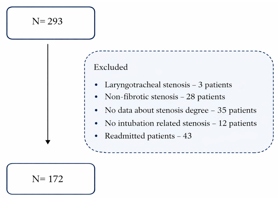
2. Materials and Methods
2.1. Study Design and Settings
2.2. Data Collection
2.3. Methods of Data Processing
3. Results
3.1. Common Characteristics for Children with PICLS
3.2. Stenosis Severity by Clinical Factors
3.3. Clinical Differences Between PICLS Patients with and Without Comorbidities
3.4. Correlation and Regression Analysis of Stenosis Grade
- Children under one year old have often been intubated (70.8%);
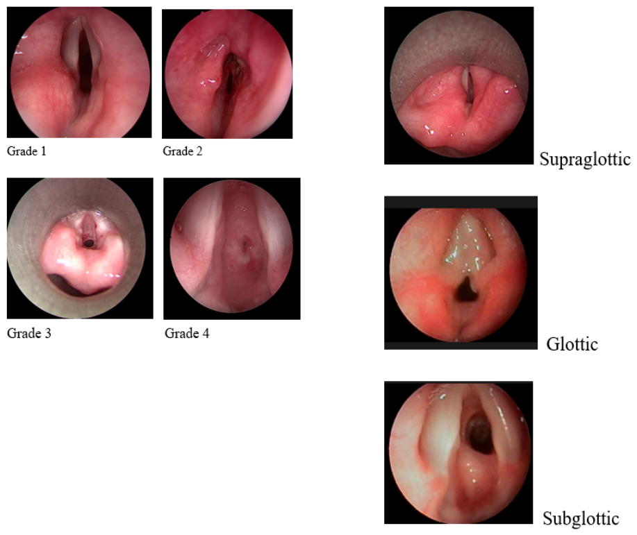
- Grade 1 PICLS (39.5%) and subglottic localization prevailed (85.3%);
- In 82.6% of cases, intubation was performed on an emergency basis, and only 31.6% of patients had a history reintubation;
- Mild forms of stenosis (Grades 1 and 2) were more frequently observed after intubations for planned surgeries and infections, whereas severe forms (Grades 3 and 4) were more commonly observed after intubations for CNS pathologies and infections;
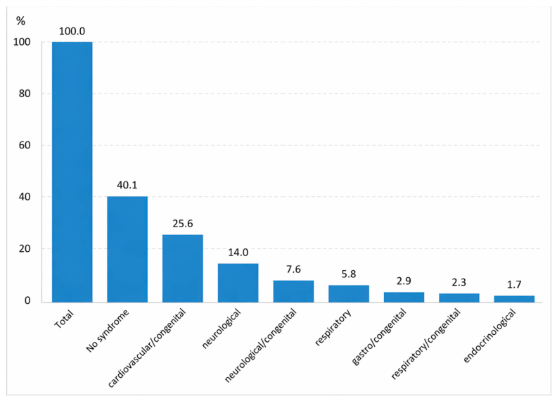
- Thirteen (65%) patients with Grade 4 stenosis had no comorbid diseases.
4. Discussion
5. Limitations and Future Directions
6. Conclusions
Author Contributions
Funding
Institutional Review Board Statement
Informed Consent Statement
Data Availability Statement
Conflicts of Interest
References
- Holzki, J.; Laschat, M.; Puder, C. Iatrogenic damage to the pediatric airway: Mechanisms and scar development. Paediatr. Anaesth. 2009, 19, 131–146. [Google Scholar] [CrossRef]
- Weiss, M.; Dave, M.; Bailey, M.; Gysin, C.; Hoeve, H.; Hammer, J.; Nicolai, T.; Spielmann, N.; Gerber, A. Endoscopic airway findings in children with or without prior endotracheal intubation. Paediatr. Anaesth. 2013, 23, 103–110. [Google Scholar] [CrossRef] [PubMed]
- Vijayasekaran, S.; Sances, R.; Cotton, R.; Elluru, R.G. Changes in the cricoarytenoid joint induced by intubation in neonates. Arch. Otolaryngol.–Head Neck Surg. 2006, 132, 1342–1345. [Google Scholar] [CrossRef]
- Stoler, N.A.; Segada, N.W.; Varghese, V.; Gowda, S.H.; Mehta, D.K. Consequences of NICU intubations: Incidence, identifications, and interventions. Otolaryngol.–Head Neck Surg. 2025, 172, 635–641. [Google Scholar] [CrossRef]
- Kruse, K.E.; Purohit, P.J.; Cadman, C.R.; Su, F.; Aghaeepour, N.; Hammer, G.B. Subglottic stenosis following cardiac surgery with cardiopulmonary bypass in infants and children. Pediatr. Crit. Care Med. 2017, 18, 429–433. [Google Scholar] [CrossRef]
- Chwa, J.S.; Shuman, E.A.; O’Dell, K. Size matters: Endotracheal tube sizes and glottic stenosis familiarity among intubating physicians. Laryngoscope Investig. Otolaryngol. 2025, 10, e70140. [Google Scholar] [CrossRef]
- Sibley, S.R.; Ball, I.M.; D’Arsigny, C.L.; Drover, J.W.; Erb, J.W.; Galvin, I.M.; Howes, D.W.; Ilan, R.; Messenger, D.W.; Moffatt, S.L.; et al. Airway injury from the presence of endotracheal tubes and association with subglottic secretion drainage. Can. J. Anaesth. 2022, 69, 1507–1514. [Google Scholar] [CrossRef]
- Costa, E.; Pazinatto, D.B.; Trevisan, L.P.; Maunsell, R. Post-extubation laryngitis in children: Diagnosis, management and follow-up. Braz. J. Otorhinolaryngol. 2024, 90, 101440. [Google Scholar] [CrossRef] [PubMed]
- Dorris, E.R.; Russell, J.; Murphy, M. Post-intubation subglottic stenosis: Aetiology at the cellular and molecular level. Eur. Respir. Rev. 2021, 30, 200218. [Google Scholar] [CrossRef]
- Cohn, J.E.; Touati, A.; Lentner, M.; Weitzel, M.; Fisher, C.; Sataloff, R.T. Self-extubation laryngeal injuries at an academic tertiary care center: A retrospective pilot study. Ann. Otol. Rhinol. Laryngol. 2017, 126, 555–560. [Google Scholar] [CrossRef] [PubMed]
- Bharti, B.; Syed, K.A.; Ebenezer, K.; Varghese, A.M.; Kurien, M. Post intubation laryngeal injuries in a pediatric intensive care unit of a tertiary hospital in India: A fibreoptic endoscopic study. Int. J. Pediatr. Otorhinolaryngol. 2016, 85, 84–90. [Google Scholar] [CrossRef]
- Rodríguez, H.; Cuestas, G.; Botto, H.; Cocciaglia, A.; Nieto, M.; Zanetta, A. Post-intubation subglottic stenosis in children. Diagnosis, treatment and prevention of moderate and severe stenosis. Acta Otorrinolaringol. Esp. 2013, 64, 339–344. [Google Scholar] [CrossRef]
- de Wit, M.; Peelen, L.M.; van Wolfswinkel, L.; de Graaff, J.C. The incidence of postoperative respiratory complications: A retrospective analysis of cuffed versus uncuffed tracheal tubes in children 0–7 years of age. Paediatr. Anaesth. 2018, 28, 210–217. [Google Scholar] [CrossRef] [PubMed]
- Bai, X.; Yu, R.; Wang, Z. Laryngeal involvement in relapsing polychondritis: Clinical and CT findings in 173 patients. RMD Open 2025, 11, e005397. [Google Scholar] [CrossRef]
- Glouvajić, D.; Bhate, J.J.; Sandu, K. Revision surgery for laryngotracheal stenosis in children: A single center’s 44 years’ experience. Laryngoscope 2023, 133, 3200–3207. [Google Scholar] [CrossRef]
- Burton, L.; Loberger, J.; Baker, M.; Prabhakaran, P.; Bhargava, V. Pre-extubation ultrasound measurement of in situ cuffed endotracheal tube laryngeal air column width difference and post-extubation stridor in children younger than 5 years. Pediatr. Crit. Care Med. 2024, 25, 222–230. [Google Scholar] [CrossRef] [PubMed]
- Sherman, J.M.; Lowitt, S.; Stephenson, C.; Ironson, G. Factors influencing acquired subglottic stenosis in infants. J. Pediatr. 1986, 109, 322–327. [Google Scholar] [CrossRef] [PubMed]
- Pinzas, L.A.; Bedwell, J.R.; Ongkasuwan, J. Glottic and subglottic injury and development of pediatric airway stenosis. Otolaryngol.–Head Neck Surg. 2023, 168, 469–477. [Google Scholar] [CrossRef]
- Schweiger, C.; Hart, C.K.; Tabangin, M.E.; Cohen, A.P.; Roetting, N.J.; de Alarcón, A. Impact of balloon diameter on dilation outcomes in a model of rabbit subglottic stenosis. Laryngoscope 2019, 129, 2409–2413. [Google Scholar] [CrossRef]
- Propst, E.J.; Wolter, N.E. Myer-Cotton grade of subglottic stenosis depends on style of endotracheal tube used. Laryngoscope 2023, 133, 2808–2812. [Google Scholar] [CrossRef]
- Manica, D.; Schweiger, C.; Maróstica, P.J.; Kuhl, G.; Carvalho, P.R. Association between length of intubation and subglottic stenosis in children. Laryngoscope 2013, 123, 1049–1054. [Google Scholar] [CrossRef] [PubMed]
- Richard, I.; Hamon, M.A.; Ferrapie, A.L.; Rome, J.; Brunel, P.; Mathé, J.F. Tracheotomy in brain injured patients: Which patients? Why? When? How? Ann. Fr. D’anesthesie Reanim. 2005, 24, 659–662. [Google Scholar] [CrossRef]
- Tsai, S.C.; Lin, F.C. Surgical treatments for post-intubation laryngotracheal stenosis in patients with central nervous system injuries. Medicine 2020, 99, e18628. [Google Scholar] [CrossRef] [PubMed]
- Veder, L.L.; Joosten, K.F.M.; Timmerman, M.K.; Pullens, B. Factors associated with laryngeal injury after intubation in children: A systematic review. Eur. Arch. Oto-Rhino-Laryngol. 2024, 281, 2833–2847. [Google Scholar] [CrossRef]
- Redondo-Sedano, J.; Antón-Pacheco, J.L.; Valverde, R.M.; Díaz, M.L.; Paredes, C.L.; Guardia, L.M.; Alelu, R.M.; Huerta, I.J.; Gordo, M.I.B.; Fraile, A.G. Laryngeal stenosis in children: Types, grades and treatment strategies. J. Pediatr. Surg. 2019, 54, 1933–1937. [Google Scholar] [CrossRef]
- Woliansky, J.; Paddle, P.; Phyland, D. Laryngotracheal stenosis management: A 16-year experience. Ear Nose Throat J. 2019, 100, 360–367. [Google Scholar] [CrossRef]
- Dutta, M.; Jana, D.; Mukherjee, D.; Panja, T.; Shukla, A.K.; Daga, D.; Yadav, K.K.; Mondal, S.; Bandyopadhyay, S.N. Endoscopic screening for laryngotracheal complications in children following prolonged mechanical ventilation. Ann. Otol. Rhinol. Laryngol. 2025, 134, 310–319. [Google Scholar] [CrossRef]
- Veder, L.L.; Joosten, K.F.M.; Schlink, K.; Timmerman, M.K.; Hoeve, L.J.; van der Schroeff, M.P.; Pullens, B. Post-extubation stridor after prolonged intubation in the pediatric intensive care unit. Eur. Arch. Oto-Rhino-Laryngol. 2020, 277, 1725–1731. [Google Scholar] [CrossRef] [PubMed]
- Alshareef, W.A.; Aldriweesh, B.A.; Almutairi, N.K.; Alsini, A.Y.; Zakzouk, A.S.; Aljasser, A.I.; Alammar, A.Y. Adverse respiratory events after pediatric endoscopic airway surgeries. Ear Nose Throat J. 2025, 104, 444–449. [Google Scholar] [CrossRef]
- Shinn, J.R.; Kimura, K.S.; Campbell, B.R.; Lowery, A.S.; Wootten, C.T.; Garrett, C.G.; Francis, D.O.; Hillel, A.T.; Du, L.; Casey, J.D.; et al. Incidence and outcomes of acute laryngeal injury after prolonged mechanical ventilation. Crit. Care Med. 2019, 47, 1699–1706. [Google Scholar] [CrossRef]
- Suresh, R.; Roohani, C.; Wang, C.S.; Kou, Y.F.; Johnson, R.F.; Chorney, S.R. Subglottic Stenosis After Pediatric Tracheostomy. Laryngoscope 2025, 135, 402–408. [Google Scholar] [CrossRef]
- Greaney, D.; Russell, J.; Dawkins, I.; Healy, M. Acquired subglottic stenosis using low-pressure, high-volume cuffed endotracheal tubes. Paediatr. Anaesth. 2018, 28, 1136–1141. [Google Scholar] [CrossRef]
- Moon, K.; Kitio, S.A.Y.; Rice-Weimer, J.; Tobias, J.D. Regulation of Intracuff Pressure by Measurement of the Ratio of Expiratory to Inspiratory Tidal Volumes. Paediatr. Anaesth. 2025, 35, 940–945. [Google Scholar] [CrossRef] [PubMed]
- Saravia, A.; Saravia, R.W.; Mudd, P.; Jones, J.W. Examining the pediatric subglottic airway by ultrasound: A systematic review. Int. J. Pediatr. Otorhinolaryngol. 2023, 166, 111482. [Google Scholar] [CrossRef]
- Thomas, R.E.; Rao, S.C.; Minutillo, C.; Vijayasekaran, S.; Nathan, E.A. Severe acquired subglottic stenosis in neonatal intensive care graduates: A case–control study. Arch. Dis. Child. Fetal Neonatal Ed. 2018, 103, F349–F354. [Google Scholar] [CrossRef] [PubMed]
- Debs, S.; Kazi, A.A.; Bastaich, D.; Thacker, L.; Petersson, R.S. Prognostic factors in the management of pediatric subglottic stenosis. Int. J. Pediatr. Otorhinolaryngol. 2021, 151, 110931. [Google Scholar] [CrossRef]
- Percul, C.; Lerendegui, L.; Lobos, P.; Liberto, D.; Moldes, J.; Urquizo, M.M. Association between subglottic stenosis and endotracheal intubation in tracheostomized pediatric patients. Cir. Pediatr. 2023, 36, 110–115. [Google Scholar] [CrossRef] [PubMed]
- Suzumura, H.; Nitta, A.; Tanaka, G.; Kuwashima, S.; Hirabayashi, H. Role of infection in the development of acquired subglottic stenosis in neonates with prolonged intubation. Pediatr. Int. 2000, 42, 508–513. [Google Scholar] [CrossRef]
- Thomas, R.E.; Erickson, S.; Hullett, B.; Minutillo, C.; Lethbridge, M.; Vijayasekaran, S.; Agrawal, S.; Bulsara, M.K.; Rao, S.C. Cuffed versus uncuffed endotracheal tubes for infants in intensive care: A pilot RCT. Arch. Dis. Child. Fetal Neonatal Ed. 2021, 106, 614–620. [Google Scholar] [CrossRef]
- Albert, D.M.; Mills, R.P.; Fysh, J.; Gamsu, H.; Thomas, J.N. Endoscopic examination of the neonatal larynx at extubation: A prospective study of variables associated with laryngeal damage. Int. J. Pediatr. Otorhinolaryngol. 1990, 20, 203–212. [Google Scholar] [CrossRef]
- Nicklaus, P.J.; Crysdale, W.S.; Conley, S.; White, A.K.; Sendi, K.; Forte, V. Evaluation of neonatal subglottic stenosis: A 3-year prospective study. Laryngoscope 1990, 100, 1185–1190. [Google Scholar] [CrossRef]
- Cakir, E.; Atabek, A.A.; Calim, O.F.; Uzuner, S.; AlShadfan, L.; Yazan, H.; Ozturan, O.; Cakir, F.B. Post-intubation subglottic stenosis in children: Analysis of clinical features and risk factors. Pediatr. Int. 2020, 62, 386–389. [Google Scholar] [CrossRef]
- Buras, M.; DeSisto, N.; Holdgraf, R. Effect of COVID-19 on the incidence of postintubation laryngeal lesions. Bayl. Univ. Med. Cent. Proc. 2023, 36, 453–457. [Google Scholar] [CrossRef]
- Moser, C.H.; Peeler, A.; Long, R.; Schoneboom, B.; Budhathoki, C.; Pelosi, P.P.; Brenner, M.J.; Pandian, V. Prevention of endotracheal tube-related pressure injury: A systematic review and meta-analysis. Am. J. Crit. Care 2022, 31, 416–424. [Google Scholar] [CrossRef]
- Lira, N.E.; Kuhl, L.P.; Maróstica, P.J.C.; Schweiger, C. Post-Intubation Acute Laryngeal Injuries: Analysis of Predictive Factors of Poor Prognosis. Laryngoscope 2025, 135, 3535–3541. [Google Scholar] [CrossRef]



| Characteristics | Overall, n (%) |
|---|---|
| Sex: | 172 |
| Male | 102 (59.3%) |
| Female | 70 (40.7%) |
| Age at primary intubation: | 171 |
| Infants (≤12 months) | 121 (70.8%) |
| children (1–12 years) | 46 (26.9%) |
| adolescent (13–18 years) | 4 (2.3%) |
| Grade of stenosis: | 172 |
| 1 | 68 (39.5%) |
| 2 | 40 (23.3%) |
| 3 | 45 (26.2%) |
| 4 | 19 (11%) |
| Localization: | 171 |
| Supraglottic | 1 (0.7%) |
| Glottic | 5 (2.9%) |
| Subglottic | 146 (85.3%) |
| Combined | 19 (11.1%) |
| Comorbidities: | 172 |
| yes | 103 (59.9%) |
| no | 69 (40.1%) |
| Intubation emergency: | 172 |
| emergency | 142 (82.6%) |
| planned | 30 (17.4%) |
| Reintubation: | 171 |
| yes | 54 (31.6%) |
| no | 117 (68.4%) |
| T-tube: | 172 |
| yes | 2 (1.2%) |
| no | 170 (98.8%) |
| Tracheostomy: | 172 |
| yes | 108 (62.8%) |
| no | 64 (37.2%) |
| Gestational age at delivery: | 170 |
| Preterm delivery | 30 (17.6%) |
| Term delivery | 140 (82.4%) |
| Delivery type: | 170 |
| vaginal | 135 (79.4%) |
| cesarean section | 35 (20.6%) |
| Residence: | 171 |
| urban | 109 (63.7%) |
| rural | 62 (36.3%) |
| - | Grade 1 (n = 68; 39.5%) | Grade 2 (n = 40; 23.3%) | Grade 3 (n = 40; 26.2%) | Grade 4 (n = 24; 11%) |
|---|---|---|---|---|
| Sex (n = 172) | ||||
| Male (n = 102) | 43 (63.2%) | 22 (55%) | 25 (55.5%) | 12 (63.2%) |
| Female (n = 70) | 25 (36.8%) | 18 (45%) | 20 (44.5%) | 7 (36.8%) |
| % | 100% | 100% | 100% | 100% |
| Age at primary intubation (n = 171) | ||||
| Infants (0 to ≤ 12 month(s) | 54 (79.4%) | 25 (62.5%) | 32 (72.7%) | 10 (52.6%) |
| Children (1–12 years) | 14 (20.6%) | 15 (37.5%) | 11 (25.0%) | 6 (31.6%) |
| Adolescents (13–18 years) | 0 | 0 | 1 (2.3%) | 3 (15.8%) |
| % | 100% | 100% | 100% | 100% |
| Reintubation (n = 171) | ||||
| Yes | 19 (27.9%) | 11 (27.5%) | 18 (41%) | 6 (31.5%) |
| No | 49 (72.1%) | 29 (72.5%) | 26 (59%) | 13 (68.5%) |
| % | 100% | 100% | 100% | 100% |
| Delivery time (n = 170) | ||||
| Preterm delivery | 9 (13.4%) | 11 (27.5%) | 6 (13.6%) | 4 (21.05%) |
| Term delivery | 58 (86.6%) | 29 (72.5%) | 38 (86.4%) | 15 (78.95%) |
| % | 100% | 100% | 100% | 100% |
| Delivery type (n = 170) | ||||
| Vaginal delivery | 54 (79.4%) | 30 (76.9%) | 37 (84.1%) | 14 (73.7%) |
| Cesarean section | 14 (20.6%) | 9 (23.1%) | 7 (15.9%) | 5 (26.3%) |
| % | 100% | 100% | 100% | 100% |
| Residence (n = 171) | ||||
| Urban residence | 45 (67.2%) | 27 (67.5%) | 27 (60%) | 10 (52.6%) |
| Rural residence | 22 (32.8%) | 13 (32.5%) | 18 (40%) | 9 (47.4%) |
| % | 100% | 100% | 100% | 100% |
| Indications for intubation (n = 171) | ||||
| Elective surgery | 26 (38.2%) | 6 (15.0%) | 9 (20.0%) | 1 (5.3%) |
| Infection | 18 (26.5%) | 23 (57.5%) | 16 (35.6%) | 4 (21.1%) |
| CNS pathology | 11 (16.2%) | 5 (12.5%) | 11 (24.4%) | 11 (57.9%) |
| Mechanical obstruction | 6 (8.8%) | 3 (7.5%) | 7 (15.6%) | 2 (10.5%) |
| Neonatal respiratory pathologies | 6 (8.8%) | 3 (7.5%) | 2 (4.4%) | 1 (5.3%) |
| No data | 1 (1.5%) | |||
| 100% | 100% | 100% | 100% | |
| Localization (n = 171) | ||||
| Supraglottic, (n = 1) | 1 (1.5%) | 0 | 0 | 0 |
| Glottic, (n = 5) | 2 (2.9%) | 2 (5%) | 1 (2.2%) | 0 |
| Subglottic (n = 146) | 60 (88.2%) | 33 (82.5%) | 36 (80.0%) | 18 (90%) |
| Combined (n = 19) | 5 (7.4%) | 5 (12.5%) | 8 (17.8%) | 2 (10%) |
| % | 100% | 100% | 100% | 100% |
| History of tracheostomy (n = 171) | ||||
| Yes | 42 (61.8%) | 19 (47.5%) | 31 (68.9%) | 17 (85%) |
| No | 26 (38.2%) | 21 (52.5%) | 14 (31.1%) | 3 (15%) |
| % | 100% | 100% | 100% | 100% |
| Intubation clinic’s level (n = 171) | ||||
| Primary | 23 (33.8%) | 13 (32.5%) | 17 (37.8%) | 4 (20%) |
| Secondary | 26 (38.2%) | 21 (52.5%) | 25 (55.6%) | 14 (70%) |
| Tertiary | 19 (28%) | 5 (12.5%) | 3 (6.6%) | 2 (10%) |
| No data | 0 | 1 (2.5%) | 0 | 0 |
| Comorbidities (n = 172) | ||||
| Patients with comorbid. | 47 (69.1%) | 23 (57.5%) | 26 (57.8%) | 7 (35%) |
| Patients without comorbid. | 21 (30.9%) | 17 (42.5%) | 19 (42.2%) | 13 (65%) |
| % | 100% | 100% | 100% | 100% |
| Patients with Comorbid. (n = 103) | Patients Without Comorbid. (n = 69) | ||
|---|---|---|---|
| Sex | Male | 61 (59.2%) | 41 (59.4%) |
| Female | 42 (40.8%) | 28 (40.6%) | |
| 100% | 100% | ||
| Age at primary int. | infants (≤12 months) | 77 (74.7%) | 44 (63.7%) |
| children (1–12 years) | 26 (25.3%) | 20 (28.9%) | |
| adolescent (13–18 years | 0 | 4 (5.7%) | |
| No data | 0 | 1 (1.7%) | |
| 100% | 100% | ||
| Localization | Supraglottic | 1 (1.0%) | 0 (0.00%) |
| Glottic | 2 (1.9%) | 3 (4.3%) | |
| Subglottic | 88 (85.4%) | 58 (84.1%) | |
| Combined | 12 (11.7%) | 8 (11.6%) | |
| 100% | 100% | ||
| Reason of intubation | CNS pathology | 22 (21.4%) | 16 (23.2%) |
| Elective surgery | 34 (33.0%) | 9 (13.0%) | |
| Infection | 36 (35.0%) | 25 (36.2%) | |
| Neonatal causes | 4 (3.8%) | 7 (10.1%) | |
| Obstruction | 7 (6.8%) | 11 (16.0%) | |
| No data | 0 (0.0%) | 1 (1.5%) | |
| 100% | 100% | ||
| Reintubation | Yes | 33 (32.1%) | 21 (30.4%) |
| No | 70 (67.9%) | 47 (68.1%) | |
| No data | 0 (0.00%) | 1 (1.5%) | |
| 100% | 100% | ||
| Emer/planned int | Emergency | 79 (76.7%) | 63 (91.3%) |
| Planned | 24 (23.3%) | 6 (8.7%) | |
| 100% | 100% | ||
| History of tracheostomy | Yes | 67 (65.0%) | 41 (59.4%) |
| No | 36 (35.0%) | 28 (40.6%) | |
| 100% | 100% |
| Regression Statistics | ||||||||
|---|---|---|---|---|---|---|---|---|
| Multiple R | 0.343010425 | |||||||
| R Square | 0.117656151 | |||||||
| Adjusted R Square | 0.050719721 | |||||||
| Standard Error | 1.04056021 | |||||||
| Observations | 157 | |||||||
| ANOVA | ||||||||
| df | SS | MS | F | Significance F | ||||
| Regression | 11 | 20.94 | 1.90 | 1.76 | 0.07 | |||
| Residual | 145 | 157.00 | 1.08 | |||||
| Total | 156 | 177.94 | ||||||
| Coefficients | Standard Error | t Stat | p-value | Lower 95% | Upper 95% | Lower 95.0% | Upper 95.0% | |
| Intercept | 1.22 | 1.19 | 1.03 | 0.30 | −1.12 | 3.57 | −1.12 | 3.57 |
| Gender | −0.07 | 0.17 | −0.41 | 0.68 | −0.41 | 0.27 | −0.41 | 0.27 |
| Age at primary intubation (months) | 0.01 | 0.00 | 2.44 | 0.02 | 0.00 | 0.01 | 0.00 | 0.01 |
| Reintubation | 0.32 | 0.18 | 1.76 | 0.08 | −0.04 | 0.68 | −0.04 | 0.68 |
| Emergency, planned intubation | 0.47 | 0.23 | 2.10 | 0.04 | 0.03 | 0.92 | 0.03 | 0.92 |
| History of tracheostomy | 0.18 | 0.19 | 0.99 | 0.33 | −0.18 | 0.55 | −0.18 | 0.55 |
| Mother’s pregnancy no. | 0.00 | 0.05 | −0.01 | 1.00 | −0.09 | 0.09 | −0.09 | 0.09 |
| Birth weight, gram | 0.00 | 0.00 | −0.73 | 0.47 | 0.00 | 0.00 | 0.00 | 0.00 |
| Height at birth, cm | 0.02 | 0.03 | 0.62 | 0.54 | −0.04 | 0.08 | −0.04 | 0.08 |
| Delivery type | −0.10 | 0.24 | −0.44 | 0.66 | −0.58 | 0.37 | −0.58 | 0.37 |
| Delivery category | −0.17 | 0.36 | −0.47 | 0.64 | −0.88 | 0.54 | −0.88 | 0.54 |
| Patient’s residence | −0.22 | 0.18 | −1.22 | 0.22 | −0.59 | 0.14 | −0.59 | 0.14 |
| Grade of Stenosis | Gender | Age at Primary Intubation (Months) | Reintubation | Emergency, Planned Intubation | History of Tracheostomy | Mother’s Pregnancy No. | Birth Weight, gram | Height at Birth, cm | Delivery Type | Delivery Category | Patient’s Residence | |
|---|---|---|---|---|---|---|---|---|---|---|---|---|
| Grade of stenosis | 1 | |||||||||||
| Gender | −0.032 | 1 | ||||||||||
| Age at primary intubation (months) | 0.199 | 0.018 | 1 | |||||||||
| Reintubation | 0.106 | −0.017 | −0.127 | 1.000 | ||||||||
| Emergency, planned intubation | 0.206 | 0.020 | 0.102 | −0.003 | 1.000 | |||||||
| History of tracheostomy | 0.128 | 0.000 | 0.156 | −0.023 | 0.118 | 1.000 | ||||||
| Mother’s pregnancy no. | 0.036 | −0.082 | −0.164 | 0.043 | 0.102 | 0.156 | 1.000 | |||||
| Birth weight, gram | −0.053 | −0.013 | 0.061 | −0.037 | −0.055 | 0.088 | −0.130 | 1.000 | ||||
| Height at birth, cm | −0.031 | −0.115 | 0.046 | −0.046 | −0.110 | 0.017 | −0.035 | 0.799 | 1.000 | |||
| Delivery type | 0.005 | −0.031 | −0.129 | 0.027 | 0.153 | 0.100 | 0.162 | −0.354 | −0.359 | 1.000 | ||
| Delivery category | −0.062 | −0.069 | 0.064 | −0.051 | −0.080 | 0.025 | −0.101 | 0.719 | 0.736 | −0.440 | 1.000 | |
| Patient’s residence | −0.091 | 0.010 | 0.158 | 0.070 | −0.115 | −0.110 | −0.208 | −0.032 | −0.025 | −0.006 | 0.071 | 1 |
Disclaimer/Publisher’s Note: The statements, opinions and data contained in all publications are solely those of the individual author(s) and contributor(s) and not of MDPI and/or the editor(s). MDPI and/or the editor(s) disclaim responsibility for any injury to people or property resulting from any ideas, methods, instructions or products referred to in the content. |
© 2026 by the authors. Licensee MDPI, Basel, Switzerland. This article is an open access article distributed under the terms and conditions of the Creative Commons Attribution (CC BY) license.
Share and Cite
Sagandykova, N.; Baurzhan, M.; Mashekova, A.; Abdildin, Y.; Baimurzayeva, M.; Mukhmetov, O.; Ng, E.Y.K.; Kairgeldina, S. Factors Associated with Severity of Post-Intubation Cicatricial Laryngeal Stenosis in Children: A Retrospective Study. J. Clin. Med. 2026, 15, 3342. https://doi.org/10.3390/jcm15093342
Sagandykova N, Baurzhan M, Mashekova A, Abdildin Y, Baimurzayeva M, Mukhmetov O, Ng EYK, Kairgeldina S. Factors Associated with Severity of Post-Intubation Cicatricial Laryngeal Stenosis in Children: A Retrospective Study. Journal of Clinical Medicine. 2026; 15(9):3342. https://doi.org/10.3390/jcm15093342
Chicago/Turabian StyleSagandykova, Nazym, Madina Baurzhan, Aigerim Mashekova, Yerkin Abdildin, Makhabat Baimurzayeva, Olzhas Mukhmetov, Eddie Yin Kwee Ng, and Sayagul Kairgeldina. 2026. "Factors Associated with Severity of Post-Intubation Cicatricial Laryngeal Stenosis in Children: A Retrospective Study" Journal of Clinical Medicine 15, no. 9: 3342. https://doi.org/10.3390/jcm15093342
APA StyleSagandykova, N., Baurzhan, M., Mashekova, A., Abdildin, Y., Baimurzayeva, M., Mukhmetov, O., Ng, E. Y. K., & Kairgeldina, S. (2026). Factors Associated with Severity of Post-Intubation Cicatricial Laryngeal Stenosis in Children: A Retrospective Study. Journal of Clinical Medicine, 15(9), 3342. https://doi.org/10.3390/jcm15093342