Predictability, Skeletal Stability, and Safety of Iliac Crest Bone Grafts in Large Maxillary Advancement with Le Fort I Osteotomy: A Systematic Review

 ,
,  , ,
, ,  and
and 
Abstract
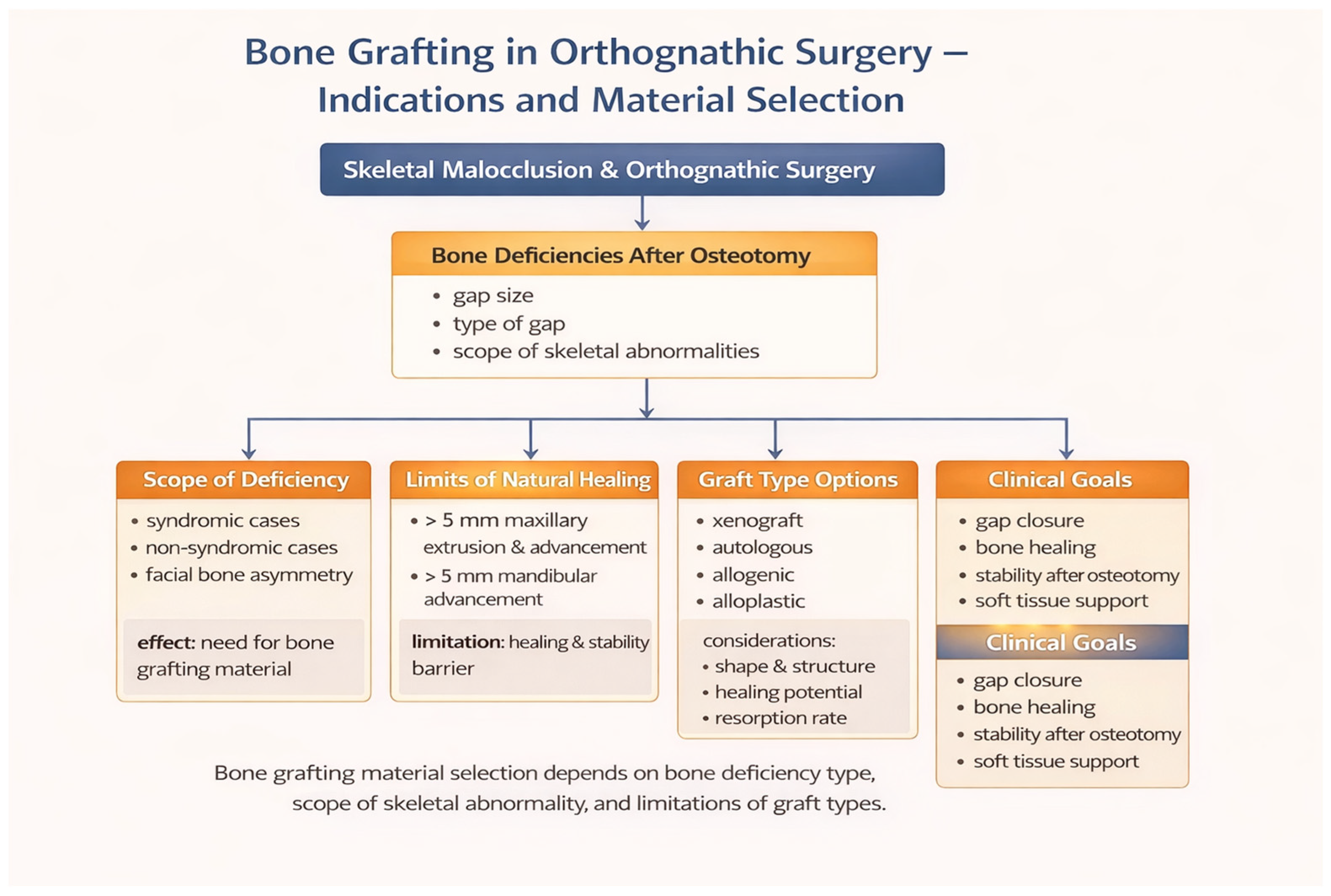
1. Introduction
2. Materials and Methods
2.1. Focused Question
2.2. Protocol
2.3. Eligibility Criteria
- Included patients undergoing Le Fort I osteotomy involving large maxillary advancement, defined as maxillary advancement and/or downgrafting greater than 5 mm, or described by the authors as extensive maxillary repositioning associated with a clinically significant osteotomy gap and insufficient bony contact between the mobilized segments, often requiring additional bone support;
- Reported the use of ICBG applied during the surgical procedure, either as interpositional or onlay grafts;
- Were clinical studies, including randomized controlled trials, prospective or retrospective cohort studies, and case series;
- Were published in English;
- Provided full-text access to allow detailed data extraction.
- Did not involve the use of ICBG during Le Fort I osteotomy;
- Were non-English publications;
- Were conference abstracts, editorials, narrative or systematic reviews, or technical notes without original clinical data;
- Lacked accessible full-text versions;
- Represented duplicate publications or overlapping patient populations, in which case only the most comprehensive or recent report was included.
2.4. Information Sources, Search Strategy, and Study Selection
- PubMed: (“Le Fort I” [Title/Abstract] OR “LeFort I” [Title/Abstract]) AND (“iliac crest” [Title/Abstract] OR autogenous” [Title/Abstract] OR “corticocancellous” [Title/Abstract]);
- Scopus: TITLE-ABS (“Le Fort I” OR “LeFort I”) AND TITLE-ABS (“iliac crest” OR “autogenous” OR “corticocancellous”);
- Embase: (‘le fort i’:ab,ti OR ‘lefort i’:ab,ti) AND (‘iliac crest’:ab,ti OR autogenous:ab,ti OR corticocancellous:ab,ti);
- Web of Science (WoS): TS = (“Le Fort I” OR “LeFort I”) AND TS = (“iliac crest” OR “autogenous” OR “corticocancellous”);
- WorldCat: (“Le Fort I” OR “LeFort I”) AND (“iliac crest” OR “autogenous” OR “corticocancellous”). WorldCat was additionally searched as a supplementary source to identify potentially relevant books, monographs, dissertations, and older records not indexed in standard biomedical databases, particularly given the historical development of surgical techniques related to this topic.
2.5. Data Collection Process and Data Items
2.6. Risk of Bias and Quality Assessment
2.7. Quality Assessment
- Is the sampling strategy relevant to address the research question?
- Is the sample representative of the target population?
- Are the measurements appropriate?
- Is the risk of nonresponse bias low?
- Is the statistical analysis appropriate to answer the research question?
3. Results
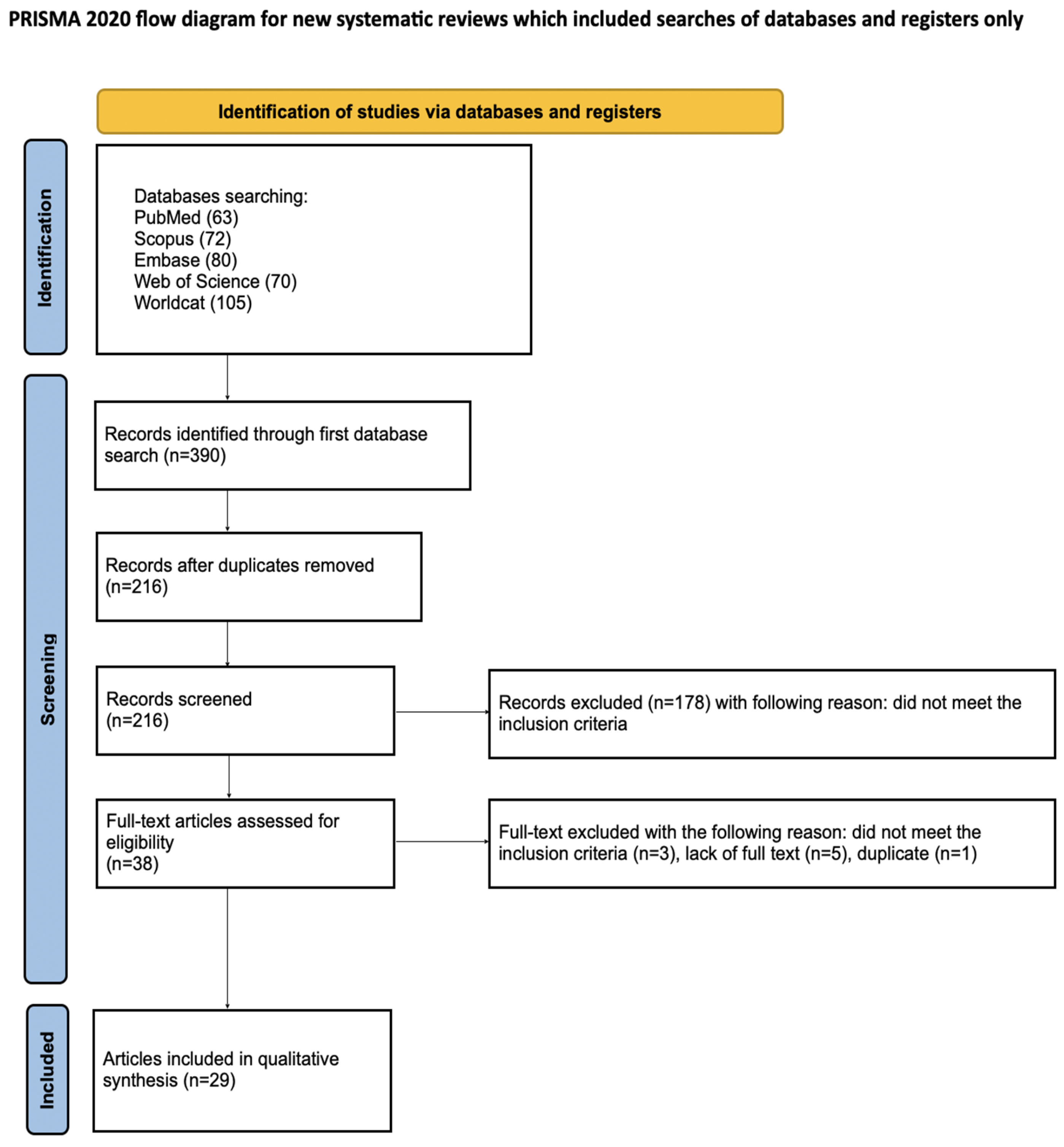
3.1. Study Selection
3.2. General Characteristic of Included Studies
3.3. Main Study Outcomes
3.3.1. Predictability of Large Maxillary Advancement
3.3.2. Skeletal Stability and Relapse Patterns
3.3.3. Graft Integration and Bone Remodeling
3.3.4. Implant-Related Outcomes
3.3.5. Safety and Complications
3.4. Quality Assessment
4. Discussion
5. Conclusions
Supplementary Materials
Author Contributions
Funding
Institutional Review Board Statement
Informed Consent Statement
Data Availability Statement
Conflicts of Interest
References
- Patel, D.; Tatum, S.A. Bone Graft Substitutes and Enhancement in Craniomaxillofacial Surgery. Facial Plast. Surg. 2023, 39, 556–563. [Google Scholar] [CrossRef] [Scilit] [PubMed]
- Yang, Z.; Zhai, S.; Liu, Y.; Wu, Y.; He, T.; Shi, X.; Chu, S. Sticky Bone: Advances and Applications. Int. J. Nanomed. 2025, 20, 10151–10175. [Google Scholar] [CrossRef] [Scilit] [PubMed]
- Bhatt, R.A.; Rozental, T.D. Bone Graft Substitutes. Hand Clin. 2012, 28, 457–468. [Google Scholar] [CrossRef] [Scilit] [PubMed]
- Giannoudis, P.V.; Dinopoulos, H.; Tsiridis, E. Bone Substitutes: An Update. Injury 2005, 36, S20–S27. [Google Scholar] [CrossRef] [Scilit]
- Golubovsky, J.L.; Ejikeme, T.; Winkelman, R.; Steinmetz, M.P. Osteobiologics. Oper. Surg. 2021, 21, S2–S9. [Google Scholar] [CrossRef] [Scilit]
- Santoro, A.; Voto, A.; Fortino, L.; Guida, R.; Laudisio, C.; Cillo, M.; D’Ursi, A.M. Bone Defect Treatment in Regenerative Medicine: Exploring Natural and Synthetic Bone Substitutes. Int. J. Mol. Sci. 2025, 26, 3085. [Google Scholar] [CrossRef] [Scilit]
- Kawecki, F.; Clafshenkel, W.P.; Fortin, M.; Auger, F.A.; Fradette, J. Biomimetic Tissue-Engineered Bone Substitutes for Maxillofacial and Craniofacial Repair: The Potential of Cell Sheet Technologies. Adv. Healthc. Mater. 2018, 7, 1700919. [Google Scholar] [CrossRef] [Scilit]
- Cheng, L.; Suresh K, S.; He, H.; Rajput, R.S.; Feng, Q.; Ramesh, S.; Wang, Y.; Krishnan, S.; Ostrovidov, S.; Camci-Unal, G.; et al. 3D Printing of Micro- and Nanoscale Bone Substitutes: A Review on Technical and Translational Perspectives. Int. J. Nanomed. 2021, 16, 4289–4319. [Google Scholar] [CrossRef] [Scilit]
- Engel, E.E.; Gava, N.F.; Santos, M.A.D.; Baldoino, L.G.; Ismael, L.K.; Modolo, L.F.M. Updates on Bone Grafts and Substitutes. Rev. Bras. Ortop. 2025, 60, s00451814114. [Google Scholar] [CrossRef] [Scilit]
- Posnick, J.C.; Kinard, B.E. Orthognathic Surgery Has a Significant Positive Effect on Perceived Personality Traits and Perceived Emotional Expressions in Long Face Patients. J. Oral Maxillofac. Surg. 2019, 77, 408.e1–408.e10. [Google Scholar] [CrossRef] [Scilit]
- Nelke, K.H.; Pawlak, W.; Morawska-Kochman, M.; Łuczak, K. Ten Years of Observations and Demographics of Hemimandibular Hyperplasia and Elongation. J. Cranio-Maxillofac. Surg. 2018, 46, 979–986. [Google Scholar] [CrossRef] [Scilit] [PubMed]
- Salem, D.; Alshihri, A.; Arguello, E.; Jung, R.; Mohmed, H.; Friedland, B. Volumetric Analysis of Allogeneic and Xenogeneic Bone Substitutes Used in Maxillary Sinus Augmentations Utilizing Cone Beam Computed Tomography: A Prospective Randomized Pilot Study. Int. J. Oral Maxillofac. Implant. 2019, 34, 920–926. [Google Scholar] [CrossRef] [Scilit] [PubMed]
- Moussa, N.T.; Dym, H. Maxillofacial Bone Grafting Materials. Dent. Clin. N. Am. 2020, 64, 473–490. [Google Scholar] [CrossRef] [Scilit] [PubMed]
- Clokie, C.M.L.; Moghadam, H.; Jackson, M.T.; Sandor, G.K.B. Closure of Critical Sized Defects with Allogenic and Alloplastic Bone Substitutes. J. Craniofacial Surg. 2002, 13, 111–121. [Google Scholar] [CrossRef] [Scilit]
- Sanz, M.; Vignoletti, F. Key Aspects on the Use of Bone Substitutes for Bone Regeneration of Edentulous Ridges. Dent. Mater. 2015, 31, 640–647. [Google Scholar] [CrossRef] [Scilit]
- Hikita, A.; Chung, U.; Hoshi, K.; Takato, T. Bone Regenerative Medicine in Oral and Maxillofacial Region Using a Three-Dimensional Printer. Tissue Eng. Part A 2017, 23, 515–521. [Google Scholar] [CrossRef] [Scilit]
- Moghadam, H.G.; Sándor, G.K.B.; Holmes, H.H.I.; Clokie, C.M.L. Histomorphometric Evaluation of Bone Regeneration Using Allogeneic and Alloplastic Bone Substitutes. J. Oral Maxillofac. Surg. 2004, 62, 202–213. [Google Scholar] [CrossRef] [Scilit]
- McAllister, B.S.; Haghighat, K. Bone Augmentation Techniques. J. Periodontol. 2007, 78, 377–396. [Google Scholar] [CrossRef] [Scilit]
- Fretwurst, T.; Gad, L.M.; Nelson, K.; Schmelzeisen, R. Dentoalveolar Reconstruction: Modern Approaches. Curr. Opin. Otolaryngol. Head Neck Surg. 2015, 23, 316–322. [Google Scholar] [CrossRef] [Scilit]
- Moon, K.-N.; Kim, S.-G.; Oh, J.-S.; Kim, C.-S.; Lim, S.-C.; Jeong, M.-A. Evaluation of Bone Formation After Grafting with Deproteinized Bovine Bone and Mineralized Allogenic Bone. Implant Dent. 2015, 24, 101–105. [Google Scholar] [CrossRef] [Scilit]
- Zhang, S.; Li, X.; Qi, Y.; Ma, X.; Qiao, S.; Cai, H.; Zhao, B.C.; Jiang, H.B.; Lee, E.-S. Comparison of Autogenous Tooth Materials and Other Bone Grafts. Tissue Eng. Regen. Med. 2021, 18, 327–341. [Google Scholar] [CrossRef] [Scilit] [PubMed]
- Ferraz, M.P. Bone Grafts in Dental Medicine: An Overview of Autografts, Allografts and Synthetic Materials. Materials 2023, 16, 4117. [Google Scholar] [CrossRef] [Scilit]
- Gabay, E.; Katorza, A.; Zigdon-Giladi, H.; Horwitz, J.; Machtei, E.E. Histological and Dimensional Changes of the Alveolar Ridge Following Tooth Extraction When Using Collagen Matrix and Collagen-embedded Xenogenic Bone Substitute: A Randomized Clinical Trial. Clin. Implant Dent. Relat. Res. 2022, 24, 382–390. [Google Scholar] [CrossRef] [Scilit] [PubMed]
- Alyahya, A.; Swennen, G.R.J. Bone Grafting in Orthognathic Surgery: A Systematic Review. Int. J. Oral Maxillofac. Surg. 2019, 48, 322–331. [Google Scholar] [CrossRef] [Scilit] [PubMed]
- Nasr, H.F.; Aichelmann-Reidy, M.E.; Yukna, R.A. Bone and Bone Substitutes. Periodontology 2000 2000, 19, 74–86. [Google Scholar] [CrossRef] [Scilit]
- Ryu, J.-I.; Yang, B.-E.; Yi, S.-M.; Choi, H.-G.; On, S.-W.; Hong, S.-J.; Lim, H.-K.; Byun, S.-H. Bone Regeneration of a 3D-Printed Alloplastic and Particulate Xenogenic Graft with rhBMP-2. Int. J. Mol. Sci. 2021, 22, 12518. [Google Scholar] [CrossRef] [Scilit]
- Musson, D.S.; Gao, R.; Watson, M.; Lin, J.-M.; Park, Y.-E.; Tuari, D.; Callon, K.E.; Zhu, M.; Dalbeth, N.; Naot, D.; et al. Bovine Bone Particulates Containing Bone Anabolic Factors as a Potential Xenogenic Bone Graft Substitute. J. Orthop. Surg. Res. 2019, 14, 60. [Google Scholar] [CrossRef] [Scilit]
- Benic, G.I.; Joo, M.; Yoon, S.; Cha, J.; Jung, U. Primary Ridge Augmentation with Collagenated Xenogenic Block Bone Substitute in Combination with Collagen Membrane and Rh BMP-2: A Pilot Histological Investigation. Clin. Oral Implant. Res. 2017, 28, 1543–1552. [Google Scholar] [CrossRef] [Scilit]
- Di Stefano, D.A.; Orlando, F.; Ottobelli, M.; Fiori, D.; Garagiola, U. A Comparison between Anorganic Bone and Collagen-Preserving Bone Xenografts for Alveolar Ridge Preservation: Systematic Review and Future Perspectives. Maxillofac. Plast. Reconstr. Surg. 2022, 44, 24. [Google Scholar] [CrossRef] [Scilit]
- Kruse, A.; Jung, R.E.; Nicholls, F.; Zwahlen, R.A.; Hämmerle, C.H.F.; Weber, F.E. Bone Regeneration in the Presence of a Synthetic Hydroxyapatite/Silica Oxide-Based and a Xenogenic Hydroxyapatite-Based Bone Substitute Material: Bone Regeneration in the Presence of a Synthetic Hydroxyapatite/Silica Oxide. Clin. Oral Implant. Res. 2011, 22, 506–511. [Google Scholar] [CrossRef] [Scilit]
- Bow, A.; Anderson, D.E.; Dhar, M. Commercially Available Bone Graft Substitutes: The Impact of Origin and Processing on Graft Functionality. Drug Metab. Rev. 2019, 51, 533–544. [Google Scholar] [CrossRef] [Scilit] [PubMed]
- Amid, R.; Kheiri, A.; Kheiri, L.; Kadkhodazadeh, M.; Ekhlasmandkermani, M. Structural and Chemical Features of Xenograft Bone Substitutes: A Systematic Review of In Vitro Studies. Biotechnol. Appl. Biochem. 2021, 68, 1432–1452. [Google Scholar] [CrossRef] [Scilit] [PubMed]
- Kadkhodazadeh, M.; Azadi, A. Management of a Severe Peri-Implantitis Case Using Titanium Mesh Technique with Xenogenic Bone Substitute and Collagen Matrix: A 4-Year Follow-Up. J. Oral Implantol. 2025, 51, 409–413. [Google Scholar] [CrossRef] [Scilit] [PubMed]
- Rogina, A.; Antunović, M.; Milovac, D. Biomimetic Design of Bone Substitutes Based on Cuttlefish Bone-Derived Hydroxyapatite and Biodegradable Polymers. J. Biomed. Mater. Res. 2019, 107, 197–204. [Google Scholar] [CrossRef] [Scilit]
- Rayad, S.; Klimas, S.; Janeczek, M.; Małyszek, A.; Bort, M.; Małysa, A.; Dominiak, M.; Dobrzyński, M. Studies on the Content of Toxic Metals in Teeth: A Narrative Review of Literature. Dent. Med. Probl. 2024, 61, 943–961. [Google Scholar] [CrossRef] [Scilit]
- Sethi, A.; Kaus, T.; Cawood, J.I.; Plaha, H.; Boscoe, M.; Sochor, P. Onlay Bone Grafts from Iliac Crest: A Retrospective Analysis. Int. J. Oral Maxillofac. Surg. 2020, 49, 264–271. [Google Scholar] [CrossRef] [Scilit]
- Schaufler, C.; Schmitt, A.-M.; Moseke, C.; Stahlhut, P.; Geroneit, I.; Brückner, M.; Meyer-Lindenberg, A.; Vorndran, E. Physicochemical Degradation of Calcium Magnesium Phosphate (Stanfieldite) Based Bone Replacement Materials and the Effect on Their Cytocompatibility. Biomed. Mater. 2023, 18, 015022. [Google Scholar] [CrossRef] [Scilit]
- Demir, Ö.; Pylostomou, A.; Loca, D. Octacalcium Phosphate Phase Forming Cements as an Injectable Bone Substitute Materials: Preparation and in Vitro Structural Study. Biomater. Adv. 2024, 157, 213731. [Google Scholar] [CrossRef] [Scilit]
- Zhao, B.; Xu, H.; Gao, Y.; Xu, J.-Z.; Yin, H.-M.; Xu, L.; Li, Z.-M.; Song, X. Promoting Osteoblast Proliferation on Polymer Bone Substitutes with Bone-like Structure by Combining Hydroxyapatite and Bioactive Glass. Mater. Sci. Eng. C 2019, 96, 1–9. [Google Scholar] [CrossRef] [Scilit]
- Cho, Y.R.; Gosain, A.K. Biomaterials in Craniofacial Reconstruction. Clin. Plast. Surg. 2004, 31, 377–385. [Google Scholar] [CrossRef] [Scilit]
- Chow, L.C. Next Generation Calcium Phosphate-Based Biomaterials. Dent. Mater. J. 2009, 28, 1–10. [Google Scholar] [CrossRef] [Scilit] [PubMed]
- Pokharel, R.K.; Paudel, S.; Lakhey, R.B. Iliac Crest Bone Graft Harvesting: Modified Technique for Reduction of Complications. J. Nepal. Med. Assoc. 2022, 60, 325–328. [Google Scholar] [CrossRef] [Scilit] [PubMed]
- Gorna, K.; Gogolewski, S. Preparation, Degradation, and Calcification of Biodegradable Polyurethane Foams for Bone Graft Substitutes. J. Biomed. Mater. Res. 2003, 67A, 813–827. [Google Scholar] [CrossRef] [Scilit] [PubMed]
- Zimmermann, G.; Moghaddam, A. Allograft Bone Matrix versus Synthetic Bone Graft Substitutes. Injury 2011, 42, S16–S21. [Google Scholar] [CrossRef] [Scilit]
- Fuchs, A.; Kreczy, D.; Brückner, T.; Gbureck, U.; Stahlhut, P.; Bengel, M.; Hoess, A.; Nies, B.; Bator, J.; Klammert, U.; et al. Bone Regeneration Capacity of Newly Developed Spherical Magnesium Phosphate Cement Granules. Clin. Oral Investig. 2022, 26, 2619–2633. [Google Scholar] [CrossRef] [Scilit]
- Profeta, A.C. Emerging Developments in the Use of Bioactive Glass for Reconstruction of Craniofacial Bone. Br. J. Oral Maxillofac. Surg. 2015, 53, 760–762. [Google Scholar] [CrossRef] [Scilit]
- Hasani, K.M.A.; Bakathir, A.A.; Al-Hashmi, A.K.; Rawahi, B.A.; Albakri, A. Modified Le Fort I Osteotomy and Genioplasty for Management of Severe Dentofacial Deformity in β-Thalassaemia Major: Case Report and Review of the Literature. Sultan Qaboos Univ. Med. J. 2020, 20, e362–e367. [Google Scholar] [CrossRef] [Scilit]
- Pingarron-Martin, L.; Arias-Gallo, J.; Ong, H.S.; Pons, M.C. Le Fort I Osteotomy with Bone Grafts in Preprosthetic Surgery: Technical Note. Craniomaxillofacial Trauma Reconstr. 2013, 6, 143–146. [Google Scholar] [CrossRef] [Scilit]
- Sullivan, S.M. Le Fort I Osteotomy. Atlas Oral Maxillofac. Surg. Clin. 2016, 24, 1–13. [Google Scholar] [CrossRef] [Scilit]
- Mommaerts, M.Y. Le Fort I–Type Osteotomy Retractor. Craniomaxillofacial Trauma Reconstr. 2017, 10, 323–324. [Google Scholar] [CrossRef] [Scilit]
- Kikuchi, A.; Sakamoto, Y.; Kishi, K. Le Fort I Osteotomy by the Precious Technique Contributes to the Reduction of Bleeding. J. Plast. Reconstr. Aesthetic Surg. 2022, 75, 2831–2870. [Google Scholar] [CrossRef] [Scilit] [PubMed]
- Atakan, A.; Ozcirpici, A.; Pamukcu, H.; Bayram, B. Does Le Fort I Osteotomy Have an Influence on Nasal Cavity and Septum Deviation? Niger. J. Clin. Pract. 2020, 23, 240. [Google Scholar] [CrossRef] [Scilit] [PubMed]
- Haas Junior, O.L.; Guijarro-Martínez, R.; De Sousa Gil, A.P.; Da Silva Meirelles, L.; De Oliveira, R.B.; Hernández-Alfaro, F. Stability and Surgical Complications in Segmental Le Fort I Osteotomy: A Systematic Review. Int. J. Oral Maxillofac. Surg. 2017, 46, 1071–1087. [Google Scholar] [CrossRef] [Scilit] [PubMed]
- Gil, A.P.S.; Machado-Fernández, A.; Guijarro-Martínez, R.; Hernández-Alfaro, F.; Haas, O.L., Jr.; De Oliveira, R.B. Le Fort I Osteotomy and Soft Tissue Response: A Retrospective Cohort Study Comparing Three Different Techniques. J. Cranio-Maxillofac. Surg. 2022, 50, 107–113. [Google Scholar] [CrossRef] [Scilit]
- Page, M.J.; McKenzie, J.E.; Bossuyt, P.M.; Boutron, I.; Hoffmann, T.C.; Mulrow, C.D.; Shamseer, L.; Tetzlaff, J.M.; Akl, E.A.; Brennan, S.E.; et al. The PRISMA 2020 Statement: An Updated Guideline for Reporting Systematic Reviews. BMJ 2021, 372, n71. [Google Scholar] [CrossRef] [Scilit]
- Homa, K.; Zakrzewski, W.; Dobrzyński, W.; Piszko, P.J.; Piszko, A.; Matys, J.; Wiglusz, R.J.; Dobrzyński, M. Surface Functionalization of Titanium-Based Implants with a Nanohydroxyapatite Layer and Its Impact on Osteoblasts: A Systematic Review. J. Funct. Biomater. 2024, 15, 45. [Google Scholar] [CrossRef] [Scilit]
- Kiryk, J.; Kiryk, S.; Kensy, J.; Świenc, W.; Palka, B.; Zimoląg-Dydak, M.; Dobrzyński, W.; Matys, J.; Dobrzyński, M. Effectiveness of Laser-Assisted Teeth Bleaching: A Systematic Review. Appl. Sci. 2024, 14, 9219. [Google Scholar] [CrossRef] [Scilit]
- Oleniacz-Trawińska, M.; Kotela, A.; Kensy, J.; Kiryk, S.; Dobrzyński, W.; Kiryk, J.; Gerber, H.; Fast, M.; Matys, J.; Dobrzyński, M. Evaluation of Factors Affecting Fluoride Release from Compomer Restorative Materials: A Systematic Review. Materials 2025, 18, 1627. [Google Scholar] [CrossRef] [Scilit]
- Tokarczuk, D.; Tokarczuk, O.; Kiryk, J.; Kensy, J.; Szablińska, M.; Dyl, T.; Dobrzyński, W.; Matys, J.; Dobrzyński, M. Fluoride Release by Restorative Materials after the Application of Surface Coating Agents: A Systematic Review. Appl. Sci. 2024, 14, 4956. [Google Scholar] [CrossRef] [Scilit]
- Małyszek, A.; Kiryk, S.; Kensy, J.; Kotela, A.; Michalak, M.; Kiryk, J.; Janeczek, M.; Matys, J.; Dobrzyński, M. Identification of Factors Influencing Fluoride Content in Tea Infusions: A Systematic Review. Appl. Sci. 2025, 15, 5974. [Google Scholar] [CrossRef] [Scilit]
- Rygas, J.; Matys, J.; Wawrzyńska, M.; Szymonowicz, M.; Dobrzyński, M. The Use of Graphene Oxide in Orthodontics—A Systematic Review. J. Funct. Biomater. 2023, 14, 500. [Google Scholar] [CrossRef] [Scilit]
- Piszko, P.J.; Piszko, A.; Kiryk, J.; Lubojański, A.; Dobrzyński, W.; Wiglusz, R.J.; Matys, J.; Dobrzyński, M. The Influence of Fluoride Gels on the Physicochemical Properties of Tooth Tissues and Dental Materials—A Systematic Review. Gels 2024, 10, 98. [Google Scholar] [CrossRef] [Scilit]
- Dobrzyński, W.; Piszko, P.J.; Kiryk, J.; Kiryk, S.; Michalak, M.; Kotela, A.; Kensy, J.; Świenc, W.; Grychowska, N.; Matys, J.; et al. Dental Resin Composites Modified with Chitosan: A Systematic Review. Mar. Drugs 2025, 23, 199. [Google Scholar] [CrossRef] [Scilit] [PubMed]
- Smołka, P.; Nelke, K.; Struzik, N.; Wiśniewska, K.; Kiryk, S.; Kensy, J.; Dobrzyński, W.; Kiryk, J.; Matys, J.; Dobrzyński, M. Discrepancies in Cephalometric Analysis Results between Orthodontists and Radiologists and Artificial Intelligence: A Systematic Review. Appl. Sci. 2024, 14, 4972. [Google Scholar] [CrossRef] [Scilit]
- Zygmunt, Ł.; Kiryk, S.; Wesołek, K.; Kiryk, J.; Nawrot-Hadzik, I.; Rybak, Z.; Sztyler, K.; Małyszek, A.; Matys, J.; Dobrzyński, M. The Role of the Oral Microbiome and Dental Caries in Respiratory Health: A Systematic Review. J. Clin. Med. 2025, 14, 7670. [Google Scholar] [CrossRef] [Scilit] [PubMed]
- Castro, M.P.; Rey, R.L.; Búa, J.A.; Santana-Mora, U.; Cembranos, J.L.L.-C. Prosthodontic Rehabilitation in Patient with Ectodermal Dysplasia Combining Preprosthetic Techniques: A Case Report. Implant Dent. 2013, 22, 460–464. [Google Scholar] [CrossRef] [Scilit]
- Pelo, S.; Gasparini, G.; Moro, A.; Boniello, R.; Amoroso, P.F. Segmental Le Fort I Osteotomy with Bone Grafting in Unilateral Severely Atrophied Maxilla. Int. J. Oral Maxillofac. Surg. 2009, 38, 246–249. [Google Scholar] [CrossRef] [Scilit]
- Posnick, J.C.; Gray, J.A. Is It Safe to Re-Harvest the Anterior Iliac Crest to Manage Le Fort I Interpositional Defects in Young Adults with a Repaired Cleft? J. Oral Maxillofac. Surg. 2015, 73, S32–S39. [Google Scholar] [CrossRef] [Scilit]
- Bayat, M.; Khobyari, M.M.; Dalband, M.; Momen-Heravi, F. Full Mouth Implant Rehabilitation of a Patient with Ectodermal Dysplasia after Orthognathic Surgery, Sinus and Ridge Augmentation: A Clinical Report. J. Adv. Prosthodont. 2011, 3, 96. [Google Scholar] [CrossRef] [Scilit]
- Sabuncuoglu, F.; Varol, A.; Şençimen, M.; Ölmez, H. Onlay Iliac Bone Grafting as an Ancillary Augmentation Procedure for Paranasal Rejuvenation during Bimaxillary Surgery. Oral Surg. Oral Med. Oral Pathol. Oral Radiol. Endodontology 2010, 109, e13–e19. [Google Scholar] [CrossRef] [Scilit]
- Piecuch, J.F.; Segal, D.; Grasso, J.E. Augmentation of the Atrophic Maxilla with Interpositional Autogenous Bone Grafts. J. Maxillofac. Surg. 1984, 12, 133–138. [Google Scholar] [CrossRef] [Scilit]
- Soehardi, A.; Meijer, G.J.; Hoppenreijs, T.J.M.; Brouns, J.J.A.; De Koning, M.; Stoelinga, P.J.W. Stability, Complications, Implant Survival, and Patient Satisfaction after Le Fort I Osteotomy and Interposed Bone Grafts: Follow-up of 5–18 Years. Int. J. Oral Maxillofac. Surg. 2015, 44, 97–103. [Google Scholar] [CrossRef] [Scilit]
- Yerit, K.C.; Posch, M.; Guserl, U.; Turhani, D.; Schopper, C.; Wanschitz, F.; Wagner, A.; Watzinger, F.; Ewers, R. Rehabilitation of the Severely Atrophied Maxilla by Horseshoe Le Fort I Osteotomy (HLFO). Oral Surg. Oral Med. Oral Pathol. Oral Radiol. Endodontology 2004, 97, 683–692. [Google Scholar] [CrossRef] [Scilit]
- Wang, X.-X.; Wang, X.; Yi, B.; Li, Z.-L.; Liang, C.; Lin, Y. Internal Midface Distraction in Correction of Severe Maxillary Hypoplasia Secondary to Cleft Lip and Palate. Plast. Reconstr. Surg. 2005, 116, 51–60. [Google Scholar] [CrossRef] [Scilit]
- Naros, A.; Bayazeed, B.; Schwarz, U.; Nagursky, H.; Reinert, S.; Schmelzeisen, R.; Sauerbier, S. A Prospective Histomorphometric and Cephalometric Comparison of Bovine Bone Substitute and Autogenous Bone Grafting in Le Fort I Osteotomies. J. Cranio-Maxillofac. Surg. 2019, 47, 233–238. [Google Scholar] [CrossRef] [Scilit] [PubMed]
- Varol, A.; Atali, O.; Sipahi, A.; Basa, S. Implant Rehabilitation for Extremely Atrophic Maxillae (Cawood Type VI) with Le Fort I Downgrafting and Autogenous Iliac Block Grafts: A 4-Year Follow-up Study. Int. J. Oral Maxillofac. Implant. 2016, 31, 1415–1422. [Google Scholar] [CrossRef] [Scilit] [PubMed]
- Isaksson, S.; Ekfeldt, A.; Alberius, P.; Blomqvist, J.-E. Early Results from Reconstruction of Severely Atrophic (Class VI) Maxillas by Immediate Endosseous Implants in Conjunction with Bone Grafting and Le Fort I Osteotomy. Int. J. Oral Maxillofac. Surg. 1993, 22, 144–148. [Google Scholar] [CrossRef] [Scilit] [PubMed]
- Nyström, E.; Lundgren, S.; Gunne, J.; Nilson, H. Interpositional Bone Grafting and Le Fort I Osteotomy for Reconstruction of the Atrophic Edentulous Maxilla. Int. J. Oral Maxillofac. Surg. 1997, 26, 423–427. [Google Scholar] [CrossRef] [Scilit]
- Stork, J.T.; Kim, R.H.; Regennitter, F.J.; Keller, E.E. Maxillary Quadrangular Le Fort I Osteotomy: Long-Term Skeletal Stability and Clinical Outcome. Int. J. Oral Maxillofac. Surg. 2013, 42, 1533–1546. [Google Scholar] [CrossRef] [Scilit]
- Nyström, E.; Nilson, H.; Gunne, J.; Lundgren, S. Reconstruction of the Atrophic Maxilla with Interpositional Bone Grafting/Le Fort I Osteotomy and Endosteal Implants: A 11–16 Year Follow-Up. Int. J. Oral Maxillofac. Surg. 2009, 38, 1–6. [Google Scholar] [CrossRef] [Scilit]
- Van Der Mark, E.L.; Bierenbroodspot, F.; Baas, E.M.; De Lange, J. Reconstruction of an Atrophic Maxilla: Comparison of Two Methods. Br. J. Oral Maxillofac. Surg. 2011, 49, 198–202. [Google Scholar] [CrossRef] [Scilit]
- Marchetti, C.; Pieri, F.; Corinaldesi, G.; Degidi, M. A Long-Term Retrospective Study of Two Different Implant Surfaces Placed after Reconstruction of the Severely Resorbed Maxilla Using Le Fort I Osteotomy and Interpositional Bone Grafting. Int. J. Oral Maxillofac. Implant. 2008, 23, 911–918. [Google Scholar]
- Sjöström, M.; Lundgren, S.; Sennerby, L. A Histomorphometric Comparison of the Bone Graft-Titanium Interface between Interpositional and Onlay/Inlay Bone Grafting Techniques. Int. J. Oral Maxillofac. Implant. 2006, 21, 52–62. [Google Scholar]
- Chiapasco, M.; Brusati, R.; Ronchi, P. Le Fort I Osteotomy with Interpositional Bone Grafts and Delayed Oral Implants for the Rehabilitation of Extremely Atrophied Maxillae: A 1–9-year Clinical Follow-up Study on Humans. Clin. Oral Implant. Res. 2007, 18, 74–85. [Google Scholar] [CrossRef] [Scilit] [PubMed]
- Posnick, J.C.; Dagys, A.P. Skeletal Stability and Relapse Patterns after Le Fort I Maxillary Osteotomy Fixed with Miniplates: The Unilateral Cleft Lip and Palate Deformity. Plast. Reconstr. Surg. 1994, 94, 924–932. [Google Scholar] [CrossRef] [Scilit]
- Eskenazi, L.B.; Schendel, S.A. An Analysis of Le Fort I Maxillary Advancement in Cleft Lip and Palate Patients. Plast. Reconstr. Surg. 1992, 90, 779–786. [Google Scholar] [CrossRef] [Scilit]
- Farmand, M. Horse-Shoe Sandwich Osteotomy of the Edentulous Maxilla as a Preprosthetic Procedure. J. Maxillofac. Surg. 1986, 14, 238–244. [Google Scholar] [CrossRef] [Scilit]
- Sjöström, M.; Lundgren, S.; Nilson, H.; Sennerby, L. Monitoring of Implant Stability in Grafted Bone Using Resonance Frequency Analysis. Int. J. Oral Maxillofac. Surg. 2005, 34, 45–51. [Google Scholar] [CrossRef] [Scilit]
- Sipos, N.; Leikola, J.; Heliövaara, A.; Kormi, E.; Suojanen, J. Comparison of Postoperative Complications and Reoperation Rates of Le Fort I Osteotomies Using Demineralized Bone Matrix (DBM) or Autogenous Bone Grafts in Patients with Orofacial Clefts and Craniofacial Malformations. Dent. J. 2025, 13, 256. [Google Scholar] [CrossRef] [Scilit]
- Posnick, J.C.; Ewing, M.P. Skeletal Stability after Le Fort I Maxillary Advancement in Patients with Unilateral Cleft Lip and Palate. Plast. Reconstr. Surg. 1990, 85, 706–710. [Google Scholar] [CrossRef] [Scilit]
- Popat, A.; Yadav, S. Air Embolism-Induced Ischemic Stroke Following Orthognathic Surgery in a Patient with Goldenhar Syndrome. Clin. Med. Res. 2024, 22, 44–48. [Google Scholar] [CrossRef] [Scilit] [PubMed]
- Kahnberg, K.E.; Nilsson, P.; Rasmusson, L. Le Fort I Osteotomy with Interpositional Bone Grafts and Implants for Rehabilitation of the Severely Resorbed Maxilla: A 2-Stage Procedure. Int. J. Oral Maxillofac. Implant. 1999, 14, 571–578. [Google Scholar]
- De Santis, D.; Trevisiol, L.; D’Agostino, A.; Cucchi, A.; De Gemmis, A.; Nocini, P.F. Guided Bone Regeneration with Autogenous Block Grafts Applied to Le Fort I Osteotomy for Treatment of Severely Resorbed Maxillae: A 4- to 6-year Prospective Study. Clin. Oral Implant. Res. 2012, 23, 60–69. [Google Scholar] [CrossRef] [Scilit] [PubMed]
- Kretschmer, W.B.; Baciut, G.; Bacuit, M.; Zoder, W.; Wangerin, K. Intraoperative Blood Loss in Bimaxillary Orthognathic Surgery with Multisegmental Le Fort I Osteotomies and Additional Procedures. Br. J. Oral Maxillofac. Surg. 2010, 48, 276–280. [Google Scholar] [CrossRef] [Scilit]
- Bajaj, V.; Kolte, A.P.; Kolte, R.; Bawankar, P.V. Comparative Evaluation of Immediate Implant Placement and Provisionalization (IIPP) with and without a Concentrated Growth Factor-Enriched Bone Graft: A Randomized Controlled Trial. Dent. Med. Probl. 2025, 62, 449–459. [Google Scholar] [CrossRef] [Scilit]
- Stroud, S.W.; Fonseca, R.J.; Sanders, G.W.; Burkes, E.J. Healing of Interpositional Autologous Bone Grafts after Total Maxillary Osteotomy. J. Oral Surg. 1980, 38, 878–885. [Google Scholar]
- Dovigo, S.; Michelon, F.; Zuffellato, N.; Montagner, G.; Trojan, D. Treatment of Maxillary Edentulism with Weldable Wing Spindles and Allogenic Bone for Sinus Lift: A Retrospective Study. Dent. Med. Probl. 2026, 63, 65–71. [Google Scholar] [CrossRef] [Scilit]
- Jensen, J.; Sindet-Pedersen, S.; Oliver, A.J. Varying Treatment Strategies for Reconstruction of Maxillary Atrophy with Implants: Results in 98 Patients. J. Oral Maxillofac. Surg. 1994, 52, 210–216. [Google Scholar] [CrossRef] [Scilit]
- Breine, U.; Brånemark, P.-I. Reconstruction of Alveolar Jaw Bone. Scand. J. Plast. Reconstr. Surg. 1980, 14, 23–48. [Google Scholar] [CrossRef] [Scilit]
- Antonowicz, B.; Borys, J.; Zalewska, A.; Żendzian-Piotrowska, M.; Łukaszuk, K.; Woźniak, Ł.; Siuta, M.; Maciejczyk, M. Circulating Biomarkers of Nitrosative Stress, Protein Glycoxidation and Inflammation in Maxillofacial Surgery Patients Treated with Titanium Implants. Dent. Med. Probl. 2025, 62, 225–236. [Google Scholar] [CrossRef] [Scilit]
- Naidu, N.S.S.; Kancharla, A.K.; Nandigam, A.R.; Tasneem, S.M.; Gummaluri, S.S.; Dey, S.; Prathipaty, R.J. Comparative Study of Demineralized Freeze-Dried Bone Allograft and Its Combination with Platelet-Rich Fibrin in the Treatment of Intrabony Defects: A Randomized Clinical Trial. Dent. Med. Probl. 2024, 61, 507–513. [Google Scholar] [CrossRef] [Scilit]
- Kanwal, L.; Qabool, H.; Idrees, W.; Sukhia, R.H.; Fida, M. Skeletal and Dental Changes after Bone-Borne versus Tooth-Borne Surgically Assisted Rapid Palatal Expansion in Subjects with Maxillary Transverse Deficiency: A Systematic Review and Meta-Analysis. Dent. Med. Probl. 2025, 62, 333–341. [Google Scholar] [CrossRef] [Scilit]
- Dulinska-Litewka, J.; Wilisowski, D.; Felkle, D. Influence of the Soft Tissue Thickness and the Levels of Vitamin D3 as Important Factors in Bone Preservation Around the Dental Implant Platform: A Retrospective Observational Study. Dent. Med. Probl. 2025, 62, 499–504. [Google Scholar] [CrossRef] [Scilit]
- Wang, H.; Lin, X.; Wu, Y.; Zhou, H.; Zhao, J.; Jiang, L. Influence of Dental Implants on the Accuracy of Measuring the Postoperative Labial Alveolar Bone Thickness in the Maxillary Anterior Region: An In Vivo CBCT Study. Dent. Med. Probl. 2025, 62, 1139–1146. [Google Scholar] [CrossRef] [Scilit]
- Pratiwi, A.R.; Hussein, Z.; Abdullah, C.A.C.; Istikharoh, F.; Fakurazi, S.; Wardani, S.C.; Abdelmonem, M. Novel Demineralized Freeze-Dried Bovine Bone Xenograft Nanoparticles (DFDBBX-NPs) for Enhanced Bone Repair. Dent. Med. Probl. 2026, 63, 209–217. [Google Scholar] [CrossRef] [Scilit]


| Study | Aim of the Study | Material and Methods | Results | Conclusions |
|---|---|---|---|---|
| Pombo Castro [66], 2013 | To demonstrate oral rehabilitation of ectodermal dysplasia using combined preprosthetic bone grafting techniques and implant-supported prosthesis. | A 27-year-old male with ectodermal dysplasia, severe jaw atrophy and oligodontia. Two-stage surgical treatment: (1) Le Fort I osteotomy, bilateral sinus lift, and 7 bone grafts (5 onlay, 2 inlay) using ICBG with titanium fixation; (2) extraction of remaining teeth and placement of 11 Straumann implants (5 maxillary, 6 mandibular) six months later. | Partial graft exposure occurred due to smoking, requiring sequestra removal but sufficient bone remained for implant placement. All 11 implants successfully osseointegrated. At 2-year follow-up, all implants were stable with no failures. | Combined preprosthetic techniques (Le Fort I osteotomy, sinus lift, onlay/inlay grafts) with subsequent implant-supported fixed prosthesis is an effective treatment for oral rehabilitation in ectodermal dysplasia |
| Popat [91] 2024 | To report a postoperative complication of air embolism-induced ischemic stroke following orthognathic surgery in a patient with Goldenhar syndrome | 20-year-old male with Goldenhar syndrome underwent orthognathic surgery (LeFort I, BSSO, ICBG). EBL 500 cc. Developed postop right arm weakness. Imaging revealed left thalamic infarct with air in cavernous sinus/interpeduncular system, consistent with air embolism. Managed conservatively with neuro monitoring. | Imaging confirmed acute left thalamic infarct with air in cavernous sinus and interpeduncular system. 3-month follow-up: improvement, only residual right arm weakness remained. | First reported air embolism stroke case in Goldenhar syndrome surgery. Air embolism is rare but potentially life-threatening. Requires prompt recognition and multidisciplinary approach. |
| Pelo [67] 2009 | To evaluate the outcome of segmental Le Fort I osteotomy with interpositional bone grafting for treating severe unilateral maxillary atrophy. | 5 patients with severe unilateral maxillary atrophy. ICBG placed between maxillary fragments. Titanium fixation. Cancellous chips for sinus lift. On-lay grafts for transversal correction. Vestibuloplasty at 7 weeks. Second surgery at 4 months: removal of hardware and placement of 25 implants in 5 patients. Follow-up: minimum 1 year. | All cases successful. Correct intermaxillary relationship achieved. Inter-arch distance reduced. 25 implants placed, 1 lost after 1 year (96% success). All patients received partial-arch fixed prostheses. | Segmental Le Fort I osteotomy with bone grafting is safe and predictable for vertical and horizontal maxillary augmentation. |
| Posnick [68] 2015 | This study evaluated safety and adequacy of re-harvesting anterior ICBG in young adults with repaired cleft undergoing Le Fort I advancement. | A retrospective review included patients under 26 who previously underwent ICBG and later required re-harvesting during Le Fort I surgery. Healing at donor and recipient sites was examined, and a questionnaire measured early and late discomfort. | Twenty-seven patients were included. All procedures yielded sufficient graft, and no complications occurred. Most patients reported easier recovery than after the first harvest, with no nerve injury or long-term functional issues. | Re-harvesting the anterior ICBG appears safe and effective for managing Le Fort I interpositional defects, providing adequate graft with minimal morbidity. |
| Bayat [69] 2011 | To describe a comprehensive oral rehabilitation approach for hypohidrotic ectodermal dysplasia with severe oligodontia and maxillofacial hypoplasia using orthognathic surgery, bone grafting, and implants. | 18 years old male with severe jaw atrophy, class III malocclusion, and single maxillary tooth treated in stages: (1) Le Fort I with bilateral sinus grafts ICBG/Bio-Oss 1:1, (2) iliac onlay grafts at 6 months, (3) placement of 14 implants (7 maxillary, 7 mandibular) at 30 N/cm torque. | Post-augmentation imaging confirmed a posterior maxillary bone height of roughly 22 mm. One mandibular implant failed during early healing but was subsequently replaced. At the 24-month review, all implants demonstrated clinical stability, preserved crestal bone levels, healthy peri-implant tissues, and stable occlusion. Maxillary repositioning remained unchanged, and no relapse was detectable. | The staged combination of orthognathic realignment, sinus lifting, and ridge augmentation can re-establish adequate bone volume for implant-supported rehabilitation in patients with ectodermal dysplasia. Over two years of follow-up, implant performance remained favorable, supporting their reliability as a long-term treatment option in individuals with severe congenital hypodontia. |
| Sabuncuoglu [70] 2010 | To demonstrate ICBG combined with bimaxillary surgery for correcting severe paranasal hollowing in midface deficiency. | A 37-year-old woman with a skeletal Class III (ANB −12°, NV-A −4 mm, NV-Pog 17 mm) and deep bilateral paranasal depressions underwent presurgical orthodontics, Le Fort I advancement (6 mm), BSSO setback (7 mm), and ICBG (1 × 3 cm) fixed with 15 mm screws to augment paranasal regions. Pre/postop cephalometry and photos documented outcomes. | At 12-month follow-up, grafts showed complete integration with no complications. Skeletal correction: SNB 87° → 78°, SNA 75° → 80°, NV-Pog 17 mm → 0 mm (10 mm sagittal improvement). Occlusion and facial aesthetics significantly improved. | Combined orthognathic surgery with ICBG effectively corrects paranasal defects and midfacial contour, providing predictable structural support and aesthetic outcomes. Long-term monitoring recommended. |
| Piecuch [71] 1984 | To assess outcomes and stability of maxillary augmentation via Le Fort I with interpositional ICBG for prosthetic rehabilitation. | 3 patients with severe maxillary atrophy. Treatment: LeFort I osteotomy + cortico-cancellous ICBG. Follow-up with sequential lateral cephalometric radiographs up to 26 months. | Improved ridge height and dentures functional in all cases. Vertical relapse occurred mainly within the first 6 months. No secondary vestibuloplasty required. | Le Fort I osteotomy with interpositional grafts provides stable and predictable augmentation. Enables successful prosthetic rehabilitation in case of severe maxillary atrophy. Long-term success depends on coordinated surgeon-prosthodontist management. |
| Soehardi [72] 20152/23/2026 6:21:00 PM | The study aimed to examine long-term effects of a 2-stage reconstruction protocol using Le Fort I osteotomy with ICBG. | 24 patients with severe maxillary atrophy (Cawood–Howell VI) treated with Le Fort I interpositional grafting, then implants at 3–6 months. Long-term follow-up evaluated bone levels, maxillary position, implant success, and patient satisfaction. | Healing was uneventful; 7 patients had minor bone defects at implant placement. 34/135 implants failed (screw-type superior); 2 patients lost all implants (multiple risk factors). Maxillary position stable with good functional outcomes reported. | The method provides stable reconstruction for advanced maxillary atrophy. Despite some implant failures, functional outcomes and patient satisfaction remain high. Newer implants designs may further improve long-term results. |
| Yerit [73] 2004 | To evaluate long-term clinical and radiographic outcomes of horseshoe Le Fort I osteotomy with ICBG for severe maxillary atrophy reconstruction and implant rehabilitation. | 36 patients with severe maxillary atrophy underwent HLFO with ICBG. Immediate group: 12 patients, 100 implants placed during surgery. Delayed group: 18/24 patients, 176 implants placed later. Follow-up assessed implant stability, bone height, peri-implant status, and functional/aesthetic outcomes. | 2-year implant survival: 95.5%; 5-year: 89.3%. One-stage: 95.9%/86.9%; two-stage: 95.0%/91.3%. 27/276 implants failed (14 in 6 one-stage patients, 13 in 9 two-stage patients). Bone loss similar between groups. No significant survival difference (p = 0.57). All patients achieved improved aesthetics and function. | HLFO with ICBG proved effective for rebuilding the severely resorbed maxilla, offering stable implant survival and predictable functional and esthetic improvement. Both surgical approaches showed comparable long-term outcomes. |
| Wang [74] 2005 | The study aimed to evaluate the effectiveness and stability of internal midface distraction in treating severe maxillary hypoplasia associated with cleft lip and/or palate. | 10 patients with severe maxillary hypoplasia after cleft repair were treated using internal midface distractors. Various device types were applied. Six patients had an ICBG during Le Fort I osteotomy, and five with mandibular prognathism underwent bilateral sagittal split ramus osteotomy to improve facial balance and occlusion. | Maxillary advancement ranged from 5 to 15 mm, and the average SNA angle increased from 71.25° to 79.05°. New bone formed within the distraction gap, and follow-up confirmed stable maxillary position and occlusion without noticeable relapse. | Internal midface distraction provides effective maxillary advancement and stable long-term results in severe cleft-related maxillary hypoplasia. |
| Naros [75] 2019 | To compare bone regeneration and stability after Le Fort I osteotomy using ICBG versus xenogenic bovine bone blocks for large defects. | 25 patients underwent Le Fort I osteotomy and were randomized to receive either autogenous bone (n = 8) or bovine blocks in INTER (n = 12) or ONLAY (n = 5) positions. Histomorphometric biopsy analysis and serial cephalometric measurements were performed after healing. | Mineralized fraction: INTER 50.2 ± 13.2%, ONLAY 46.5 ± 12.3%, autogenous 57.1 ± 20.6%. New bone: 23.3 ± 14.1%, 14.9 ± 18.2%, 48.7 ± 27.8%, respectively. Maxillary advancement (SNA): INTER 3.6 ± 1.22°, ONLAY 4.7 ± 3.47°, BONE 2.4 ± 1.15°. Relapse rates: 20.5%, 30.3%, 33.0%. | Bovine bone blocks achieved mineralized fractions comparable to autogenous grafts. INTER positioning provided the lowest relapse rate. Xenogenic blocks represent a clinically effective alternative for large Le Fort I osteotomy gaps. |
| Varol [76] 2016 | To assess implant survival and marginal bone loss after rehabilitation of severely atrophic maxillae (Cawood VI) using Le Fort I downgrafting with ICBG. | 10 edentulous patients (mean age 50.4 ± 12.55 years) with Cawood VI atrophy underwent Le Fort I with ICBG (2009–2015). Mean advancement: 9 ± 1.4 mm; inferior repositioning: 8 ± 1.0 mm. 98 implants placed (80 maxillary, 18 mandibular) after 5.9 ± 0.73 months healing. Radiographic follow-up: 4 years. | Mean follow-up: 47.8 ± 3.4 months. Implant survival: 93.75% (9 failures). Marginal bone loss: 1.8 ± 1.0 mm at 1 year, 3.75 ± 0.85 mm at 4 years (p < 0.05). 1-year resorption significantly higher in 8-implant group vs. 6- and 10-implant groups (p = 0.045, p = 0.026). | Le Fort I downgrafting with ICBG achieves predictable long-term implant success in severe maxillary atrophy (Cawood VI), with high survival rates and acceptable bone loss over 4 years. |
| Isaksson [77] 1993 | To assess early outcomes of simultaneous implant placement, bone grafting, and Le Fort I osteotomy in severe maxillary atrophy (Cawood VI). | 12 patients (7 males, 5 females) with Cawood VI atrophy underwent Le Fort I with ICBG and immediate implant placement: 59 implants in grafted bone, 8 in native bone. Prosthetic loading at 9–12 months. Follow-up: 11–24 months. | Grafted bone: 14/59 implants failed (21% failure rate; mostly in 2 patients). Nongrafted bone: 8/8 implants succeeded. No implant loss post-loading. Hospital stay: 2–5 days. Minimal donor site morbidity; full ambulation within 1 month. | Immediate implant placement with Le Fort I and ICBG enables rehabilitation in severe maxillary atrophy, but requires high primary stability. Two-stage approach recommended when rigid fixation is unattainable to reduce early failure risk. |
| Nystrom [78] 1997 | To evaluate outcomes of two-stage Le Fort I interpositional bone grafting for severe maxillary atrophy with delayed implant placement. | Ten edentulous patients (6 men, 4 women; mean age 50 years, range 38–58) with Cawood class VI maxillary atrophy underwent Le Fort I osteotomy with ICBG. Six months later, 60 titanium implants were placed. Follow-up ranged from 15 to 39 months after implant insertion. | Mean maxillary repositioning was 5 ± 3 mm horizontally and 5 ± 3 mm vertically. Relapse of 10–28% occurred in 6 patients during the first 6 months. Of 60 implants, 3 failed during initial healing (5% failure rate), with no further losses during a mean 27-month follow-up. All patients received fixed prostheses with full functional restoration. | Two-stage Le Fort I interpositional grafting reliably reconstructs severe maxillary atrophy with high implant survival and acceptable skeletal stability despite moderate early relapse. |
| Stork [79] 2013 | To assess long-term skeletal stability and patient satisfaction after quadrangular Le Fort I osteotomy (QLF-I) with/without interpositional iliac grafts in cleft and non-cleft patients. | 53 patients (mean age 18.6 ± 4.1 years; 60%F) from 212 treated (1984–2010) met criteria. 34 patients (64%) received ICBG, 19 did not. Serial cephalometry at T1, T2 (≤6 months), T3 (>12 months; mean 5.2 ± 5.2 y follow-up) assessed skeletal (A point, PNS) and dental (CI, 2 M) stability. | Mean advancement: 7.2 mm (A point), 7.7 mm (PNS). Horizontal relapse: 1.2 mm (A point), 0.9 mm (PNS); 86.5% had ≤3 mm relapse. Vertical augmentation: 3.1 mm with 1.4 mm relapse; impaction: 3.9 mm with 0.3 mm relapse. Patient satisfaction: 9.2/10; 95% reported improved occlusion. | Quadrangular Le Fort I osteotomy achieves high long-term skeletal stability in both planes with minimal relapse, even after large advancements. High patient satisfaction confirms this as a predictable technique for midfacial deficiency correction. |
| Nystrom [80] 2009 | To evaluate long-term implant survival and marginal bone loss after Le Fort I interpositional grafting with delayed implants in severe maxillary atrophy, analyzing gender and smoking effects. | Twenty-six edentulous patients (13 men, 13 women; mean age 54.7 years, range 38–70) with Cawood class VI maxillary atrophy underwent two-stage Le Fort I osteotomy with ICBG. A total of 167 Brånemark implants were placed after 6 months of graft healing. Mean follow-up was 13 years (range 11–16 years). | Twenty-four of 167 implants failed (19 early, 5 late), yielding a cumulative survival rate of 85% at 10 years. Mean marginal bone loss reached 2.5 mm at 1 year, 2.9 mm at 2 years, and 3.1 mm at 10 years, with stabilization after 2 years. No significant differences in implant survival were found between smokers and non-smokers (86.1% vs. 84.6%) or between genders (p > 0.05). | Le Fort I interpositional grafting provides stable long-term implant survival and limited marginal bone loss in severe maxillary atrophy, with bone stabilization after 2 years, enabling predictable functional and aesthetic rehabilitation over > 10 years. |
| Van der Mark [81] 2011 | To compare implant survival after severe maxillary atrophy reconstruction using Le Fort I interpositional grafting versus sinus floor elevation with onlay grafts. | 27 patients (mean edentulism: 17.5 y, range 12–22) treated 2004–2007. Le Fort I group: 10 patients (5 M/5 F, age 53 ± 8 y) with ICBG. Sinus lift group: 17 patients (2 M/15 F, age 53 ± 7 y) with onlay grafts. 140 implants placed at 5–6 months post-grafting. Follow-up: 7–36 months. | Le Fort I group: 3/54 implants failed (6%). Onlay group: 2/86 failed (2%). Overall survival: 135/140 (96%); no significant difference (p = 0.3). Uneventful healing in all patients; no post-loading failures. | Both techniques achieve comparable high short-term implant survival in severe maxillary atrophy. Selection between Le Fort I and sinus lift with onlay grafts should prioritize interarch relationships and soft tissue needs over survival rates. |
| Kretschmer [94] 2010 | To assess impact of additional procedures and ICBG on intraoperative blood loss during bimaxillary orthognathic surgery with multisegmental Le Fort I osteotomies. | 225 patients (134 females, 91 males; mean age 26 y, range 16–54) underwent bimaxillary surgery with multisegmental Le Fort I (2006–2009). 49 received ICBG; 64 had additional osteotomies. Hemoglobin and PCV measured preoperatively and postoperatively (day 1). | Mean operative time: 258 ± 57 min. Postop hemoglobin decreased 25% (range 3–57%); PCV decreased 26% (−3% to 58%). Additional procedures caused significantly greater reductions (p < 0.001). 4 patients (2%) required transfusion, all from additional-procedure group. | Additional procedures with multisegmental Le Fort I significantly increase blood loss; operative time strongly correlates with hematological reduction. Despite this, transfusion rate remains low (2%), not justifying routine preoperative blood donation. |
| Marchetti [82] 2008 | To compare long-term implant survival, success, and marginal bone loss in severely atrophic maxillae reconstructed with Le Fort I interpositional ICBG using machined versus TPS-surface implants. | 12 edentulous patients (7 females, 5 males; age 55.1 ± 5.1 y, range 47–63) with Cawood VI atrophy underwent Le Fort I with ICBG. 104 implants placed at 5–6 months (53 machined, 51 TPS). Follow-up: 72–144 months (mean 102 ± 24.4 months). | 11/104 implants failed; cumulative survival: 89.4% (machined 86.8%, TPS 92.2%; NS). Success rate: 67.3% overall (machined 66.0%, TPS 68.7%). Mean marginal bone loss: machined 2.91 ± 0.77 mm, TPS 2.72 ± 0.84 mm (NS). | Le Fort I interpositional grafting enables predictable long-term implant rehabilitation (6–12 years) in severe maxillary atrophy. Implant surface type (machined vs. TPS) does not significantly affect survival, success, or bone loss. Stable functional and aesthetic outcomes maintained despite progressive remodeling. |
| Sjostrom [83] 2006 | To histomorphometrically compare implant integration in Le Fort I interpositional grafts versus onlay/inlay grafts, and assess timing of implant placement (simultaneous vs. delayed). | 23 patients (14 females, 9 males) with severe maxillary atrophy: interpositional grafts (n = 8, age 54 y) or onlay/inlay grafts (n = 15, age 56 y). 68 titanium microimplants placed simultaneously or at 6 months, retrieved at 6–14 months. 3 control microimplants in nongrafted bone. | No significant differences between interpositional and onlay/inlay grafts in bone-implant contact, bone area in threads, or new bone (e.g., delayed bone-implant contact: 37.7 ± 20.5% vs. 25.2 ± 1.5%, p = 0.214). Delayed placement showed significantly higher bone-implant contact and new bone than simultaneous (p < 0.05). Nongrafted bone had significantly higher bone in threads (58.0 ± 13.7% vs. 25.7 ± 11.0%, p = 0.003) and new bone (75.0 ± 8.0% vs. 56.5 ± 10.9%, p = 0.009). | Implant integration is comparable between interpositional and onlay/inlay grafting techniques. Delayed implant placement after a 6-month healing period results in superior osseointegration compared with simultaneous placement. Timing of implantation plays a more critical role than the grafting technique itself. |
| Chiapasco [84] 2007 | To evaluate the clinical outcome of osseointegrated implants placed in severely atrophied edentulous maxillae after Le Fort I osteotomy with interpositional ICBG | 39 patients (18 males/21 females, age 32–76, mean 53.3 y) with severe maxillary atrophy (Class VI) from 3 centers (1995–2004). Le Fort I with ICBG and titanium fixation. Additional procedures: 2 BSSO setbacks, 5 buccal onlay grafts. 281 implants (4–10/patient) placed at 4–8 months. Prosthetic loading at 4–8 months: 19 fixed, 20 overdentures. Mean follow-up: 45.9 months (12–108 range) | 38/39 reconstructions successful. 6 patients (42 implants) dropped out. 15 implants removed (5 pre-loading, 10 post-loading 1–3 years). 32 implants: excessive bone resorption but integrated. Survival rate: 94.5%. Success rate: 82.9%. Clinical parameters comparable to native bone. Patient satisfaction: 92.3%. Highest resorption within 3 years, then stabilized. | Le Fort I osteotomy with interpositional grafts and delayed implants is acceptable for severe maxillary atrophy. S |
| Posnick [85] 1994 | Assessment of long-term skeletal stability and recurrence patterns in patients with unilateral cleft lip and palate who underwent Le Fort I surgery. | 35 cleft lip/palate patients (mean age 18 y, range 14–25) underwent modified Le Fort I with ICBG and miniplate fixation (1987–1990). 24 maxillary-only, 11 bimaxillary procedures; 13 had concurrent pharyngoplasty. Intermaxillary fixation 6 weeks, splint 8 weeks. Pureed diet. Follow-up: 1.5–4.5 y (mean 2.5 y). | Mean horizontal advancement: 6.9 mm; maintained 5.3 mm at 1 y (relapse 1.6 mm). Vertical change: 2.1 mm initially, 1.7 mm at 1 y (relapse 0.4 mm). Positive overjet in all; positive overbite in 86% (30/35). No relapse difference between maxillary-only vs. bimaxillary (p = 0.05). Pharyngoplasty group: greater advancement (8.2 mm vs. 6.4 mm), similar relapse. | Miniplate fixation in Le Fort I for unilateral cleft patients provides acceptable stability with 6.9 mm horizontal advancement and 1.6 mm relapse. Pharyngoplasty does not significantly affect relapse despite larger initial advancement. |
| Eskenazi [86] 1992 | To analyze Le Fort I maxillary advancement in cleft lip and palate patients comparing two fixation methods (wire vs. miniplate). | 24 cleft patients (age 16–46, mean 27 years): 12 wire fixation, 12 miniplate fixation. Each group: 10 unilateral, 2 bilateral clefts. Modified high-level Le Fort I with autogenous grafts (iliac n = 14, mandibular n = 2, cranial n = 8). Onlay grafts for contour in selected cases. Wire group: intermaxillary fixation → elastics at 24 h → 3 weeks max. Miniplate group: rigid fixation, 6–8 wks wiring. Preop: 1–2 y orthodontics, alveolar grafting (n = 18). | Mean advancement: 7.8 mm horizontal (range 3 mm–2 cm), 2.3 mm vertical lengthening (range −5 mm to +1.5 cm). Wire group: 6.3 mm/0.8 mm initial → 4.2 mm/−0.5 mm at 1 y (33%/62% relapse). Plated group: 7.8 mm/4.3 mm → 7.5 mm/4.1 mm at 1 year (4%/5% relapse). Plated group significantly more stable; wire group showed significant dental relapse within 1 year. | Miniplate fixation superior to wire fixation for maxillary advancement stability in cleft patients (4–5% vs. 33–62% relapse). Relapse occurred mainly within first postoperative year (wire group). Pharyngeal flaps increase relapse tendency. Miniplate fixation with autogenous grafting provides sufficient stability without pterygomaxillary grafts. |
| Kahnberg [92] 1999 | To evaluate combined treatment with Le Fort I osteotomy, interpositional bone grafts, and delayed implant placement. | 25 patients (17 F, 8 M, mean age 56 y, range 38–77): development group (n = 5, 7 years follow-up), routine group (n = 20, up to 5 y). Inclusion: severe atrophy (Cawood–Howell IV–VI), 1–4 mm alveolar height, sagittal discrepancy. Stage 1: Le Fort I with ICBG (cortical/cancellous) in sinuses/nasal cavity, wire/miniplate fixation, 3–4 mo healing. Stage 2: plate removal, 181 Brånemark implants (6–8/patient) placed with guide splint, 6 mo healing. Temporary acrylic prosthesis 6 mo. 3 smokers quit 9 mo preoperatively. | Initial graft healing uncomplicated. 6 patients had sinus infections; 2 needed exploration for loose fragments. Iliac crest pain 1–2 weeks common. 30/181 implants lost: 14 at abutment connection/initial prosthetics, 16 between 1–4 y. Losses concentrated in 3 patients (15 implants); 14 had no losses. Final prostheses: 22 fixed, 2 overdentures, 1 prong denture. | Le Fort I with interpositional ICBG and delayed implant placement reliably reconstructs severe maxillary atrophy (Types IV–VI), correcting horizontal/vertical discrepancies. Two-stage protocol safe. Routine group: 85.6% 5-year implant survival. Recommend initial acrylic prosthesis to moderate forces during 1–2 y graft maturation. Smoking may negatively affect outcomes. |
| Farmand [87] 1986 | To describe and evaluate horse-shoe sandwich osteotomy procedure for treating severe maxillary atrophy | 15 patients (5 M, 10 F, mean age 52 y, range 36–65), 1990–1997. Indications: severe maxillary atrophy (13 with retromaxillism). Horse-shoe alveolar segment mobilized inferior/anteriorly via splint. Interpositional grafts: iliac crest (n = 13, L-shaped blocks) or ribs (n = 2). Osteosynthesis wires. Simultaneous vestibuloplasty when mucosa adequate. Temporary prosthesis day 7, definitive at 2 mo. Follow-up: 3–15 mo. | 3 cases: maxilla advanced/lowered ~10 mm. 2 cases: inferior positioning only. Complications minimal: 3 superficial mucosal necrosis (scarring), 2 small oro-nasal fistulae (closed). No fractures of alveolar process or palatal roof-septum junction. Palatal height: increased in 13 patients, minimal in 2 (thick palatal mucosa). | Horse-shoe sandwich osteotomy avoids disadvantages of direct augmentation and standard Le Fort I. Single-session procedure increases vestibular and palatal height while improving mucosal contour. Little bone resorption observed (1–2 mm average). May be method of choice for extreme maxillary atrophy. |
| De Santis [93] 2012 | Evaluation of the applicability of guided bone regeneration (GBR) using barrier membranes in Le Fort I osteotomy with interpositional bone grafts. | 20 patients (5 M, 15 F, mean age 58.9 y, range 43–81) with severe atrophy (Cawood–Howell VI), unfavorable intermaxillary relations, ≥4 y follow-up. Exclusions: heavy smoking (>20 cigs/day), systemic disease, scarred tissues. Stage 1: Le Fort I with iliac cortico-cancellous blocks, cancellous/Bio-oss packing, Bio-Gide membrane coverage. Stage 2 (4 mo): 154 implants with surgical guide. Stage 3 (4 mo): healing abutments, temporary acrylic prosthesis 6 mo, then definitive fixed full-arch. | 20/20 Le Fort I successful. 18 maxillary-only; 2 required BSSO; 6 had vestibular onlay. Mean advancement: 4.2 ± 0.5 cm (range 3.1–5 cm), stable at 4–6 y (4.1 ± 0.4 cm). Complications: iliac pain 1–2 wks common, 2 soft tissue dehiscences, no infections/sequestration. Implants: 152/154 osseointegrated; 2 failed integration. After loading: 2 bone loss (overload), 2 peri-implantitis (treated). Total failures: 6/154 in 5 patients. Success: 96.1%; cumulative 95.8%. Mean bone loss: 1.3 ± 0.4 mm (0.8–2.4 mm) | Le Fort I with interpositional grafts covered by barrier membranes predictably treats severe maxillary atrophy, compensating sagittal/vertical discrepancies with minimal resorption. GBR protocol achieves 95.8% implant success—comparable to native bone, superior to conventional Le Fort I—by preventing soft tissue interference during bone remodeling. |
| Sjostrom [88] 2005 | To compare implants placed in grafted and normal non-grafted maxilla using resonance frequency analysis (RFA). | 39 patients. Grafted group: 29 (21 females, 8 males, age 58 y, range 48–73) with severe atrophy (Cawood–Howell IV–VI). Stage 1:ICBG, 6 mo healing. Techniques: (1) onlay/nasal inlay (n = 24; 6 with sinus grafts, 18 posterior onlay); (2) Le Fort I interpositional (n = 5, reversed intermaxillary). Stage 2 (6 mo): 222 Brånemark implants (193 Standard, 29 Mark II; 10–18 mm × 3.75 mm, final drill 2.85 mm). Stage 3 (6–8 mo): abutment connection. Control: 10 non-grafted, 75 implants (final drill 3 mm). RFA/ISQ measured at: placement, abutment connection, 6 mo post-loading. | 28/29 grafted patients completed (1 refused final). RFA on 212/222 implants. Failures: grafted 17/222 (8%; 13 early, 4 post-loading), non-grafted 1/75 (1%). Overall survival: 92%. RFA values (ISQ): Grafted: placement 61.5 ± 9.0, abutment 60.2 ± 6.9, loaded 62.5 ± 5.2 (p = 0.045 loaded vs. abutment). Non-grafted: placement 58.5 ± 4.7, abutment 60.9 ± 4.3, loaded 63.0 ± 5.6 (p = 0.022 loaded vs. placement). No significant difference between groups; both showed increasing stability over time. | Two-stage implants in grafted bone achieve stability comparable to non-grafted bone. RFA shows similar pattern in both groups with increasing stability over time. Six-month graft healing allows revascularization, enabling healing similar to native bone. All implants reach similar stability after 6 months loading, regardless of initial differences. |
| Sipos [89] 2025 | To evaluate postoperative complications and reoperation rates after Le Fort I using demineralized bone matrix (DBM) versus autogenous bone grafts (ABG) in orofacial cleft and craniofacial malformation patients. | 138 consecutive patients. DBM group (n = 103) received DBX, ABG group (n = 35) received ICBG. DBM: 53 females, 50 males, mean age 20.5 years. ABG: 19 females, 16 males, mean age 20.4 years. Diagnoses: Total cleft patients 113 (88 DBM, 25 ABG), craniofacial malformations/syndromes 25 (15 DBM, 10 ABG). Surgical procedures: Le Fort I osteotomy (84.8% cases), bimaxillary osteotomy (16.0% cases). | No statistical differences: age (p = 0.939), sex (p = 0.772), diagnosis (p = 0.063) between groups. DBM: 13.6% patients with complications (14/103), 17.5% overall complication rate. ABG: 20.0% patients with complications (7/35), 22.9% overall complication rate. Reoperation rate—Total: 6.5% (9/138). DBM: 6.8% (7/103). ABG: 5.7% (2/35) | No significant difference in complication or reoperation rates between DBM and ABG in maxillary osteotomies for cleft/craniofacial patients. DBM is a viable alternative offering: ready availability, no donor site morbidity, ~30 min shorter anesthesia, 30% lower cost, easier recovery. |
| Posnik [90] 1990 | To investigate long-term skeletal stability after Le Fort I maxillary advancement in unilateral cleft lip and palate patients. | 30 adults (mean age 18.0 y, range 13.4–23.3). All had Le Fort I; 15 bimaxillary (3 BSSO setback, 5 vertical genioplasty, 7 vertical genioplasty ± advancement). Grafts: iliac (n = 11) for residual clefts/interpositional/onlay; rib (n = 19, inframammary). Fixation: wire/intermaxillary 6–8 wks (n = 25), miniplates (n = 5). | Mean horizontal advancement: 1 wk 6.7 ± 2.6 mm (1.7–12.2 mm) → 6 wks 5.7 ± 2.5 mm → 1 y 4.9 ± 2.6 mm → 2 y 4.8 ± 2.6 mm (1.0–10.7 mm). Mean relapse at 2 y: 2.0 mm (0–3.7 mm). Wire group (n = 25): 1 wk 6.5 ± 2.7 mm → 1 y 4.6 ± 2.7 mm, relapse 1.9 mm. Miniplate (n = 5): 1 wk 8.0 ± 1.7 mm → 1 y 6.4 ± 2.2 mm, relapse 1.6 mm. | Le Fort I in unilateral cleft patients shows horizontal relapse primarily in first year, then stable. Mean 1.4 mm relapse at 2 years. Miniplate fixation achieved greater advancement but similar relapse. No significant differences (p > 0.05) between: maxillary-only vs. bimaxillary, iliac vs. rib grafts, segmentalized vs. non-segmentalized, with/without perioperative orthodontics. |
| Study | Study Type/Number of Patients | Maxillary Advancement (mm) | Type/Size of ICBG | Follow-Up Duration | Primary Outcomes | Complications |
|---|---|---|---|---|---|---|
| Pombo Castro [66], 2013 | Case study, 1 patient | Data not included in the report | Autologous onlay and inlay ICBG (corticocancellous blocks for the Le Fort osteotomy and cancellous bone for sinus lift). | 2 years | Balanced occlusion, Unilateral chewing (protecting the non-working side of TMJ) Satisfying Aesthetic result with lip support improvement, better self-esteem for the patient | Partial graft exposure with suppuration, which were removed under local anesthesia (patient smoked during the healing process) |
| Popat [91] 2024 | Case presentation, 1 patient | Data not included in the report | anterior ICBG | Data not included for surgical outcomes, 3 months with the neurological assessment | Data not included for surgical outcomes | Data not included for surgical outcomes; air embolism treated conservatively |
| Pelo [67] 2009 | Study of 5 patients with severe unilateral atrophy of the maxilla | Data not included in the report | cortical-cancellous bone graft from the anterior portion of the iliac crest and chips of cancellous bone (for sinus lift and onlay bone grafts) | Data not included in the study. | Achieved: improved maxillary contour with firm augmented ridges, corrected intermaxillary relationship and inter-arch distance, restored bone volume for implant placement (24/25 successful), adequate vertical augmentation and palatal-buccal dimension, corrected caudal fragment rotation via buccal onlay grafts. | Vestibuloplasty about 7 weeks after surgery |
| Posnick [68] 2015 | Retrospective studies, 27 patients | 7.4 mm horizontal, 2.4 mm vertical (at the incisors), 2.6 mm transverse (at the first molars) | Interpositional block graft, corticocancellous bone from anterior iliac crest | Min. 12 months after surgery | Graft rigidly fixed at osteotomy site | none |
| Bayat [69] 2011 | Case report, 1 patient | Data not included in the report | Onlay graft from iliac corticocancellous bone, sinus augmentation from mixture of iliac bone and Bio-Oss (1:1) | 24 months after delivery of final restoration | Appropriate healing with the stable bone height (22 mm) in the posterior maxillary area, one implant failure (out of 14), better intermaxillary relationship and facial profile, good functional and aesthetic results for the patient | Replacement of failed implant |
| Sabuncuoglu [70] 2010 | Case report, 1 patient | 6 mm | ICBG from anterior ilium (particulated spongious grafts, corticocancellous block bone grafts) | 12 months | improved overall facial balance, successful incorporation of iliac block grafts, postoperative normalization of the cephalometric variables, improved speech and self-esteem for the patient | none |
| Piecuch [71] 1984 | Case study 3 patients | Inferior repositioning (vertical): anterior 9–14 mm, posterior 3–4 mm; horizontal advancement not quantified. | Interpositional autogenous iliac cortico-cancellous grafts plus cancellous strips; graft size was not specified. | Follow-up reached up to 26 months (individual: 18, 24, 26 months). | New bone: not mentioned. Relapse: minor early vertical. Stability: stable augmentation, good implant survival. Outcome: improved denture function/aesthetics. Fixation: transosseous wire. | Specific complications were not reported. No reoperation was described; notably no secondary vestibuloplasty was needed before denture construction. |
| Soehardi [72] 20152/23/2026 6:21:00 PM | Retrospective study 24 patients | Maxillary advancement was evaluated cephalometrically using changes in the SNA angle; linear measurements in millimetres were not reported. | Interpositional corticocancellous ICBG were used; block size was approximately 5 × 6 cm, supplemented with particulate bone. | The follow-up ranged from 5 to 18 years. | New bone: not reported. Relapse: none clinically relevant. Stability: 74.8% implant survival; stable maxillary position long-term. Outcome: markedly improved patient satisfaction and function. Fixation: microplates/screws; sinus/nasal floors sealed with cortical plates. | Early sinus bone defects occurred in seven patients and required secondary grafting; no major long-term surgical complications were reported. |
| Yerit [73] 2004 | Retrospective case series 36 patients | Maxillary advancement was not reported in millimeters; it was determined by cephalometric analysis and model surgery. | A corticocancellous ICBG was used as an interpositional (sandwich) graft; graft size was not specified. | The average follow-up was 94 months for group A and 45 months for group B, with an overall mean of 62 months. The follow-up range across both groups was approximately 0.3 to 126 months. | New bone: not reported. Relapse: assessed as vertical bone loss—mean 3.67 ± 2.77 mm (canine), 4.42 ± 2.72 mm (molar). Implant survival: 2 y: 95.9% (one-stage) vs. 95.0% (two-stage); 5 y: 86.9% vs. 91.3%. Outcome: all patients reported improved mastication and facial aesthetics. Fixation: micro- and miniplates. | Complications were mostly minor; secondary surgery was occasionally required for soft-tissue management. 27 of 276 implants failed overall. Only small corrective procedures were required, with no major secondary surgery. |
| Wang [74] 2005 | Case series 10 patients | 5–15 mm (mean 10.8 mm; ~9.6 mm left, 11.9 mm right), measured on pre- and post-distraction cephalograms | The size and exact type of the ICBG were not reported. | Mean follow-up 38.4 months (from 4 to 69 months). | New bone: qualitatively assessed (dense new bone), no percentage. Relapse: none obvious; ~8% based on SNA change. Stability: stable maxillary position/occlusion; no implant data. Outcome: improved occlusion and soft-tissue profile. Fixation: miniplates spanning cleft with graft. | Complications included TMJ pain caused by condylar displacement requiring immediate reoperation, local infection and pain, mucosal dehiscence leading to partial graft bone loss, and mechanical failure of the distractor requiring replacement. |
| Naros [75] 2019 | Prospective clinical study with random allocation; 25 patients undergoing Le Fort I osteotomy: BONE (n = 8), INTER Bio-Oss® block (n = 12), ONLAY Bio-Oss® block (n = 5). | Mean SNA change (T1–T2): INTER 3.58° ± 1.22°, ONLAY 4.70° ± 3.47°, BONE 2.35° ± 1.15°. | ICBG placed into the osteotomy gap; graft size not numerically specified (block graft adapted intraoperatively to the defect). Comparator: bovine Bio-Oss® block in INTER or ONLAY configuration. | Mean healing/observation period to hardware removal and biopsy: 11.6 ± 5.7 months (minimum ≥ 6 months). Radiological evaluation performed at T1, T2, and T3. | Histomorphometric and cephalometric outcomes: –Mineralized fraction: INTER 50.2% ± 13.2, ONLAY 46.5% ± 12.3, BONE 57.1% ± 20.6 –New bone formation: INTER 23.3% ± 14.1, ONLAY 14.9% ± 18.2, BONE 48.7% ± 27.8 –Postoperative stability (relapse based on SNA): INTER 20.5%, ONLAY 30.3%, BONE 33.0% | One inter-group patient developed late abscess and pseudarthrosis |
| Varol [76] 2016 | Retrospective clinical study; 10 patients | Mean maxillary advancement 9 ± 1.4 mm; mean inferior repositioning 8 ± 1.0 mm | ICBG harvested from anterior ilium (n = 7) and posterior ilium (n = 3); some grafts sculpted into a horseshoe-shaped interpositional block. Numerical graft dimensions not reported. | Mean follow-up 47.8 ± 3.4 months; mean graft healing period before implant placement 5.9 ± 0.73 months. | –Implant survival rate 93.75%; failure rate 6.25% after 4 years (9 implant losses) –Peri-implant marginal bone resorption: 1 year: 1.8 ± 1.0 mm, 4 years: 3.75 ± 0.85 mm –98 implants placed total; 80 in maxillae, 18 in mandibles; fixed full-arch prosthetic rehabilitation achieved. | Complications: 2 palatal suture fractures during downfracture, 1 iliac seroma (conservative management). Implant failures: 5 maxillary (after healing cap), 4 mandibular (poor hygiene). No graft loss from infection/non-union. |
| Isaksson [77] 1993 | Prospective clinical case series; 12 patients | Maxilla repositioned anteroinferiorly (~10 mm) to correct sagittal and vertical discrepancies; exact mean advancement not numerically reported. | ICBG placed to the nasal floor and maxillary sinuses; three graft blocks measuring approximately 2 × 0.8 × 0.8 cm each were used per patient. | 11–24 months after surgery | –59 implants placed in grafted areas and 8 implants in nongrafted bone –14 implants (21%) failed due to lack of osseointegration (all within the first postoperative year); 10 failures occurred in 2 patients | 2 complete implant failures per patient in two cases due to inability to achieve rigid fixation; |
| Nystrom [78] 1997 | Prospective clinical case series; 10 patients | Mean maxillary repositioning: ~5 ± 3 mm anteriorly and ~5 ± 3 mm inferiorly. Relapse during 6-month healing observed in six patients, 10–28% | Autogenous corticocancellous anterior ICBG, harvested as a block approximately 4 × 2 × 1.5 cm | subsequent clinical follow-up 15–39 months | –60 implants placed (6 per patient) –3 implants failed to osseointegrate during healing and were removed at abutment surgery | Donor-site morbidity generally mild: postoperative pain, discomfort and gait disturbance mostly resolved within 3 weeks; at 2 months, pain in 2, discomfort in 1, gait disturbance in 3 patients. |
| Stork [79] 2013 | Retrospective cohort: 121 questionnaire respondents, 53 met cephalometric criteria; 34/53 received interpositional iliac grafts. | Mean horizontal advancement (A-point): 7.2 mm; relapse 1.2 mm (50% ≤ 1 mm, 86.5% ≤ 3 mm; some 3–5 mm). Vertical augmentation: 3.1 mm; relapse 1.4 mm. Impaction relapse: 0.3 mm. | Interpositional corticocancellous ICBG, numerical graft dimensions not reported | T1 preoperative → T2 ≤ 6 months postoperative → T3 > 12 months postoperative (long-term). | Skeletal stability/relapse: Horizontal (A-point): mean 1.2 mm; 50% ≤ 1 mm, 86.5% ≤ 3 mm; some 3–5 mm (mainly cleft). Vertical: augmentation relapse 1.4 mm (A-point); impaction relapse 0.3–0.6 mm. Outcome: Mean satisfaction 9.2/10. | –Sensory disturbance in some patients -Iliac crest donor site morbidity limited: –77% had difficulty ambulating ≤ 2 weeks –75% donor site pain ≤ 4–6 weeks |
| Nystrom [80] 2009 | Prospective follow-up study 26 patients | Not reported | Interpositional corticocancellous ICBG, numerical graft dimensions not reported. | Mean follow-up 13 years (range 11–16 years). | 167 implants; 24 failures. Survival: 90.4% (1 y), 88.6% (2 y), 86.2% (5 y), 84.7% (10 y). Marginal bone loss: 2.5 mm (1 y), 2.9 mm (2 y, p = 0.01 vs. 1 y), 3.0 mm (5 y), 3.1 mm (10 y); stabilized after 2 y. | Supplementary implant placement performed in 14 of 24 failed implant cases |
| Van der Mark [81] 2011 | retrospective comparative clinical study; 27 patients Le Fort I interpositional graft group: 10 patients | Mean forward repositioning of the maxilla: 5 mm Range: 2–8 mm (SD 1.9 mm) | Interpositional corticocancellous iliac blocks + inlay sinus cortical closure (posterior iliac crest). Particulate iliac bone mixed 4:1 with Bio-Oss®. Graft dimensions not reported. | Implants placed 5–6 months after grafting. Follow-up after implant insertion: 7–36 months. | Implant outcomes (Le Fort I group) Number of implants: 54 Failed implants: 3-failure 6% Implant losses occurred within first postoperative months; no further failures during follow-up. | (Le Fort I group) –One palatal fracture intraoperatively, but advancement completed successfully –Temporary sensory disturbance or donor site pain in a few cases, resolving ≤ 3 months |
| Kretschmer [94] 2010 | Retrospective cohort: 225 consecutive patients with bimaxillary orthognathic surgery (multisegmental Le Fort I). | Not reported | ICBG. Additional procedures group (n = 93): iliac grafts n = 49, additional osteotomies n = 44, both n = 20. No additional procedures: n = 132. Numerical graft dimensions not reported. | Clinical follow-up period up to 17 months (range 7–36 months) | Not reported | 4 transfusions required (2%) |
| Marchetti [82] 2008 | Retrospective clinical study; 12 patients | 4–8 mm anterior advancement 2–4 mm inferior repositioning | ICBG placed bilaterally in maxillary sinuses + premaxillary buccal onlay grafts for contour/stability. Exact dimensions not reported. | Follow-up after prosthetic loading: 6–12 years Mean follow-up: 102 ± 24.42 months Machined implant group: 104 ± 26.53 months TPS group: 100 ± 18.06 months | 104 implants (53 machined, 51 TPS); 11 failures. Cumulative survival: 89.4% (6–12 y); machined 86.8%, TPS 92.2% (NS). Success: 67.3% overall (machined 66.0%, TPS 68.7%). MBR: machined 2.91 ± 0.77 mm (0.6–4.9 mm), TPS 2.72 ± 0.84 mm (0.7–5.3 mm); NS. | -Late infection in 2 patients (after 4–6 weeks) requiring removal of one miniplate each -Localized graft resorption at plate removal site but did not prevent implant placement |
| Sjostrom [83] 2006 | Prospective histomorphometric clinical study; 23 patients Interpositional bone graft (IBG) after Le Fort I: n = 8 Onlay/inlay bone graft (OBG): n = 15 | Not reported | ICBG: Interpositional corticocancellous grafts (Le Fort I group) Onlay + nasal floor inlay grafts, with additional sinus inlay grafts in 9 patients Numerical graft dimensions not reported. | Histomorphometry via scheduled titanium microimplant retrieval: simultaneous → 6 mo, simultaneous → 12–14 mo, delayed (after 6 mo graft healing) → 6–8 mo. | Bone-implant contact (BIC %) Interpositional vs. Onlay/Inlay: A (simultaneous, 6 mo): 14.6 ± 8.2 vs. 20.8 ± 17.6; B (simultaneous, 12–14 mo): 28.4 ± 16.7 vs. 23.2 ± 8.8; C (delayed, ≥6 mo): 37.7 ± 20.5 vs. 25.2 ± 1.5. No difference IBG vs. OBG; delayed higher than simultaneous (p < 0.05). Bone in threads (%): A: 22.1 ± 9.2 vs. 25.2 ± 17.9; B: 33.5 ± 16.7 vs. 24.0 ± 9.0; C: 41.8 ± 22.3 vs. 28.1 ± 17.6. NFB (%): A: 57.5 ± 7.1 vs. 63.2 ± 9.0; B: 67.3 ± 9.1 vs. 60.2 ± 8.6; C: 75.3 ± 12.3 vs. 66.3 ± 14.3. Nongrafted vs. grafted: Bone in threads 58.0 ± 13.7 vs. 25.7 ± 11.0 (p = 0.003); NFB 75.0 ± 8.0 vs. 56.5 ± 10.9 (p = 0.009). | –3 microimplants damaged during retrieval/processing –A few microimplants showed marginal bone resorption; majority showed none or minor |
| Chiapasco [84] 2007 | Clinical follow-up study 39 patients (18 males, 21 females), aged: 32–76 years (mean: 53.3 years) | No data | Bicortical bone blocks: interpositional inlay shaped for anterior/lateral nasal floor and maxillary sinuses. Particulated bone filled remaining spaces. Buccal onlay grafts in 5 patients with extreme horizontal atrophy. | Mean follow-up: 45.9 months post-loading (range 12–108 mo; 1–9 y). Evaluated at: 1, 3, 6, 12 months post-loading, then annually. | New bone: 38/39 successful reconstruction (97.4%); 1 partial failure (graft exposure/infection), 2 buccal onlay resorptions. 281 implants (4–10/patient): 94.5% survival, 82.9% success; 15 removed (5 pre-loading, 10 post-loading). Patient satisfaction: 92.3%. | Reoperation: 25.6% (10/39). Graft-related: 1 resorption, 1 infection, 1 implant dehiscence. Implant-related: 3 patients/4 implants replaced; 7 patients/11 implants failed without replacement (prosthesis adapted). |
| Posnick [85] 1994 | Prospective study 35 patients (mean age 18 years, range 14–25 years) | Horizontal: mean 6.9 mm initially achieved, 5.3 mm 1 year after, mean relapse: 1,6 mm horizontally Vertical: mean change 2,1 mm initially, 1.7 mm 1 year after, mean vertical relapse 0.4 mm | Corticocancellous grafts | Radiographic: preop, immediate postop (3–7 d), 6–8 wks, 1 y. Clinical follow-up: 1.5–4.5 y (mean 2.5 y), range up to 5 y (mean 1.5 y). Exclusions: 10 patients (7 incomplete records, 3 < 3 mm advancement). | Relapse: horizontal mean 1.6 mm, <1 mm in 11/35 patients (31%), vertical mean 0,4 mm stability: 100% positive overjet, 86% positive overbite (30/35) | Need for reoperation not reported |
| Eskenazi [86] 1992 | Comparative study 24 patients (14–46 years, mean age 27) | Horizontal: Wire: 6.3 mm → 4.2 mm at 1 y, relapse 2.1 mm (33%). Miniplate: 7.8 mm → 7.5 mm at 1 y, relapse 0.3 mm (4%). Vertical: Wire: 0.8 mm → −0.5 mm at 1 y, relapse 1.3 mm (162%). Miniplate: 4.3 mm → 4.1 mm at 1 y, relapse 0.2 mm (5%). | Graft: cranial bone: 8 patients, iliac bone: 14 patients, mandibular bone: 2 patients (from chin) | Minimum: 1 y cephalometric follow-up. Long-term (>2 y): 20 patients. Radiographic intervals: preop, immediate postop, 6 mo, 1 y. | Relapse—miniplate group more stable in both dimensions Class I occlusion 1 year after in all patients | -1 patient: Required subsequent procedure to correct velopharyngeal insufficiency after maxillary advancement -4 patients: Developed incisor angulation requiring compensation (wire group) -3 patients: Transverse collapse requiring management (wire group) |
| Kahnberg [92] 1999 | Prospective study 25 patients (17 females, 8 males; aged 38–77 years, mean 56 years) | Maximum forward repositioning: 10 mm Vertical correction: Achieved by rotating maxilla inferiorly | Graft: -cortical bone -cancellous bone -interpositional grafts | Follow-up: 6 patients 7 y, 6 patients 4 y, 4 patients 3 y, 4 patients 2 y, 3 patients 1 y, 2 patients < 1 y. Total 20 patients followed > 2 y. | Development: 60.0% (5 y). Routine group: 85.6% (5 y, life table). Total: 30/181 losses (16.6%): 14 at abutment/initial prosthetics, 16 between 1–4 y. Concentrated: 15/30 in 3 patients. 14/25 patients: no failures. | Sinus Infections -6 patients: sinus infections developed -2 patients (8%): Exploration and removal of loose bone fragments 1 patient: prong denture after losing 5/6 implants (heavy bruxer) |
| Farmand [87] 1986 | Case series 15 patients (5 males, 10 females, 36–65 years, mean 52 years) | Advancement and lowering: 10 mm (13 patients) Vertical only: 2 patients (no advancement) Intraoperative vertical increase: 10 mm bony height | Graft: -ICBG (13), L-shaped cortical blocks -ribs (2), whole rib -interpositional (between mobilized alveolar process and midface) | Range: 3–15 months postoperatively | -100% improved facial appearance -vestibular height: 11/15 very good, 3/15 good and 1/15 moderate result -palatal height 13/15 achieved -bone resorption: 0–3 mm (average 1–2 mm) at first year range | -3 patients superficial mucosal necrosis (secondary suturing) -2 patients with small fistula (easily closed) -4 patients secondary vestibuloplasty |
| De Santis [93] 2012 | Prospective study 20 patients (5 males, 15 females, 43–81 years, mean 58.9 years) | Mean advancement: 42 mm (4.2 cm) Range: 31–50 mm (3.1–5 cm) At 4–6 year follow-up: 41 mm (4.1 cm) maintained | Anterior iliac crest for all patients and posterior if larger volume needed Graft: -corticocancellous blocks: -cancellous fragments: Packed around blocks | Patient: mean 51.5 ± 9.3 mo (35–65 mo); ≥4 y: 100% (20/20), ≥5 y: 80% (16/20), ≥6 y: 30% (6/20). Implant: mean 66.4 ± 18.4 mo (48–74 mo); ≥4 y: 100% (152), ≥5 y: 81.6% (124), >6 y: 31.6% (48). No dropouts; all completed. | Le Fort I with 100% success, mean advancement maintained: 4.2 cm → 4.1 cm at 4–6 years. Bone resorption: mean peri-implant bone loss 1.3 ± 0.4 mm during loading (range 0.8–2.4 mm), implant success 96.1% | -1 patient peri-implantitis(reoperated) -2 patients overloading (adjustments) -2 patients with 2 implants failed integration (removed without replacement) |
| Sjostrom [88] 2005 | Prospective study 29 patients (21 females, 8 males, 48–73 years, mean 58 years) | No measurements provided | -24 patients L-shaped onlay grafts -5 patients interpositional grafts | Implant healing: 6 mo (n = 10, standard); 8 mo (n = 19, prolonged due to low stability/previous failures/bruxism). Follow-up: minimum 1 y post-loading. RFA: 3 timepoints—placement, abutment connection (6–8 mo), 6 mo post-loading. | Grafted bone: 17/222 failed (8%); 92% survival (13 early, 4 late). Non-grafted: 1/75 failed (1%); 99% survival. Primary stability: Mobile implants (20/222, 9%): 52.8 ± 13.1 ISQ (p = 0.020); stable: 62.3 ± 10.4 ISQ. 7/20 mobile failed (35%). Failed vs. successful: 54.6 ± 12.0 vs. 62.0 ± 10.8 ISQ at placement (p = 0.072). Grafting: Interpositional (n = 5): 65.7→61.4→61.4 ISQ; Onlay (n = 24): 60.6→59.9→62.7 ISQ. | -1 patient graft resorption (reoperation) -implant failures: early—13 implants (4 replaced), late—4 implants lost after loading |
| Sipos [89] 2025 | Retrospective study 138 patients (DBM 103 patients, ABG 35 patients, 72 females, 66 males) | No measurements provided | DBM group: demineralized bone matrix ABG: iliac crest (cortico-cancellous) | Time period: 2014–2022 (8 years) | Complication Rates -Overall: 18.8% -DBM group: 13.6% of patients (17.5% overall complication rate) -ABG group: 20.0% of patients (22.9% overall complication rate) | Reoperation Rates -overall: 9/138 (6.5%) -DBM group: 7/103 (6.8%) -ABG group: 2/35 (5.7%) |
| Posnik [90] 1990 | Restrospective study 30 patients with unilateral cleft lip and palate | Horizontal: mean 4.8 mm at 2 years (range 1–10.7 mm), direct wire fixation: 6,5 mm (1 week), 4.6 mm (1 year); miniplate fixation: 8.0 mm (1 week), 6.4 mm (1 year) Vertical: mean downward: 2.6 mm immediately post-op; mean 1.2 mm at 1 year | ABG: all 30 patients. Anterior iliac crest: 11 patients, Rib graft: 19 patients. | Minimum 2 years postoperatively follow-up. Serial cephalometric radiographs at: preoperative, immediate post-op, 6–8 weeks, 1 year, and 2 years | Graft stability: no correlation between advancement and relapse, maxillae stable after first year | Not mentioned of complications or reoperations. |
| Authors | 1. Is the Sampling Strategy Relevant to Address the Research Question? | 2. Is the Sample Representative of the Target Population? | 3. Are the Measurements Appropriate? | 4. Is the Risk of Nonresponse Bias Low? | 5. Is the Statistical Analysis Appropriate to Answer the Research Question? |
|---|---|---|---|---|---|
| Pombo Castro [66], 2013 | No | No | Yes | Yes | No |
| Popat [91] 2024 | No | No | Yes | Yes | No |
| Pelo [67] 2009 | No | No | Yes | Yes | No |
| Posnick [68] 2015 | Yes | Yes | Yes | No | No |
| Bayat [69] 2011 | No | No | Yes | Yes | No |
| Sabuncuoglu [70] 2010 | No | No | Yes | Yes | No |
| Piecuch [71] 1984 | No | No | Yes | Yes | No |
| Soehardi [72] 20152/23/2026 6:21:00 PM | Yes | Yes | Yes | Yes | Yes |
| Yerit [73] 2004 | Yes | Yes | Yes | Yes | Yes |
| Wang [74] 2005 | Yes | No | Yes | Yes | Yes |
| Naros [75] 2019 | Yes | No | Yes | Yes | No |
| Varol [76] 2016 | Yes | No | Yes | Yes | Yes |
| Isaksson [77] 1993 | Yes | Yes | Yes | No | Yes |
| Nystrom [78] 1997 | Yes | Yes | Yes | Yes | Yes |
| Stork [79] 2013 | Yes | Yes | Yes | Yes | Yes |
| Nystrom [80] 2009 | Yes | Yes | Yes | Yes | Yes |
| Van der Mark [81] 2011 | Yes | Yes | Yes | Yes | Yes |
| Kretschmer [94] 2010 | Yes | Yes | Yes | Yes | Yes |
| Marchetti [82] 2008 | Yes | Yes | Yes | Yes | Yes |
| Sjostrom [83] 2006 | Yes | Yes | Yes | Yes | Yes |
| Chiapasco [84] 2007 | Yes | Yes | Yes | Yes | Yes |
| Posnick [85] 1994 | Yes | Yes | Yes | No | Yes |
| Eskenazi [86] 1992 | Yes | Yes | Yes | No | Yes |
| Kahnberg [92] 1999 | Yes | Yes | Yes | Yes | Yes |
| Farmand [87] 1986 | Yes | Yes | Yes | Yes | Yes |
| De Santis [93] 2012 | Yes | Yes | Yes | Yes | Yes |
| Sjostrom [88] 2005 | Yes | Yes | Yes | Yes | Yes |
| Sipos [89] 2025 | Yes | Yes | Yes | Yes | Yes |
| Posnik [90] 1990 | Yes | Yes | Yes | No | Yes |
Disclaimer/Publisher’s Note: The statements, opinions and data contained in all publications are solely those of the individual author(s) and contributor(s) and not of MDPI and/or the editor(s). MDPI and/or the editor(s) disclaim responsibility for any injury to people or property resulting from any ideas, methods, instructions or products referred to in the content. |
© 2026 by the authors. Licensee MDPI, Basel, Switzerland. This article is an open access article distributed under the terms and conditions of the Creative Commons Attribution (CC BY) license.
Share and Cite
Nelke, K.; Kotela, A.; Majchrzak, Z.; Wesołek, K.; Małyszek, A.; Laszczyńska, M.; Matys, J.; Dobrzyński, M. Predictability, Skeletal Stability, and Safety of Iliac Crest Bone Grafts in Large Maxillary Advancement with Le Fort I Osteotomy: A Systematic Review. J. Clin. Med. 2026, 15, 2586. https://doi.org/10.3390/jcm15072586
Nelke K, Kotela A, Majchrzak Z, Wesołek K, Małyszek A, Laszczyńska M, Matys J, Dobrzyński M. Predictability, Skeletal Stability, and Safety of Iliac Crest Bone Grafts in Large Maxillary Advancement with Le Fort I Osteotomy: A Systematic Review. Journal of Clinical Medicine. 2026; 15(7):2586. https://doi.org/10.3390/jcm15072586
Chicago/Turabian StyleNelke, Kamil, Agnieszka Kotela, Zuzanna Majchrzak, Kamil Wesołek, Agata Małyszek, Marzena Laszczyńska, Jacek Matys, and Maciej Dobrzyński. 2026. "Predictability, Skeletal Stability, and Safety of Iliac Crest Bone Grafts in Large Maxillary Advancement with Le Fort I Osteotomy: A Systematic Review" Journal of Clinical Medicine 15, no. 7: 2586. https://doi.org/10.3390/jcm15072586
APA StyleNelke, K., Kotela, A., Majchrzak, Z., Wesołek, K., Małyszek, A., Laszczyńska, M., Matys, J., & Dobrzyński, M. (2026). Predictability, Skeletal Stability, and Safety of Iliac Crest Bone Grafts in Large Maxillary Advancement with Le Fort I Osteotomy: A Systematic Review. Journal of Clinical Medicine, 15(7), 2586. https://doi.org/10.3390/jcm15072586