Digitally Guided Frontal Sinus Fracture Fixation: A Point-of-Care “In-House” Biomodel Protocol with Cyanoacrylate-Assisted Fragment Stabilization

 , , , , ,
  ,
,
Abstract
1. Introduction
2. Materials and Methods
2.1. Study Design
2.2. Ethics, Consent, and Privacy
2.3. Patient Assessment and Imaging Acquisition
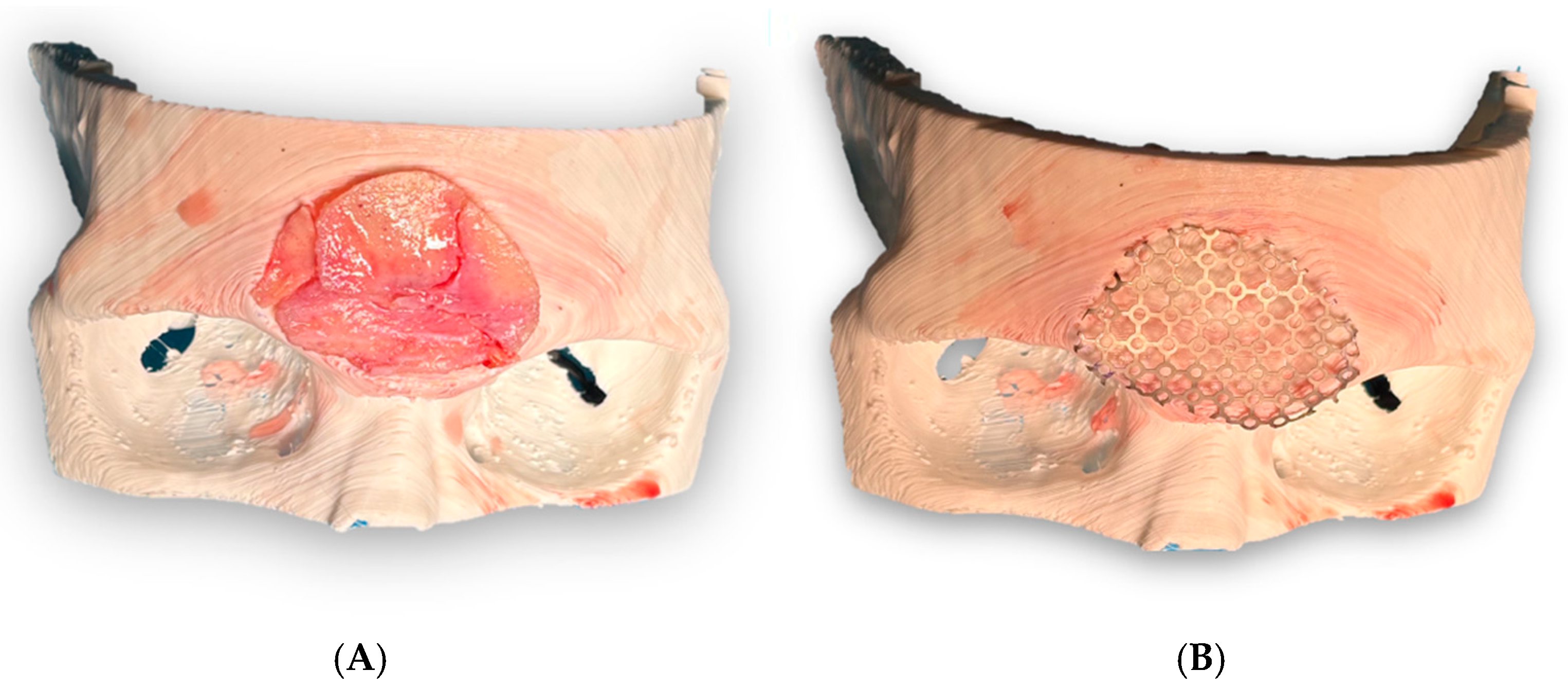
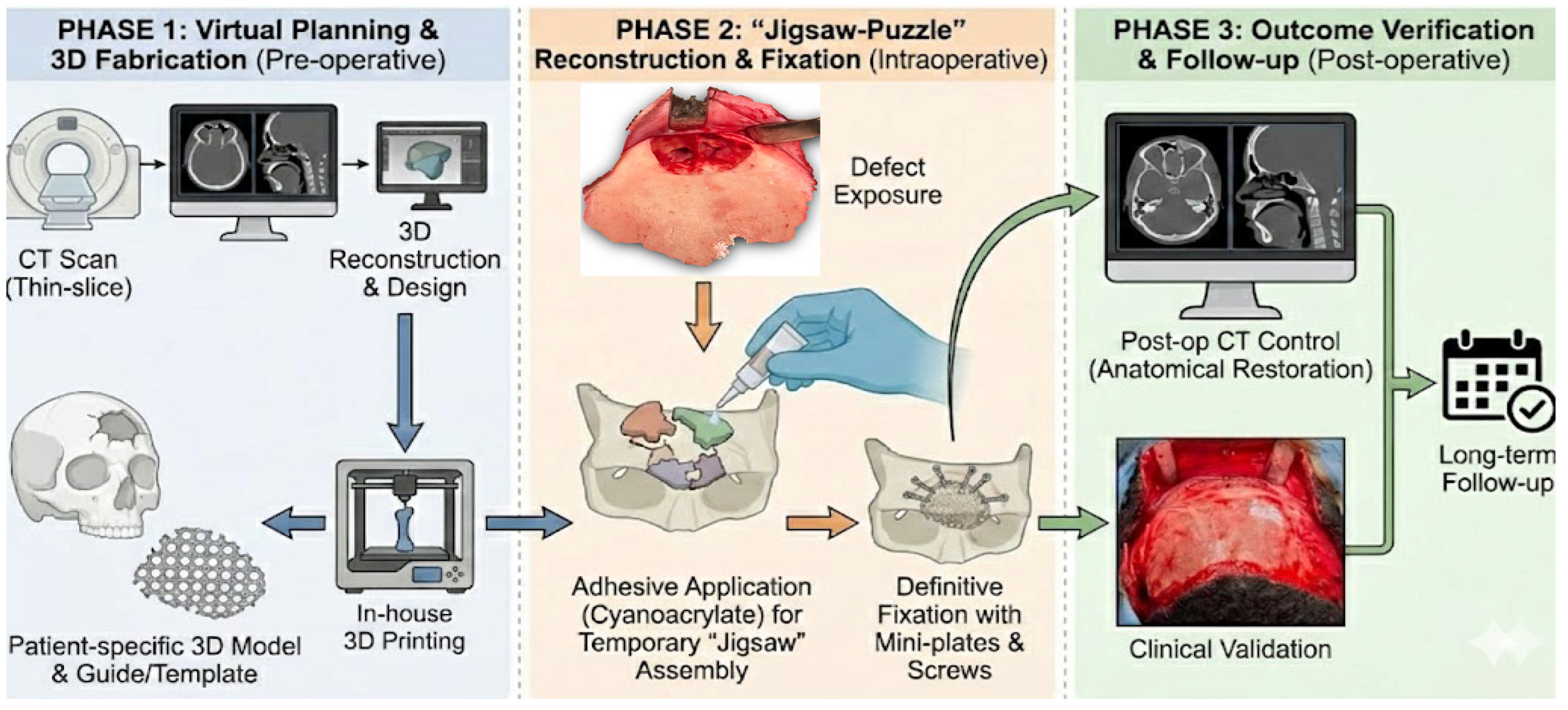
2.4. Digital Planning and 3D Biomodel Design (POC Workflow)
- Defect/hollow model: reproducing the depressed region and fracture void to improve the three-dimensional understanding of the defect.
- Reconstruction/template model: representing the intended anatomical anterior table contour to guide extracorporeal reassembly of the comminuted fragments against a stable reference surface.
2.5. Materials (Fixation Systems and Adhesive)
- Adhesive: Glubran2® (GEM S.r.l., Viareggio, Italy), a modified N-butyl-2-cyanoacrylate.
- Rigid fixation: titanium microplates and screws (standard cranio-maxillofacial osteosynthesis system), with configuration selected intraoperatively according to fragment size and bone thickness.
- Backup strategy: patient-adapted/custom titanium mesh designed from the digital model (prepared but not used in this case).
2.6. Surgical Procedure
2.6.1. Surgical Approach and Exposure
2.6.2. Assessment of Frontal Sinus Drainage and Sinus Preservation Strategy
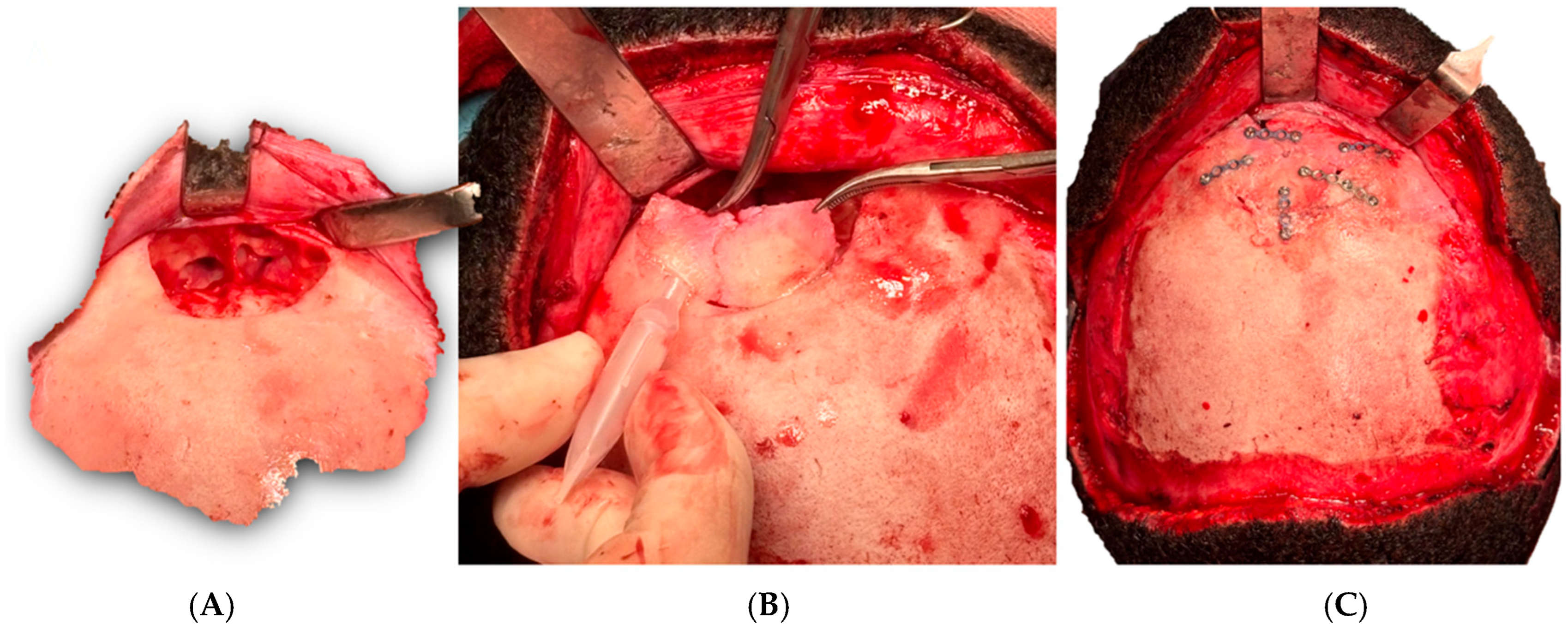
2.6.3. Extracorporeal Fragment Reassembly Using the 3D Template
2.6.4. Adhesive-Assisted Stabilization and Definitive Fixation
- The adhesive was applied in minimal, targeted amounts at selected interfragmentary interfaces.
- The construct was held immobile during polymerization to prevent micro-displacement.
- Titanium microplates were then applied to provide long-term mechanical stability, aiming to achieve stable fixation with limited hardware.
2.6.5. Closure and Immediate Postoperative Care
2.7. Outcome Measures and Follow-Up Assessment
- Clinical endpoints: restoration of frontal contour; wound complications (infection, dehiscence, hematoma/seroma); sensory disturbances; and patient-reported satisfaction.
- Radiological endpoints: postoperative CT assessment of anterior table alignment and contour restoration.
- Quantitative endpoint (recommended): objective CT-based metrics can complement visual assessment, including (i) craniometric measurements of anterior table displacement/step-off and (ii) 3D surface deviation analysis (deviation maps) by rigidly registering the postoperative CT to the preoperative virtual plan/template and reporting mean absolute/RMS deviation and the percentage of surface within predefined tolerances (e.g., ±1–2 mm). In this initial case report, outcome assessment was primarily qualitative and these quantitative metrics will be prioritized in future series.
- Follow-up: clinical surveillance for sinonasal symptoms and delayed complications.
2.8. Data, Materials, and Protocol Availability
- Data: Anonymized imaging excerpts and intraoperative photographs are provided within the manuscript figures. Full raw DICOM data are not publicly deposited due to patient privacy regulations but may be made available upon reasonable request to the corresponding author, subject to institutional review and data-sharing agreements.
- Materials: The adhesive and fixation systems are commercially available as specified.
- Protocols: The digital planning and POC manufacturing workflow is described step-by-step in Section 2.4, Section 2.5 and Section 2.6 to enable replication.
3. Results
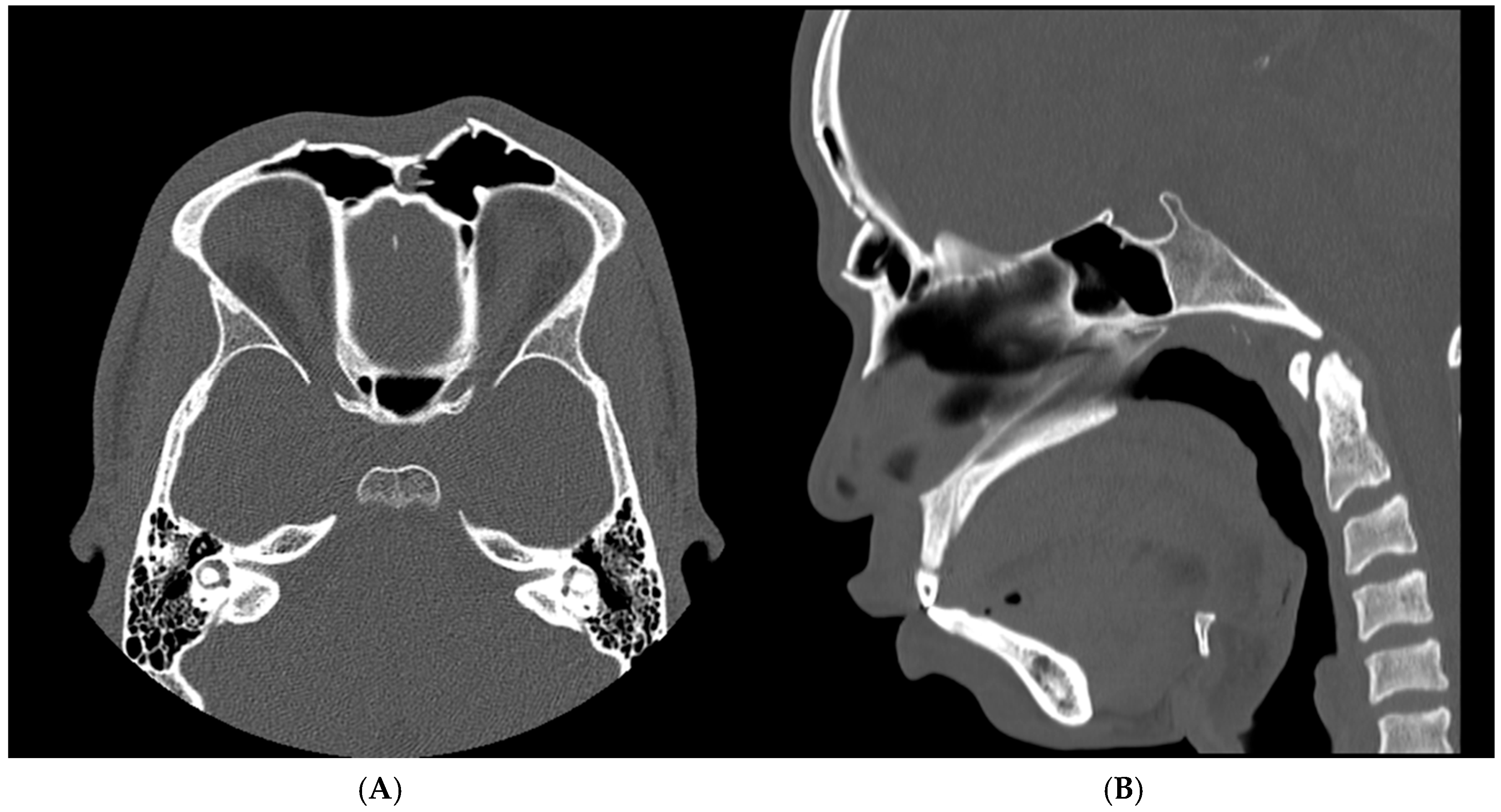
3.1. Preoperative Findings
3.2. Point-of-Care Planning Output and Intraoperative Feasibility
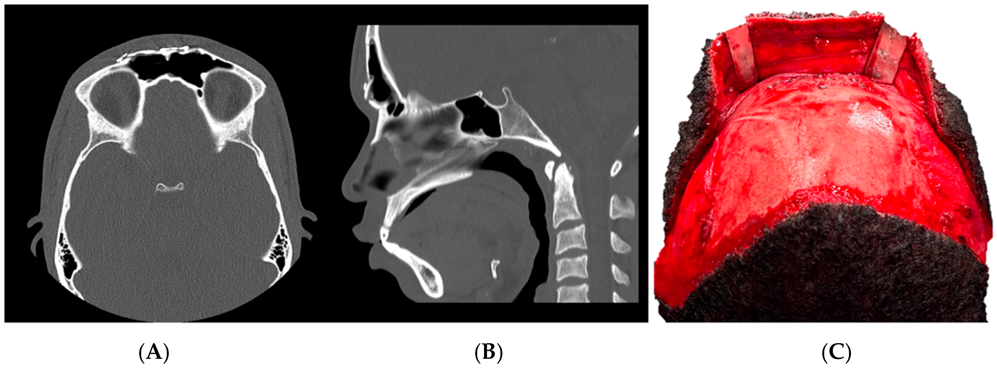
3.3. Postoperative Outcomes and Follow-Up
4. Discussion
4.1. Biosafety Profile of Cyanoacrylate Surgical Adhesives: Rationale, Benefits, and Limitations
4.2. Novelty and Current Evidence Gap in Cranio-Maxillofacial Osteosynthesis
4.3. Evidence Suggesting Inferior Osteosynthetic Performance Compared with Screws/Titanium
4.4. Cyanoacrylate as a Coadjuvant: Where It May Add Value
4.5. Comparative Perspective by Cranio-Maxillofacial Subsite
- Mandible (load-bearing): High functional loads and micromotion make adhesive-only fixation less attractive. Mandibular studies (pilot clinical and osteotomy fixation) illustrate feasibility but reinforce that rigid stability remains critical, and adhesive-only constructs should be interpreted cautiously [34,35].
- Upper third/frontal sinus anterior table (non-load-bearing contour unit): This region is primarily contour-bearing rather than masticatory load-bearing. Key endpoints are contour restoration, stable bony union, and avoidance of sinus complications. In this setting, cyanoacrylate as a coadjuvant is most defensible: it can stabilize small segments without extensive hardware, potentially reducing palpability while rigid fixation provides definitive stability [1,2,3,4,5].
4.6. Expected Behavior in Load-Bearing vs. Non-Load-Bearing Facial Bone
4.7. Technical Novelty: Extracorporeal Reduction Guided by POC Biomodel and Academic Manufacturing
4.8. Future Directions
4.9. Limitations and Need for Validation
5. Conclusions
Supplementary Materials
Author Contributions
Funding
Institutional Review Board Statement
Informed Consent Statement
Data Availability Statement
Acknowledgments
Conflicts of Interest
References
- Jing, X.L.; Luce, E. Frontal Sinus Fractures: Management and Complications. Craniomaxillofac. Trauma Reconstr. 2019, 12, 241–248. [Google Scholar] [CrossRef]
- Calis, M.; Kaplan, G.O.; Küçük, K.Y.; Altunbulak, A.Y.; Akgöz Karaosmanoğlu, A.; Işıkay, A.İ.; Mavili, M.E.; Tunçbilek, G. Algorithms for the management of frontal sinus fractures: A retrospective study. J. Craniomaxillofac. Surg. 2022, 50, 749–755. [Google Scholar] [CrossRef] [PubMed]
- Schultz, K.; Braun, T.L.; Truong, T.A. Frontal Sinus Fractures. Semin. Plast. Surg. 2017, 31, 80–84. [Google Scholar] [CrossRef] [PubMed]
- Dedhia, R.D.; Morisada, M.V.; Tollefson, T.T.; Strong, E.B. Contemporary management of frontal sinus fractures. Curr. Opin. Otolaryngol. Head Neck Surg. 2019, 27, 253–260. [Google Scholar] [CrossRef]
- Sakat, M.S.; Kilic, K.; Altas, E.; Gozeler, M.S.; Ucuncu, H. Comminuted Frontal Sinus Fracture Reconstructed With Titanium Mesh. J. Craniofac. Surg. 2016, 27, e207–e208. [Google Scholar] [CrossRef]
- Foresta, E.; Torroni, A.; Gasparini, G.; Saponaro, G.; Longo, G.; Boniello, R.; Cervelli, D.; Marianetti, T.M.; Pelo, S.; Moro, A. Use of N-Butyl-2-Cyanoacrylate (Glubran2®) in Fractures of Orbital-Maxillo-Zygomatic Complex. J. Maxillofac. Oral Surg. 2015, 14, 761–764. [Google Scholar] [CrossRef] [PubMed]
- Dadaş, B.; Alkan, S.; Cifci, M.; Başak, T. Treatment of tripod fracture of zygomatic bone by N-2-butyl cyanoacrylate glue fixation, and its effects on the tissues. Eur. Arch. Otorhinolaryngol. 2007, 264, 539–544. [Google Scholar] [CrossRef]
- Hafeez, A.A.; Furmeen, S.S.; Durairaj, D.; Rajasekaran, M.A.; Rajiah, D. Application of N-Butyl-2-Cyanoacrylate for the Treatment of Comminuted Fractures in the Anterior Wall of the Maxillary Sinus: A Prospective Clinical Study. Cureus 2023, 15, e35487. [Google Scholar] [CrossRef]
- Baş, B.; Ozden, B.; Bekçioğlu, B.; Sanal, K.O.; Gülbahar, M.Y.; Kabak, Y.B. Screw fixation is superior to N-butyl-2-cyanoacrylate in onlay grafting procedure: A histomorphologic study. Int. J. Oral Maxillofac. Surg. 2012, 41, 537–543. [Google Scholar] [CrossRef]
- Wang, B.H.; Boulton, M.; Lee, D.H.; Pelz, D.M.; Lownie, S.P. A systematic characterization of the factors influencing polymerization and dynamic behavior of n-butyl cyanoacrylate. J. Neurointerv. Surg. 2018, 10, 150–155. [Google Scholar] [CrossRef]
- Shermak, M.A.; Wong, L.; Inoue, N.; Crain, B.J.; Im, M.J.; Chao, E.Y.; Manson, P.N. Fixation of the craniofacial skeleton with butyl-2-cyanoacrylate and its effects on histotoxicity and healing. Plast. Reconstr. Surg. 1998, 102, 309–318. [Google Scholar] [CrossRef]
- Kaplan, M.; Bozkurt, S.; Kut, M.S.; Kullu, S.; Demirtas, M.M. Histopathological effects of ethyl 2-cyanoacrylate tissue adhesive following surgical application: An experimental study. Eur. J. Cardiothorac. Surg. 2004, 25, 167–172. [Google Scholar] [CrossRef] [PubMed]
- Miranda, M.; Gianfreda, F.; Molica, G.; Martelli, M.; Gargari, M.; Bollero, P. The Use of Cyanoacrylate and Glubran in Dentistry: A Review of Clinical Applications and Outcomes. Materials 2025, 18, 2642. [Google Scholar] [CrossRef]
- Wang, T.H.; Yu, W.C.; Shih, Y.C.; Chen, C.E.; Wang, S.J.; Ma, H.; Chen, W.M. 3D-Printed “Jigsaw Puzzle” in Craniomaxillofacial Comminuted Fracture Reduction. Ann. Plast. Surg. 2025, 94, S3–S8. [Google Scholar] [CrossRef] [PubMed]
- Calvo-Haro, J.A.; Pascau, J.; Asencio-Pascual, J.M.; Calvo-Manuel, F.; Cancho-Gil, M.J.; Del Cañizo López, J.F.; Fanjul-Gómez, M.; García-Leal, R.; González-Casaurrán, G.; González-Leyte, M.; et al. Point-of-care manufacturing: A single university hospital’s initial experience. 3D Print. Med. 2021, 7, 11. [Google Scholar] [CrossRef]
- Tousidonis, M.; Ruiz-de-Leon, G.; Navarro-Cuellar, C.; Ochandiano, S.; Salmeron, J.I.; Franco Herrera, R.; Calvo-Haro, J.A.; Perez-Mañanes, R. Academic Point-of-Care Manufacturing in Oral and Maxillofacial Surgery: A Retrospective Review at Gregorio Marañón University Hospital. Medicina 2026, 62, 234. [Google Scholar] [CrossRef]
- Ruiz-de-Leon, G.; Navarro-Cuellar, C.; Tousidonis, M. Maxillary Jaw-in-a-Day: The Role of Point-of-Care Manufacturing and Implant Stability Quotient in Centrofacial Chondrosarcoma. J. Craniofac. Surg. 2025, 36, e481–e483. [Google Scholar] [CrossRef]
- Tousidonis, M.; Navarro-Cuellar, C.; Fariña, R. Proportional Condylectomy for the Treatment of Unilateral Condylar Hyperplasia With Malocclusion: Role of Point-of-Care Manufacturing Surgical Guides and Intraoperative Computed Tomography to Avoid Secondary Orthognathic Surgery. J. Craniofac. Surg. 2025, 36, e251–e253. [Google Scholar] [CrossRef] [PubMed]
- Giray, C.B.; Atasever, A.; Durgun, B.; Araz, K. Clinical and electron microscope comparison of silk sutures and n-butyl-2-cyanoacrylate in human mucosa. Aust. Dent. J. 1997, 42, 255–258. [Google Scholar] [CrossRef]
- Joshi, A.D.; Saluja, H.; Mahindra, U.; Halli, R. A comparative study: Efficacy of tissue glue and sutures after impacted mandibular third molar removal. J. Maxillofac. Oral Surg. 2011, 10, 310–315. [Google Scholar] [CrossRef]
- Bozkurt, M.K.; Saydam, L. The use of cyanoacrylates for wound closure in head and neck surgery. Eur. Arch. Otorhinolaryngol. 2008, 265, 331–335. [Google Scholar] [CrossRef]
- Soni, A.; Narula, R.; Kumar, A.; Parmar, M.; Sahore, M.; Chandel, M. Comparing cyanoacrylate tissue adhesive and conventional subcuticular skin sutures for maxillofacial incisions—A prospective randomized trial considering closure time, wound morbidity, and cosmetic outcome. J. Oral Maxillofac. Surg. 2013, 71, 2152.e1–2152.e8. [Google Scholar] [CrossRef]
- DeRenzis, F.A.; Aleo, J.J. An in vitro bioassay of cyanoacrylate cytotoxicity. Oral Surg. Oral Med. Oral Pathol. 1970, 30, 803–808. [Google Scholar] [CrossRef] [PubMed]
- Inal, S.; Yilmaz, N.; Nisbet, C.; Güvenç, T. Biochemical and histopathological findings of N-butyl-2-cyanoacrylate in oral surgery: An experimental study. Oral Surg. Oral Med. Oral Pathol. Oral Radiol. Endod. 2006, 102, e14–e17. [Google Scholar] [CrossRef]
- Sonis, S.; Jandinski, J.J., 3rd; Qiunta, J. Cyanoacrylate as an adhesive for other medicaments: A study with triamcinalone on the healing of experimental oral ulcers. Pharmacol. Ther. Dent. 1975, 2, 147–156. [Google Scholar]
- Kim, P.H.; Tsauo, J.; Shin, J.H.; Yun, S.C. Transcatheter Arterial Embolization of Gastrointestinal Bleeding with N-Butyl Cyanoacrylate: A Systematic Review and Meta-Analysis of Safety and Efficacy. J. Vasc. Interv. Radiol. 2017, 28, 522–531.e5. [Google Scholar] [CrossRef]
- Kull, S.; Martinelli, I.; Briganti, E.; Losi, P.; Spiller, D.; Tonlorenzi, S.; Soldani, G. Glubran2 surgical glue: In vitro evaluation of adhesive and mechanical properties. J. Surg. Res. 2009, 157, e15–e21. [Google Scholar] [CrossRef]
- Pérez, M.; Fernández, I.; Márquez, D.; Bretaña, R.M. Use of N-butyl-2-cyanoacrylate in oral surgery: Biological and clinical evaluation. Artif. Organs 2000, 24, 241–243. [Google Scholar] [CrossRef]
- Salata, L.A.; Mariguela, V.C.; Antunes, A.A.; Grossi-Oliveira, G.; Almeida, A.; Taba, M., Jr. Short-term evaluation of grafts fixed with either N-butyl-2-cyanoacrylate or screws. J. Oral Maxillofac. Surg. 2014, 72, 676–682. [Google Scholar] [CrossRef] [PubMed]
- Gosain, A.K.; Song, L.; Corrao, M.A.; Pintar, F.A. Biomechanical evaluation of titanium, biodegradable plate and screw, and cyanoacrylate glue fixation systems in craniofacial surgery. Plast. Reconstr. Surg. 1998, 101, 582–591. [Google Scholar] [CrossRef] [PubMed]
- Gosain, A.K.; Plastic Surgery Educational Foundation DATA Committee. The current status of tissue glues: I. For bone fixation. Plast. Reconstr. Surg. 2002, 109, 2581–2583. [Google Scholar] [CrossRef]
- Rezende, M.L.; Cunha, P.O.; Damante, C.A.; Santana, A.C.; Greghi, S.L.; Zangrando, M.S. Cyanoacrylate Adhesive as an Alternative Tool for Membrane Fixation in Guided Tissue Regeneration. J. Contemp. Dent. Pract. 2015, 16, 512–518. [Google Scholar] [CrossRef]
- Choi, B.H.; Kim, B.Y.; Huh, J.Y.; Lee, S.H.; Zhu, S.J.; Jung, J.H.; Li, J. Cyanoacrylate adhesive for closing sinus membrane perforations during sinus lifts. J. Craniomaxillofac. Surg. 2006, 34, 505–509. [Google Scholar] [CrossRef]
- Mehta, M.J.; Shah, K.H.; Bhatt, R.G. Osteosynthesis of mandibular fractures with N-butyl cyanoacrylate: A pilot study. J. Oral Maxillofac. Surg. 1987, 45, 393–396. [Google Scholar] [CrossRef] [PubMed]
- Shermak, M.A.; Wong, L.; Inoue, N.; Chao, E.Y.; Manson, P.N. Butyl-2-cyanoacrylate fixation of mandibular osteotomies. Plast. Reconstr. Surg. 1998, 102, 319–324. [Google Scholar] [CrossRef] [PubMed]
- Perin, L.F.; Helene, A., Jr.; Fraga, M.F. Sutureless closure of the upper eyelids in blepharoplasty: Use of octyl-2-cyanoacrylate. Aesthet. Surg. J. 2009, 29, 87–92. [Google Scholar] [CrossRef] [PubMed]
- Ranson, J.M.; Amin, K.; Schechter, E.M.; Kosutic, D. Haemostatic property of cyanoacrylate in pedicled flaps. Br. J. Oral Maxillofac. Surg. 2016, 54, 1046–1047. [Google Scholar] [CrossRef]
- De Santis, E.; Silva, E.R.; Martins, E.N.C.; Favero, R.; Botticelli, D.; Xavier, S.P. Healing at the Interface Between Autologous Block Bone Grafts and Recipient Sites Using n-Butyl-2-Cyanoacrylate Adhesive as Fixation: Histomorphometric Study in Rabbits. J. Oral Implantol. 2017, 43, 447–455. [Google Scholar] [CrossRef]
- Negre, A.; Sy, K.; Sabri, H.; Kalani, K.; Wang, H.L.; Agossa, K. Cyanoacrylate Adhesive Versus Sutures for Free Gingival Graft Fixation: A Systematic Review and Meta-Analysis of Clinical Outcomes. J. Esthet. Restor. Dent. 2025, 37, 2368–2378. [Google Scholar] [CrossRef]



| Workflow Step | Key Actions (Replicable Details) | Output(s) | Clinical Endpoint(S)/Decision Points |
|---|---|---|---|
| 1. Clinical assessment | CMF trauma evaluation; inspection/palpation of forehead depression; surgical indication based on deformity and fracture pattern | Clinical baseline | Indication for anatomical contour restoration and stable fixation |
| 2. CT imaging acquisition | High-resolution craniofacial CT with thin-slice protocol | DICOM dataset | Define fracture pattern; assess anterior/posterior table involvement; plan sinus preservation strategy |
| 3. Image segmentation | Thresholding + region growing; manual refinement of fracture margins; artifact correction | 3D bone/fracture segmentation | Accurate delineation of comminuted fragments for planning and modeling |
| 4. 3D model generation | STL creation; mesh repair (holes/non-manifold); minimal smoothing preserving landmarks | Printable STL model | Reliable geometric substrate for template design and intraop guidance |
| 5. Biomodel design | Two-part approach: (i) defect/hollow model; (ii) reconstruction/template model representing intended premorbid contour | Defect model + anatomical reconstruction template | Enable extracorporeal “puzzle” reduction; reduce intraoperative guesswork |
| 6. Backup fixation planning | Design of patient-adapted titanium mesh from digital model (contingency strategy) | Backup mesh design | Decision point if fragment viability/stability is insufficient for reconstruction |
| 7. POC manufacturing | In-house 3D printing; post-processing (supports removal/finishing); dimensional QC (visual inspection + fitting) | Physical biomodel(s) available for planning/intraop reference | Improve reduction accuracy and efficiency; provide tangible contour reference in comminution |
| 8. Surgical exposure | Coronal approach; subgaleal/subperiosteal elevation; careful fragment mobilization/removal; bed preparation | Exposed defect and free fragments | Optimize conditions for precise reduction; avoid malreduction by debris/interposed bone |
| 9. Sinus drainage assessment | Intraoperative evaluation of sinonasal drainage pathway | Drainage status (intact vs. compromised) | Decision: preserve sinus vs. obliterate/cranialize; in this case, preservation |
| 10. Extracorporeal reduction (“puzzle”) | Fragment reassembly against reconstruction template outside operative field | Reassembled fragment construct | Reproduce anatomical curvature; minimize repeated in-field manipulation |
| 11. Adhesive-assisted stabilization | Targeted, minimal Glubran2® at selected interfaces; immobilization during polymerization | Temporary interfragmentary stability | Maintain alignment of small/irregular fragments that are difficult to screw/plate alone |
| 12. Definitive rigid fixation | Titanium microplates/screws placed to reinforce construct with limited hardware | Stable osteosynthesis | Long-term mechanical stability; minimize palpability/hardware burden |
| 13. Closure and immediate care | Layered closure; no drain; routine analgesia/wound care | Uneventful early recovery | Early endpoint: absence of wound issues/CSF leak/sinonasal symptoms |
| 14. Postoperative imaging | CT to assess contour restoration and fragment alignment | Postoperative CT confirmation | Radiological endpoint: restored anterior table contour and alignment |
| 15. Follow-up | Clinical surveillance including sinonasal symptoms and signs of delayed complications | 6-month clinical follow-up | Endpoint: asymptomatic course; no infection, instability, or sinus-related complaints |
Disclaimer/Publisher’s Note: The statements, opinions and data contained in all publications are solely those of the individual author(s) and contributor(s) and not of MDPI and/or the editor(s). MDPI and/or the editor(s) disclaim responsibility for any injury to people or property resulting from any ideas, methods, instructions or products referred to in the content. |
© 2026 by the authors. Licensee MDPI, Basel, Switzerland. This article is an open access article distributed under the terms and conditions of the Creative Commons Attribution (CC BY) license.
Share and Cite
Tousidonis, M.; Khayat, S.; Maza-Muela, C.; Franco-Herrera, R.; Pérez-Mañanes, R.; Calvo-Haro, J.-A.; Troulis, M.J.; Navarro-Cuellar, C.; Salmeron, J.-I.; Ochandiano, S. Digitally Guided Frontal Sinus Fracture Fixation: A Point-of-Care “In-House” Biomodel Protocol with Cyanoacrylate-Assisted Fragment Stabilization. J. Clin. Med. 2026, 15, 2057. https://doi.org/10.3390/jcm15052057
Tousidonis M, Khayat S, Maza-Muela C, Franco-Herrera R, Pérez-Mañanes R, Calvo-Haro J-A, Troulis MJ, Navarro-Cuellar C, Salmeron J-I, Ochandiano S. Digitally Guided Frontal Sinus Fracture Fixation: A Point-of-Care “In-House” Biomodel Protocol with Cyanoacrylate-Assisted Fragment Stabilization. Journal of Clinical Medicine. 2026; 15(5):2057. https://doi.org/10.3390/jcm15052057
Chicago/Turabian StyleTousidonis, Manuel, Saad Khayat, Cristina Maza-Muela, Rocio Franco-Herrera, Ruben Pérez-Mañanes, Jose-Antonio Calvo-Haro, Maria J. Troulis, Carlos Navarro-Cuellar, Jose-Ignacio Salmeron, and Santiago Ochandiano. 2026. "Digitally Guided Frontal Sinus Fracture Fixation: A Point-of-Care “In-House” Biomodel Protocol with Cyanoacrylate-Assisted Fragment Stabilization" Journal of Clinical Medicine 15, no. 5: 2057. https://doi.org/10.3390/jcm15052057
APA StyleTousidonis, M., Khayat, S., Maza-Muela, C., Franco-Herrera, R., Pérez-Mañanes, R., Calvo-Haro, J.-A., Troulis, M. J., Navarro-Cuellar, C., Salmeron, J.-I., & Ochandiano, S. (2026). Digitally Guided Frontal Sinus Fracture Fixation: A Point-of-Care “In-House” Biomodel Protocol with Cyanoacrylate-Assisted Fragment Stabilization. Journal of Clinical Medicine, 15(5), 2057. https://doi.org/10.3390/jcm15052057