Systematic Review

Effect of Magnesium-Modified Titanium Implants on Osseointegration: A Systematic Review and Meta-Analysis of Preclinical Studies

1
Department of Prosthetic Dental Sciences, College of Dentistry, Qassim University, Buraydah 51452, Saudi Arabia
2
Department of Periodontology and Implant Dentistry, College of Dentistry, Qassim University, Buraydah 51452, Saudi Arabia
*
Authors to whom correspondence should be addressed.
J. Clin. Med. 2026, 15(5), 1987; https://doi.org/10.3390/jcm15051987
Submission received: 29 January 2026 / Revised: 22 February 2026 / Accepted: 3 March 2026 / Published: 5 March 2026
(This article belongs to the Special Issue Current Trends in Implant Dentistry)

Abstract

Objectives: This study systematically evaluated and quantitatively synthesized preclinical evidence on the effects of magnesium (Mg) incorporation into or coating of titanium dental implants on osseointegration and peri-implant bone formation. Methods: Electronic searches of PubMed, Scopus, and Web of Science were performed up to May 2025 to identify animal studies evaluating Mg-modified titanium implants. Eligible studies compared Mg-incorporated or Mg-coated implants with non-modified titanium controls and reported quantitative histomorphometric outcomes. Primary outcomes included the values of bone-to-implant contact (BIC) and bone area (BA) around implants. Study quality was assessed using the ARRIVE 2.0 guidelines. Meta-analyses were performed using weighted mean differences with 95% confidence intervals under fixed- or random-effects models based on heterogeneity. Results: Eleven preclinical animal studies conducted in rabbit and rat models were included. Mg was incorporated using various surface-modification techniques, including ion implantation, Mg-substituted hydroxyapatite coatings, mesoporous titania layers, and nanotubular structures. Overall, the studies’ quality was high, with most studies rated as excellent and with a low-to-moderate risk of bias. Furthermore, the meta-analysis revealed a significant increase in BIC for Mg-modified implants compared with uncoated implants (Z = 4.38, p < 0.001), implying improved osseointegration. Meanwhile, pooled BA values showed no significant differences between the groups (Z = 0.93, p = 0.35). Conclusions: Mg coating onto or incorporation into titanium implant surfaces can improve BIC in preclinical models, indicating improved osseointegration in the early stages.

1. Introduction

Dental implants are the favored treatment option for replacing missing teeth in many cases, with success rates exceeding 90% in one long-term evaluation [1,2]. The cornerstone of implant success is the establishment of osseointegration, defined as a direct, load-bearing interface between bone and an implant surface without intervening fibrous tissue [3,4]. However, systemic and local risk factors, such as osteoporosis, diabetes mellitus, radiation therapy, smoking, and peri-implant infections, may negatively affect bone healing and increase the risk of biological complications or early implant failure [5]. Therefore, advances in implant surface engineering aimed at enhancing biological responses at the bone–implant interface have remained a major research priority.
Various surface modification approaches, such as micro-roughening, sandblasting, acid-etching, anodization, and laser structuring, have been developed to improve cellular adhesion, accelerate bone apposition, and enhance early implant stability [6,7]. Moreover, bioactive implant coatings, such as hydroxyapatite (HA), have been investigated extensively due to their chemical nature being similar to natural bone minerals [4]. However, some reports raised concerns related to their long-term reliability and limited clinical success [8]. Recently, there have been more attempts to incorporate ions or elements using new strategies to enhance osteogenic and immune responses at the molecular level.
Magnesium (Mg) has recently emerged as one of the most promising elements for dental implant surface functionalization [9,10]. As the fourth most abundant positively charged ion in the human body, Mg plays an essential role in bone metabolism, cellular homeostasis, osteoblast differentiation, and mineral deposition [11]. Strong evidence suggests that Mg ions can stimulate crucial pro-osteogenic pathways—including Wnt/β-catenin and VEGF/HIF-1α—thereby enhancing angiogenesis and improving bone tissue formation around implants [12]. Additionally, Mg exhibits immunomodulatory properties by reducing inflammatory cytokine activity and favoring M2 macrophage polarization, ultimately supporting a regenerative peri-implant environment [13].
Mg has been extensively investigated as an implant coating due to it sustained biodegradability, which permits gradual ion release directly at the bone–implant interface [14,15]. The localized release of Mg ions has been shown to enhance osteoblast proliferation, control osteoclast differentiation, and limit bacterial adhesion on implant surfaces [16]. These activates can facilitate osseointegration and improve resistance to implant-associated infections [16,17]. These effects can be crucial since peri-implant mucositis and peri-implantitis are major causes of implant loss in the long term [18].
Several preclinical reports revealed the favorable effects of Mg incorporation or coatings on implant osseointegration. For instance, Tao et al. reported that Mg-, Zn-, and Sr-incorporated HA coatings can significantly enhance bone formation in osteopenic rats [19]. Yang et al. reported that Mg-incorporated Ti nanotube implants demonstrated better integration with bone with lower risks of infection-induced osteolysis [20]. Furthermore, Shen et al. found higher bone regeneration and antibacterial activities when coating implant with a Mg/zinc framework [21]. Some reports suggested that titanium implants modified with Mg with different microstructures have a positive influence on implant integration with bone especially in compromised bone conditions [9]. This was confirmed by some in vivo studies that found greater bone formation and stronger biomechanical anchorage in osteoporotic animal models [9,17]. In their in vivo investigation, Okuzu et al. reported improved early stability for implants with Mg- and Sr-releasing surfaces in a rabbit implant model [22]. Another animal experiment investigated titanium (Ti) implants coated with Mg ions and reported enhanced peri-implant bone quality compared with control implants without a coating in osteoporotic bone [23].
However, in addition to these favorable findings, other studies found conflicting results. For instance, Galli et al. found that Mg-releasing mesoporous coatings did not significantly influence early bone maturation in rabbits [24]. Variations in coating techniques, including micro-arc oxidation, ion implantation, sol–gel processing, and nanoscale deposition, may result in differing Mg release kinetics, degradation behavior, and biological responses [25,26]. Accordingly, the available research evidence remains fragmented due to heterogeneous study designs, animal models, coating formulations, and outcome evaluation methods.
Given the increasing adoption of Mg-incorporated implant technologies and the need to apply these findings in human patients, a systematic and quantitative appraisal of the existing evidence is warranted. Therefore, this systematic review and meta-analysis aims to comprehensively assess the effects of Mg-coated dental implants on osseointegration while exploring potential sources of heterogeneity across preclinical studies.

2. Results

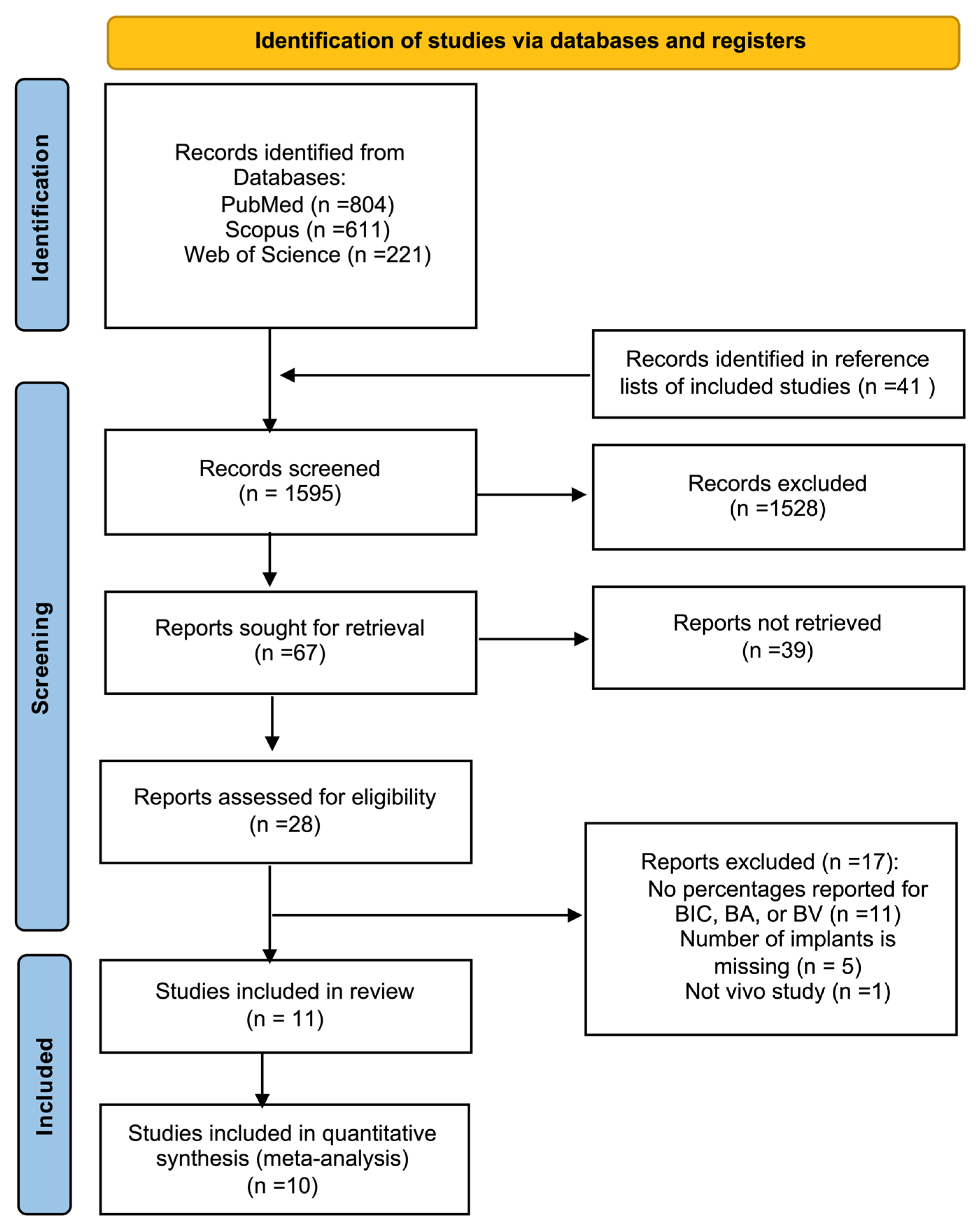
Eleven preclinical animal studies that evaluated Mg-incorporated or Mg-coated Ti implants were included in this review (Figure 1) [19,20,21,22,24,27,28,29,30,31,32]. The implants were evaluated in rabbit or rat models, primarily in tibial and femoral bone sites, with healing periods of 2–24 weeks. Multiple surface-modification strategies are represented, including Mg-ion implantation, Mg-substituted HA coatings, Mg-loaded mesoporous titania layers, and Mg-incorporated nanotubular structures (Table 1).

2.1. Quality Assessment

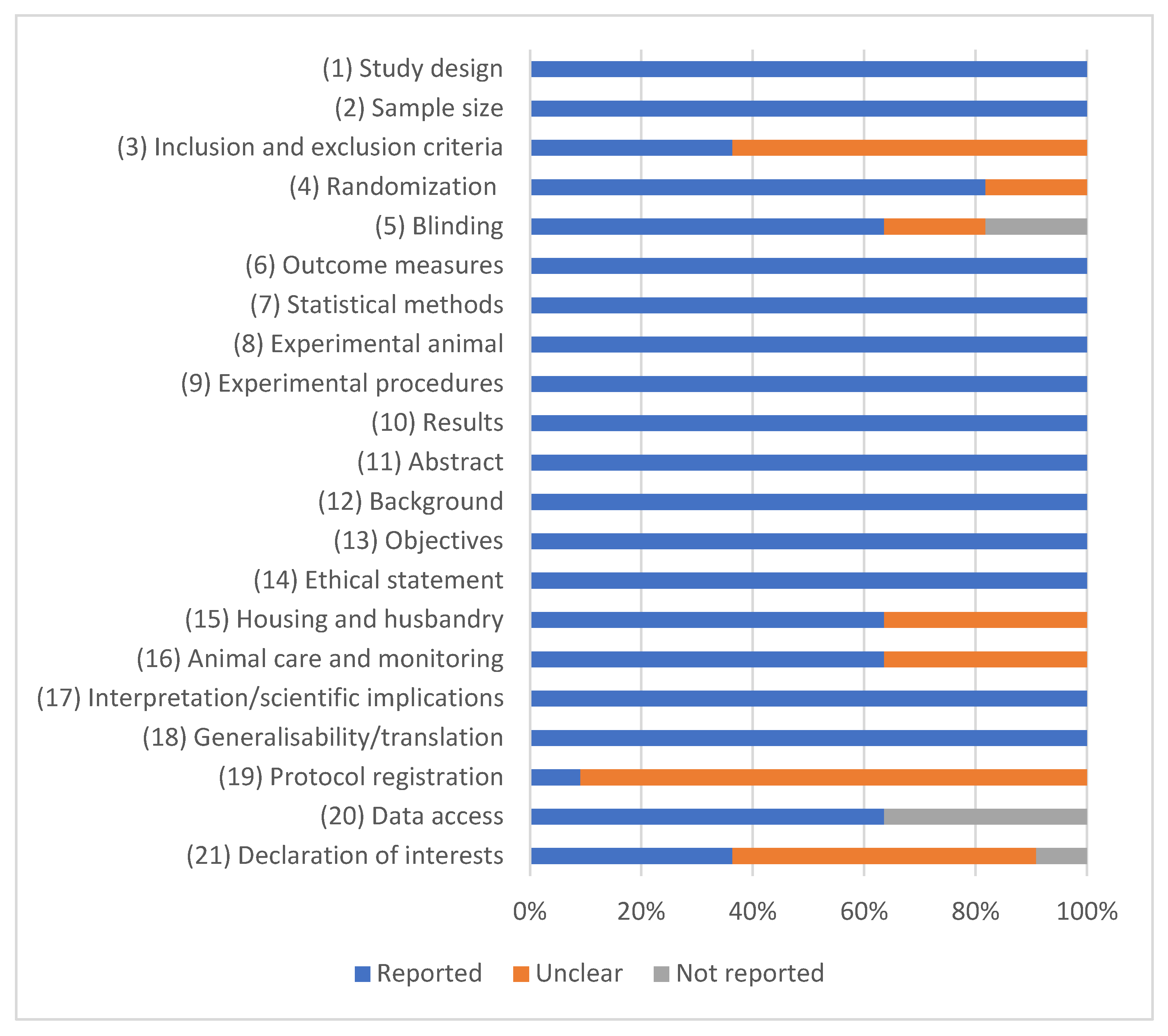
An ARRIVE 2.0 evaluation revealed an overall high study quality, with eight studies rated as excellent and three studies rated as average (Figure 2; Table 2). No studies were excluded due to quality concerns.

2.2. Risk-of-Bias Assessment

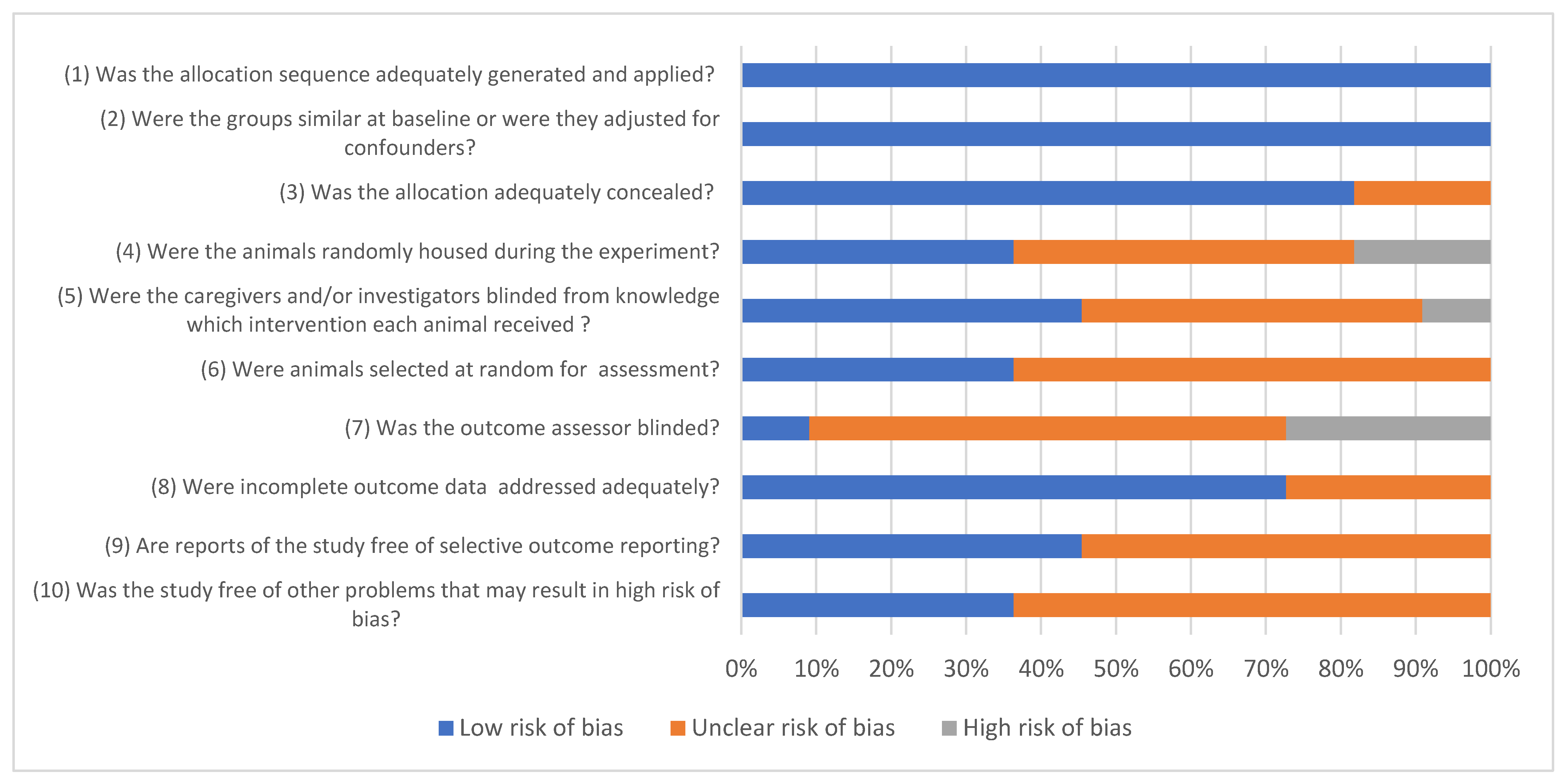
The risk-of-bias assessment using the SYRCLE tool revealed an overall low-to-moderate risk of bias across the included preclinical studies (Figure 3). Most of the studies demonstrated a low risk of bias for domains related to baseline characteristics, outcome assessment, and selective outcome reporting. Outcome data were generally complete, and histomorphometric analyses, such as BIC and BA fraction, were clearly defined and consistently reported.

2.3. Meta-Analysis of BIC

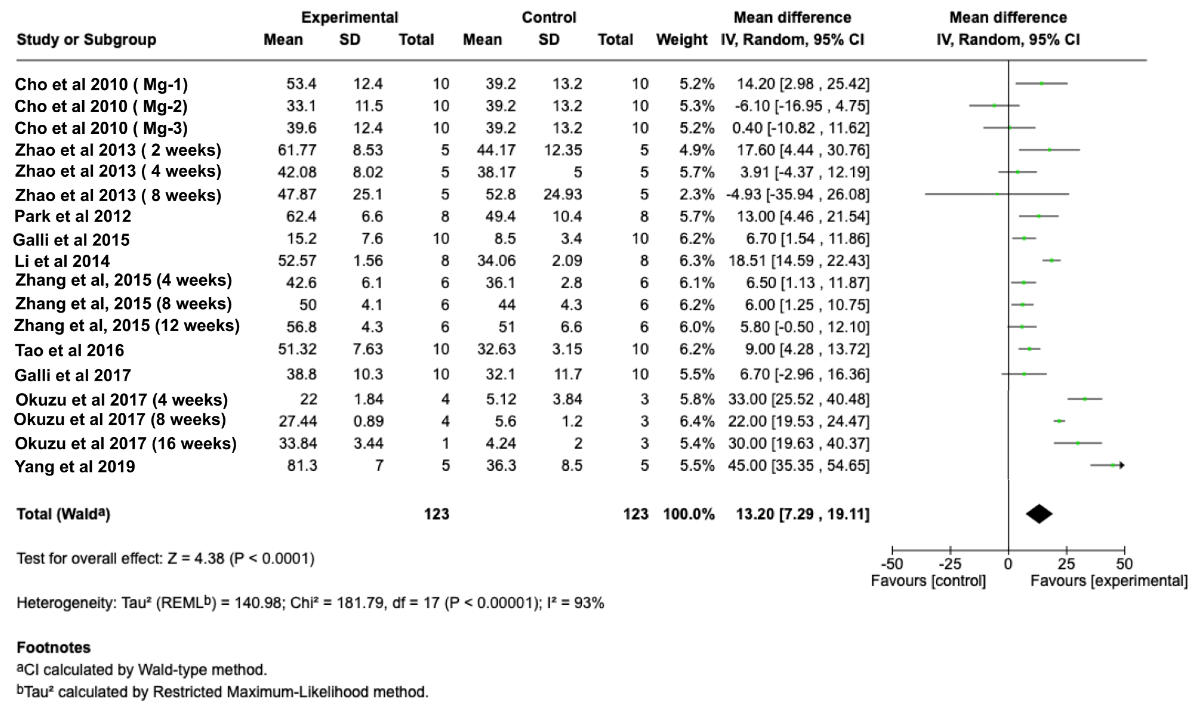
Quantitative synthesis demonstrated a significant increase in BIC for Mg-incorporated implants when compared with non-Mg controls. The pooled analysis showed a favorable effect of Mg-modified surfaces (Z = 4.38, p < 0.001) (Figure 4). Improvements in BIC were observed across the included studies; however, no formal subgroup analyses were conducted to assess the influence of specific coating strategies.

2.4. Meta-Analysis of BA

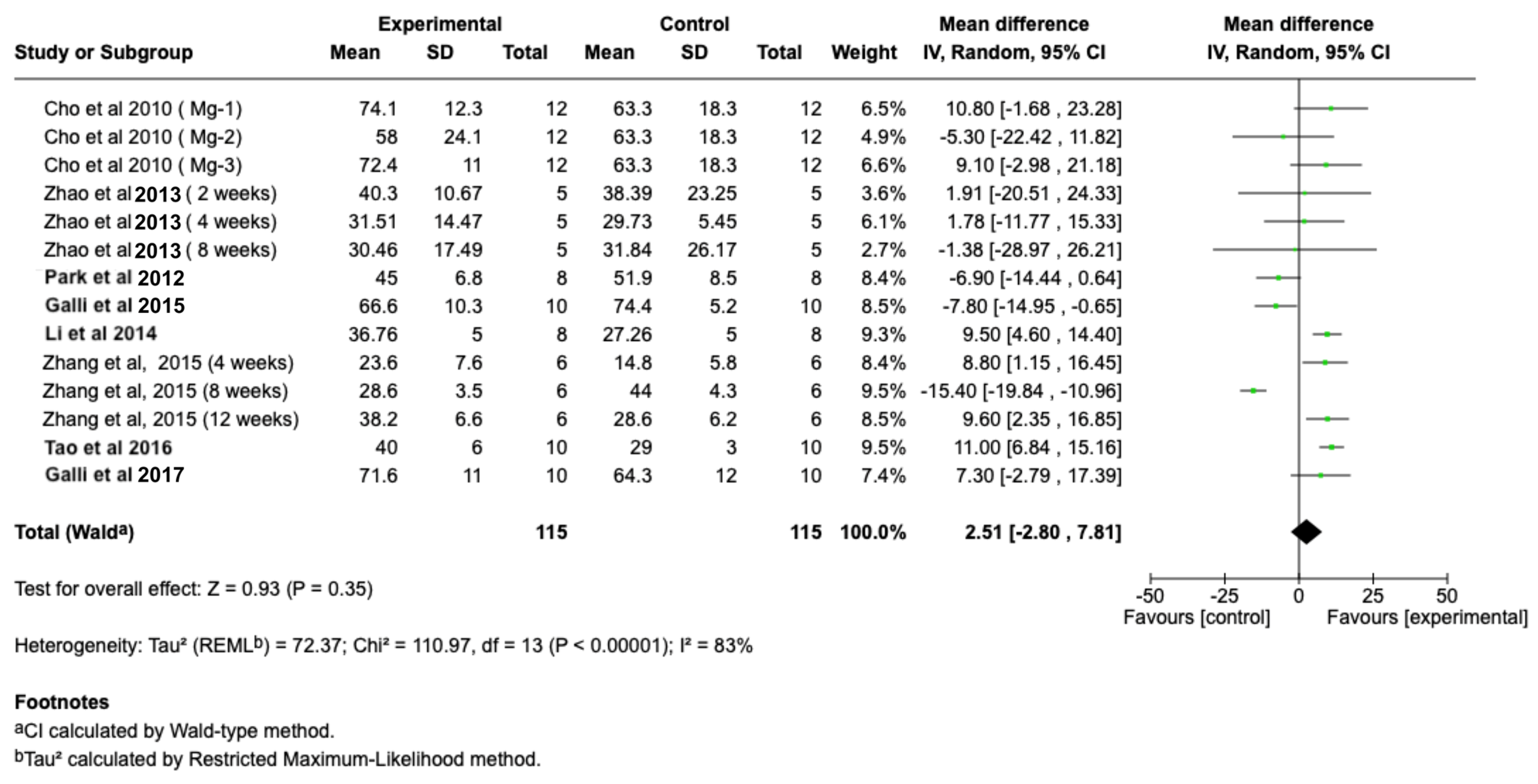
Pooled BA (%) outcomes did not show a significant difference within the experimental and control groups (Z = 0.93, p = 0.35) (Figure 5). However, a trend toward increased peri-implant bone formation was noted, particularly in studies with extended healing durations (≥8 weeks).

3. Discussion

This review demonstrates that Mg incorporation into Ti implant surfaces significantly enhances BIC, indicating improved early osseointegration compared with unmodified controls [19,20,21,22,24,27,28,29,30,31,32]. These findings are consistent with preclinical literature showing that Mg supports osteoblast function and mineralization by modulating integrin signaling, RUNX2 expression, and alkaline phosphatase activity [33,34]. Improved BIC may reflect Mg’s capacity to accelerate contact osteogenesis during early healing.
Although BA did not show a significant pooled improvement, this may be due to BIC and BA reflecting different biological phenomena: BIC captures the quality of direct bone contact at an implant interface, whereas BA represents regional bone volume that may require longer remodeling periods to change significantly [35]. The released Mg concentrates near implant surfaces, creating localized effects that significantly improve bone–implant contact without immediately changing the broader bone architecture [22]. Indeed, the studies using controlled-release systems, such as Mg-loaded mesoporous titania and nanotubes, reported enhanced bone volume, indicating more mature bone formation with optimized ion delivery [20,36].
Mg may also support osseointegration via immunomodulatory mechanisms by promoting M2 macrophage polarization and angiogenesis via HIF-1α and VEGF pathways [37,38]. Although few of the included studies assessed immune or vascular responses directly, the consistent increase in BIC supports the presence of a favorable early peri-implant environment [20,21].
Furthermore, a main cause of heterogeneity within the included studies is the variation in Mg coating methods. The evaluated studies used diverse techniques, such as ion implantation, micro-arc oxidation, mesoporous titania carriers, and nanotubular structures [39,40]. These methods produce coatings that differ considerably in coating features, degradation behavior, and surface topography. For instance, ion implantation or incorporation into the surface can produces thin layers with a controlled Mg concentration but reduced release duration [31]. Meanwhile, loading Mg into surfaces with mesoporous or nanotubular features may allow for sustained ion release, which may extend its biological activity [20]. It has been suggested that hydroxyapatite-based systems exhibit osteoconductive properties, though concerns were raised regarding degradation behavior and coating stability of HA in the long term. Since subgroup analyses were not performed in this review, definitive conclusions regarding the advantage of one method over another cannot be drawn. Nevertheless, the inconsistency in biological effects likely reflects differences in release behavior and surface stability between the implants produced using different modification techniques.
Another important consideration for Mg’s effects is their dose-dependence. Moderate Mg ion release is believed to promote osteogenesis and angiogenesis. However, high concentrations of Mg may enhance corrosion and be associated with cytotoxicity [41]. No comparisons were mentioned in the included studies regarding the optimal release behavior for Mg and the ideal therapeutic amount for Mg incorporation and release remains unclear.
From a clinical prospective, Mg-coated implants could provide significant advantages, especially in compromised bone conditions, such as osteoporosis, where improved osseointegration has been confirmed in preclinical models [19,31]. These results indicate that this is a promising surface modification method. However, concerns remain regarding coating stability and loading protocols, which require further investigation in clinical trials with functionally loaded jawbone models. In this review, the included studies were performed mainly on small animal models and without functional loading, and human clinical data remain absent. These promising preclinical findings require validation using large-animal studies and clinical evaluation before general implementation in dental practice.
Limitations of this study include methodological heterogeneity across studies and inadequate reporting of biomechanical outcomes. Furthermore, the ideal Mg concentration and release profiles remain uncertain, since high Mg concentrations may lead to corrosion or cytotoxicity [15,42]. Future studies should aim to evaluate standardize concentration levels and evaluate dose–response effects under controlled experimental conditions.

4. Materials and Methods

4.1. PICO Framework

This systematic review was according to the guidelines of the Preferred Reporting Items for Systematic Reviews and Meta-Analyses (PRISMA) (Supplementary Table S1) [43]. Additionally, it was recorded in the International Platform of Registered Systematic Review and Meta-Analysis Protocols (INPLASY) (registration number: INPLASY202610101). The PICO framework was established at beginning of the search process as follows:
  • Population (P): animal models;
  • Intervention (I): Ti implants coated or incorporated with Mg;
  • Control (C): Ti implants without Mg incorporation or coating;
  • Outcome (O): bone formation around implants.

4.2. Search Strategies

An electronic search was conducted in May 2025 in three main databases (PubMed, Scopus, and Web of Science) to identify animal studies published not before 2005. The search terms included four key concepts: bone formation, Ti implants, Mg coating, and animal experimentation. The search strategy was as follows: (“bone formation” OR “bone remodeling” OR “osseointegration”) AND (“titanium implants” OR “bone-implant interface”) AND (“magnesium” OR “Mg coating” OR “antimicrobial surfaces” OR “implant surface modification”) AND (“animal models” OR “in vivo”).

4.3. Inclusion and Exclusion Criteria

Studies were eligible if they investigated Mg-modified endosseous Ti implants in animal models. The Mg had to be applied locally, either as a coating or as a releasable agent from an implant surface, during or before implant insertion. Eligible studies reported quantitative bone outcomes, such as bone-to-implant contact (BIC), the percent bone volume (BV/TV), or bone area (BA). The uncoated (control) and Mg-modified (test) implants groups were required to have implants with similar surface characteristics except for the incorporation of Mg. Only articles published in English were included.

4.4. Study Selection

Titles and abstracts retrieved from the search results were screened against the eligibility criteria. Two independent reviewers evaluated the abstracts, and when relevance was uncertain, the full-text articles were assessed.

4.5. Data Extraction

A standardized form was used for data extraction to collect information related to study characteristics and publication date. The collected experimental information included animal species, number of subjects and implants, healing duration, and Mg-coating technique. Furthermore, the mean ± SD values for BIC and BA results for both the test and control groups were collected.

4.6. Quality Assessment

Study quality was determined using the ARRIVE 2.0 reporting guidelines for animal research [44]. These guidelines include 21 evaluation items. Every item was scored 2 points (reported), 1 point (unclear), or 0 points (not reported). Following that, a quality coefficient was calculated to categorize the studies as excellent (0.8–1), average (0.5–0.8), or poor (<0.5).

4.7. Risk-of-Bias Assessment

The SYRCLE risk-of-bias tool was used to assess potential biases and evaluate methodological reliability among the included experimental studies [45].

4.8. Data Analysis

The analysis examined two quantitative measurements: BA and BIC percentages. In the statistical calculations, weighted mean differences used the number of implants as the unit of analysis. Studies lacking adequate reporting of the primary outcomes were excluded. Heterogeneity was assessed using the I2 statistic. For the heterogeneity, a random-effects with inverse-variance weighting was used when significant heterogeneity was found; otherwise, a fixed-effects approach was applied. The effect size was reported as the mean difference with 95% confidence intervals. All statistical tests were conducted using Review Manager software (Version 5.3.3, The Nordic Cochrane Centre).

5. Conclusions

This systematic review and meta-analysis revealed that incorporating Mg or coating Ti implant surfaces with Mg may significantly improve BIC, indicating enhanced early tissue integration at an implant interface. Although the pooled BA data did not show a statistically significant difference, trends toward increased peri-implant bone formation were observed in several studies, particularly when Mg was delivered using controlled-release coating systems.
These findings collectively highlight the beneficial role of Mg in accelerating osseointegration and improving the quality of interfacial bone contact around Ti implants. Nevertheless, all the included studies were conducted in small-animal long-bone models, with limited evaluation of functional loading or long-term stability. Future research in jawbone environments and well-designed clinical trials is necessary to confirm the translatability of these promising preclinical outcomes into routine dental implant practice.

Supplementary Materials

The following supporting information can be downloaded at: https://www.mdpi.com/article/10.3390/jcm15051987/s1, Table S1: PRISMA checklist.

Author Contributions

Conceptualization and methodology, A.A.; software, A.A.; validation, A.A.; formal analysis, A.A.; investigation, A.A. and D.A.; resources, A.A.; data curation, A.A. and D.A.; writing—review and editing, A.A. and D.A.; visualization, A.A. and D.A.; supervision, A.A.; project administration, A.A.; All authors have read and agreed to the published version of the manuscript.

Funding

This research received no external funding.

Institutional Review Board Statement

Not applicable.

Informed Consent Statement

Not applicable.

Data Availability Statement

Data collection and interpretation in this study are maintained by the authors and available upon request.

Acknowledgments

The researchers would like to thank the Deanship of Graduate Studies and Scientific Research at Qassim University for financial support (QU-APC-2026).

Conflicts of Interest

The researchers declare that they have no competing interests.

Correction Statement

This article has been republished with a minor correction to the Document Type. This change does not affect the scientific content of the article.

Abbreviations

The following abbreviations are used in this manuscript:
Mgmagnesium
Tititanium
BICbone-to-implant contact
BAbone area
PRISMAPreferred Reporting Items for Systematic Reviews and Meta-Analyses

References

  1. Pjetursson, B.E.; Thoma, D.; Jung, R.; Zwahlen, M.; Zembic, A. A systematic review of the survival and complication rates of implant-supported fixed dental prostheses (FDP s) after a mean observation period of at least 5 years. Clin. Oral Implant. Res. 2012, 23, 22–38. [Google Scholar] [CrossRef] [PubMed]
  2. Howe, M.-S.; Keys, W.; Richards, D. Long-term (10-year) dental implant survival: A systematic review and sensitivity meta-analysis. J. Dent. 2019, 84, 9–21. [Google Scholar] [CrossRef] [PubMed]
  3. Albrektsson, T.; Wennerberg, A. Oral implant surfaces: Part 1—Review focusing on topographic and chemical properties of different surfaces and in vivo responses to them. Int. J. Prosthodont. 2004, 17, 536–543. [Google Scholar] [PubMed]
  4. Albrektsson, T.; Chrcanovic, B.; Östman, P.O.; Sennerby, L. Initial and long-term crestal bone responses to modern dental implants. Periodontol. 2000 2017, 73, 41–50. [Google Scholar]
  5. Chrcanovic, B.; Albrektsson, T.; Wennerberg, A. Reasons for failures of oral implants. J. Oral Rehabil. 2014, 41, 443–476. [Google Scholar]
  6. Buser, D.; Janner, S.F.; Wittneben, J.G.; Brägger, U.; Ramseier, C.A.; Salvi, G.E. 10-year survival and success rates of 511 titanium implants with a sandblasted and acid-etched surface: A retrospective study in 303 partially edentulous patients. Clin. Implant Dent. Relat. Res. 2012, 14, 839–851. [Google Scholar]
  7. Le Guéhennec, L.; Soueidan, A.; Layrolle, P.; Amouriq, Y. Surface treatments of titanium dental implants for rapid osseointegration. Dent. Mater. 2007, 23, 844–854. [Google Scholar] [CrossRef]
  8. Neto, J.V.C.; Teixeira, A.B.V.; Dos Reis, A.C. Hydroxyapatite coatings versus osseointegration in dental implants: A systematic review. J. Prosthet. Dent. 2025, 134, 92–99. [Google Scholar]
  9. Li, Y.-b.; Zhang, H.-q.; Lu, Y.-p.; Yang, X.-j.; Wang, G.-d.; Wang, Y.-y.; Tang, K.-l.; Huang, S.-y.; Xiao, G.-y. Construction of magnesium phosphate chemical conversion coatings with different microstructures on titanium to enhance osteogenesis and angiogenesis. ACS Appl. Mater. Interfaces 2024, 16, 21672–21688. [Google Scholar] [CrossRef]
  10. Liu, L.; Wang, F.; Song, W.; Zhang, D.; Lin, W.; Yin, Q.; Wang, Q.; Li, H.; Yuan, Q.; Zhang, S. Magnesium promotes vascularization and osseointegration in diabetic states. Int. J. Oral Sci. 2024, 16, 10. [Google Scholar] [CrossRef]
  11. Castiglioni, S.; Cazzaniga, A.; Albisetti, W.; Maier, J.A. Magnesium and osteoporosis: Current state of knowledge and future research directions. Nutrients 2013, 5, 3022–3033. [Google Scholar] [CrossRef]
  12. Nie, X.; Sun, X.; Wang, C.; Yang, J. Effect of magnesium ions/Type I collagen promote the biological behavior of osteoblasts and its mechanism. Regen. Biomater. 2020, 7, 53–61. [Google Scholar] [CrossRef] [PubMed]
  13. Lee, C.-H.; Kim, Y.-J.; Jang, J.-H.; Park, J.-W. Modulating macrophage polarization with divalent cations in nanostructured titanium implant surfaces. Nanotechnology 2016, 27, 085101. [Google Scholar] [CrossRef] [PubMed]
  14. Staiger, M.P.; Pietak, A.M.; Huadmai, J.; Dias, G. Magnesium and its alloys as orthopedic biomaterials: A review. Biomaterials 2006, 27, 1728–1734. [Google Scholar] [CrossRef] [PubMed]
  15. Chen, Y.; Xu, Z.; Smith, C.; Sankar, J. Recent advances on the development of magnesium alloys for biodegradable implants. Acta Biomater. 2014, 10, 4561–4573. [Google Scholar] [CrossRef]
  16. Nie, X.; Shi, Y.; Wang, L.; Abudureheman, W.; Yang, J.; Lin, C. Study on the mechanism of magnesium calcium alloys/mineralized collagen composites mediating macrophage polarization to promote bone repair. Heliyon 2024, 10, e30279. [Google Scholar] [CrossRef]
  17. Park, J.-W.; Hanawa, T.; Chung, J.-H. The relative effects of Ca and Mg ions on MSC osteogenesis in the surface modification of microrough Ti implants. Int. J. Nanomed. 2019, 14, 5697–5711. [Google Scholar] [CrossRef]
  18. Berglundh, T.; Armitage, G.; Araujo, M.G.; Avila-Ortiz, G.; Blanco, J.; Camargo, P.M.; Chen, S.; Cochran, D.; Derks, J.; Figuero, E. Peri-implant diseases and conditions: Consensus report of workgroup 4 of the 2017 World Workshop on the Classification of Periodontal and Peri-Implant Diseases and Conditions. J. Clin. Periodontol. 2018, 45, S286–S291. [Google Scholar] [CrossRef]
  19. Tao, Z.-S.; Zhou, W.-S.; He, X.-W.; Liu, W.; Bai, B.-L.; Zhou, Q.; Huang, Z.-L.; Tu, K.-k.; Li, H.; Sun, T. A comparative study of zinc, magnesium, strontium-incorporated hydroxyapatite-coated titanium implants for osseointegration of osteopenic rats. Mater. Sci. Eng. C 2016, 62, 226–232. [Google Scholar] [CrossRef]
  20. Yang, Y.; Liu, L.; Luo, H.; Zhang, D.; Lei, S.; Zhou, K. Dual-Purpose Magnesium-Incorporated Titanium Nanotubes for Combating Bacterial Infection and Ameliorating Osteolysis to Realize Better Osseointegration. ACS Biomater. Sci. Eng. 2019, 5, 5368–5383. [Google Scholar] [CrossRef]
  21. Shen, X.; Zhang, Y.; Ma, P.; Sutrisno, L.; Luo, Z.; Hu, Y.; Yu, Y.; Tao, B.; Li, C.; Cai, K. Fabrication of magnesium/zinc-metal organic framework on titanium implants to inhibit bacterial infection and promote bone regeneration. Biomaterials 2019, 212, 1–16. [Google Scholar] [CrossRef]
  22. Okuzu, Y.; Fujibayashi, S.; Yamaguchi, S.; Yamamoto, K.; Shimizu, T.; Sono, T.; Goto, K.; Otsuki, B.; Matsushita, T.; Kokubo, T. Strontium and magnesium ions released from bioactive titanium metal promote early bone bonding in a rabbit implant model. Acta Biomater. 2017, 63, 383–392. [Google Scholar] [CrossRef] [PubMed]
  23. Galli, S.; Stocchero, M.; Andersson, M.; Karlsson, J.; He, W.; Lilin, T.; Wennerberg, A.; Jimbo, R. The effect of magnesium on early osseointegration in osteoporotic bone: A histological and gene expression investigation. Osteoporos. Int. 2017, 28, 2195–2205. [Google Scholar] [CrossRef] [PubMed]
  24. Galli, S.; Naito, Y.; Karlsson, J.; He, W.; Andersson, M.; Wennerberg, A.; Jimbo, R. Osteoconductive potential of mesoporous titania implant surfaces loaded with magnesium: An experimental study in the rabbit. Clin. Implant Dent. Relat. Res. 2015, 17, 1048–1059. [Google Scholar] [CrossRef] [PubMed]
  25. Ming, X.; Wu, Y.; Zhang, Z.; Li, Y. Micro-arc oxidation in titanium and its alloys: Development and potential of implants. Coatings 2023, 13, 2064. [Google Scholar] [CrossRef]
  26. Wan, P.; Tan, L.; Yang, K. Surface modification on biodegradable magnesium alloys as orthopedic implant materials to improve the bio-adaptability: A review. J. Mater. Sci. Technol. 2016, 32, 827–834. [Google Scholar] [CrossRef]
  27. Cho, L.R.; Kim, D.G.; Kim, J.H.; Byon, E.S.; Jeong, Y.S.; Park, C.J. Bone response of Mg ion-implanted clinical implants with the plasma source ion implantation method. Clin. Oral Implant. Res. 2010, 21, 848–856. [Google Scholar] [CrossRef]
  28. Zhao, S.f.; Jiang, Q.h.; Peel, S.; Wang, X.x.; He, F.m. Effects of magnesium-substituted nanohydroxyapatite coating on implant osseointegration. Clin. Oral Implant. Res. 2013, 24, 34–41. [Google Scholar] [CrossRef]
  29. Park, J.W.; An, C.H.; Jeong, S.H.; Suh, J.Y. Osseointegration of commercial microstructured titanium implants incorporating magnesium: A histomorphometric study in rabbit cancellous bone. Clin. Oral Implant. Res. 2012, 23, 294–300. [Google Scholar] [CrossRef]
  30. Li, X.; Li, Y.; Liao, Y.; Li, J.; Zhang, L.; Hu, J. The effect of magnesium-incorporated hydroxyapatite coating on titanium implant fixation in ovariectomized rats. Int. J. Oral Maxillofac. Implant. 2014, 29, 196–202. [Google Scholar] [CrossRef]
  31. Zhang, J.; Liu, L.; Zhao, S.; Wang, H.; Yang, G. Characterization and In Vivo Evaluation of Trace Element-Loaded Implant Surfaces in Ovariectomized Rats. Int. J. Oral Maxillofac. Implant. 2015, 30, 1105–1112. [Google Scholar] [CrossRef] [PubMed]
  32. Galli, S.; Andersson, M.; Jinno, Y.; Karlsson, J.; He, W.; Xue, Y.; Mustafa, K.; Wennerberg, A.; Jimbo, R. Magnesium release from mesoporous carriers on endosseus implants does not influence bone maturation at 6 weeks in rabbit bone. J. Biomed. Mater. Res. Part B Appl. Biomater. 2017, 105, 2118–2125. [Google Scholar] [CrossRef] [PubMed]
  33. Wang, S.; Zhao, X.; Hsu, Y.; He, Y.; Wang, F.; Yang, F.; Yan, F.; Xia, D.; Liu, Y. Surface modification of titanium implants with Mg-containing coatings to promote osseointegration. Acta Biomater. 2023, 169, 19–44. [Google Scholar] [CrossRef]
  34. Nourisa, J.; Zeller-Plumhoff, B.; Helmholz, H.; Luthringer-Feyerabend, B.; Ivannikov, V.; Willumeit-Römer, R. Magnesium ions regulate mesenchymal stem cells population and osteogenic differentiation: A fuzzy agent-based modeling approach. Comput. Struct. Biotechnol. J. 2021, 19, 4110–4122. [Google Scholar] [CrossRef] [PubMed]
  35. Buser, D.; Sennerby, L.; De Bruyn, H. Modern implant dentistry based on osseointegration: 50 years of progress, current trends and open questions. Periodontol. 2000 2017, 73, 7–21. [Google Scholar] [CrossRef]
  36. Chen, Y.; Zhou, L.; Guan, M.; Jin, S.; Tan, P.; Fu, X.; Zhou, Z. Multifunctionally disordered TiO(2) nanoneedles prevent periprosthetic infection and enhance osteointegration by killing bacteria and modulating the osteoimmune microenvironment. Theranostics 2024, 14, 6016–6035. [Google Scholar] [CrossRef]
  37. Tang, H.; Zhao, D.; Tian, J. Enhanced osteogenic activities of magnesium-containing nanowire modified-titanium surface via macrophage immunomodulation. Biointerphases 2025, 20, 061002. [Google Scholar] [CrossRef]
  38. Bessa-Gonçalves, M.; Silva, A.M.; Brás, J.P.; Helmholz, H.; Luthringer-Feyerabend, B.J.C.; Willumeit-Römer, R.; Barbosa, M.A.; Santos, S.G. Fibrinogen and magnesium combination biomaterials modulate macrophage phenotype, NF-kB signaling and crosstalk with mesenchymal stem/stromal cells. Acta Biomater. 2020, 114, 471–484. [Google Scholar] [CrossRef]
  39. He, Y.; Zhang, Y.; Shen, X.; Tao, B.; Liu, J.; Yuan, Z.; Cai, K. The fabrication and in vitro properties of antibacterial polydopamine-LL-37-POPC coatings on micro-arc oxidized titanium. Colloids Surf. B Biointerfaces 2018, 170, 54–63. [Google Scholar] [CrossRef]
  40. Alenezi, A.; Galli, S.; Atefyekta, S.; Andersson, M.; Wennerberg, A. Osseointegration effects of local release of strontium ranelate from implant surfaces in rats. J. Mater. Sci. Mater. Med. 2019, 30, 116. [Google Scholar] [CrossRef]
  41. Witte, F. The history of biodegradable magnesium implants: A review. Acta Biomater. 2010, 6, 1680–1692. [Google Scholar] [CrossRef]
  42. Lu, Y.; Deshmukh, S.; Jones, I.; Chiu, Y.L. Biodegradable magnesium alloys for orthopaedic applications. Biomater. Transl. 2021, 2, 214–235. [Google Scholar] [CrossRef]
  43. Moher, D.; Liberati, A.; Tetzlaff, J.; Altman, D.G. Preferred reporting items for systematic reviews and meta-analyses: The PRISMA statement. PLoS Med. 2009, 6, e1000097. [Google Scholar] [CrossRef]
  44. Du Sert, N.P.; Ahluwalia, A.; Alam, S.; Avey, M.T.; Baker, M.; Browne, W.J.; Clark, A.; Cuthill, I.C.; Dirnagl, U.; Emerson, M. Reporting animal research: Explanation and elaboration for the ARRIVE guidelines 2.0. PLoS Biol. 2020, 18, e3000411. [Google Scholar]
  45. Hooijmans, C.R.; Rovers, M.M.; De Vries, R.B.; Leenaars, M.; Ritskes-Hoitinga, M.; Langendam, M.W. SYRCLE’s risk of bias tool for animal studies. BMC Med. Res. Methodol. 2014, 14, 43. [Google Scholar] [CrossRef]
Figure 1. PRISMA flow diagram showing the search process for this systematic review.
Jcm 15 01987 g001
Figure 2. Quality assessments of the studies based on ARRIVE 2.0 guidelines. Obtained results are expressed as percentages.
Jcm 15 01987 g002
Figure 3. Risk-of-bias distribution based on SYRCLE assessment. Obtained results are presented as percentages.
Jcm 15 01987 g003
Figure 4. Forest plot showing the BIC values on Ti implants with Mg coatings (test) or without coatings (control). The pooled analysis showed a statistically significant overall effect for Mg-coated implants (Z = 4.38.01, p < 0.001) [19,20,22,24,27,28,29,30,31,32].
Jcm 15 01987 g004
Figure 5. Forest plot showing BA values from the included studies on implants with Mg coatings (test) or without coatings (control). The pooled analysis revealed no significant difference between Mg-coated and control implants (Z = 0.93, p = 0.35) [19,24,27,28,29,30,31,32].
Jcm 15 01987 g005
Table 1. Overview of the included studies in this systematic review.
AuthorsCoating TechniqueAnimal ModelImplant SiteEvaluation Method(s)Healing PeriodControl GroupTest Group(s)
Cho et al. 2010 [27]Mg ion-implanted
implants treated with
resorbable blasting media (RBM)
New Zealand
White rabbits
TibiaBIC and BA6 weeksTi implants treated with
RBM
Implants incorporating Mg-1 (a), Mg-2 (b), Mg-3 (c)
Zhao et al. 2013 [28]Magnesium-substituted
nanohydroxyapatite coating on implant
New Zealand White rabbitsFemursBIC and BA2, 4, and 8 weeksElectrochemically deposited pure hydroxyapatite (EDHA) coatings on the surface of
Ti implants
Electrochemically deposited
magnesium-substituted hydroxyapatite (EDMHA) coatings on the surface of pure
Ti implants
Park et al. 2012 [29]Commercial
microstructured Ti implants
incorporating magnesium
New Zealand White rabbitsFemursBIC and BA4 weeksTi implants
produced by hydroxyapatite grit blasting (RBM
implant)
Ti implants with
Mg-incorporated nanoporous oxide layer (RBM/
Mg implant)
Galli et al. 2015 [24]Mesoporous titania
surfaces loaded with magnesium
New Zealand White rabbitsTibiaBIC and BA3 weeksTi implants coated with thin films of mesoporous TiO2Mg-loaded mesoporous implants
Li et al. 2014 [30]Magnesium-incorporated hydroxyapatite (HA) coatingSprague Dawley ratsFemursBIC and BA12 weeksHA-coated implantsMg-HA coated implants
Zhang et al. 2015 [31]Hydroxyapatite (HA) coating with zinc (Zn), magnesium (Mg), or strontium (Sr) Ovariectomized Sprague Dawley ratsTibiaBIC and BA4, 8, and 12 weeksHA coatingZn-HA, Mg-HA, and Sr-HA coatings
Tao et al. 2016 [19]Zinc, magnesium, strontium-incorporated
hydroxyapatite-coated Ti implants
Sprague Dawley ratsFemursBIC, BA, and
BV/TV
12 weeksHA coatingZn-HA, Mg-HA coatings
Galli et al. 2017 [32]Ti implants were coated with mesoporous
titania layers and loaded with Mg
New Zealand White rabbitsTibiaBIC and BA6 weeksControl implantsMg-loaded implants
Okuzu et al. 2017 [22]Alkali and heat treatment followed by Mg (and Sr) ion incorporationJapanese White rabbitsTibiaBIC4, 8, 16, and 24 weeksCp-Ti implantsMg-Ti implants
Yang et al. 2019 [20]Magnesium-incorporated Ti nanotubesRatsFemursBIC35 dNanotube-modified Ti implants (NT)(Mg)-incorporated NT implants (NT-Mg)
Shen et al. 2019 [21]Magnesium/zinc metal–organic framework on Ti
implants
RatsFemursBV/TV4 weeksAlkali–heat-treated Ti (AT) implantsAT-Mg/Zn3 implants
Table 2. Quality coefficients of the included studies.
AuthorsYearAnimal ModelQuality CoefficientCategory
Cho et al. [27]2010New Zealand White rabbits0.833Excellent
Zhao et al. [28]2013New Zealand White rabbits0.738Average
Park et al. [29]2012New Zealand White rabbits0.833Excellent
Galli et al. [24]2015New Zealand White rabbits0.880Excellent
Li et al. [30]2014Sprague Dawley rats0.952Excellent
Zhang et al. [31]2015Sprague Dawley rats0.952Excellent
Tao et al. [19]2016Sprague Dawley rats0.833Excellent
Galli et al. [32]2017New Zealand White rabbits0.904Excellent
Okuzu et al. [22]2017Japanese White rabbits0.761Average
Yang et al. [20]2019Rats0.976Excellent
Shen et al. [21]2019Rats0.928Excellent
Disclaimer/Publisher’s Note: The statements, opinions and data contained in all publications are solely those of the individual author(s) and contributor(s) and not of MDPI and/or the editor(s). MDPI and/or the editor(s) disclaim responsibility for any injury to people or property resulting from any ideas, methods, instructions or products referred to in the content.