Artificial Intelligence in Orthopaedics: Clinical Performance, Limitations, and Translational Readiness—A Review



Abstract
1. Introduction
Technical Foundations of AI in Orthopaedics
2. Materials and Methods
2.1. Study Design
2.2. Search Strategy
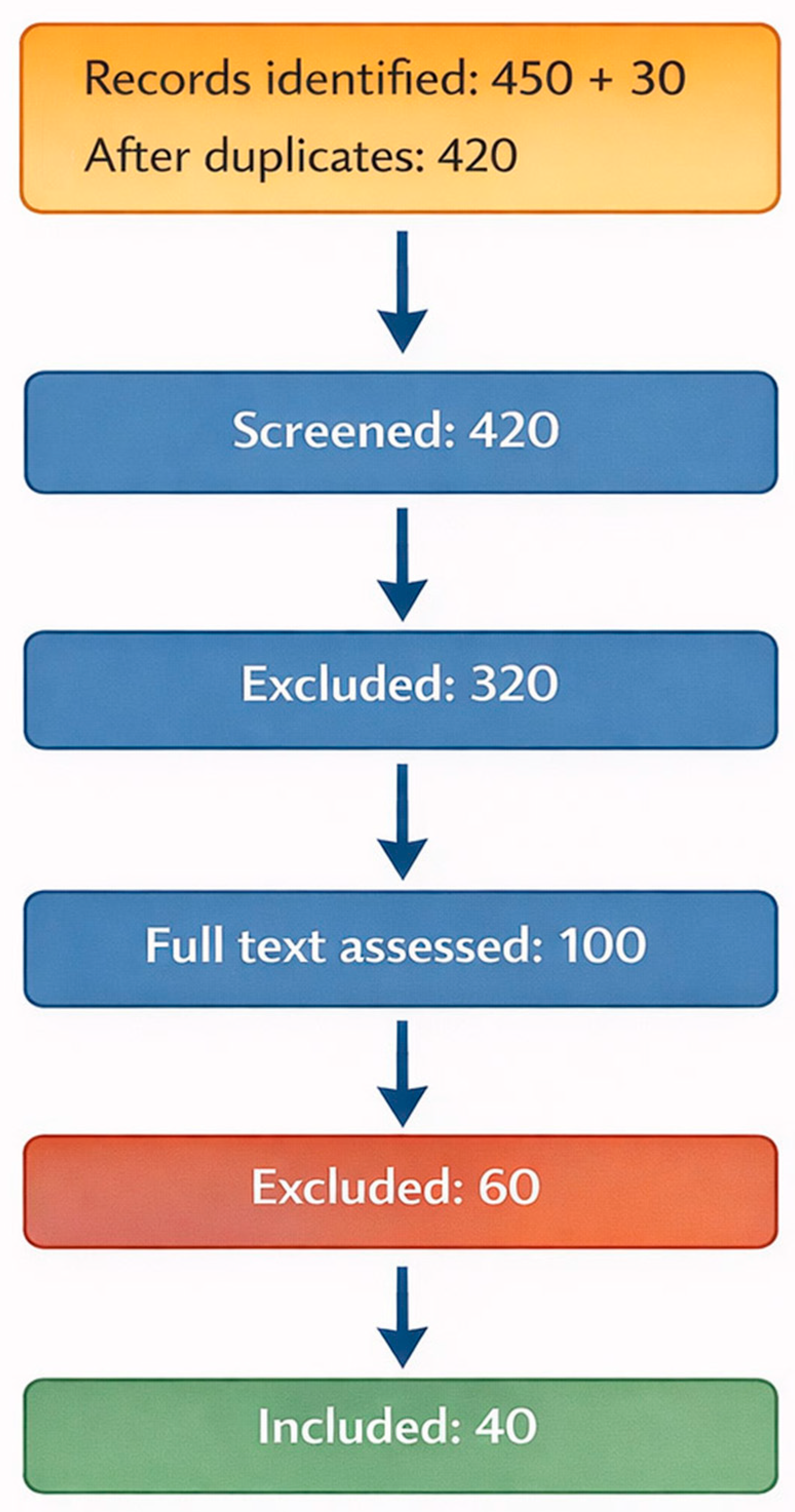
2.3. Eligibility Criteria and Study Selection
2.4. Data Extraction and Synthesis
2.5. Organization and Evaluation of Evidence
3. Results
3.1. Evidence-Based Review
3.2. Diagnostic Imaging Using AI
3.3. Surgical Planning and Intraoperative Augmentation
3.4. Predictive Analysis and Precise Stratification
3.5. Rehabilitation Intelligence and Teleorthopaedics
3.6. System-Level Governance, Ethics, and Future Trajectories
4. Discussion
4.1. Key Findings and Maturity of Evidence
4.2. Comparison with Previous Works
4.3. Technical Challenges in AI Implementation
4.4. Clinical and Healthcare System Implications
4.5. Economic and Implementation Considerations
4.6. Strengths and Limitations of This Review
4.7. Generative AI
4.8. Directions for the Future
5. Conclusions
Supplementary Materials
Author Contributions
Funding
Institutional Review Board Statement
Informed Consent Statement
Data Availability Statement
Acknowledgments
Conflicts of Interest
References
- Nguyen, A.; Lee, P.; Rodriguez, E.K.; Chahal, K.; Freedman, B.R.; Nazarian, A. Addressing the growing burden of musculoskeletal diseases in the ageing US population: Challenges and innovations. Lancet Healthy Longev. 2025, 6, 100707. [Google Scholar] [CrossRef]
- Malik, K.M.; Beckerly, R.; Imani, F. Musculoskeletal Disorders a Universal Source of Pain and Disability Misunderstood and Mismanaged: A Critical Analysis Based on the U.S. Model of Care. Anesth. Pain Med. 2018, 8, e85532. [Google Scholar] [CrossRef]
- Kumar, R.; Marla, K.; Sporn, K.; Paladugu, P.; Khanna, A.; Gowda, C.; Ngo, A.; Waisberg, E.; Jagadeesan, R.; Tavakkoli, A. Emerging Diagnostic Approaches for Musculoskeletal Disorders: Advances in Imaging, Biomarkers, and Clinical Assessment. Diagnostics 2025, 15, 1648. [Google Scholar] [CrossRef]
- Gill, T.K.; Tabish, M. Global, regional, and national burden of other musculoskeletal disorders, 1990–2020, and projections to 2050: A systematic analysis of the Global Burden of Disease Study 2021. Lancet Rheumatol. 2023, 5, e670–e682. [Google Scholar] [CrossRef]
- Lee, Y.K.; Yoon, E.-J.; Kim, T.H.; Kim, J.-I.; Kim, J.-H. Musculoskeletal Digital Therapeutics and Digital Health Rehabilitation: A Global Paradigm Shift in Orthopedic Care. JCM 2025, 14, 8467. [Google Scholar] [CrossRef] [PubMed]
- Meara, J.G.; Leather, A.J.M.; Hagander, L.; Alkire, B.C.; Alonso, N.; Ameh, E.A.; Bickler, S.W.; Conteh, L.; Dare, A.J.; Davies, J.; et al. Global Surgery 2030: Evidence and solutions for achieving health, welfare, and economic development. Lancet 2015, 386, 569–624. [Google Scholar] [CrossRef] [PubMed]
- Zarghami, A. Role of Artificial Intelligence in Surgical Decision-Making: A Comprehensive Review: Role of AI in SDM. Galen Med. J. 2024, 13, e3332. [Google Scholar] [CrossRef]
- Zhang, X.; Yang, Y.; Shen, Y.W.; Zhang, K.R.; Jiang, Z.K.; Ma, L.T.; Ding, C.; Wang, B.Y.; Meng, Y.; Liu, H. Diagnostic accuracy and potential covariates of artificial intelligence for diagnosing orthopedic fractures: A systematic literature review and meta-analysis. Eur. Radiol. 2022, 32, 7196–7216. [Google Scholar] [CrossRef]
- Liu, Y.; Wu, X.; Sang, Y.; Zhao, C.; Wang, Y.; Shi, B.; Fan, Y. Evolution of Surgical Robot Systems Enhanced by Artificial Intelligence: A Review. Adv. Intell. Syst. 2024, 6, 2300268. [Google Scholar] [CrossRef]
- Yuan, D.; Zhang, H.; Tong, L.; Chen, D. Artificial Intelligence for Bone: Theory, Methods, and Applications. Adv. Intell. Discov. 2025, 3, 202500070. [Google Scholar] [CrossRef]
- Xue, D.; Wang, K.; He, H.; Wang, L.; Dai, Y.; Shen, G.; Chen, Y.; Chen, J.; Yang, Y.; Chen, Z.; et al. Comparison of Artificial Intelligence and Traditional Methods in Preoperative Planning for Primary Total Hip Arthroplasty: A Systematic Review and Meta-Analysis. Orthop. Surg. 2025, 17, 2823–2834. [Google Scholar] [CrossRef] [PubMed]
- Vaishya, R.; Sibal, A.; Kar, S.; Reddy, S. Integrating artificial intelligence into orthopedics: Opportunities, challenges, and future directions. J. Hand Microsurg. 2025, 17, 100257. [Google Scholar] [CrossRef]
- Tian, C.; Gao, Y.; Rui, C.; Qin, S.; Shi, L.; Rui, Y. Artificial intelligence in orthopaedic trauma. EngMedicine 2024, 1, 100020. [Google Scholar] [CrossRef]
- Song, J.; Wang, G.C.; Wang, S.C.; He, C.R.; Zhang, Y.Z.; Chen, X.; Su, J.C. Artificial intelligence in orthopedics: Fundamentals, current applications, and future perspectives. Mil. Med. Res. 2025, 12, 42. [Google Scholar] [CrossRef]
- Sharma, S. Artificial intelligence for fracture diagnosis in orthopedic X-rays: Current developments and future potential. SICOT-J 2023, 9, 21. [Google Scholar] [CrossRef]
- Sharaf, Y. Application of Artificial Intelligence in Pediatric Orthopaedic. SVOAOR 2023, 3, 88811–88858. [Google Scholar] [CrossRef]
- Olczak, J.; Fahlberg, N.; Maki, A.; Razavian, A.S.; Jilert, A.; Stark, A.; Sköldenberg, O.; Gordon, M. Artificial intelligence for analyzing orthopedic trauma radiographs. Acta Orthop. 2017, 88, 581–586. [Google Scholar] [CrossRef] [PubMed]
- Lakhani, A. Revolutionizing orthopedic care: The impact of ai in predictive analysis, surgical precision, and personalized rehabilitation. JCHM 2024, 11, 120–132. [Google Scholar] [CrossRef]
- Kumar, R.; Sporn, K.; Ong, J.; Waisberg, E.; Paladugu, P.; Vaja, S.; Hage, T.; Sekhar, T.C.; Vadhera, A.S.; Ngo, A.; et al. Integrating Artificial Intelligence in Orthopedic Care: Advancements in Bone Care and Future Directions. Bioengineering 2025, 12, 513. [Google Scholar] [CrossRef]
- Bakİ, H. Transforming Orthopedic Decision-Making and Patient Care Through Artificial Intelligence. JMS 2025, 94, 94–95. [Google Scholar] [CrossRef]
- Hansen, V.; Jensen, J.; Kusk, M.W.; Gerke, O.; Tromborg, H.B.; Lysdahlgaard, S. Deep learning performance compared to healthcare experts in detecting wrist fractures from radiographs: A systematic review and meta-analysis. Eur. J. Radiol. 2024, 174, 111399. [Google Scholar] [CrossRef]
- Tieu, A.; Kroen, E.; Kadish, Y.; Liu, Z.; Patel, N.; Zhou, A.; Yilmaz, A.; Lee, S.; Deyer, T. The Role of Artificial Intelligence in the Identification and Evaluation of Bone Fractures. Bioengineering 2024, 11, 338. [Google Scholar] [CrossRef]
- Zech, J.R.; Santomartino, S.M.; Yi, P.H. Artificial Intelligence (AI) for Fracture Diagnosis: An Overview of Current Products and Considerations for Clinical Adoption, From the AJR Special Series on AI Applications. Am. J. Roentgenol. 2022, 219, 869–878. [Google Scholar] [CrossRef]
- Debs, P.; Fayad, L.M. The promise and limitations of artificial intelligence in musculoskeletal imaging. Front. Radiol. 2023, 3, 1242902. [Google Scholar] [CrossRef] [PubMed]
- Elkohail, A.; Soffar, A.; Paul, A.; Radu, L.; Wasim Shaffe Ahamed, M.; Swealem, A.; Mohideen Ahamed Sha, A.A.; Mandoth Veetil, H.H.; Millat, M.S.; Shah, R. Artificial Intelligence in Bone Fracture Detection: A Review of Evidence, Limitations, and Clinical Integration. Cureus 2025, 17, e97674. [Google Scholar] [CrossRef]
- Sayed, A.; Elkohail, A.; Soffar, A.; Elbanna, M.; Radu, L.; Wasim Shaffe Ahamed, M.; Shah, R. Current Concepts in Artificial Intelligence-Assisted Arthroplasty: A Review of the Perioperative Pathway. Cureus 2025, 17, e99946. [Google Scholar] [CrossRef]
- Li, P.; Wang, Y.; Zhao, R.; Hao, L.; Chai, W.; Jiying, C.; Feng, Z.; Ji, Q.; Zhang, G. The Application of artificial intelligence in periprosthetic joint infection. J. Adv. Res. 2026, 79, 633–659. [Google Scholar] [CrossRef]
- Zampogna, B.; Torre, G.; Zampoli, A.; Parisi, F.; Ferrini, A.; Shanmugasundaram, S.; Franceschetti, E.; Papalia, R. Can machine learning predict the accuracy of preoperative planning for total hip arthroplasty, basing on patient-related factors? An explorative investigation on Supervised machine learning classification models. J. Clin. Orthop. Trauma. 2024, 53, 102470. [Google Scholar] [CrossRef]
- Min, M.; Wang, X.; Urba, R.; Zhang, W.; Gao, J.; Fan, L. Comparison of traditional template measurements and artificial intelligence preoperative planning in total knee arthroplasty. Front. Surg. 2025, 12, 1573148. [Google Scholar] [CrossRef] [PubMed]
- Lan, Q.; Li, S.; Zhang, J.; Guo, H.; Yan, L.; Tang, F. Reliable prediction of implant size and axial alignment in AI-based 3D preoperative planning for total knee arthroplasty. Sci. Rep. 2024, 14, 16971. [Google Scholar] [CrossRef] [PubMed]
- Anwar, A.; Zhang, Y.; Zhang, Z.; Li, J. Artificial intelligence technology improves the accuracy of preoperative planning in primary total hip arthroplasty. Asian J. Surg. 2024, 47, 2999–3006. [Google Scholar] [CrossRef]
- Wongsak, S.; Janyawongchot, T.; Sri-Utenchai, N.; Owasirikul, D.; Jaovisidha, S.; Woratanarat, P.; Sa-Ngasoongsong, P. Development of a Deep Learning Model for Hip Arthroplasty Templating Using Anteroposterior Hip Radiograph. JCM 2025, 14, 8689. [Google Scholar] [CrossRef]
- Hu, Y.; Fan, Y.; Sun, Z.; Song, H. AI-assisted preoperative planning combined with robotic-assisted total knee arthroplasty vs. conventional surgery: A retrospective controlled study. BMC Musculoskelet. Disord. 2026, 27, 129. [Google Scholar] [CrossRef] [PubMed]
- Li, S.; Jiang, J.; Zhang, J.; Yan, L.; Guo, H.; Tang, F. Advantages and effectiveness of AI three-dimensional reconstruction technology in the preoperative planning of total hip arthroplasty. Sci. Rep. 2025, 15, 24630. [Google Scholar] [CrossRef]
- Velasquez Garcia, A.; Bukowiec, L.G.; Yang, L.; Nishikawa, H.; Fitzsimmons, J.S.; Larson, A.N.; Taunton, M.J.; Sanchez-Sotelo, J.; O’Driscoll, S.W.; Wyles, C.C. Artificial intelligence–based three-dimensional templating for total joint arthroplasty planning: A scoping review. Int. Orthop. (SICOT) 2024, 48, 997–1010. [Google Scholar] [CrossRef] [PubMed]
- Gu, Y.; Yang, Y.; Gao, X.; Wang, Y.; Yang, L.; Wei, Y. Application of artificial intelligence in risk assessment and management of venous thromboembolism: Scoping review. Front. Physiol. 2025, 16, 1664470. [Google Scholar] [CrossRef]
- Danilatou, V.; Dimopoulos, D.; Kostoulas, T.; Douketis, J. Machine Learning-Based Predictive Models for Patients with Venous Thromboembolism: A Systematic Review. Thromb. Haemost. 2024, 124, 1040–1052. [Google Scholar] [CrossRef]
- Naser, A.M.; Vyas, R.; Morgan, A.A.; Kalaiger, A.M.; Kharawala, A.; Nagraj, S.; Agarwal, R.; Maliha, M.; Mangeshkar, S.; Singh, N.; et al. Role of Artificial Intelligence in the Diagnosis and Management of Pulmonary Embolism: A Comprehensive Review. Diagnostics 2025, 15, 889. [Google Scholar] [CrossRef]
- Simon, B.D.; Ozyoruk, K.B.; Gelikman, D.G.; Harmon, S.A.; Türkbey, B. The future of multimodal artificial intelligence models for integrating imaging and clinical metadata: A narrative review. Diagn. Interv. Radiol. 2025, 31, 303–312. [Google Scholar] [CrossRef]
- Federer, S.J.; Jones, G.G. Artificial intelligence in orthopaedics: A scoping review. PLoS ONE 2021, 16, e0260471. [Google Scholar] [CrossRef] [PubMed]
- Luo, Z.; Wang, Y.; Zhang, T.; Wang, J. Effectiveness of AI-assisted rehabilitation for musculoskeletal disorders: A network meta-analysis of pain, range of motion, and functional outcomes. Front. Bioeng. Biotechnol. 2025, 13, 1660524. [Google Scholar] [CrossRef] [PubMed]
- Moldovan, F.; Moldovan, L. An Innovative Assessment Framework for Remote Care in Orthopedics. Healthcare 2025, 13, 736. [Google Scholar] [CrossRef]
- Olawade, D.B.; Adeleye, K.K.; Egbon, E.; Nwabuoku, U.S.; Clement David-Olawade, A.; Boussios, S.; Vanderbloemen, L. Enhancing home rehabilitation through AI-driven virtual assistants: A narrative review. Ann. Transl. Med. 2025, 13, 61. [Google Scholar] [CrossRef]
- Han, F.; Huang, X.; Wang, X.; Chen, Y.f.; Lu, C.; Li, S.; Lu, L.; Zhang, D.W. Artificial Intelligence in Orthopedic Surgery: Current Applications, Challenges, and Future Directions. MedComm 2025, 6, e70260. [Google Scholar] [CrossRef]
- Guo, T.-Y.; Deng, J.-H.; Zhou, Z.-M.; Chen, J.-Y.; Zhou, H.-F.; Zhang, X.; Qi, T.-T.; Zeng, H.; Yu, F. Advancements in deep learning-based image screening for orthopedic conditions: Emphasis on osteoporosis, osteoarthritis, and bone tumors. Ageing Res. Rev. 2025, 111, 102840. [Google Scholar] [CrossRef]
- Alzubaidi, L.; Al-Dulaimi, K.; Salhi, A.; Alammar, Z.; Fadhel, M.A.; Albahri, A.S.; Alamoodi, A.H.; Albahri, O.S.; Hasan, A.F.; Bai, J.; et al. Comprehensive review of deep learning in orthopaedics: Applications, challenges, trustworthiness, and fusion. Artif. Intell. Med. 2024, 155, 102935. [Google Scholar] [CrossRef]
- Luiken, I.; Lemke, T.; Komenda, A.; Marka, A.W.; Kim, S.H.; Graf, M.M.; Ziegelmayer, S.; Weller, D.; Mertens, C.J.; Bressem, K.K.; et al. Evaluation of commercial AI algorithms for the detection of fractures, effusions, and dislocations on real-world clinical data: A prospective registry study. Radiography 2025, 31, 103189. [Google Scholar] [CrossRef] [PubMed]
- Ruitenbeek, H.C.; Oei, E.H.G.; Visser, J.J.; Kijowski, R. Artificial intelligence in musculoskeletal imaging: Realistic clinical applications in the next decade. Skelet. Radiol. 2024, 53, 1849–1868. [Google Scholar] [CrossRef]
- Nowroozi, A.; Salehi, M.A.; Shobeiri, P.; Agahi, S.; Momtazmanesh, S.; Kaviani, P.; Kalra, M.K. Artificial intelligence diagnostic accuracy in fracture detection from plain radiographs and comparing it with clinicians: A systematic review and meta-analysis. Clin. Radiol. 2024, 79, 579–588. [Google Scholar] [CrossRef] [PubMed]
- Langerhuizen, D.W.G.; Janssen, S.J.; Mallee, W.H.; van den Bekerom, M.P.J.; Ring, D.; Kerkhoffs, G.; Jaarsma, R.L.; Doornberg, J.N. What Are the Applications and Limitations of Artificial Intelligence for Fracture Detection and Classification in Orthopaedic Trauma Imaging? A Systematic Review. Clin. Orthop. Relat. Res. 2019, 477, 2482–2491. [Google Scholar] [CrossRef]
- Kutbi, M. Artificial Intelligence-Based Applications for Bone Fracture Detection Using Medical Images: A Systematic Review. Diagnostics 2024, 14, 1879. [Google Scholar] [CrossRef]
- Kuo, R.Y.L.; Harrison, C.; Curran, T.A.; Jones, B.; Freethy, A.; Cussons, D.; Stewart, M.; Collins, G.S.; Furniss, D. Artificial Intelligence in Fracture Detection: A Systematic Review and Meta-Analysis. Radiology 2022, 304, 50–62. [Google Scholar] [CrossRef]
- Litjens, G.; Kooi, T.; Bejnordi, B.E.; Setio, A.A.A.; Ciompi, F.; Ghafoorian, M.; van der Laak, J.; van Ginneken, B.; Sánchez, C.I. A survey on deep learning in medical image analysis. Med. Image Anal. 2017, 42, 60–88. [Google Scholar] [CrossRef]
- Mienye, I.D.; Swart, T.G.; Obaido, G.; Jordan, M.; Ilono, P. Deep Convolutional Neural Networks in Medical Image Analysis: A Review. Information 2025, 16, 195. [Google Scholar] [CrossRef]
- Al-Nafjan, A.; Aljuhani, A.; Alshebel, A.; Alharbi, A.; Alshehri, A. Artificial Intelligence in Predictive Healthcare: A Systematic Review. JCM 2025, 14, 6752. [Google Scholar] [CrossRef]
- Kus, I.; Kocak, C.; Keles, A. A systematic review of vision transformer and explainable AI advances in multimodal facial expression recognition. Intell. Syst. Appl. 2026, 29, 200615. [Google Scholar] [CrossRef]
- Halder, A.; Gharami, S.; Sadhu, P.; Singh, P.K.; Woźniak, M.; Ijaz, M.F. Implementing vision transformer for classifying 2D biomedical images. Sci. Rep. 2024, 14, 12567. [Google Scholar] [CrossRef] [PubMed]
- Tang, H.; Chen, Y.; Wang, T.; Zhou, Y.; Zhao, L.; Gao, Q.; Du, M.; Tan, T.; Zhang, X.; Tong, T. HTC-Net: A hybrid CNN-transformer framework for medical image segmentation. Biomed. Signal Process. Control 2024, 88, 105605. [Google Scholar] [CrossRef]
- Aburass, S.; Dorgham, O.; Al Shaqsi, J.; Abu Rumman, M.; Al-Kadi, O. Vision Transformers in Medical Imaging: A Comprehensive Review of Advancements and Applications Across Multiple Diseases. J. Imaging Inform. Med. 2025, 38, 3928–3971. [Google Scholar] [CrossRef] [PubMed]
- Yao, W.; Bai, J.; Liao, W.; Chen, Y.; Liu, M.; Xie, Y. From CNN to Transformer: A Review of Medical Image Segmentation Models. J. Imaging Inform. Med. 2024, 37, 1529–1547. [Google Scholar] [CrossRef]
- Jang, R.; Kim, N.; Jang, M.; Lee, K.H.; Lee, S.M.; Lee, K.H.; Noh, H.N.; Seo, J.B. Assessment of the Robustness of Convolutional Neural Networks in Labeling Noise by Using Chest X-Ray Images From Multiple Centers. JMIR Med. Inform. 2020, 8, e18089. [Google Scholar] [CrossRef]
- Gong, Z.; Fu, Y.; He, M.; Fu, X. Automated identification of hip arthroplasty implants using artificial intelligence. Sci. Rep. 2022, 12, 12179. [Google Scholar] [CrossRef] [PubMed]
- Patel, R.; Thong, E.H.E.; Batta, V.; Bharath, A.A.; Francis, D.; Howard, J. Automated Identification of Orthopedic Implants on Radiographs Using Deep Learning. Radiol. Artif. Intell. 2021, 3, e200183. [Google Scholar] [CrossRef]
- Wang, C.-T.; Huang, B.; Thogiti, N.; Zhu, W.-X.; Chang, C.-H.; Pao, J.-L.; Lai, F. Successful real-world application of an osteoarthritis classification deep-learning model using 9210 knees—An orthopedic surgeon’s view. J. Orthop. Res. 2023, 41, 737–746. [Google Scholar] [CrossRef]
- Pi, S.-W.; Lee, B.-D.; Lee, M.S.; Lee, H.J. Ensemble deep-learning networks for automated osteoarthritis grading in knee X-ray images. Sci. Rep. 2023, 13, 22887. [Google Scholar] [CrossRef]
- Kijowski, R.; Fritz, J.; Deniz, C.M. Deep learning applications in osteoarthritis imaging. Skelet. Radiol. 2023, 52, 2225–2238. [Google Scholar] [CrossRef]
- Shwartz-Ziv, R.; Armon, A. Tabular data: Deep learning is not all you need. Inf. Fusion 2022, 81, 84–90. [Google Scholar] [CrossRef]
- Sun, Z.; Lin, M.; Zhu, Q.; Xie, Q.; Wang, F.; Lu, Z.; Peng, Y. A scoping review on multimodal deep learning in biomedical images and texts. J. Biomed. Inform. 2023, 146, 104482. [Google Scholar] [CrossRef]
- Obuchowicz, R.; Lasek, J.; Wodziński, M.; Piórkowski, A.; Strzelecki, M.; Nurzynska, K. Artificial Intelligence-Empowered Radiology-Current Status and Critical Review. Diagnostics 2025, 15, 282. [Google Scholar] [CrossRef] [PubMed]
- Slim, K.; Nini, E.; Forestier, D.; Kwiatkowski, F.; Panis, Y.; Chipponi, J. Methodological index for non-randomized studies (minors): Development and validation of a new instrument. ANZ J. Surg. 2003, 73, 712–716. [Google Scholar] [CrossRef]
- Karnuta, J.M.; Shaikh, H.J.F.; Murphy, M.P.; Brown, N.M.; Pearle, A.D.; Nawabi, D.H.; Chen, A.F.; Ramkumar, P.N. Artificial Intelligence for Automated Implant Identification in Knee Arthroplasty: A Multicenter External Validation Study Exceeding 3.5 Million Plain Radiographs. J. Arthroplast. 2023, 38, 2004–2008. [Google Scholar] [CrossRef]
- Lee, L.S.; Chan, P.K.; Wen, C.; Fung, W.C.; Cheung, A.; Chan, V.W.K.; Cheung, M.H.; Fu, H.; Yan, C.H.; Chiu, K.Y. Artificial intelligence in diagnosis of knee osteoarthritis and prediction of arthroplasty outcomes: A review. Arthroplasty 2022, 4, 16. [Google Scholar] [CrossRef]
- Ren, M.; Yi, P.H. Artificial intelligence in orthopedic implant model classification: A systematic review. Skelet. Radiol. 2022, 51, 407–416. [Google Scholar] [CrossRef] [PubMed]
- Lambrechts, A.; Wirix-Speetjens, R.; Maes, F.; Van Huffel, S. Artificial Intelligence Based Patient-Specific Preoperative Planning Algorithm for Total Knee Arthroplasty. Front. Robot. AI 2022, 9, 840282. [Google Scholar] [CrossRef] [PubMed]
- Gurung, B.; Liu, P.; Harris, P.D.R.; Sagi, A.; Field, R.E.; Sochart, D.H.; Tucker, K.; Asopa, V. Artificial intelligence for image analysis in total hip and total knee arthroplasty. Bone Jt. J. 2022, 104-B, 929–937. [Google Scholar] [CrossRef]
- Hernigou, P.; Barbier, O.; Chenaie, P. Hip arthroplasty dislocation risk calculator: Evaluation of one million primary implants and twenty-five thousand dislocations with deep learning artificial intelligence in a systematic review of reviews. Int. Orthop. 2023, 47, 557–571. [Google Scholar] [CrossRef]
- Bernard de Villeneuve, F.; Jacquet, C.; El Kadim, B.; Donnez, M.; Coue, O.; Poujade, T.; Khakha, R.; Argenson, J.-N.; Ollivier, M. An artificial intelligence based on a convolutional neural network allows a precise analysis of the alignment of the lower limb. Int. Orthop. (SICOT) 2022, 47, 511–518. [Google Scholar] [CrossRef] [PubMed]
- Shah, A.K.; Lavu, M.S.; Hecht, C.J.; Burkhart, R.J.; Kamath, A.F. Understanding the use of artificial intelligence for implant analysis in total joint arthroplasty: A systematic review. Arthroplasty 2023, 5, 54. [Google Scholar] [CrossRef]
- Li, S.; Liu, X.; Chen, X.; Xu, H.; Zhang, Y.; Qian, W. Development and Validation of an Artificial Intelligence Preoperative Planning and Patient-Specific Instrumentation System for Total Knee Arthroplasty. Bioengineering 2023, 10, 1417. [Google Scholar] [CrossRef]
- Tiwari, A.; Poduval, M.; Bagaria, V. Evaluation of artificial intelligence models for osteoarthritis of the knee using deep learning algorithms for orthopedic radiographs. World J. Orthop. 2022, 13, 603–614. [Google Scholar] [CrossRef]
- Chen, X.; Liu, X.; Wang, Y.; Ma, R.; Zhu, S.; Li, S.; Li, S.; Dong, X.; Li, H.; Wang, G.; et al. Development and Validation of an Artificial Intelligence Preoperative Planning System for Total Hip Arthroplasty. Front. Med. 2022, 9, 841202. [Google Scholar] [CrossRef]
- Bonnin, M.; Müller-Fouarge, F.; Estienne, T.; Bekadar, S.; Pouchy, C.; Ait Si Selmi, T. Artificial Intelligence Radiographic Analysis Tool for Total Knee Arthroplasty. J. Arthroplast. 2023, 38, S199–S207.e192. [Google Scholar] [CrossRef] [PubMed]
- Karnuta, J.M.; Luu, B.C.; Roth, A.L.; Haeberle, H.S.; Chen, A.F.; Iorio, R.; Schaffer, J.L.; Mont, M.A.; Patterson, B.M.; Krebs, V.E.; et al. Artificial Intelligence to Identify Arthroplasty Implants From Radiographs of the Knee. J. Arthroplast. 2021, 36, 935–940. [Google Scholar] [CrossRef]
- Wang, J.; Zhao, Z.; Liang, H.; Zhang, R.; Liu, X.; Zhang, J.; Singh, S.; Guo, W.; Yan, T.; Hoang, B.H.; et al. Artificial intelligence assisted preoperative planning and 3D-printing guiding frame for percutaneous screw reconstruction in periacetabular metastatic cancer patients. Front. Bioeng. Biotechnol. 2024, 12, 1404937. [Google Scholar] [CrossRef]
- Longo, U.G.; De Salvatore, S.; Piccolomini, A.; Ullman, N.S.; Salvatore, G.; D’Hooghe, M.; Saccomanno, M.; Samuelsson, K.; Papalia, R.; Pareek, A. Revolutionizing total hip arthroplasty: The role of artificial intelligence and machine learning. J. Exp. Orthop. 2025, 12, e70195. [Google Scholar] [CrossRef]
- Zadoo, N.; Tak, N.; Reddy, A.J.; Patel, R. Enhancing Pediatric Bone Age Assessment Using Artificial Intelligence: Implications for Orthopedic Surgery. Cureus 2025, 17, e79507. [Google Scholar] [CrossRef] [PubMed]
- Lind, A.; Akbarian, E.; Olsson, S.; Nåsell, H.; Sköldenberg, O.; Razavian, A.S.; Gordon, M. Artificial intelligence for the classification of fractures around the knee in adults according to the 2018 AO/OTA classification system. PLoS ONE 2021, 16, e0248809. [Google Scholar] [CrossRef]
- Liu, P.-R.; Zhang, J.-Y.; Xue, M.-D.; Duan, Y.-Y.; Hu, J.-L.; Liu, S.-X.; Xie, Y.; Wang, H.-L.; Wang, J.-W.; Huo, T.-T.; et al. Artificial Intelligence to Diagnose Tibial Plateau Fractures: An Intelligent Assistant for Orthopedic Physicians. Curr. Med. Sci. 2021, 41, 1158–1164. [Google Scholar] [CrossRef]
- Musbahi, O.; Ahmed, A.; Hall, T.; Kutyla, K.; Sadiq, M.; Cobb, J.; Bottle, A.; Van Arkel, R.; Jones, G. Deep learning classification models demonstrate high accuracy and clinical potential in radiograph interpretation in the arthroplasty clinical pathway: A systematic review and meta-analysis. J. Exp. Orthop. 2025, 12, e70342. [Google Scholar] [CrossRef]
- Karnuta, J.M.; Murphy, M.P.; Luu, B.C.; Ryan, M.J.; Haeberle, H.S.; Brown, N.M.; Iorio, R.; Chen, A.F.; Ramkumar, P.N. Artificial Intelligence for Automated Implant Identification in Total Hip Arthroplasty: A Multicenter External Validation Study Exceeding Two Million Plain Radiographs. J. Arthroplast. 2023, 38, 1998–2003.e1991. [Google Scholar] [CrossRef] [PubMed]
- Smolle, M.A.; Goetz, C.; Maurer, D.; Vielgut, I.; Novak, M.; Zier, G.; Leithner, A.; Nehrer, S.; Paixao, T.; Ljuhar, R.; et al. Artificial intelligence-based computer-aided system for knee osteoarthritis assessment increases experienced orthopaedic surgeons’ agreement rate and accuracy. Knee Surg. Sports Traumatol. Arthrosc. 2022, 31, 1053–1062. [Google Scholar] [CrossRef]
- Wu, D.; Liu, X.; Zhang, Y.; Chen, J.; Tang, P.; Chai, W. Research and application of artificial intelligence based three-dimensional preoperative planning system for total hip arthroplasty. Chin. J. Reparative Reconstr. Surg. 2020, 34, 1077. [Google Scholar] [CrossRef]
- Al Zoubi, F.; Gold, R.; Poitras, S.; Kreviazuk, C.; Brillinger, J.; Fallavollita, P.; Beaulé, P.E. Artificial intelligence-driven prescriptive model to optimize team efficiency in a high-volume primary arthroplasty practice. Int. Orthop. (SICOT) 2022, 47, 343–350. [Google Scholar] [CrossRef] [PubMed]
- Houserman, D.J.; Berend, K.R.; Lombardi, A.V., Jr.; Fischetti, C.E.; Duhaime, E.P.; Jain, A.; Crawford, D.A. The Viability of an Artificial Intelligence/Machine Learning Prediction Model to Determine Candidates for Knee Arthroplasty. J. Arthroplast. 2023, 38, 2075–2080. [Google Scholar] [CrossRef] [PubMed]
- Dalil, D.; Esmaeili, S.; Safaee, E.; Asgari, S.; Kejani, N. The Prediction of Venous Thromboembolism Using Artificial Intelligence and Machine Learning in Lower Extremity Arthroplasty: A Systematic Review. Arthroplast. Today 2025, 33, 101672. [Google Scholar] [CrossRef]
- Breu, R.; Avelar, C.; Bertalan, Z.; Grillari, J.; Redl, H.; Ljuhar, R.; Quadlbauer, S.; Hausner, T. Artificial intelligence in traumatology. Bone Jt. Res. 2024, 13, 588–595. [Google Scholar] [CrossRef]
- Passias, P.G.; Tretiakov, P.; Williamson, T.; Krol, O.; Imbo, B.; Joujon-Roche, R.; McFarland, K.; Passfall, L.; Diebo, B.G.; Vira, S.N.; et al. 141. Comparative analysis of utilization of artificial intelligence in minimally invasive adult spinal deformity surgery. Spine J. 2022, 22, S74–S75. [Google Scholar] [CrossRef]
- Benhenneda, R.; Brouard, T.; Charousset, C.; Berhouet, J. Can artificial intelligence help decision-making in arthroscopy? Part 2: The IA-RTRHO model—A decision-making aid for long head of the biceps diagnoses in small rotator cuff tears. Orthop. Traumatol. Surg. Res. 2023, 109, 103652. [Google Scholar] [CrossRef]
- Chen, X.; Li, S.; Wang, Y.; Liu, X.; Zhang, Y.; Qiu, G.; Qian, W. Artificially Intelligent Three-Dimensionally-Printed Patient-Specific Instrument Improves Total Hip Arthroplasty Accuracy. J. Arthroplast. 2023, 38, 2060–2067.e2061. [Google Scholar] [CrossRef]
- Wilhelm, N.J.; von Schacky, C.E.; Lindner, F.J.; Feucht, M.J.; Ehmann, Y.; Pogorzelski, J.; Haddadin, S.; Neumann, J.; Hinterwimmer, F.; von Eisenhart-Rothe, R.; et al. Multicentric development and validation of a multi-scale and multi-task deep learning model for comprehensive lower extremity alignment analysis. Artif. Intell. Med. 2024, 150, 102843. [Google Scholar] [CrossRef]
- Murphy, M.P.; Killen, C.J.; Winfrey, S.R.; Schmitt, D.R.; Hopkinson, W.J.; Wu, K.; Brown, N.M. Artificial Intelligence Autonomously Measures Cup Orientation, Corrects for Pelvis Orientation, and Identifies Retroversion From Antero-Posterior Pelvis Radiographs. J. Arthroplast. 2023, 38, S319–S323. [Google Scholar] [CrossRef]
- Udombuathong, P.; Srisawasdi, R.; Kesornsukhon, W.; Ratanasanya, S. Application of Artificial Intelligence to Assist Hip Fracture Diagnosis Using Plain Radiographs. J. Southeast Asian Med. Res. 2022, 6, e0111. [Google Scholar] [CrossRef]
- Zhang, G.; Liu, Z.; Wang, D.; Tian, Z.; Yao, Q. The advantages of artificial intelligence-assisted total hip arthroplasty: A randomized controlled trial followed by 12 months. Heliyon 2024, 10, e39664. [Google Scholar] [CrossRef]
- Zhang, B.; Li, W.; Li, M.; Ding, X.; Huo, J.; Wu, T.; Han, Y. The role of 3-dimensional preoperative planning for primary total hip arthroplasty based on artificial intelligence technology to different surgeons: A retrospective cohort study. Medicine 2023, 102, e34113. [Google Scholar] [CrossRef]
- Humayun, A.; Rehman, M.; Zainulabideen, M. Artificial intelligence in knee osteoarthritis imaging and total knee arthroplasty: Advances, challenges, and segmentation methods—A review. Knee 2026, 60, 104327. [Google Scholar] [CrossRef]
- Ou, J.; Zhang, J.; Alswadeh, M.; Zhu, Z.; Tang, J.; Sang, H.; Lu, K. Advancing osteoarthritis research: The role of AI in clinical, imaging and omics fields. Bone Res. 2025, 13, 48. [Google Scholar] [CrossRef] [PubMed]
- Alwzwazy, H.A.; Alzubaidi, L.; Zhao, Z.; Gu, Y. FracNet: An end-to-end deep learning framework for bone fracture detection. Pattern Recognit. Lett. 2025, 190, 1–7. [Google Scholar] [CrossRef]
- Antonson, N.W.; Buckner, B.C.; Konigsberg, B.S.; Hartman, C.W.; Garvin, K.L.; Kildow, B.J. Novel Technique for the Identification of Hip Implants Using Artificial Intelligence. J. Arthroplast. 2024, 39, 1178–1183. [Google Scholar] [CrossRef] [PubMed]
- Amjad, J.; Sajid, M.Z.; Amjad, A.; Hamid, M.F.; Youssef, A.; Sharif, M.I. Knee Osteoarthritis Detection and Classification Using Autoencoders and Extreme Learning Machines. AI 2025, 6, 151. [Google Scholar] [CrossRef]
- Belton, N.; Lawlor, A.; Curran, K.M. An AI system for continuous knee osteoarthritis severity grading: An anomaly detection inspired approach with few labels. Artif. Intell. Med. 2025, 167, 103138. [Google Scholar] [CrossRef]
- Nurzynska, K.; Wodzinski, M.; Piórkowski, A.; Strzelecki, M.; Obuchowicz, R.; Kamiński, P. Automated determination of hip arthrosis on the Kellgren-Lawrence scale in pelvic digital radiographs scans using machine learning. Comput. Methods Programs Biomed. 2025, 266, 108742. [Google Scholar] [CrossRef]
- Misir, A.; Yuce, A. AI in Orthopedic Research: A Comprehensive Review. J. Orthop. Res. 2025, 43, 1508–1527. [Google Scholar] [CrossRef]
- Ariateja, D.; Ardiyanto, I.; Soesanti, I. A Review of Contrast Enhancement Techniques in Digital Image Processing. In Proceedings of the 2018 4th International Conference on Science and Technology (ICST), Yogyakarta, Indonesia, 7–8 August 2018; IEEE: New York, NY, USA, 2018; pp. 1–6. [Google Scholar]
- Smolikowska, A.; Kamiński, P.; Obuchowicz, R.; Piórkowski, A. Effect of Adaptive Histogram Equalization of Orthopedic Radiograph Images on the Accuracy of Implant Delineation Using Active Contours; Springer International Publishing: Cham, Germany, 2021; pp. 136–144. [Google Scholar]
- Mozafari, J.K.; Moshtaghioon, S.A.; Mahdavi, S.M.; Ghaznavi, A.; Behjat, M.; Yeganeh, A. The role of artificial intelligence in preoperative planning for Total Hip Arthroplasty: A systematic review. Front. Artif. Intell. 2024, 7, 1417729. [Google Scholar] [CrossRef] [PubMed]
- Tafat, W.; Budka, M.; McDonald, D.; Wainwright, T.W. Artificial intelligence in orthopaedic surgery: A comprehensive review of current innovations and future directions. Comput. Struct. Biotechnol. Rep. 2024, 1, 100006. [Google Scholar] [CrossRef]
- Sharma, A.C.; Azeem, A.; Omari, I.H.; Premkumar, A. Artificial Intelligence for Predicting Postoperative Complications in Orthopedics: A Review of Clinical Applications, Challenges, and Future Directions. Cureus 2025, 17, e100254. [Google Scholar] [CrossRef]
- Hofer, I.S.; Wax, D.B. Unveiling the Future of Postoperative Outcomes Prediction: The Role of Machine Learning and Trust in Healthcare. J. Med. Syst. 2024, 48, 91. [Google Scholar] [CrossRef]
- Zaniletti, I.; Larson, D.R.; Lewallen, D.G.; Berry, D.J.; Maradit Kremers, H. How to Develop and Validate Prediction Models for Orthopedic Outcomes. J. Arthroplast. 2023, 38, 627–633. [Google Scholar] [CrossRef] [PubMed]
- Chiasakul, T.; Lam, B.D.; McNichol, M.; Robertson, W.; Rosovsky, R.P.; Lake, L.; Vlachos, I.S.; Adamski, A.; Reyes, N.; Abe, K.; et al. Artificial intelligence in the prediction of venous thromboembolism: A systematic review and pooled analysis. Eur. J. Haematol. 2023, 111, 951–962. [Google Scholar] [CrossRef]
- Rouzrokh, P.; Ramazanian, T.; Wyles, C.C.; Philbrick, K.A.; Cai, J.C.; Taunton, M.J.; Maradit Kremers, H.; Lewallen, D.G.; Erickson, B.J. Deep Learning Artificial Intelligence Model for Assessment of Hip Dislocation Risk Following Primary Total Hip Arthroplasty From Postoperative Radiographs. J. Arthroplast. 2021, 36, 2197–2203.e2193. [Google Scholar] [CrossRef] [PubMed]
- Ye, M.; Zhang, H.; Li, L. Research on Data Mining Application of Orthopedic Rehabilitation Information for Smart Medical. IEEE Access 2019, 7, 177137–177147. [Google Scholar] [CrossRef]
- Liu, H.; Xie, Q. Applications of artificial intelligence in rehabilitation: Technological innovation and transformation of clinical practice. SLAS Technol. 2025, 35, 100360. [Google Scholar] [CrossRef]
- Han, X.; Guffanti, D.; Brunete, A. A Comprehensive Review of Vision-Based Sensor Systems for Human Gait Analysis. Sensors 2025, 25, 498. [Google Scholar] [CrossRef]
- Shajari, S.; Kuruvinashetti, K.; Komeili, A.; Sundararaj, U. The Emergence of AI-Based Wearable Sensors for Digital Health Technology: A Review. Sensors 2023, 23, 9498. [Google Scholar] [CrossRef]
- Mahajan, A.; Heydari, K.; Powell, D. Wearable AI to enhance patient safety and clinical decision-making. npj Digit. Med. 2025, 8, 176. [Google Scholar] [CrossRef]
- Wei, S.; Wu, Z. The Application of Wearable Sensors and Machine Learning Algorithms in Rehabilitation Training: A Systematic Review. Sensors 2023, 23, 7667. [Google Scholar] [CrossRef] [PubMed]
- Rossi, M.; Rehman, S. Integrating Artificial Intelligence Into Telemedicine: Evidence, Challenges, and Future Directions. Cureus 2025, 17, e90829. [Google Scholar] [CrossRef]
- Chaturvedi, U.; Chauhan, S.B.; Singh, I. The impact of artificial intelligence on remote healthcare: Enhancing patient engagement, connectivity, and overcoming challenges. Intell. Pharm. 2025, 3, 323–329. [Google Scholar] [CrossRef]
- Eskandar, K. The role of artificial intelligence in orthopedic surgery: Current applications and future perspectives—A systematic review of the literature. Rev. Esp. Cir. Ortop. Traumatol. 2025; in press. [Google Scholar] [CrossRef]
- Aydın, M.; Orhan, F. Evaluating the Impact of AI in Orthopedics: A Quantitative Analysis of Advancements and Challenges. Bratisl. Med. J. 2025, 126, 1858–1875. [Google Scholar] [CrossRef]
- Reddy, S. Global Harmonization of Artificial Intelligence-Enabled Software as a Medical Device Regulation: Addressing Challenges and Unifying Standards. Mayo Clin. Proc. Digit. Health 2025, 3, 100191. [Google Scholar] [CrossRef] [PubMed]
- Abozeed, M.; Junck, K.; Lirette, S.; Kimpe, T.; Xthona, A.; Tridandapani, S.; Perchik, J. Interpretation time efficiency with radiographs: A comparison study between standard 6 and 12 MP high-resolution display monitors. J. Med. Imaging 2024, 11, 035502. [Google Scholar] [CrossRef]
- Fleishon, H.B.; Bhargavan, M.; Meghea, C. Radiologists’ Reading Times Using PACS and Using Films: One Practice’s Experience. Acad. Radiol. 2006, 13, 453–460. [Google Scholar] [CrossRef]
- Bachmann, R.; Gunes, G.; Hangaard, S.; Nexmann, A.; Lisouski, P.; Boesen, M.; Lundemann, M.; Baginski, S.G. Improving traumatic fracture detection on radiographs with artificial intelligence support: A multi-reader study. BJR|Open 2024, 6, tzae011. [Google Scholar] [CrossRef]
- Qin, H.; Ding, Y.; Ju, J.; Qu, Z.; Peng, L. Enhanced fracture detection on radiographs with AI assistance for clinicians: A systematic review and meta-analysis. Ann. Med. 2026, 58, 2610079. [Google Scholar] [CrossRef] [PubMed]
- Ramadanov, N.; John, P.; Hable, R.; Schreyer, A.G.; Shabo, S.; Prill, R.; Salzmann, M. Artificial intelligence-guided distal radius fracture detection on plain radiographs in comparison with human raters. J. Orthop. Surg. Res. 2025, 20, 468. [Google Scholar] [CrossRef] [PubMed]
- Elbahi, M.K.; Muhammed, A.; Fadlelmola Abdalla Mohamednour, M.; Mukhtar, F.S. Artificial Intelligence in Fracture Diagnosis on Radiographs: Evidence, Pitfalls, and Pathways for Clinical Integration (2020–2025). Cureus 2025, 17, e93124. [Google Scholar] [CrossRef]
- Anttila, T.T.; Karjalainen, T.V.; Mäkelä, T.O.; Waris, E.M.; Lindfors, N.C.; Leminen, M.M.; Ryhänen, J.O. Detecting Distal Radius Fractures Using a Segmentation-Based Deep Learning Model. J. Digit. Imaging 2023, 36, 679–687. [Google Scholar] [CrossRef] [PubMed]
- Russe, M.F.; Rebmann, P.; Tran, P.H.; Kellner, E.; Reisert, M.; Bamberg, F.; Kotter, E.; Kim, S. AI-based X-ray fracture analysis of the distal radius: Accuracy between representative classification, detection and segmentation deep learning models for clinical practice. BMJ Open 2024, 14, e076954. [Google Scholar] [CrossRef]
- Tiwari, A.; Yadav, A.K.; Bagaria, V. Application of deep learning algorithm in automated identification of knee arthroplasty implants from plain radiographs using transfer learning models: Are algorithms better than humans? J. Orthop. 2022, 32, 139–145. [Google Scholar] [CrossRef]
- Sharma, S.; Batta, V.; Chidambaranathan, M.; Mathialagan, P.; Mani, G.; Kiruthika, M.; Datta, B.; Kamineni, S.; Reddy, G.; Masilamani, S.; et al. Knee Implant Identification by Fine-Tuning Deep Learning Models. Indian J. Orthop. 2021, 55, 1295–1305. [Google Scholar] [CrossRef]
- Huo, J.; Huang, G.; Han, D.; Wang, X.; Bu, Y.; Chen, Y.; Cai, D.; Zhao, C. Value of 3D preoperative planning for primary total hip arthroplasty based on artificial intelligence technology. J. Orthop. Surg. Res. 2021, 16, 156. [Google Scholar] [CrossRef]
- Ding, X.; Zhang, B.; Li, W.; Huo, J.; Liu, S.; Wu, T.; Han, Y. Value of preoperative three-dimensional planning software (AI-HIP) in primary total hip arthroplasty: A retrospective study. J. Int. Med. Res. 2021, 49, 03000605211058874. [Google Scholar] [CrossRef] [PubMed]
- Al-Zubaidi, O.; Alzuabidi, A.; Alkurdi, B.; Alkurdi, D.; Aslam-Pervez, N. Accuracy of AI-Assisted Methods in Preoperative Templating Compared to the Standard X-Ray-Based Surgeon-Performed Templating in Total Hip Replacement Surgery: A Systematic Review and Meta-Analysis. Cureus 2025, 17, e95328. [Google Scholar] [CrossRef]
- Song, P.; Fan, Z.; Zhi, X.; Cao, Z.; Min, S.; Liu, X.; Zhang, Y.; Kong, X.; Chai, W. Study on the accuracy of automatic segmentation of knee CT images based on deep learning. Chin. J. Reparative Reconstr. Surg. 2022, 36, 534–539. [Google Scholar] [CrossRef]
- Marsilio, L.; Moglia, A.; Rossi, M.; Manzotti, A.; Mainardi, L.; Cerveri, P. Combined Edge Loss UNet for Optimized Segmentation in Total Knee Arthroplasty Preoperative Planning. Bioengineering 2023, 10, 1433. [Google Scholar] [CrossRef] [PubMed]
- Sing, D.; Cummins, D.D.; Burch, S.; Theologis, A.A. Computer-assisted Navigation in Lumbar Spine Instrumented Fusions: Comparison of In-hospital and 30-Day Postoperative Complications With Nonnavigated Fusions in a National Database. J. Am. Acad. Orthop. Surg. 2023, 31, e638–e644. [Google Scholar] [CrossRef] [PubMed]
- Orosz, L.D.; Poulter, G.T.; Haines, C.M.; Lee, N.J.; Rafiqzad, Y.; Lerebo, W.T.; Roy, R.T.; Jazini, E.; Gum, J.L.; Lehman, R.A.; et al. Comprehensive Outcomes Following Navigated Robotics in Thoracolumbar Spine Surgery: The PRoGRSS Final Analysis. Glob. Spine J. 2026, 21925682261421190. [Google Scholar] [CrossRef]
- Tragaris, T.; Benetos, I.S.; Vlamis, J.; Pneumaticos, S. Machine Learning Applications in Spine Surgery. Cureus 2023, 15, e48078. [Google Scholar] [CrossRef]
- Kuchtaruk, A.; Yu, S.S.Y.; Iansavichene, A.; Davidson, J.; Wilson, C.A.; Symonette, C. Telerehabilitation Technology Used for Remote Wrist/Finger Range of Motion Evaluation: A Scoping Review. Plast. Reconstr. Surg. Glob. Open 2023, 11, e5147. [Google Scholar] [CrossRef]
- Livingston, N.; Khurana, N.; Chouman, A.; McConnell, J.; Latack, K.; Lindahl, A.; Day, C.S. Do teleconsultations increase patient access to orthopaedic care? Digit. Health 2025, 11, 20552076251390575. [Google Scholar] [CrossRef]
- Capodici, A.; Noci, F.; Nuti, S.; Emdin, M.; Dalmiani, S.; Passino, C.; Hernandez-Boussard, T.; Giannoni, A. Reducing outpatient wait times through telemedicine: A systematic review and quantitative analysis. BMJ Open 2025, 15, e088153. [Google Scholar] [CrossRef]
- Glinkowski, W.M. Telemedicine Orthopedic Consultations Duration and Timing in Outpatient Clinical Practice During the COVID-19 Pandemic. Telemed. e-Health 2023, 29, 778–787. [Google Scholar] [CrossRef]
- Meng, Y.; Lee, M.; Clark, H.-J.Y.; Nguyen, C.; Kwon, S.; Schultz, S.; Pham, N.S.; Schirmer, D.; Roh, E.Y. Evaluation of range of motion using vision artificial intelligence (AI) in musculoskeletal medicine. J. Med. Artif. Intell. 2024, 7, 22. [Google Scholar] [CrossRef]
- Obuchowicz, R.; Piórkowski, A.; Nurzyńska, K.; Obuchowicz, B.; Strzelecki, M.; Bielecka, M. Will AI Replace Physicians in the Near Future? AI Adoption Barriers in Medicine. Diagnostics 2026, 16, 396. [Google Scholar] [CrossRef]
- Yu, X. Progress and Future Direction of Artificial Intelligence-assisted 3D MRI Analysis in Orthopedics. TNS 2025, 128, 14–20. [Google Scholar] [CrossRef]
- González-Pola, R.; Herrera-Lozano, A.; Graham-Nieto, L.F.; Zermeño-García, G. Deep learning applications in orthopaedics: A systematic review and future directions. Acta Ortop. Mex. 2025, 39, 152–163. [Google Scholar] [CrossRef]
- Gitto, S.; Serpi, F.; Albano, D.; Risoleo, G.; Fusco, S.; Messina, C.; Sconfienza, L.M. AI applications in musculoskeletal imaging: A narrative review. Eur. Radiol. Exp. 2024, 8, 22. [Google Scholar] [CrossRef] [PubMed]
- Chen, K.; Stotter, C.; Klestil, T.; Nehrer, S. Artificial Intelligence in Orthopedic Radiography Analysis: A Narrative Review. Diagnostics 2022, 12, 2235. [Google Scholar] [CrossRef] [PubMed]
- Baghbani, S.; Mehrabi, Y.; Movahedinia, M.; Babaeinejad, E.; Joshaghanian, M.; Amiri, S.; Shahrezaee, M. The revolutionary impact of artificial intelligence in orthopedics: Comprehensive review of current benefits and challenges. J. Robot. Surg. 2025, 19, 511. [Google Scholar] [CrossRef] [PubMed]
- Lu, Z.; Yuan, C.; Xu, Q.; Feng, Y.; Xia, Q.; Wang, X.; Zhu, J.; Wu, J.; Wang, T.; Chen, J.; et al. AI-assisted 3D versus conventional 2D preoperative planning in total hip arthroplasty for Crowe type II-IV high hip dislocation: A two-year retrospective study. J. Orthop. Surg. Res. 2025, 20, 777. [Google Scholar] [CrossRef]
- Cho, S.-H.; Kim, Y.-S. An overview of artificial intelligence and machine learning in shoulder surgery. Clin. Shoulder Elb. 2025, 28, 242–250. [Google Scholar] [CrossRef]
- Salimi, M.; Parry, J.A.; Shahrokhi, R.; Mosalamiaghili, S. Application of artificial intelligence in trauma orthopedics: Limitation and prospects. World J. Clin. Cases 2023, 11, 4231–4240. [Google Scholar] [CrossRef]
- Bousson, V.; Benoist, N.; Guetat, P.; Attané, G.; Salvat, C.; Perronne, L. Application of artificial intelligence to imaging interpretations in the musculoskeletal area: Where are we? Where are we going? Jt. Bone Spine 2023, 90, 105493. [Google Scholar] [CrossRef] [PubMed]
- Bhatnagar, A.; Kekatpure, A.L.; Velagala, V.R.; Kekatpure, A. A Review on the Use of Artificial Intelligence in Fracture Detection. Cureus 2024, 16, 58364. [Google Scholar] [CrossRef] [PubMed]
- Yi, P.H.; Mutasa, S.; Fritz, J. AI MSK clinical applications: Orthopedic implants. Skelet. Radiol. 2021, 51, 305–313. [Google Scholar] [CrossRef]
- Mishra, A.; Ramanathan, A.; Batta, V.; Malathy, C.; Kundu, S.S.; Gayathri, M.; Vathana, D.; Kamineni, S. Harnessing the Potential of Deep Learning for Total Shoulder Implant Classification: A Comparative Study. In Lecture Notes in Computer Science; Springer Nature: Cham, Switzerland, 2023; pp. 119–132. [Google Scholar]
- Ghose, S.; Datta, S.; Batta, V.; Malathy, C.; M, G. Artificial Intelligence Based Identification of Total Knee Arthroplasty Implants; IEEE: New York, NY, USA, 2020; pp. 302–307. [Google Scholar]
- Andriollo, L.; Picchi, A.; Iademarco, G.; Fidanza, A.; Perticarini, L.; Rossi, S.M.P.; Logroscino, G.; Benazzo, F. The Role of Artificial Intelligence and Emerging Technologies in Advancing Total Hip Arthroplasty. JPM 2025, 15, 21. [Google Scholar] [CrossRef]
- Ali, S.M.; Nara, S.; Ramanathan, A.; Malathy, C.; Athilakshmi, R.; Gayathri, M.; Batta, V. Automatic Identification of Make and Model of Ankle Implants Using Artificial Intelligence; IEEE: New York, NY, USA, 2023; pp. 1–7. [Google Scholar]
- P, P.; Mandal, P.K.; Hussain, D.; Roy, D.; Chakraborty, M.; Khan, A.; Sarkar, S. Intersection of Orthopaedics and Artificial Intelligence: A Review. SSR-IIJLS 2024, 10, 5544–5552. [Google Scholar] [CrossRef]
- Kumar Yadav, A.; Joshi, P.; Tiwari, A.; Watarkar, S.; Paul, I.; Bhandari, G. From data to precision: The transformative role of AI and machine learning in modern orthopaedic practice. J. Clin. Orthop. Trauma 2025, 69, 103101. [Google Scholar] [CrossRef]
- Yick, H.T.V.; Chan, P.K.; Wen, C.; Fung, W.C.; Yan, C.H.; Chiu, K.Y. Artificial intelligence reshapes current understanding and management of osteoarthritis: A narrative review. J. Orthop. Trauma Rehabil. 2022, 29, 22104917221082315. [Google Scholar] [CrossRef]
- Lisacek-Kiosoglous, A.B.; Powling, A.S.; Fontalis, A.; Gabr, A.; Mazomenos, E.; Haddad, F.S. Artificial intelligence in orthopaedic surgery. Bone Jt. Res. 2023, 12, 447–454. [Google Scholar] [CrossRef] [PubMed]
- G, B.; M.P, M.; H. B, R.; Kumar, G.; S, C. An Examination of the Use of Artificial Intelligence in Orthopaedic Surgery. Int. J. Innov. Sci. Res. Technol. (IJISRT) 2024, 9, 2054–2058. [Google Scholar] [CrossRef]
- Laur, O.; Wang, B. Musculoskeletal trauma and artificial intelligence: Current trends and projections. Skelet. Radiol. 2021, 51, 257–269. [Google Scholar] [CrossRef]
- Dean, M.C.; Oeding, J.F.; Diniz, P.; Seil, R.; Samuelsson, K. Leveraging digital twins for improved orthopaedic evaluation and treatment. J. Exp. Orthop. 2024, 11, e70084. [Google Scholar] [CrossRef]
- Liang, W.; Zhou, C.; Bai, J.; Zhang, H.; Jiang, B.; Wang, J.; Fu, L.; Long, H.; Huang, X.; Zhao, J.; et al. Current advancements in therapeutic approaches in orthopedic surgery: A review of recent trends. Front. Bioeng. Biotechnol. 2024, 12, 1328997. [Google Scholar] [CrossRef]
- The European Federation Of Radiographer Societies. Artificial Intelligence and the Radiographer/Radiological Technologist Profession: A joint statement of the International Society of Radiographers and Radiological Technologists and the European Federation of Radiographer Societies. Radiography 2020, 26, 93–95. [Google Scholar] [CrossRef]
- Dubey, A.; Uldin, H.; Khan, Z.; Panchal, H.; Iyengar, K.P.; Botchu, R. Role of Artificial Intelligence in Musculoskeletal Interventions. Cancers 2025, 17, 1615. [Google Scholar] [CrossRef]
- Khojastehnezhad, M.A.; Youseflee, P.; Moradi, A.; Ebrahimzadeh, M.H.; Jirofti, N. Artificial Intelligence and the State of the Art of Orthopedic Surgery. Arch. Bone Jt. Surg. 2025, 13, 17–22. [Google Scholar] [CrossRef] [PubMed]
- Woodward, C.; Green, J.; Reed, M.R.; Beard, D.J.; Williams, P.R. Risk stratification in hip and knee replacement using Artificial Intelligence: A dual centre study to support the utility of high-volume low-complexity hubs and ambulatory surgery centres. Intell.-Based Med. 2025, 12, 100256. [Google Scholar] [CrossRef]
- Nathan, L.; Muthusamy, V. Uses, benefits and future of artificial intelligence (AI) in orthopedics. Indian J. Med Sci. 2024, 76, 95–96. [Google Scholar] [CrossRef]
- Hornung, A.L.; Hornung, C.M.; Mallow, G.M.; Barajas, J.N.; Rush, A., III; Sayari, A.J.; Galbusera, F.; Wilke, H.-J.; Colman, M.; Phillips, F.M.; et al. Artificial intelligence in spine care: Current applications and future utility. Eur. Spine J. 2022, 31, 2057–2081. [Google Scholar] [CrossRef]
- Foni, N.O.; Costa, L.A.V.; Velloso, L.M.R.; Pedrotti, C.H.S. Telemedicine: Is It a Tool for Orthopedics? Curr. Rev. Musculoskelet. Med. 2020, 13, 797–801. [Google Scholar] [CrossRef] [PubMed]
- Rudin, C. Stop explaining black box machine learning models for high stakes decisions and use interpretable models instead. Nat. Mach. Intell. 2019, 1, 206–215. [Google Scholar] [CrossRef] [PubMed]
- Lowe, C.; Sephton, R.; Marsh, W.; Morrissey, D. Evaluation of a Musculoskeletal Digital Assessment Routing Tool (DART): Crossover Noninferiority Randomized Pilot Trial. JMIR Form. Res. 2024, 8, e56715. [Google Scholar] [CrossRef]
- Pawelczyk, J.; Kraus, M.; Voigtlaender, S.; Siebenlist, S.; Rupp, M.-C. Advancing Musculoskeletal Care Using AI and Digital Health Applications: A Review of Commercial Solutions. HSS J.® Musculoskelet. J. Hosp. Spec. Surg. 2025, 21, 331–341. [Google Scholar] [CrossRef]
- Johnson, M.A.; Khilnani, T.; Hyun, A.; Amen, T.B.; Varady, N.H.; Nwachukwu, B.U.; Dines, J.S. The State of Telemedicine, Telerehabilitation, and Virtual Care in Musculoskeletal Health: A Narrative Review. Hss J. 2025, 21, 15563316251341229. [Google Scholar] [CrossRef]
- Kim, J.Y.; Hasan, A.; Kueper, J.; Tang, T.; Hayes, C.; Fine, B.; Balu, S.; Sendak, M. Establishing organizational AI governance in healthcare: A case study in Canada. npj Digit. Med. 2025, 8, 522. [Google Scholar] [CrossRef]
- Bodnari, A.; Travis, J. Scaling enterprise AI in healthcare: The role of governance in risk mitigation frameworks. npj Digit. Med. 2025, 8, 272. [Google Scholar] [CrossRef]
- Simo, J.K.; Patel, A.V.; White, R.C.; Bustamante, G.C.; Dopirak, M.R.; Wilson, S.; Barnett, J.S.; Todd, C.P.; Bishop, J.Y.; Cvetanovich, G.L.; et al. Emerging trends and future directions of machine learning in arthroplasty: A narrative review. AIH 2025, 2, 11. [Google Scholar] [CrossRef]
- Lopez, C.D.; Gazgalis, A.; Boddapati, V.; Shah, R.P.; Cooper, H.J.; Geller, J.A. Artificial Learning and Machine Learning Decision Guidance Applications in Total Hip and Knee Arthroplasty: A Systematic Review. Arthroplast. Today 2021, 11, 103–112. [Google Scholar] [CrossRef]
- Farhadi, F.; Barnes, M.R.; Sugito, H.R.; Sin, J.M.; Henderson, E.R.; Levy, J.J. Applications of artificial intelligence in orthopaedic surgery. Front. Med. Technol. 2022, 4, 995526. [Google Scholar] [CrossRef] [PubMed]
- Oettl, F.C.; Zsidai, B.; Oeding, J.F.; Samuelsson, K. Artificial Intelligence and Musculoskeletal Surgical Applications. HSS J.® Musculoskelet. J. Hosp. Spec. Surg. 2025, 21, 267–273. [Google Scholar] [CrossRef] [PubMed]
- Burnham, J.M.; Meta, F.; Lizzio, V.; Makhni, E.C.; Bozic, K.J. Technology assessment and cost-effectiveness in orthopedics: How to measure outcomes and deliver value in a constantly changing healthcare environment. Curr. Rev. Musculoskelet. Med. 2017, 10, 233–239. [Google Scholar] [CrossRef]
- Jayakumar, P.; Moore, M.L.G.; Bozic, K.J. Value-based Healthcare: Can Artificial Intelligence Provide Value in Orthopaedic Surgery? Clin. Orthop. Relat. Res. 2019, 477, 1777–1780. [Google Scholar] [CrossRef]
- El Arab, R.A.; Al Moosa, O.A. Systematic review of cost effectiveness and budget impact of artificial intelligence in healthcare. npj Digit. Med. 2025, 8, 548. [Google Scholar] [CrossRef]
- Shah, R.; Bozic, K.J.; Jayakumar, P. Artificial Intelligence in Value-Based Health Care. HSS J.® Musculoskelet. J. Hosp. Spec. Surg. 2025, 21, 307–313. [Google Scholar] [CrossRef]
- Tordjman, M.; Fritz, J.; Regnard, N.E.; Kijowski, R.; Mihoubi, F.; Taouli, B.; Mei, X.; Huang, M.; Guermazi, A. Artificial intelligence in musculoskeletal radiology: Practical aspects and latest perspectives. BJR Open 2025, 7, tzaf029. [Google Scholar] [CrossRef]
- Guermazi, A.; Omoumi, P.; Tordjman, M.; Fritz, J.; Kijowski, R.; Regnard, N.E.; Carrino, J.; Kahn, C.E., Jr.; Knoll, F.; Rueckert, D.; et al. How AI May Transform Musculoskeletal Imaging. Radiology 2024, 310, e230764. [Google Scholar] [CrossRef]
- Mahe, P.; Shank, S.; de Gast, A.; Reynier, M. Auto-planning in preoperative shoulder arthroplasty software reduces implant planning time and number of actions. Digit. Health 2026, 12, 20552076261418902. [Google Scholar] [CrossRef]
- Thompson, Y.L.E.; Fergus, J.; Chung, J.; Delfino, J.G.; Chen, W.; Levine, G.M.; Samuelson, F.W. Impact of Artificial Intelligence Triage on Radiologist Report Turnaround Time: Real-World Time Savings and Insights From Model Predictions. J. Am. Coll. Radiol. 2026, 23, 63–73. [Google Scholar] [CrossRef] [PubMed]
- Razavian, N.; Batchu-Green, P.; Chowdhry, V.; Elemento, O.; Rajpurkar, P.; Saria, S.; Shah, N.H.; Topol, E.J. Catalyzing Health AI by Fixing Payment Systems. NEJM AI 2025, 2, aipc2500871. [Google Scholar] [CrossRef] [PubMed]
- Kakatum Rao, S.; Gupta, P.; Mohammed, A.; Zakhmi, K.; Ranjan Mohanty, M.; Prasad Jalaja, P. The Impact of Artificial Intelligence on Financial Systems in Healthcare: A Systematic Review of Economic Evaluation Studies. Cureus 2025, 17, e86279. [Google Scholar] [CrossRef] [PubMed]
- Clusmann, J.; Kolbinger, F.R.; Muti, H.S.; Carrero, Z.I.; Eckardt, J.N.; Laleh, N.G.; Löffler, C.M.L.; Schwarzkopf, S.C.; Unger, M.; Veldhuizen, G.P.; et al. The future landscape of large language models in medicine. Commun. Med. 2023, 3, 141. [Google Scholar] [CrossRef] [PubMed]
- Sánchez-Rosenberg, G.; Magnéli, M.; Barle, N.; Kontakis, M.G.; Müller, A.M.; Wittauer, M.; Gordon, M.; Brodén, C. ChatGPT-4 generates orthopedic discharge documents faster than humans maintaining comparable quality: A pilot study of 6 cases. Acta Orthop. 2024, 95, 152–156. [Google Scholar] [CrossRef] [PubMed]



| Study | Was the Full Text Used for Data Extraction? | Study Type | Primary AI Application | Anatomical Region |
|---|---|---|---|---|
| D. Langerhuizen et al., 2019 [50] | Yes | Systematic review | Fracture detection/classification | Ankle, hand, hip, spine, wrist, proximal humerus |
| Xiang Zhang et al., 2022 [8] | No | Systematic review/meta-analysis | Fracture diagnosis | Multiple (fractures) |
| J. Karnuta et al., 2023 [71] | No | Validation study | Knee implant identification | Knee |
| Jakub Olczak et al., 2017 [17] | Yes | Validation study | Fracture detection | Wrist, hand, ankle |
| Lok Sze Lee et al., 2022 [72] | Yes | Narrative review | OA diagnosis, TKA outcome prediction | Knee |
| M. Ren et al., 2021 [73] | No | Systematic review | Implant identification | Not specified |
| Adriaan Lambrechts et al., 2022 [74] | Yes | Development study | TKA preoperative planning | Knee |
| B. Gurung et al., 2022 [75] | No | Systematic review | THA/TKA image analysis | Hip, knee |
| P. Hernigou et al., 2022 [76] | No | Systematic review of review | Dislocation risk prediction | Hip |
| Florent Bernard de Villeneuve et al., 2022 [77] | No | Development study | Lower limb alignment analysis | Lower limb |
| Aakash K. Shah et al., 2023 [78] | Yes | Systematic review | TJA implant analysis | Hip, knee |
| Songlin Li et al., 2023 [79] | Yes | Retrospective cohort | TKA preoperative planning/PSI | Knee |
| Anjali Tiwari et al., 2022 [80] | Yes | Validation study | Knee OA grading | Knee |
| Xi Chen et al., 2022 [81] | Yes | Prospective cohort | THA preoperative planning | Hip |
| M. Bonnin et al., 2023 [82] | No | Development/validation | TKA radiographic analysis | Knee |
| J. Karnuta et al., 2021 [83] | No | Validation study | Knee implant identification | Knee |
| Di Xue et al., 2025 [11] | No | Systematic review/meta-analysis | THA preoperative planning | Hip |
| Jichuan Wang et al., 2024 [84] | Yes | Proof-of-concept | Screw reconstruction planning | Pelvis |
| U. Longo et al., 2025 [85] | Yes | Systematic review | THA outcome prediction | Hip |
| Nalin Zadoo et al., 2025 [86] | No | Validation study | Pediatric bone age assessment | Hand/wrist |
| Anna Lind et al., 2021 [87] | Yes | Validation study | Knee fracture classification | Knee |
| Pengran Liu et al., 2021 [88] | No | Validation study | Tibial plateau fracture diagnosis | Knee |
| O. Musbahi et al., 2025 [89] | No | Systematic review/meta-analysis | OA classification and prognosis | Not specified |
| J. Karnuta et al., 2023 [90] | No | Validation study | THA implant identification | Hip |
| M. Smolle et al., 2022 [91] | Yes | Controlled trial | Knee OA assessment | Knee |
| Dong Wu et al., 2020 [92] | No | Clinical trial | THA preoperative planning | Hip |
| Farid Al Zoubi et al., 2022 [93] | No | Cohort study | OR efficiency optimization | Hip, knee |
| D. Houserman et al., 2022 [94] | No | Validation study | Arthroplasty candidacy prediction | Knee |
| Davood Dalil et al., 2025 [95] | No | Systematic review | VTE prediction | Hip, knee |
| Rosmarie Breu et al., 2024 [96] | Yes | Retrospective study | Distal radius fracture detection | Wrist |
| P. Passias et al., 2022 [97] | No | Retrospective cohort | Spinal deformity surgery | Spine |
| Rayane Benhenneda et al., 2023 [98] | No | Diagnostic study | Arthroscopic LHB diagnosis | Shoulder |
| Adeel Anwar et al., 2024 [31] | No | Prospective study | THA preoperative planning | Hip |
| Xi Chen et al., 2022 [99] | No | Prospective study | THA PSI-assisted surgery | Hip |
| Qing Lan et al., 2024 [30] | Yes | Retrospective cohort | TKA preoperative planning | Knee |
| Nikolas J. Wilhelm et al., 2024 [100] | No | Multicenter validation | Lower extremity alignment | Knee |
| Michael P. Murphy et al., 2023 [101] | Yes | Retrospective cohort | Cup orientation measurement | Hip |
| Phichai Udombuathong et al., 2022 [102] | Yes | Retrospective diagnostic | Hip fracture diagnosis | Hip |
| Gang Zhang et al., 2024 [103] | No | RCT | THA preoperative planning | Hip |
| Bingshi Zhang et al., 2023 [104] | No | Retrospective cohort | THA preoperative planning | Hip |
| Application Domain | Model Types | Reported Performance | Clinical Benefit | Key Limitations |
|---|---|---|---|---|
| Fracture detection | CNNs (ResNet, DenseNet, VGG), transfer learning | Sensitivity 90%, specificity 92% | Reduced diagnostic error; fast execution (<1 s/image) | Mostly single-centre training; limited external validation |
| Distal radius fracture analysis | Deep learning radiograph interpreters | ↑ Sensitivity 80→87%; ↓ error 14→9% | Performance boost for non-experts | Trust and liability concerns; data heterogeneity |
| Implant identification | CNN classifiers & radiograph feature extractors | Accuracy 97–99% | Faster revision planning; component sourcing | Limited integration into PACS/theatre workflow |
| Osteoarthritis grading (KL, MRI) | CNNs/ensemble learning | Accuracy up to 93%; improved inter-observer agreement | Standardisation, reproducibility | However, the black box” reasoning remains unexplained in this study. |
| Thermal imaging for inflammation | AI segmentation & pattern recognition | Early feasibility only | Potential remote monitoring tool | Lacks robust clinical validation |
| Surgical Area | Key Functions | Evidence of Benefit | Magnitude of Effect | Constraints |
|---|---|---|---|---|
| THA planning | Component templating, segmentation, sizing | More accurate sizing vs. manual | Acetabular matching ↑ from 30–57% to 66–90% | AI platform integration into conventional OR planning |
| THA operative metrics | Pre-op planning support | Improved intraoperative performance | −12 min OR time, −50 mL blood loss, ↓ correction rate by 40%, HHS + 1.42 | Lack of prospective trials |
| TKA templating | Femoral & tibial sizing | Higher accuracy vs. manual | 92.9% vs. ~45% | Variability between centres |
| Segmentation/PSI creation | Accelerated 3D workflow | Reduced processing time | Segmentation 129 → 4 min; PSI planning 160 → 35 min | Workflow dependency on vendor systems |
| Spine deformity surgery | Intraoperative decision support | Fewer complications and shorter stay | ↓ blood loss (p = 0.001), ↓ LOS (p = 0.012) | No standard regulatory pathway |
| Surgical navigation | Automated positioning and orientation calculations. | Real-time validation | Cup angle calculation in 0.22 s | Explainability gaps |
| Predictive Target | Performance Reported | Clinical Utility | Validation Maturity |
|---|---|---|---|
| VTE risk after arthroplasty | AUC 0.71–0.982 | Tailored prophylaxis evaluation | Limited external validation |
| THA dislocation risk | Accuracy 95% | Component alignment strategy | Minimal generalisability studies |
| Arthroplasty candidacy prediction | Accuracy 87.8%; AUC 0.97–0.98 | Supports shared decision-making | No prospective testing |
| Satisfaction/recovery modeling | Feasibility demonstrated | Early expectation management | Feature attribution unclear |
| Biologic responder prediction (conceptual) | Theoretical modeling | Precision orthobiologics | No validated clinical tool |
| Function | Use Case | Evidence Strength | Benefits | Barriers |
|---|---|---|---|---|
| Computer vision gait analysis | Asymmetry detection, movement scoring | Moderately strong feasibility | Remote access, reproducibility | No RCT linking to outcomes |
| Adaptive rehab planning | Wearables + AI coaching | Emerging | Personalisation + early detection | Adherence variability |
| Remote triage/ROM assessment | Teleconsultation screening | Early clinical deployment | Improved access | Digital divide |
| Thermal imaging monitoring | Diabetic foot, inflammation | Conceptual translation | Non-invasive early alerts | Needs validation trials |
| Issue | Evidence Described | Implication | Research Need |
|---|---|---|---|
| Explainability gap | A barrier to surgeon trust | Slows adoption | XAI models for planning + diagnostics |
| Data heterogeneity | Single-centre datasets | Poor generalisability | Multicentre training datasets |
| Workflow fragmentation | Lack of PACS/robotics interoperability | Translation gap | Implementation science |
| Algorithmic bias | Risk of unequal access | Ethical hazard | Bias detection frameworks |
| Undefined liability | No clear accountability | Legal risk | Regulatory standards |
| Priority Area | Rationale | Target Outcome |
|---|---|---|
| Digital twins | Surgery simulation before intervention | Individualised pre-trial planning |
| PROMIS–AI fusion | Underused dataset for modeling | Phenotype-driven rehab + decision aids |
| Precision orthobiologics | Variable outcomes of PRP/IAHA | Algorithmic responder identification |
| Teleorthopaedics learning loops | Continuous monitoring data streams | Real-time model updating |
| Fair-AI governance | Safety > accuracy | Bias-safe clinical deployment |
| Domain | Conventional | AI-Enhanced | Observed Gain |
|---|---|---|---|
| Fracture detection | 0.5–3.0 min (average 1.2–1.5 min)/X-ray [133,134] | 0.5–2.5 min (average 1.0–1.3 min)/X-ray [135,136] | Significant efficiency boost with reduced missed fractures. Improved sensitivity, 29% fewer missed fractures, and optimized working time through AI-supported prioritization rather than raw interpretation speed alone. |
| Distal radius interpretation accuracy | 80% sensitivity; 14% error [137,138] | 87% sensitivity; 9% error [139,140] | ↑ precision, especially for non-expert readers. Improved sensitivity and fewer missed fractures, particularly with segmentation-based AI support. |
| Implant identification | ~60–80% accuracy; time-consuming, and operator-dependent [141] | ~96–99% accuracy (manufacturer + model), AUROC ≈ 0.99 [142] | Rapid revision planning; reduced uncertainty |
| Arthroplasty templating accuracy | ~40–60% exact size match [143,144] | ~70–90% exact size match; ~3–4× higher odds of correct sizing vs. manual 2D [143,145] | Improved implant size prediction and positioning, enabling better THA planning and potentially shorter operative time. |
| CT segmentation for TKA/PSI | 129 min processing [79,146] | 4 min with AI [79,146] | Reduction in CT processing time (≈20–30×), enabling rapid 3D planning and PSI generation without compromising segmentation accuracy. Processing time refers to computational segmentation time and does not represent total clinical workflow duration |
| PSI planning time | 160 min [147] | 35 min [147] | Improved scheduling, accelerated PSI planning and prototyping (substantially shortened planning time) while maintaining sub-millimetric accuracy of cutting guides. |
| Spinal surgery complication reduction | Standard complication risk and length of stay (e.g., transfusions ~28%, LOS ~5.1 days in short lumbar fusions) [148] | ↓ complications with AI (p = 0.021) lower rate of selected complications (e.g., transfusions 23% vs. 28%) and shorter hospital stays (4.8 vs. 5.1 days), ↑ accuracy in screw placement and fewer reoperations [149,150] | Lower complication rates and shorter stay with AI-supported navigation and risk prediction, enhancing safety in spine surgery |
| Teleorthopaedic triage | In-person review required; manual ROM goniometry; limited reach [151,152,153,154] | Remote screening + ROM estimation [155] | Expanded access, triage efficiency, and early feasibility evidence |
Disclaimer/Publisher’s Note: The statements, opinions and data contained in all publications are solely those of the individual author(s) and contributor(s) and not of MDPI and/or the editor(s). MDPI and/or the editor(s) disclaim responsibility for any injury to people or property resulting from any ideas, methods, instructions or products referred to in the content. |
© 2026 by the authors. Licensee MDPI, Basel, Switzerland. This article is an open access article distributed under the terms and conditions of the Creative Commons Attribution (CC BY) license.
Share and Cite
Glinkowski, W.M.; Spalińska, A.; Wołk, A.; Wołk, K. Artificial Intelligence in Orthopaedics: Clinical Performance, Limitations, and Translational Readiness—A Review. J. Clin. Med. 2026, 15, 1751. https://doi.org/10.3390/jcm15051751
Glinkowski WM, Spalińska A, Wołk A, Wołk K. Artificial Intelligence in Orthopaedics: Clinical Performance, Limitations, and Translational Readiness—A Review. Journal of Clinical Medicine. 2026; 15(5):1751. https://doi.org/10.3390/jcm15051751
Chicago/Turabian StyleGlinkowski, Wojciech Michał, Antonina Spalińska, Agnieszka Wołk, and Krzysztof Wołk. 2026. "Artificial Intelligence in Orthopaedics: Clinical Performance, Limitations, and Translational Readiness—A Review" Journal of Clinical Medicine 15, no. 5: 1751. https://doi.org/10.3390/jcm15051751
APA StyleGlinkowski, W. M., Spalińska, A., Wołk, A., & Wołk, K. (2026). Artificial Intelligence in Orthopaedics: Clinical Performance, Limitations, and Translational Readiness—A Review. Journal of Clinical Medicine, 15(5), 1751. https://doi.org/10.3390/jcm15051751