Article

The Clinical, Histological, and Genetic Spectrum of RYR1 Variants—A Multi-Center Israeli Cohort Study


1
Pediatric Neuromuscular Clinic, Pediatric Neurology Unit, Wolfson Medical Center, Holon 5822012, Israel
2
Gray Faculty of Medical and Health Sciences, Tel Aviv University, Tel Aviv 6997801, Israel
3
Pediatric Neurology Unit, Schneider Children Medical Center, Petch-Tikva 49202, Israel
4
Pediatric Neurology Institute, Dana-Dwek Children’s Hospital, Tel Aviv Sourasky Medical Center, Tel Aviv 6423906, Israel
5
Pediatric Neurology Unit, Wolfson Medical Center, Holon 5822012, Israel
6
Independent Researcher, Ness-Ziona 740511, Israel
7
Department of Neurology, Wolfson Medical Center, Holon 5822012, Israel
8
Institute of Medical Genetics, Wolfson Medical Center, Holon 5822012, Israel
9
Hadassa Medical Center, Jerusalem 9112001, Israel
10
Faculty of Medicine, Hebrew University, Jerusalem 9112102, Israel
*
Author to whom correspondence should be addressed.
J. Clin. Med. 2026, 15(4), 1388; https://doi.org/10.3390/jcm15041388
Submission received: 9 December 2025 / Revised: 31 January 2026 / Accepted: 5 February 2026 / Published: 10 February 2026
(This article belongs to the Section Clinical Neurology)

Abstract

Background: Variants in the ryanodine receptor 1 (RYR1) gene have been linked to a range of disorders, from congenital myopathy to adult-onset manifestations, with phenotypes varying from mild to severe. Methods: A retrospective review was conducted on an Israeli cohort of 36 individuals with RYR1 variants, identified through genetic testing as part of a national collaboration among multiple pediatric and adult neuromuscular clinics. Clinical features, molecular data, laboratory results, electromyographic findings, and muscle histology were analyzed. Each variant was classified according to its respective domain within the RYR1 gene. Results: Thirty-six cases were included in the analysis; 31 were from 11 unrelated families, and 5 were sporadic. Nine individuals were asymptomatic with normal CK levels. Most of the 27 affected patients presented with variable degrees of perinatal weakness, often accompanied by respiratory impairment or arthrogryposis. Weakness was predominantly proximal, with clinical courses that included deterioration, improvement, or stabilization. Three cases of King–Denborough syndrome were identified. Additional presentations included malignant hyperthermia and, in isolated cases, periodic paralysis. Muscle biopsies demonstrated considerable histologic heterogeneity, including fiber-size variation, internal nuclei, multiminicores, and fibrosis or dystrophic features. The pathogenic RYR1 variants included five compound-heterozygous genotypes, two homozygous variants, and two heterozygous variants. There was a positive correlation between variants located in the Bsol domain and disease severity. Conclusions: This cohort confirms and expands the clinical and histological diversity associated with RYR1 variants in Israel. Variants in the Bsol domain appear to be indicative of disease severity.

1. Introduction

Ryanodine receptor 1 (RYR1) is a homotetrameric sarcoplasmic reticulum protein that functions as a channel releasing Ca2+ from intracellular stores. This process is triggered by conformational changes in the dihydropyridine receptor (DHPR), secondary to activation of acetylcholine receptors at the neuromuscular junction [1,2]. Thus, RYR1 plays a vital role in excitation–contraction coupling [3].
Variants in the RYR1 gene (MIM #180901), which encodes the RYR1 protein, are the most prevalent cause of congenital myopathies. RYR1-related myopathies represent a heterogeneous group of autosomal recessive or autosomal dominant disorders, characterized by variable clinical presentations, diverse disease courses, and a wide range of histologic findings. The phenotypic spectrum includes congenital severe myopathy, which may occasionally show spontaneous improvement; syndromic myopathy such as King–Denborough syndrome (KDS) (a rare genetic disorder characterized by congenital myopathy, dysmorphic facial features, skeletal abnormalities, and a high susceptibility to malignant hyperthermia); myalgia; and malignant hyperthermia (MH) [4,5]. Moreover, with the advent of next-generation sequencing, additional conditions associated with pathogenic RYR1 variants have been identified, including lethal multiple pterygium syndrome and periodic paralysis (PP) [6,7].
The most common histopathologic features of RYR1-related myopathies are central core disease (CCD) and multiminicore disease (MmD) [8]. However, other findings—such as nemaline rods (NRs), centronuclear myopathy (CNM), congenital fiber-type disproportion (CFTD), and dystrophic changes—have also been reported [9]. As in other myopathies, similar genetic variants, whether dominant or recessive, may present with diverse clinical manifestations, even within the same family [10,11]. Furthermore, clinical overlap exists between phenotypes caused by variants in RYR1 and other genes; for example, MH may also result from variants in CACNA1S [12,13].
The RYR1 complex is divided into several domains, including the N-terminal domain (residues 1–627), SPRY1 (residues 628–849), RY1 and RY2 (residues 850–1054), SPRY2 (residues 1055–1241), SPRY3 (residues 1242–1656), the junctional solenoid (Jsol; residues 1657–2144), and the bridging solenoid (Bsol) [14].
The aim of this study is to characterize the clinical, histopathologic, and electrophysiological findings, seeking correlations with the genetic heterogeneity and localization of the pathogenic variant of RYR1-related disorders in Israel.

2. Methods

2.1. Patients

The clinical, pathologic, and genetic findings obtained from Israeli individuals with RYR1 variants were retrospectively analyzed. This cohort of patients was gathered through a national collaboration among multiple pediatric and adult neuromuscular clinics. The study was approved by the ethics committee of Wolfson Medical Center, Holon Israel. The cohort comprised 5 sporadic cases, 11 families with recessive inheritance, and 2 families with dominant inheritance. The clinical assessment included data regarding medical and family history, age of onset, initial presentation, muscle weakness distribution, skeletal deformities, dysmorphic features, and the clinical course. Laboratory evaluation included serum creatine kinase (CK) levels. HyperCKemia was defined as a CK level greater than twice the normal upper limit. Results of electromyography, brain MRI, and cardiorespiratory screening were also collected.
The severity of the disease was rated according to the following scale: 0—no symptoms; 1—myalgia and/or fatigability and/or skeletal deformity not including scoliosis; 2—mild weakness; 3—moderate weakness; 4—severe weakness with ambulation still possible and/or scoliosis; 5—wheelchair-bound; 6—wheelchair and respiratory support required.

2.2. Pathologic Assessment

Muscle biopsy samples were frozen in isopentane chilled in liquid nitrogen. Seven-micrometer-thick transverse sections were cut and stained with hematoxylin and eosin (H&E); modified Gomori Trichome; PAS; Oil Red O; NADH–Tetrazolium reductase; cytochrome oxidase with succinate dehydrogenase; ATPase at PH 9.4 and after preincubation at PH 4.3 and 4.6; and Congo red. Immunohistochemical staining for dystrophin 1–3; dysferlin; alfa-, beta-, gamma-, and delta-sarcoglycan; merosin; and caveolin 3 (all from Novoscastra Lab, Newcastle, UK) was performed using a Ventana Nexes automatic stainer—Roche diagnostics Mannheim Deutschland Gmbh (Mannheim, Germany). Staining was based on an indirect biotin–avidin system, resulting in dark-red precipitates at the antigens sites.

2.3. Evaluation of RYR1 Variants

RYR1 variants were identified via exome or genome sequencing. Segregation in the families was validated via Sanger sequencing. The variants were classified according to the American College of Medical Genetics and Genomics (ACMG) guidelines [15]. Western blot analysis and quantification of RYR1 protein extracted from muscle biopsies were performed for one family only, using routine methods.

3. Results

3.1. Characterization of the Patients

Thirty-six pediatric and adult patients with homozygous or heterozygous (inherited or de novo) RYR1 variants were assessed. The cohort consisted of 31 familial cases from 11 unrelated families and 5 unrelated sporadic cases. Two families (F-A and F-B) had been previously investigated [16,17]. Genetic data, clinical features, muscle morphology, and electrodiagnostic findings are summarized in Table 1 and Table 2 (families) and Table 3 (sporadic cases). In Table 1 and Table 2, only a single patient from families H, I, and J was included, as the other family members were not examined by our team. The familial cohort consisted of 24 affected and 8 unaffected individuals. Cases defined as affected included both symptomatic patients and those with asymptomatic hyperCKemia. In addition, seven carriers were identified through familial segregation analysis. The clinical, histopathological, and electrophysiological data refer only to the affected individuals. Among the affected familial patients, no difference in clinical severity scores was observed between males (n = 11) and females (n = 13). All familial and sporadic patients except one (P17) presented with muscle weakness at birth or during early infancy, often accompanied by delayed motor milestones. Decreased fetal movement suggesting antenatal involvement was not reported. The patients from families A, C, F, and I exhibited a progressive clinical course, whereas those from families E, G, and J had a stable course. Patients from families B, H, and K showed a benign course with noticeable improvement over the years. In family D, one patient experienced progression, while another remained stable. Most patients had proximal limb muscle weakness: 10 (34%) had lower-limb involvement, and 4 (14%) had both lower- and upper-limb involvement. Distal limb involvement was noted in seven (24%) patients; in three (10%) of them, it was accompanied by axial weakness. Neck muscles were affected in seven (24%) patients from birth or early infancy. Only three (10%) patients reported myalgia in adulthood, and in one patient (P11) it was the only clinical manifestation. Facial weakness was present in seven (24%) patients, with eye closure weakness in three, ophthalmoplegia in three, and ptosis in one. One patient experienced periodic paralysis attacks without weakness between episodes. Respiratory function was impaired in eight patients (28%): two (7%) had recurrent pneumonia in early childhood, and three (10%) developed gradual respiratory deterioration requiring nocturnal respiratory support. All affected members of family B required invasive ventilation during the neonatal period but were successfully weaned later in life. In family C, one patient exhibited ophthalmoplegia, while another presented with a rigid spine.
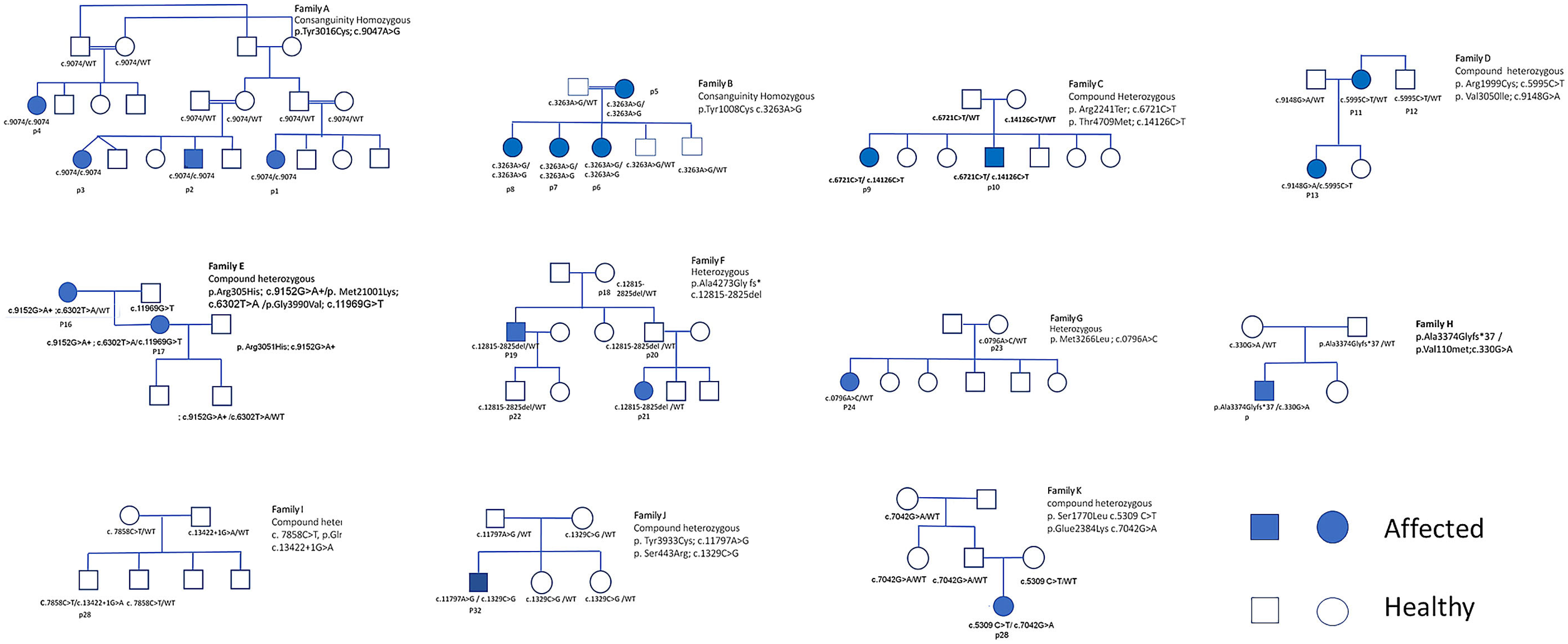
No feeding or swallowing difficulties were noted, and no cardiac involvement was observed. Three patients (P16, P19, and P36) had additional clinical features consistent with KDS (e.g., short stature and facial dysmorphism). Two patients (P5 and P16) experienced MH during surgery, the latter of whom was diagnosed with KDS. Mobility was preserved in most patients, regardless of their genetic variant. Serum CK levels were within the normal range for most patients, except for five (17%) who exhibited mild to moderate elevations. The familial pedigrees are presented in Figure 1.

3.2. Histopathological Findings

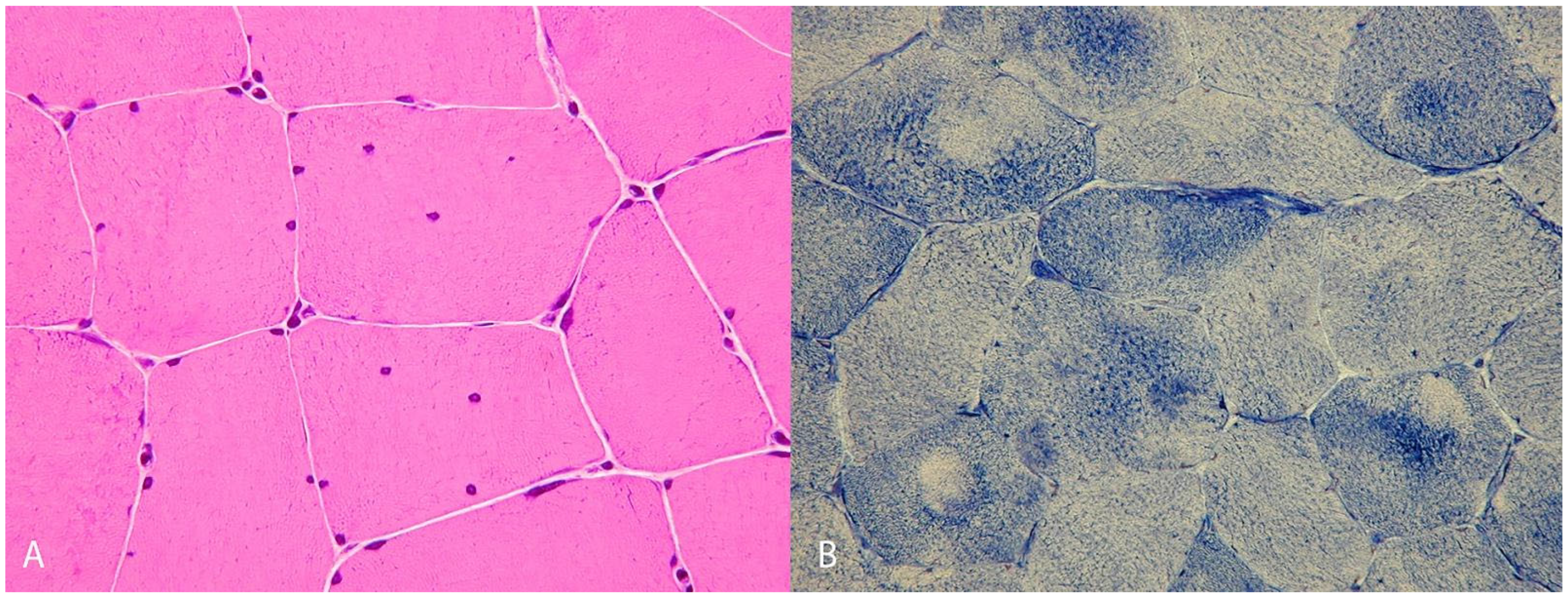
Muscle biopsies were performed on 18 patients. All showed non-specific myopathic changes, including variability in fiber size and internal nuclei. In two cases, NADH staining showed areas of decreased enzymatic activity in type I fibers (Figure 2). In five biopsies, mild endomysial fibrosis was also observed. In two biopsies, electron microscopy revealed multiminicores. Neither vacuoles nor abnormal protein aggregates were detected.

3.3. Molecular and Genetic Studies

In this cohort of 36 individuals, 23 distinct variants were identified, comprising 19 missense variants, one duplication (P25), and one intronic/splice-site variant (P26). Three nonsense variants resulted in premature stop codons: one caused by a single amino acid substitution (P9, P10, and P26), and another arising from a frameshift mutation (P18–P22).
A homozygous variant was detected in two inbred families: c.9047A>G, p.Tyr3016Cys in FA and c.3263A>G, p.Tyr1088Cys in FB, both of which showed severe neonatal presentations followed by significant improvement over time.
Variants in the Bsol domain were detected in 11 patients, either as homozygous variants in the four affected members of FA or as heterozygous variants in six familial cases (FC: P9, P10; FD: P13; FH: P25; FI: P26; FJ: P27; FK: P28) and one sporadic case (P34).
In eight patients, variants were identified in a compound heterozygous state, involving seven familial cases (FC-P9 and P10; FD-P13; FE-P16; FH-P25; FI-P26; FJ-P27; FK-P28) and one sporadic case (P34). Among the familial cases, individuals carrying a variant in the Bsol domain in combination with an additional variant exhibited a more rapidly progressive disease course and more severe myopathy—often accompanied by respiratory involvement and/or scoliosis (P9, P10, P13, P26, P25, and P28)—compared with those who carried a single variant located outside the Bsol domain, some of whom had only mild symptoms or remained asymptomatic. Similarly, the sporadic case P34 demonstrated a progressive course with scoliosis and respiratory difficulties.

4. Discussion

The RYR1 protein is located on the membrane of the sarcoplasmic reticulum within muscle cells and plays a pivotal role in regulating intracellular calcium by mediating its release from sarcoplasmic stores. The released calcium ions bind to troponin C, inducing a conformational change in the troponin complex. This, in turn, triggers the formation of cross-bridges between the contractile proteins actin and myosin, enabling their sliding interaction and ultimately generating muscle contraction [27,28].
The emergence of high-resolution cryo-electron microscopy (cryo-EM) and X-ray crystallography has provided valuable insights into the structure and function of RYR1. By resolving its three-dimensional architecture, these techniques have enabled researchers to better understand both normal and mutated conformations of the protein [29]. RYR1 is composed of four subunits, each containing multiple functional domains, and is divided into two major components: a cytosolic shell formed by the N-terminal domains, and a channel and activation core comprising the remaining C-terminal segments. Bridging these components are the solenoid structures from each protomer, known as the junctional solenoid (JSol) and the bridging solenoid (BSol) [14] (Figure 3).
The BSol domain plays a central role in the conformational transitions between the open and closed states of the RYR1 channel through its interactions with other domains, including Nsol. Variants in the BSol domain can disrupt the structural conformation of the RyR1 channel and impair calcium homeostasis. Such disruption may result in pathological calcium release, either through leakage due to incomplete channel closure or through insufficient opening, leading to reduced calcium release and consequent muscle weakness and myopathy. Indeed, studies have shown that biallelic variants in the BSol domain are frequently associated with severe clinical phenotypes, including marked muscle weakness, respiratory insufficiency, and feeding difficulties during infancy [1,2].
These observations were also confirmed in our cohort, in which most patients who were wheelchair-bound or exhibited a severe progressive course were found to carry at least one variant in the BSol domain. Notably, in Family F and in a sporadic case (P34), the variant p.Arg1999Cys (c.5995C>T), when present in combination with another variant, was associated with a severe and progressive disease trajectory.
Our series encompassed a heterogeneous range of clinical presentations. Most patients exhibited a phenotype characterized by neonatal or early-onset progressive muscle weakness. In the most severe cases, the disease progressed to the point of requiring wheelchair use and mechanical ventilation. Conversely, at the milder end of the spectrum, some patients demonstrated a disease course marked by clinical improvement. Across this variability, muscular symptoms such as myalgia and muscle weakness were common, and a subset of patients experienced malignant hyperthermia (Table 1, Table 2 and Table 3).
The high prevalence of severe cases in our series may be somewhat biased, as milder presentations could be underdiagnosed. Notably, five asymptomatic individuals with normal CK levels carried heterozygous variants, similar to their affected family members. Reduced penetrance and clinical heterogeneity of RYR1-related disorders have been observed in other studies and are suggested to result from exposure to external factors, including physical exercise, heat, fever, or polymorphisms that modulate calcium homeostasis [30]. The existence of asymptomatic and mildly symptomatic individuals with RYR1 variants complicates the interpretation of genetic testing. Functional studies, when available, may help confirm the pathogenicity of the variant.
The most frequent pathological finding in muscle biopsies of patients with RYR1 variants is the presence of central cores, which have been particularly associated with dominant inheritance [31]. Other pathological findings, such as multiminicores [32], centronuclear myopathy [33], and fiber-type disproportion [9], have been primarily linked to recessive disease. However, in our cohort, most biopsies demonstrated non-specific myopathic changes, including fiber size variation, internal nuclei, endomysial fibrosis, and type 1 fiber predominance. Only three biopsies from familial patients showed multiminicores (P9, P27, and P28), while one biopsy from a sporadic case revealed fiber-type disproportion (P34). The absence of typical RYR1-related myopathy features in most biopsies underscores that muscle biopsy cannot reliably diagnose RYR1-related conditions, particularly when genetic studies identify variants of uncertain significance. Genetic testing is especially valuable when muscle biopsy findings are inconclusive or normal, as observed in atypical presentations such as rhabdomyolysis, myalgia, or periodic paralysis. Nonetheless, interpretation of genetic results should be integrated with clinical, histological, and imaging data to achieve an optimal and accurate diagnosis.
Two of our patients experienced malignant hyperthermia (MH). One patient (P5) had been previously reported in a Samaritan family with benign congenital myopathy [17], while the other (P16) carried a newly identified variant that has not been previously described. To date, 29 different variants have been associated with MH [30,34], most of which are located within known hotspot regions of RYR1 [30]. However, the two variants identified in our cohort were situated outside these hotspot domains.
Two familial patients and one sporadic patient presented with KDS (P16, P19, and P36). All exhibited dysmorphic features, short stature, skeletal abnormalities, and myopathy. One patient had MH susceptibility confirmed via a caffeine test. Muscle biopsy findings varied and included fiber size variation, internal nuclei, fat and connective tissue replacement, and dystrophic features. Central cores were not observed. One patient (P16) carried three heterozygous missense variants, one (P19) carried a heterozygous nonsense variant, and one (P36) harbored a de novo heterozygous missense variant. None of these variants were located in the Bsol domain.
In family F, all members carried the c.12815_12825, p.Ala4272Glyfs*307 variant, yet one member exhibited a mild phenotype while three others were asymptomatic, despite carrying the same pathogenic variant. Similar discrepancies, where the proband with KDS harbored a heterozygous missense variant, while other family members with the same variant displayed only mild symptoms or were clinically asymptomatic, have been reported [35,36].
Phenotypic variability in RYR1 patients results from the combined effects of variant type, inheritance pattern, affected protein domains, environmental factors, modifier genes, and post-translational modifications. Single heterozygous variants are often linked to milder phenotypes like central core disease and malignant hyperthermia susceptibility [10]. In contrast, biallelic variants have been associated with early onset generalized weakness, respiratory insufficiency, and multisystem involvement [11]. Compound heterozygosity and the location of variants beyond well-defined regions increased phenotypic complexity [11]. Environmental factors, modifier genes, and post translational changes to myosin further contribute to the variability [37].
The reason for the phenotypic variability among patients within the same family, including those with KDS, remains unclear. Proposed explanations include the presence of a genetic modifier that may influence the expression of the unaffected RYR1 allele and the preferential expression of the non-mutated allele in asymptomatic or mildly symptomatic family members [35].
Similar to our findings, recent large cohort studies of RYR1-related myopathies from England and Italy report considerable clinical heterogeneity, with phenotypes ranging from asymptomatic or mildly affected individuals (including isolated hyperCKemia) to patients with severe muscle weakness, loss of ambulation, skeletal deformities, and significant respiratory involvement [8,38]. In accordance with our cohort, severe phenotypes were more frequently observed in patients with early disease onset, typically at birth, during infancy, or in early childhood. Furthermore, most severe cases were associated with autosomal recessive inheritance, predominantly involving homozygous or compound heterozygous pathogenic variants. These observations support an association between biallelic pathogenic variants and greater disease severity, although genotype–phenotype correlations remain variable.

5. Conclusions

The presented data expands our knowledge about the clinical, histological, and genetic spectrum of RYR1 variants in Israel and suggests that the range of variants and clinical presentations is broader than previously recognized. Variants in the Bsol domain are characterized by greater disease severity, suggesting that this region may serve as a potential target for therapeutic intervention.

Author Contributions

Conceptualization, M.G., M.S. and R.D.; methodology, M.G., M.S. and R.D. software, M.G., M.S., R.D. and V.F.; validation, M.M., S.A., L.S. and Y.M.; formal analysis, D.R. and K.Y.; investigation, M.G., Z.A. and A.N.; resources, R.D., D.L. and M.S.; data curation, M.M. and S.A.; writing—original draft preparation, M.G., R.D. and M.S.; writing—review and editing, M.G., R.D., M.S. and D.L.; visualization, V.F. and K.Y.; supervision, R.D.; project administration, M.G. and R.D.; funding acquisition, R.D. All authors have read and agreed to the published version of the manuscript.

Funding

This research did not receive any specific grant from funding agencies in the public, commercial, or not-for-profit sectors.

Institutional Review Board Statement

The study was conducted according to the guidelines of the Declaration of Helsinki, and approved by the Ethics Committee of the Edith Wolfson Medical Center on 26 June 2019 (Study 0079-19-WOMC) at Wolfson Medical Center.

Informed Consent Statement

This is a retrospective research study that received waiver from the informed consent form.

Data Availability Statement

The data are not publicly available due to privacy and ethical restrictions related to the sensitive genetic and clinical information of the participant. All relevant data generated or analyzed during this study are included in this published article. Further inquiries can be directed to the corresponding author.

Conflicts of Interest

The authors declare no competing interests.

References

  1. Zucchi, R.; Ronca-Testoni, S. The sarcoplasmic reticulum Ca2+ channel/ryanodine receptor: Modulation by endogenous effectors, drugs and disease states. Pharmacol. Rev. 1997, 49, 1–51. [Google Scholar] [CrossRef]
  2. Cheng, W.; Altafaj, X.; Ronjat, M.; Coronado, R. Interaction between the dihydropyridine receptor Ca2+ channel β-subunit and ryanodine receptor type 1 strengthens excitation-contraction coupling. Proc. Natl. Acad. Sci. USA 2005, 102, 19225–19230. [Google Scholar] [CrossRef]
  3. Iyer, K.A.; Hu, Y.; Klose, T.; Murayama, T.; Samsó, M. Molecular mechanism of the severe MH/CCD variant Y522S in skeletal ryanodine receptor (RyR1) by cryo-EM. Proc. Natl. Acad. Sci. USA 2022, 119, e2122140119. [Google Scholar] [CrossRef]
  4. Witting, N.; Laforet, P.; Voermans, N.C.; Roux-Buisson, N.; Bompaire, F.; Rendu, J.; Duno, M.; Feillet, F.; Kamsteeg, E.J.; Poulsen, N.S.; et al. Phenotype and genotype of muscle ryanodine receptor rhabdomyolysis-myalgia syndrome. Acta Neurol. Scand. 2018, 137, 452–461. [Google Scholar] [CrossRef] [PubMed]
  5. Snoeck, M.; Van Engelen, B.G.; Küsters, B.; Lammens, M.; Meijer, R.; Molenaar, J.P.; Raaphorst, J.; Verschuuren-Bemelmans, C.C.; Straathof, C.S.M.; Sie, L.T.L.; et al. RYR1-related myopathies: A wide spectrum of phenotypes throughout life. Eur. J. Neurol. 2015, 22, 1094–1112. [Google Scholar] [CrossRef] [PubMed]
  6. McKie, A.B.; Alsaedi, A.; Vogt, J.; Stuurman, K.E.; Weiss, M.M.; Shakeel, H.; Tee, L.; Morgan, N.V.; Nikkels, P.G.; van Haaften, G.; et al. Germline variants in RYR1 are associated with foetal akinesia deformation sequence/lethal multiple pterygium syndrome. Acta Neuropath. Commun. 2014, 2, 148. [Google Scholar] [CrossRef]
  7. Dlamini, N.; Voermans, N.C.; Lillis, S.; Stewart, K.; Kamsteeg, E.J.; Drost, G.; Quinlivan, R.; Snoeck, M.; Norwood, F.; Radunovic, A.; et al. Variants in RYR1 are a common cause of exertional myalgia and rhabdomyolysis. Neuromuscul. Disord. 2013, 23, 540–548. [Google Scholar] [CrossRef] [PubMed]
  8. Fusto, A.; Cassandrini, D.; Fiorillo, C.; Codemo, V.; Astrea, G.; D’Amico, A.; Maggi, L.; Magri, F.; Pane, M.; Tasca, G.; et al. Expanding the clinical-pathological and genetic spectrum of RYR1-related congenital myopathies with cores and minicores: An Italian population study. Acta Neuropath. Commun. 2022, 10, 20. [Google Scholar] [CrossRef]
  9. Clarke, N.F.; Waddell, L.B.; Cooper, S.T.; Perry, M.; Smith, R.L.; Kornberg, A.J.; Muntoni, F.; Lillis, S.; Straub, V.; Bushby, K.; et al. Recessive mutations in RYR1 are a common cause of congenital fiber type disproportion. Hum. Mutat. 2010, 31, E1544–E1550. [Google Scholar] [CrossRef]
  10. Zhou, H.; Jungbluth, H.; Sewry, C.A.; Feng, L.; Bertini, E.; Bushby, K.; Straub, V.; Roper, H.; Rose, M.R.; Brockington, M.; et al. Molecular mechanisms and phenotypic variation in RYR1-related congenital myopathies. Brain 2007, 130, 2024–2036. [Google Scholar] [CrossRef]
  11. Amburgey, K.; Bailey, A.; Hwang, J.H.; Tarnopolsky, M.A.; Bonnemann, C.G.; Medne, L.; Mathews, K.D.; Collins, J.; Daube, J.R.; Wellman, G.P.; et al. Genotype-phenotype correlations in recessive RYR1-related myopathies. Orphanet J. Rare Dis. 2013, 8, 117. [Google Scholar] [CrossRef] [PubMed]
  12. Beam, T.A.; Loudermilk, E.F.; Kisor, D.F. Pharmacogenetics and pathophysiology of CACNA1S mutations in malignant hyperthermia. Physiol. Genom. 2017, 49, 81–87. [Google Scholar] [CrossRef] [PubMed]
  13. Fiszer, D.; Shaw, M.A.; Fisher, N.A.; Carr, I.M.; Gupta, P.K.; Watkins, E.J.; de Sa, D.R.; Kim, J.H.; Hopkins, P.M. Next-generation sequencing of RYR1 and CACNA1S in malignant hyperthermia and exertional heat illness. Anesthesiology 2015, 122, 1033–1046. [Google Scholar] [CrossRef] [PubMed]
  14. des Georges, A.; Clarke, O.B.; Zalk, R.; Yuan, Q.; Condon, K.J.; Grassucci, R.A.; Hendrickson, W.A.; Marks, A.R.; Frank, J. Structural basis for gating and activation of RyR1. Cell 2016, 167, 145–157. [Google Scholar] [CrossRef]
  15. Guha, S.; Reddi, H.V.; Aarabi, M.; DiStefano, M.; Wakeling, E.; Dungan, J.S.; Gregg, A.R.; ACMG Laboratory Quality Assurance Committee. Laboratory testing for preconception/prenatal carrier screening: A technical standard of the American College of Medical Genetics and Genomics (ACMG). Genet. Med. 2024, 26, 101137. [Google Scholar] [CrossRef]
  16. Attali, R.; Aharoni, S.; Treves, S.; Rokach, O.; Becker Cohen, M.; Fellig, Y.; Straussberg, R.; Dor, T.; Daana, M.; Mitrani-Rosenbaum, S.; et al. Variable myopathic presentation in a single family with novel skeletal RYR1 mutation. PLoS ONE 2013, 8, e69296. [Google Scholar] [CrossRef] [PubMed]
  17. Böhm, J.; Leshinsky-Silver, E.; Vassilopoulos, S.; Le Gras, S.; Lerman-Sagie, T.; Ginzberg, M.; Jost, B.; Lev, D.; Laporte, J. Samaritan myopathy, an ultimately benign congenital myopathy, is caused by a RYR1 mutation. Acta Neuropathol. 2012, 124, 575–581. [Google Scholar] [CrossRef]
  18. Zhou, H.; Lillis, S.; Loy, R.E.; Ghassemi, F.; Rose, M.R.; Norwood, F.; Mills, K.; Al-Sarraj, S.; Lane, R.J.; Feng, L.; et al. Multi-minicore disease and atypical periodic paralysis associated with novel mutations in the skeletal muscle ryanodine receptor (RYR1) gene. Neuromuscul. Disord. 2010, 20, 166–173. [Google Scholar] [CrossRef]
  19. Robinson, R.; Carpenter, D.; Shaw, M.A.; Halsall, J.; Hopkins, P. Mutations in RYR1 in malignant hyperthermia and central core disease. Hum. Mutat. 2006, 27, 977–989. [Google Scholar] [CrossRef]
  20. Janssens, L.; De Puydt, J.; Milazzo, M.; Symoens, S.; De Bleecker, J.L.; Herdewyn, S. Risk of malignant hyperthermia in patients carrying a variant in the skeletal muscle ryanodine receptor 1 gene. Neuromuscul. Disord. 2022, 32, 864–869. [Google Scholar] [CrossRef]
  21. Duarte, S.T.; Oliveira, J.; Santos, R.; Pereira, P.; Barroso, C.; Conceição, I.; Evangelista, T. Dominant and recessive RYR1 mutations in adults with core lesions and mild muscle symptoms. Muscle Nerve 2011, 44, 102–108. [Google Scholar] [CrossRef]
  22. Avnon, T.; Svirsky, R.; Orr-Urtreger, A.; Sagie, L.; Fattal-Valevski, A.; Fellig, Y.; Ben-Shachar, S. Clinical Observation: Effect of a Second Transpositioned Variant in a Family with Autosomal Dominant Ryanodine Receptor-1–Related Disease. J. Pediatr. Genet. 2020, 9, 121–124. [Google Scholar] [CrossRef]
  23. Biancalana, V.; Rendu, J.; Chaussenot, A.; Mecili, H.; Bieth, E.; Fradin, M.; Mercier, S.; Michaud, M.; Nougues, M.C.; Pasquier, L.; et al. A recurrent RYR1 mutation associated with early-onset hypotonia and benign disease course. Acta Neuropathol. Commun. 2021, 9, 155. [Google Scholar] [CrossRef]
  24. Lester, E.B.; Larsen, M.J.; Laulund, L.W.; Illum, N.; Dunkhase-Heinl, U.; Schrøder, H.D.; Fagerberg, C.R. Ryanodine receptor 1 related myasthenia like myopathy responsive to pyridostigmine. Eur. J. Med. Genet. 2023, 66, 104706. [Google Scholar] [CrossRef] [PubMed]
  25. Driver, K.; Vo, C.; Scriba, C.K.; Saker, S.; Larmonier, T.; Malfatti, E.; Romero, N.B.; Ravenscroft, G.; Laing, N.G.; Taylor, R.L.; et al. Generation of two induced pluripotent stem cell lines from a 33-year-old central core disease patient with a heterozygous dominant c. 14145_14156delCTACTGGGACA (p. Asn4715_Asp4718del) deletion in the RYR1 gene. Stem Cell Res. 2023, 73, 103258. [Google Scholar] [CrossRef]
  26. Benucci, S.; Ruiz, A.; Franchini, M.; Ruggiero, L.; Zoppi, D.; Sitsapesan, R.; Lindsay, C.; Pelczar, P.; Pietrangelo, L.; Protasi, F.; et al. A novel, patient-derived RyR1 mutation impairs muscle function and calcium homeostasis in mice. J. Gen. Physiol. 2024, 156, e202313486. [Google Scholar] [CrossRef] [PubMed]
  27. Franzini-Armstrong, C.; Protasi, F. Ryanodine receptors of striated muscles: A complex channel capable of multiple interactions. Physiol. Rev. 1997, 77, 699–729. [Google Scholar] [CrossRef]
  28. Santulli, G.; Marks, A.R. Essential roles of intracellular calcium release channels in muscle, brain, metabolism, and aging. Curr. Mol. Pharmacol. 2015, 8, 206–222. [Google Scholar] [CrossRef] [PubMed]
  29. Woll, K.A.; Haji-Ghassemi, O.; Van Petegem, F. Pathological conformations of disease mutant ryanodine receptors revealed by cryo-EM. Nat. Comm. 2021, 12, 807. [Google Scholar] [CrossRef]
  30. Segarra-Casas, A.; Iruzubieta, P.; Kapetanovic, S.; Hernández-Laín, A.; Jericó, I.; Fernández-Torrón, R.; Maneiro, M.; Marco-Moreno, P.; Zelaya-Huerta, M.V.; Rodríguez-Santiago, B.; et al. A founder variant in the RYR1 gene is associated with hyperCKemia, myalgia and muscle cramps. Eur. J. Neurol. 2025, 32, e16471. [Google Scholar] [CrossRef]
  31. Wu, S.; Ibarra, M.C.; Malicdan, M.C.; Murayama, K.; Ichihara, Y.; Kikuchi, H.; Nonaka, I.; Noguchi, S.; Hayashi, Y.K.; Nishino, I. Central core disease is due to RYR1 mutations in more than 90% of patients. Brain 2006, 129, 1470–1480. [Google Scholar] [CrossRef]
  32. Jungbluth, H. Multi-minicore disease. Orphanet J. Rare Dis. 2007, 2, 31. [Google Scholar] [CrossRef]
  33. Abath Neto, O.; Moreno, C.A.M.; Malfatti, E.; Donkervoort, S.; Böhm, J.; Guimarães, J.B.; Foley, A.R.; Mohassel, P.; Dastgir, J.; Bharucha-Goebel, D.X. Common and variable clinical, histological, and imaging findings of recessive RYR1-related centronuclear myopathy patients. Neuromuscul. Disord. 2017, 27, 975–985. [Google Scholar] [CrossRef]
  34. Monnier, N.; Kozak-Ribbens, G.; Krivosic-Horber, R.; Nivoche, Y.; Qi, D.; Kraev, N.; Loke, J.; Sharma, P.; Tegazzin, V.; Figarella-Branger, D.; et al. Correlations between genotype and pharmacological, histological, functional, and clinical phenotypes in malignant hyperthermia susceptibility. Hum. Mutat. 2005, 26, 413–425. [Google Scholar] [CrossRef] [PubMed]
  35. Dowling, J.J.; Lillis, S.; Amburgey, K.; Zhou, H.; Al-Sarraj, S.; Buk, S.J.; Wraige, E.; Chow, G.; Abbs, S.; Leber, S.; et al. King-Denborough syndrome with and without mutations in the skeletal muscle ryanodine receptor (RYR1) gene. Neuromuscul. Disord. 2011, 21, 420–427. [Google Scholar] [CrossRef]
  36. Rueffert, H.; Olthoff, D.; Deutrich, C.; Schober, R.; Froster, U.G. A new mutation in the skeletal ryanodine receptor gene (RYR1) is potentially causative of malignant hyperthermia, central core disease, and severe skeletal malformation. Am. J. Med. Genet. Part A 2004, 124, 248–254. [Google Scholar] [CrossRef] [PubMed]
  37. Todd, J.J.; Sagar, V.; Lawal, T.A.; Allen, C.; Razaqyar, M.S.; Shelton, M.S.; Chrismer, I.C.; Zhang, X.; Cosgrove, M.M.; Kuo, A.; et al. Correlation of phenotype with genotype and protein structure in RYR1-related disorders. J. Neurol. 2018, 265, 2506–2524. [Google Scholar] [CrossRef] [PubMed]
  38. Sarkozy, A.; Sa, M.; Ridout, D.; Fernandez-Garcia, M.A.; Distefano, M.G.; Main, M.; Sheehan, J.; Manzur, A.Y.; Munot, P.; Robb, S.; et al. Long-term Natural History of Pediatric Dominant and Recessive RYR1-Related Myopathy. Neurology 2023, 101, 1495–1508. [Google Scholar] [CrossRef]
Figure 1. Pedigrees.
Jcm 15 01388 g001
Figure 2. Characterization of the histological features of the muscle biopsy of P5: (A) H&E staining revealed an increased number of internal nuclei. (B) NADH staining shows areas of decreased enzymatic activity in type I fibers.
Jcm 15 01388 g002
Figure 3. Two-Two-dimensional representations of the domain boundaries of the RYR1 monomer described in the work of des Georges et al. [14] (obtained with permission).
Jcm 15 01388 g003
Table 1. Genetic characteristics of families with RYR1 variants. Variants classification is according to ACMG criteria.
Segregation
Affected/Unaffected
Genetic Variation
NM_000540.3
DomainOnsetCurrent Age/
Gender
Reference
Family A4 affected (P1–P4)
Homozygous Consanguinity
P1–P4:
c.9047A>G; p.Tyr3016Cys
Missense, P
(PS4, PP1, PS3, PM2, PP3, PP2)
Clinvar ID RCV000558544
BSolP1: neonatal
P2: 1.5 Y
P3: 6 Y
P4: 4 Y
P1: 38 Y/F
P2: 25 Y/M
P3: 18 Y/M
P4: 17 Y/M
[16]
Family B4 affected (P5–P8)
Homozygous Consanguinity
P5–P8:
c.3263A>G; p.Tyr1088Cys
Missense, LP
(PM2, PP3, PP2, PP5)
UniProt ID VAR_068512
SPRY2P5–P8:
neonatal
P5: 43 Y/F
P6: 21 Y/F
P7: 21 Y/F
P8: 17 Y/F
[17]
Family C2 affected (P9, P10)
Compound heterozygous
P9, P10:
c.6721C>T; p. Arg2241Ter
Nonsense, P
(PM3, PS3, PVS1, PM2)
ClinVar ID RCV000147436
c.14126C>T; p.Thr4709Met
Missense, LP
(PM2, PM5, PP3PP2, PP5)
ClinVar ID RCV000119498
I. BSol
II. pVSD
P9, P10:
neonatal
P9: 15 Y/M
P10: 22 Y/F
[18]
Family D2 affected (P11, P13)
1 Heterozygous
1 Compound heterozygous
1 unaffected (P12)
Heterozygous
P11, P13:
c.5995C>T; p.Arg1999Cys
Missense, LP
(PM2, PP3, PP2, PM3)
ClinVar ID RCV001047012
P12, P13
c.9148G>A; p.Val3050lle
Missense, VUS
(PM2, PM5, PP2, BP6 PP1)
ClinVar ID RCV001660679
I. JSol
II. BSol
P11: 10 Y
P13: neonatal
P11: 66 Y/F
P12: 68 Y/M
P13: 40 Y/F
Family E2 affected:
1 heterozygous (2 mutations P17) **
1 Compound heterozygous
(3 mutations-P16)
2 unaffected
1 heterozygous (1 mutation -P14)
1 heterozygous (2 mutation-P15) **
P15–P17:
c.9152G>A; p. Arg3051His
Missense, LP
(PM2, PP2, PM3)
ClinVar ID RCV001803928
c.6302T>A; p. Met2101Lys
Missense, VUS(PM2, PP2, PM3)
ClinVar ID RCV001592839
P14, P16
c.11969G>T; p.Gly3990Val
Missense, LP
(PP1, PS3, PM2, PP3, PP2, PP5 PM3)
ClinVar ID RCV002281939
I. NTD-B
II. JSol
III. CSol
P14, P15:
asymptomatic
P16: 15 Y
P17: 4 Y
P14: 69 Y/M
P15: 66 Y/F
P16: 31 Y/F
P17: 4 Y/M
III
[19]
Family F2 affected (P19, P21)
Heterozygous
3 unaffected (P18, P20, P22)
Heterozygous
P18–22:
c.12815_12825del
p.Ala4272Glyfs*307, c.12815_12825del NM_000540.3 LP/LP
Nonsense, LP
(PVs1, PM2)
TaFP19: neonatal
P21: 1.5 Y
P18, P20, P22;
asymptomatic
P18: 70 Y/F
P19: 45 Y/M
P20: 39 Y/M
P21: 17 Y/F
P22: 16 Y/M
Family G1 affected (P24)
Heterozygous
1 unaffected (P23)
Heterozygous
P23, P24:
c.9796A>C; p. Met3266Leu
Missense, VUS
(PM2, PP2)
ClinVar ID RCV001368477
BSolP24: 14.5 YP23: 46 Y/F
P24: 16 Y/F
[20]
Family H1 affected (P25)
Compound heterozygous
P 25:
c.11320dup; p. Ala3374Gly fs*37
(PS4, PVS1, PM2)
ClinVar ID RCV000721230
c.3301G>A; p. Val1101Met
Missense LP
PM2, PP3, PP2, PP5
ClinVar ID RCV001852321
I BSol
II NTD-A
P25:
neonatal
P25: 1.5 Y/M
Family I1 affected (P26)
Compound heterozygous
P26:
c.13437+1G>A
Splicing, LP
(PVS1, PM2, PP5)
ClinVar ID RCV000721317
c.7858C>T; p.Gln2620Ter
Nonsense, P
(PS4, PVS1, PM2, PM3)
ClinVar
RCV001219907
I. BSol
II.SPRY2
P26:
neonatal
P26: 37 Y/M
Family J1 affected (P27)
Compound heterozygous
P27:
c.11798A>G; p.Tyr3933Cys
Missense, LP
(PP3, PM2, PP2)
ClinVar ID RCV000148797
c.1329C>G; p. Ser443Arg
Missense, LP
(PP1, PM2, PP2, PM3)
I Nsol
II CSol
P27:
Neonatal
P27: 15.5 Y/M[21]
Family K4 affected (P28–P31)
1 Compound heterozygous
3 Heterozygote
P28:
c.3509C>T; p. Ser1770Leu;
Missense, LP
(PM2, PP2, PM3, PP1)
ClinVar ID RCV000079149
P28–P31
c.7042G>A; p. Glu2348Lys
Missense, LP
(PM1, PP2, PM2, PM5, PP3)
ClinVar ID RCV000721635
I. Bsol
II. Jsol
P28:
Neonatal
P28: 7.5 Y/F
P29: M
P30: F
[22]
ACMG—American College of Medical Genetics and Genomics: P pathogenic variant, LP likely pathogenic. ** Two heterozygous variants located on the same allele.
Table 2. Clinical and ancillary characteristics of familial patients with RYR1 variants.
Muscle InvolvementFacial InvolvementRespiratory
Involvement
Skeletal
Involvement
CK LevelsEMGBiopsy FindingsSeverityMotor Development/Course
Family A
(P1–P4)
P1P4:
neck and limb girdle muscle weakness
P1: wheelchair bound since 11 Y
P1P4:
facial muscles and eye closure weakness,
dysmorphic features: elongated face
P1:
recurrent severe
pneumonia
P1:
scoliosis
P1:
mildly elevated
P2, P3, P4:
normal
P1:
myopathic changes
P1–P4;
Fiber size variation, internalized and central nuclei,
endomysial fibrosis
P1: 5
P2: 5
P3: 4
P4: 4
P1:
rapid progression since birth
P2–P4:
variable progressive
Family B
(P5–P8)
P5–P8: congenital hypotonia &weakness,
by 2 Y improved to have only distal limbs weakness
P5: MH during surgery
P5–P8:
facial weakness, bi-temporal Narrowing,
epicanthal folds,
hypertelorism
P5P8:
neonatal, improved gradually within 2 weeks
P5–P7:
normal
P8:
NA
P5:
myopathic changes
P5:
Internalized and central nuclei, central areas devoid of oxidative enzyme activity and moth-eaten appearance
P5: 1
P6: 1
P7: 1
P8: 1
P5–P8:
delayed/Improving
Family C
(P9, P10)
P9, P10: congenital hypotonia, neck and limb weakness,
P9: never walked and wheelchair bound
P10: wheelchair bound since age 7 Y
P9:
ophthalmoplegia
P9:
night BI-PAP since 7 y
P10:
neonatal respiratory weakness, needing ventilation for 4 days, night BI-PAP since 12 Y
P9:
scoliosis& rigid spine, operated at 15 y
P10:
scoliosis, surgery pending
P9, P10:
normal
P9, P10:
ND
P9:
LM: great variability in fiber size
EM: minicores and large mitochondria
P9: 6
P10: 5
P9:
delayed/progressive
P10:
severely delayed/progressive
Family D
(P11–P13)
P11:
myalgia and fatigability since early age
P13:
congenital proximal muscle weakness, wheelchair bound since 13 Y
P13:
eye closure
weakness
NoP13:
scoliosis
P11, P13:
normal
P11:
normal
P13:
myopathic changes-severe
P13:
type 1 fiber predominant, atrophy and grouping
P11: 1
P12: 0
P13: 5
P11:
delayed/stable
P13: Delayed/
progressive:
Family E
(P14–P17)
P16:
mild weakness,
MH during surgery
P16:
KDS: dysmorphic face, high arch palate
NoP15:
short stature
P16:
mild scoliosis,
joints deformity,
pes cavus
P14, P15:
NA
P16:
moderately elevated
P17:
mildly elevated
P15:
normal
P14–p17:
ND
P16:
fiber size variation internal nuclei
positive findings with caffeine exposure
P14: 0
P15: 1
P16: 0
P16:
normal/stable
Family F
(P18–P22)
P19: proximal and distal
lower &upper limbs weakness
P21: proximal lower limbs weakness
P19:
KDS: dysmorphic faces
P21:
weakness eye closure
NoP19:
short stature,
arthrogryposis
P19:
NA
P21: normal
P19:
severe myopathic feature
P21:
normal
P19:
fiber size variation, internal nuclei. perimysium replaced by fat and connective tissue
P21:
myopathic features
P19: 5
P21: 2
P19: severely delayed/progressive
P21: mildly delayed/
slowly progressive
Family G
(P23, P24)
NormalNoNoP24;
episodic weakness
NormalP24:
normal
NDP24:
variable
P24:
normal/stable
Family H
(P25)
General hypertonia,
poor sucking and crying,
no head control
NoC-PAP night ventilation since 20 yNoMildly elevated NDND4P25:
mildly delayed/Improving
Family I
(P26)
Proximal and distal lower limbs weakness, muscle atrophy,
wheelchair bound since age 7 Y
Limited upper gaze &ptosis Joint contractions,
scoliosis
Mildly elevatedMyopathic featuresFiber type variation and internal nuclei−5P26:
severely delayed/progressive
Family J
(P27)
Proximal weaknessNo NoNormalNDFiber type variation and internal nuclei
EM: multi mini- core
2P27:
severely delayed/stable
Family K
(P28–P30)
P28:
proximal
weakness
NoNo P28:
scoliosis operated
P28:
Normal
P29–P30
moderate
elevated
NDP28:
Multi mini core
2P28: moderately delayed/Improving
P29–P31: normal/Improving
CK Serum creatine kinase level; NA None available; EMG electromyography; ND Not done; EM electron microscopy; LM light microscopy, M malignant hyperthermia, KDS King–Denborough syndrome.
Table 3. Genetic and clinical characteristics of sporadic patients with RYR1 variants.
PatientP32P33P34P35P36
GenderMaleFemaleFemaleMaleMale
Current age/Onset66 Y/50 Y10 Y/neonatal35 Y/neonatal20 Y/18 Y26 Y/Neonatal
Genetic variants
Variant
NM_000540.3
Heterozygous
c.528G>T; p. Glu176Asp
missense, LP
(PM2, PM1, PP2, PP4)
ClinVar ID RCV001580415
* De novo heterozygous
c.12083C>T; p. Ser4028Leu
missense, P
(PP1, PS3, PM2, PP)
ClinVar ID RCV000721259
Compound heterozygous
c.5995C>T; p.Arg1999Cys
missense, LP
(PM2, PP3, PP2, PM3)
ClinVar ID RCV001047012
c.6721C>T nonsense, P
p. Arg2241 *
(PM3, PS3, PVS1, PM2)
ClinVar ID RCV000147436
* De novo heterozygous
c.15067T>C missense, LP
p. Phe5023Leu;
PP3, PM2, PP2)
ClinVar ID RCV001225325
* De novo heterozygous
c.14818G>A missense, LP
p. Ala4940Thr;
(PM1, PP2, PM2, PP3)
ClinvarID RCV000119566
DomainNTD-ACSolBsol, JSolCTDS6c
Muscle involvementLower limbs muscle weakness, myalgia,
atrophy of quadriceps and lumbar and upper thoracic muscles
Proximal lower limbs weakness
delayed motor milestones
Proximal and distal lower limbsMyalgiaLower and upper limbs
Respiratory involvementNoRecurrent pneumonia in early childhoodRespiratory difficultiesNoNo
Skeletal involvementNoNoScoliosis/operatedNoKDS: scoliosis,
Short stature
CK levels1000–3000 **normalNA800–100NA
EMGMyopathic featuresNDNANAND
Biopsy findingsMuscular dystrophyFiber size variationFiber type disproportionNDDystrophic features
Courseprogressivestableprogressivestableprogressive
Severity42615
Reference [23][24][25][26]
ACMG—Variant classification is according to ACMG criteria, CKserum creatine kinase; NA—Not available; EMG—electromyography; ND—Not done. * Family segregation did not detect mutation in other family members. ** Other family members suffered from malignant hyperthermia.
Disclaimer/Publisher’s Note: The statements, opinions and data contained in all publications are solely those of the individual author(s) and contributor(s) and not of MDPI and/or the editor(s). MDPI and/or the editor(s) disclaim responsibility for any injury to people or property resulting from any ideas, methods, instructions or products referred to in the content.