Foramen Ovale Measurements and Venous Hemodynamic Changes Assessed by Inferior Vena Cava Doppler Parameters in Early- and Late-Onset Fetal Growth Restriction
 , , ,
, , ,
Abstract
1. Introduction
2. Materials and Methods
Statistical Analysis
3. Results
4. Discussion
5. Conclusions
Author Contributions
Funding
Institutional Review Board Statement
Informed Consent Statement
Data Availability Statement
Conflicts of Interest
Abbreviations
| AUC | Area Under the Curve |
| BMI | Body Mass Index |
| CAPO | Composite Adverse Perinatal Outcome |
| CI | Confidence Interval |
| EFW | Estimated Fetal Weight |
| EO-FGR | Early-Onset Fetal Growth Restriction |
| FO | Foramen Ovale |
| FO/RA | Foramen Ovale-to-Right Atrium Ratio |
| FGR | Fetal Growth Restriction |
| GA | Gestational Age |
| IVC | Inferior Vena Cava |
| LO-FGR | Late-Onset Fetal Growth Restriction |
| NICU | Neonatal Intensive Care Unit |
| OR | Odds Ratio |
| PI | Pulsatility Index |
| PLI | Preload Index |
| PVIV | Peak Velocity Index for Veins |
| RA | Right Atrium |
| ROC | Receiver Operating Characteristic |
| UA | Umbilical Artery |
References
- Malhotra, A.; Allison, B.J.; Castillo-Melendez, M.; Jenkin, G.; Polglase, G.R.; Miller, S.L. Neonatal Morbidities of Fetal Growth Restriction: Pathophysiology and Impact. Front. Endocrinol. 2019, 10, 55. [Google Scholar] [CrossRef] [Scilit]
- Lee, A.C.; Kozuki, N.; Cousens, S.; Stevens, G.A.; Blencowe, H.; Silveira, M.F.; Sania, A.; Rosen, H.E.; Schmiegelow, C.; Adair, L.S.; et al. Estimates of burden and consequences of infants born small for gestational age in low and middle income countries with INTERGROWTH-21st standard: Analysis of CHERG datasets. BMJ 2017, 358, j4229. [Google Scholar] [CrossRef] [Scilit]
- Figueras, F.; Gratacós, E. Update on the diagnosis and classification of fetal growth restriction and proposal of a stage-based management protocol. Fetal Diagn. Ther. 2014, 36, 86–98. [Google Scholar] [CrossRef] [Scilit]
- de Onis, M.; Blössner, M.; Villar, J. Levels and patterns of intrauterine growth retardation in developing countries. Eur. J. Clin. Nutr. 1998, 52, S5–S15. [Google Scholar]
- Gumina, D.L.; Su, E.J. Mechanistic insights into the development of severe fetal growth restriction. Clin. Sci. 2023, 137, 679–695. [Google Scholar] [CrossRef] [Scilit]
- Coutinho, C.M.; Giorgione, V.; Thilaganathan, B.; Patey, O. Cardiovascular Adaptation in Fetal Growth Restriction: A Longitudinal Study From Fetuses at Term to the First Year of Life. BJOG 2025, 132, 189–196. [Google Scholar] [CrossRef] [Scilit] [PubMed]
- Verburg, B.O.; Jaddoe, V.W.V.; Wladimiroff, J.W.; Hofman, A.; Witteman, J.C.M.; Steegers, E.A.P. Fetal hemodynamic adaptive changes related to intrauterine growth: The Generation R Study. Circulation 2008, 117, 649–659. [Google Scholar] [CrossRef] [Scilit] [PubMed]
- Baschat, A.A. Pathophysiology of fetal growth restriction: Implications for diagnosis and surveillance. Obstet. Gynecol. Surv. 2004, 59, 617–627. [Google Scholar] [CrossRef] [Scilit]
- Srisupundit, K.; Luewan, S.; Tongsong, T. Prenatal Diagnosis of Fetal Heart Failure. Diagnostics 2023, 13, 779. [Google Scholar] [CrossRef] [Scilit]
- Akira, M.; Noa, U.; Atsuko, T.; Kanako, M.; Mikio, M. The relationship between fetal inferior vena cava diameter pulse and flow velocity waveforms in normal and compromised pregnancies. Early Hum. Dev. 2008, 84, 129–135. [Google Scholar] [CrossRef] [Scilit] [PubMed]
- Alfirevic, Z.; Stampalija, T.; Dowswell, T. Fetal and umbilical Doppler ultrasound in high-risk pregnancies. Cochrane Database Syst. Rev. 2017, 6, CD007529. [Google Scholar] [CrossRef] [Scilit] [PubMed]
- Duygulu Bulan, D.; Dayanan, R.; Kahraman, N.C.; Kunt, S.; Celen, S. Pulmonary Vascular Doppler and Fetal Lung Biometry as Predictors of Neonatal Respiratory Complications in Early- and Late-Onset Fetal Growth Restriction. Pediatr. Pulmonol. 2025, 60, e71333. [Google Scholar] [CrossRef] [Scilit] [PubMed]
- Kiserud, T.; Chedid, G.; Rasmussen, S. Foramen ovale changes in growth-restricted fetuses. Ultrasound Obstet. Gynecol. 2004, 24, 141–146. [Google Scholar] [CrossRef] [Scilit] [PubMed]
- DeVore, G.R.; Putra, M.; Hobbins, J.C. Assessment of Atrial Size, Shape, and Contractility in Growth-Restricted and Small-for-Gestational-Age Fetuses. J. Ultrasound Med. 2025, 44, 831–843. [Google Scholar] [CrossRef] [Scilit]
- Nader, Â.R.L.; Zielinsky, P.; Naujorks, A.A.; Nicoloso, L.H.S.; Junior, A.L.P.; Sulis, N.M.; van der Sand, L.F.; Antunes, V.D.B.; Marinho, G.D.S.; dos Santos, F.G.; et al. Behaviour of the Foramen Ovale Flow in Fetuses with Intrauterine Growth Restriction. Obstet. Gynecol. Int. 2018, 2018, 1496903. [Google Scholar] [CrossRef] [Scilit]
- Nardozza, L.M.M.; Caetano, A.C.R.; Zamarian, A.C.P.; Mazzola, J.B.; Silva, C.P.; Marçal, V.M.G.; Lobo, T.F.; Peixoto, A.B.; Júnior, E.A. Fetal growth restriction: Current knowledge. Arch. Gynecol. Obstet. 2017, 295, 1061–1077. [Google Scholar] [CrossRef] [Scilit]
- van de Meent, M.; Schuit, E.; Ganzevoort, W.; Al-Nasiry, S.; Bekker, M.N.; Derks, J.B.; Duvekot, J.J.; Gordijn, S.J.; Groenendaal, F.; Jellema, R.; et al. Temporal Changes in Fetal and Maternal Parameters in Early-Onset Fetal Growth Restriction: A Multicenter, Retrospective Cohort Study. BJOG Int. J. Obstet. Gynaecol. 2025. [Google Scholar] [CrossRef] [Scilit]
- Gordijn, S.J.; Beune, I.M.; Thilaganathan, B.; Papageorghiou, A.; Baschat, A.A.; Baker, P.N.; Silver, R.M.; Wynia, K.; Ganzevoort, W. Consensus definition of fetal growth restriction: A Delphi procedure. Ultrasound Obstet. Gynecol. 2016, 48, 333–339. [Google Scholar] [CrossRef] [Scilit]
- Kiserud, T.; Rasmussen, S. Ultrasound assessment of the fetal foramen ovale. Ultrasound Obstet. Gynecol. 2001, 17, 119–124. [Google Scholar] [CrossRef] [Scilit]
- Schneider, C.; McCrindle, B.W.; Carvalho, J.S.; Hornberger, L.K.; McCarthy, K.P.; Daubeney, P.E.F. Development of Z-scores for fetal cardiac dimensions from echocardiography. Ultrasound Obstet. Gynecol. 2005, 26, 599–605. [Google Scholar] [CrossRef] [Scilit]
- Rock, C.R.; White, T.A.; Piscopo, B.R.; Sutherland, A.E.; Miller, S.L.; Camm, E.J.; Allison, B.J. Cardiovascular and Cerebrovascular Implications of Growth Restriction: Mechanisms and Potential Treatments. Int. J. Mol. Sci. 2021, 22, 7555. [Google Scholar] [CrossRef] [Scilit]
- Bellotti, M.; Pennati, G.; De Gasperi, C.; Bozzo, M.; Battaglia, F.C.; Ferrazzi, E. Simultaneous measurements of umbilical venous, fetal hepatic, and ductus venosus blood flow in growth-restricted human fetuses. Am. J. Obstet. Gynecol. 2004, 190, 1347–1358. [Google Scholar] [CrossRef] [PubMed]
- Lantto, J.; Erkinaro, T.; Haapsamo, M.; Huhta, H.; Alanne, L.; Kokki, M.; Ohtonen, P.; Bhide, A.; Acharya, G.; Räsänen, J. Peripheral chemoreflex activation and cardiac function during hypoxemia in near-term fetal sheep without placental compromise. J. Appl. Physiol. 2021, 131, 1486–1495. [Google Scholar] [CrossRef] [Scilit] [PubMed]
- Giussani, D.A. The fetal brain sparing response to hypoxia: Physiological mechanisms. J. Physiol. 2016, 594, 1215–1230. [Google Scholar] [CrossRef] [Scilit] [PubMed]
- Uzun Çilingir, I.; Sayın, C.; Sutcu, H.; İnan, C.; Erzincan, S.; Varol, F. Evaluation of Inferior and Superior Vena Cava and the Vena Cava Ratio in Growth Restricted Fetuses. J. Ultrasound Med. 2023, 42, 2653–2659. [Google Scholar] [CrossRef] [Scilit]
- Fouron, J.-C.; Absi, F.; Skoll, A.; Proulx, F.; Gosselin, J. Changes in flow velocity patterns of the superior and inferior venae cavae during placental circulatory insufficiency. Ultrasound Obstet. Gynecol. 2003, 21, 53–56. [Google Scholar] [CrossRef] [Scilit]
- Kaponis, A.; Harada, T.; Makrydimas, G.; Kiyama, T.; Arata, K.; Adonakis, G.; Tsapanos, V.; Iwabe, T.; Stefos, T.; Decavalas, G.; et al. The importance of venous Doppler velocimetry for evaluation of intrauterine growth restriction. J. Ultrasound Med. 2011, 30, 529–545. [Google Scholar] [CrossRef] [Scilit]
- Baschat, A.A.; Güclü, S.; Kush, M.L.; Gembruch, U.; Weiner, C.P.; Harman, C.R. Venous Doppler in the prediction of acid-base status of growth-restricted fetuses with elevated placental blood flow resistance. Am. J. Obstet. Gynecol. 2004, 191, 277–284. [Google Scholar] [CrossRef] [Scilit]
- Maršál, K. Physiological adaptation of the growth-restricted fetus. Best Pract. Res. Clin. Obstet. Gynaecol. 2018, 49, 37–52. [Google Scholar] [CrossRef] [Scilit]


| Total FGR n = 120 | Control n = 120 | p Value | |
|---|---|---|---|
| Maternal age (years) | 27.0 (7.0) | 28.0 (7.0) | 0.776 |
| Nulliparous | 48 (40.0%) | 52 (43.3%) | 0.356 |
| BMI (kg/m2) | 28.13 (6.03) | 29.75 (5.64) | 0.276 |
| Early-onset FGR n (%) | 60 (50.0%) | ||
| Late-onset FGR n (%) | 60 (50.0%) | ||
| Gestational age at USG examination (weeks) | 34.0 (5.0) | 33.3 (3.5) | 0.154 |
| Gestational age at birth | 37.0 (1) | 38.2 (2) | <0.001 |
| Mode of Delivery | 0.641 | ||
| Vaginal delivery | 44 (36.6%) | 42 (35.0%) | |
| Primary cesarean section | 35 (29.2%) | 30 (25.0%) | |
| Repeat cesarean section | 41 (34.2%) | 48 (40.0%) | |
| Birth weight (grams) | 2280 (403) | 3150 (468) | <0.001 |
| Estimated Fetal Weight percentile | 4.15 (7.6) | 36.5 (23.9) | <0.001 |
| Preterm birth | 28 (23.3%) | 14 (11.6%) | 0.136 |
| Fetal Distress | 28 (23.3%) | 9 (7.5%) | <0.001 |
| Apgar score at 1st minute | 8 (2) | 9 (1) | <0.001 |
| Apgar score at 5th minute | 9 (2) | 9 (1) | <0.001 |
| NICU admission | 40 (33.3%) | 15 (12.5%) | <0.001 |
| CAPO | 61 (50.8%) | 27 (22.5%) | <0.001 |
| LO-FGR n = 60 | Control n = 60 | p Value | EO-FGR n = 60 | Control n = 60 | p Value | |
|---|---|---|---|---|---|---|
| Maternal age (years) | 27.17 ± 5.36 | 28.01 ± 4.79 | 0.870 | 27.68 ± 4.85 | 27.55 ± 4.26 | 0.304 |
| Nulliparous | 28 (46.6%) | 28 (46.6%) | 1.000 | 20 (33.3%) | 24 (40.0%) | 0.450 |
| BMI (kg/m2) | 28.17 ± 4.44 | 29.88 ± 3.56 | 0.306 | 29.60 ± 4.55 | 28.82 ± 3.74 | 0.755 |
| Gestational age at diagnosis (weeks) | 35.6 (2.5) | 29.2 (4.4) | ||||
| Gestational age at USG examination (weeks) | 36.0 (1.5) | 35.5 (2.0) | 0.120 | 31.1 (3.2) | 31.6 (1.6) | 0.156 |
| Gestational age at birth | 37.1 (0.4) | 38.2 (1.2) | <0.001 | 37 (1.6) | 38.5 (3.0) | <0.001 |
| Mode of Delivery | 0.178 | 0.017 | ||||
| Vaginal delivery | 26 (43.3%) | 23 (38.3%) | 18 (30.0%) | 19 (31.7%) | ||
| Primary cesarean section | 15 (25.0%) | 17 (28.4%) | 20 (33.3%) | 13 (21.6%) | ||
| Repeat cesarean section | 19 (31.7%) | 20 (33.3%) | 22 (36.7%) | 28 (46.7%) | ||
| Birth weight (grams) | 2350 (350) | 3180 (510) | <0.001 | 2100 (770) | 3240 (460) | <0.001 |
| Preterm birth | 8 (13.3%) | 6 (10.0%) | 0.381 | 20 (33.3%) | 8 (13.3%) | 0.010 |
| Fetal Distress | 13 (21.6%) | 2 (3.3%) | <0.001 | 15 (25.0%) | 7 (11.6%) | 0.062 |
| Apgar score at 1st minute | 8 (2) | 9 (1) | 0.009 | 7 (2) | 9 (1) | <0.001 |
| Apgar score at 5th minute | 9 (2) | 9 (1) | 0.029 | 8 (3) | 10 (1) | <0.001 |
| NICU admission | 13 (21.6%) | 7 (11.6%) | 0.063 | 27 (45.0%) | 8 (13.3%) | <0.001 |
| CAPO | 28 (46.6%) | 13 (21.6%) | <0.001 | 33 (55.0%) | 14 (23.3%) | <0.001 |
| Total FGR n = 120 | Control n = 120 | p Value | |
|---|---|---|---|
| FO width (mm) | 5.10 (1.35) | 5.32 (1.23) | <0.001 |
| FO Z score | −0.73 (1.23) | 0.55 (0.86) | 0.042 |
| RA width (mm) | 12.20 (2.5) | 13.95 (2.65) | 0.061 |
| RA Z-score | −2.45 (2.25) | −0.55 (0.89) | 0.016 |
| FO/RA | 0.40 (0.10) | 0.42 (0.15) | 0.021 |
| IVC diameter (mm) | 3.62 (0.96) | 4.31 (0.99) | <0.001 |
| IVC Z-score | −0.64 (1.27) | 0.56 (1.36) | <0.001 |
| UA S/D | 2.62 (0.88) | 2.58 (0.49) | 0.471 |
| UA PI | 0.99 (0.26) | 0.94 (0.19) | 0.181 |
| UA PI percentile | 60.0 (62.0) | 47.0 (51.0) | 0.424 |
| IVC PLI | 0.78 (0.13) | 0.72 (0.15) | <0.001 |
| IVC PI | 1.38 (0.40) | 1.14 (0.48) | <0.001 |
| LO-FGR n = 60 | Control n = 60 | p Value | EO-FGR n = 60 | Control n = 60 | p Value | |
|---|---|---|---|---|---|---|
| FO width (mm) | 5.45 (0.98) | 5.78 (1.13) | 0.008 | 4.79 (1.62) | 5.14 (0.86) | 0.002 |
| FO Z score | −0.38 (0.88) | −0.25 (0.85) | 0.543 | −0.94 (1.48) | −0.59 (0.73) | 0.005 |
| RA width (mm) | 13.14 (2.17) | 14.15 (2.67) | 0.165 | 11.33 (2.11) | 13.80 (2.64) | 0.002 |
| RA Z-score | −1.58 (0.96) | −0.54 (1.04) | 0.123 | −3.58 (2.46) | −0.57 (1.03) | <0.001 |
| FO/RA | 0.41 (0.08) | 0.45 (0.14) | 0.023 | 0.39 (0.13) | 0.40 (0.15) | 0.188 |
| IVC diameter (mm) | 3.77 (0.98) | 4.39 (1.06) | 0.004 | 3.50 (1.09) | 4.27 (1.12) | <0.001 |
| IVC Z-score | −0.70 (1.14) | 0.30 (1.03) | <0.001 | −0.38 (1.08) | 0.68 (1.50) | <0.001 |
| UA S/D | 2.46 (0.65) | 2.48 (0.48) | 0.412 | 2.89 (1.20) | 2.73 (0.74) | 0.471 |
| UA PI | 0.91 (0.25) | 0.89 (0.16) | 0.236 | 1.04 (0.38) | 0.99 (0.20) | 0.181 |
| UA PI percentile | 56.0 (61.0) | 47.0 (53.0) | 0.062 | 63.0 (74.0) | 48.5 (51.0) | 0.474 |
| IVC PLI | 0.81 (0.12) | 0.70 (0.15) | <0.001 | 0.75 (0.15) | 0.73 (0.14) | 0.067 |
| IVC PI | 1.41 (0.61) | 1.17 (0.54) | <0.001 | 1.33 (0.47) | 1.12 (0.41) | 0.005 |
| IVC PVIV | 1.69 (0.96) | 1.29 (0.90) | <0.001 | 1.45 (1.04) | 1.40 (0.13) | 0.059 |
| Cut-Off | Sensitivity (%) | Specificity (%) | AUC | 95% CI | p-Value | |
|---|---|---|---|---|---|---|
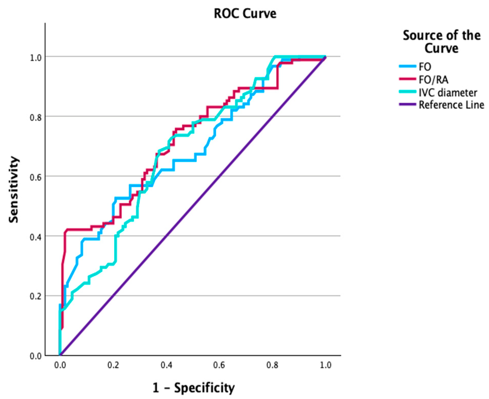
| FO width | <5.21 | 62.1% | 62.2% | 0.701 | 0.614–0.759 | <0.001 |
| FO/RA | <0.41 | 67.4% | 64.6% | 0.722 | 0.652–0.792 | <0.001 |
| IVC diameter | <3.89 | 68.4% | 63.7% | 0.682 | 0.609–0.759 | <0.001 |
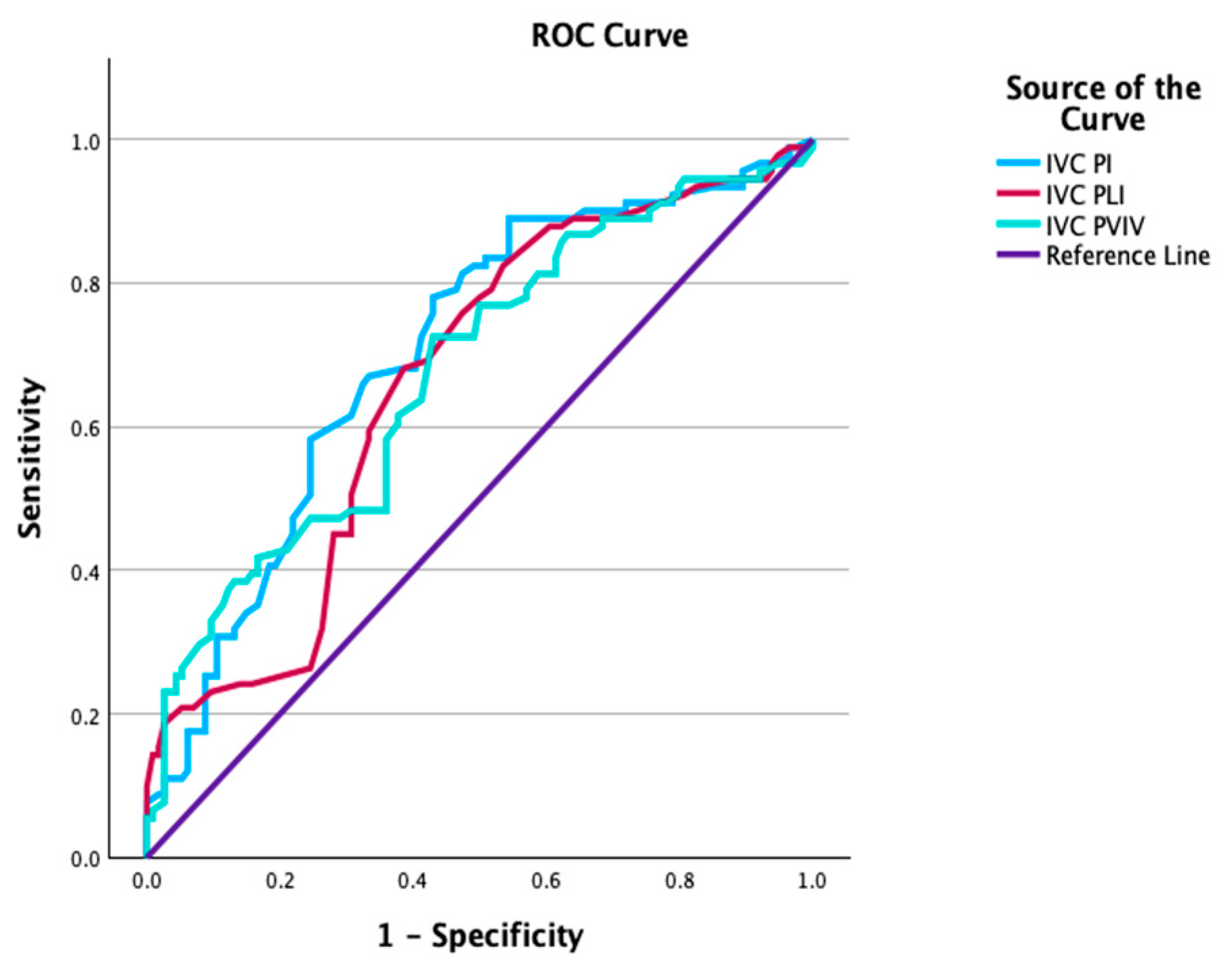
| IVC PI | >1.29 | 67.0% | 66.7% | 0.704 | 0.632–0.776 | <0.001 |
| IVC PLI | >0.75 | 68.1% | 62.4% | 0.662 | 0.588–0.737 | <0.001 |
| IVC PVIV | >1.45 | 61.5% | 63.3% | 0.676 | 0.602–0.750 | <0.001 |
| Variables | OR (95% CI) | p Value |
|---|---|---|
| Maternal age (years) | 1.02 (0.96–1.08) | 0.501 |
| BMI (kg/m2) | 1.02 (0.94–1.09) | 0.603 |
| GA at USG examination (weeks) | 0.90 (0.81–0.99) | 0.045 |
| FO/RA ratio | 0.57 (0.39–0.82) | 0.003 |
| IVC PI | 2.64 (1.28–5.43) | 0.008 |
Disclaimer/Publisher’s Note: The statements, opinions and data contained in all publications are solely those of the individual author(s) and contributor(s) and not of MDPI and/or the editor(s). MDPI and/or the editor(s) disclaim responsibility for any injury to people or property resulting from any ideas, methods, instructions or products referred to in the content. |
© 2026 by the authors. Licensee MDPI, Basel, Switzerland. This article is an open access article distributed under the terms and conditions of the Creative Commons Attribution (CC BY) license.
Share and Cite
Ayas Ozkan, M.; Ozkan, H.D.; Dayanan, R.; Sarı, H.; Akın, F.; Dağdeviren, G.; Çağlar, A.T. Foramen Ovale Measurements and Venous Hemodynamic Changes Assessed by Inferior Vena Cava Doppler Parameters in Early- and Late-Onset Fetal Growth Restriction. J. Clin. Med. 2026, 15, 980. https://doi.org/10.3390/jcm15030980
Ayas Ozkan M, Ozkan HD, Dayanan R, Sarı H, Akın F, Dağdeviren G, Çağlar AT. Foramen Ovale Measurements and Venous Hemodynamic Changes Assessed by Inferior Vena Cava Doppler Parameters in Early- and Late-Onset Fetal Growth Restriction. Journal of Clinical Medicine. 2026; 15(3):980. https://doi.org/10.3390/jcm15030980
Chicago/Turabian StyleAyas Ozkan, Merve, Halis Doğukan Ozkan, Ruken Dayanan, Hilal Sarı, Furkan Akın, Gülşah Dağdeviren, and Ali Turhan Çağlar. 2026. "Foramen Ovale Measurements and Venous Hemodynamic Changes Assessed by Inferior Vena Cava Doppler Parameters in Early- and Late-Onset Fetal Growth Restriction" Journal of Clinical Medicine 15, no. 3: 980. https://doi.org/10.3390/jcm15030980
APA StyleAyas Ozkan, M., Ozkan, H. D., Dayanan, R., Sarı, H., Akın, F., Dağdeviren, G., & Çağlar, A. T. (2026). Foramen Ovale Measurements and Venous Hemodynamic Changes Assessed by Inferior Vena Cava Doppler Parameters in Early- and Late-Onset Fetal Growth Restriction. Journal of Clinical Medicine, 15(3), 980. https://doi.org/10.3390/jcm15030980