The Eight-Chop Technique in Nonagenarian Patients: Real-World Intraoperative and Postoperative Outcomes


Abstract
1. Introduction
2. Materials and Methods
2.1. Study Design and Patients
2.2. Preoperative and Postoperative Examinations
2.3. Surgical Technique
2.4. Intraoperative and Postoperative Outcome Measures
2.5. Statistical Analysis
2.6. GenAI Statement
3. Results
3.1. Baseline Characteristics
3.2. Intraoperative Parameters
3.3. Postoperative Outcomes
3.4. Complications
4. Discussion
4.1. Principal Findings
4.2. Comparison with Previous Studies
4.3. Clinical Implications
4.4. Limitations and Future Directions
5. Conclusions
Supplementary Materials
Funding
Institutional Review Board Statement
Informed Consent Statement
Data Availability Statement
Acknowledgments
Conflicts of Interest
Abbreviations
| BCVA | Best corrected visual acuity |
| CCT | Central corneal thickness |
| CDE | Cumulative dissipated energy |
| CECD | Corneal endothelial cell density |
| CV | Coefficient of variation in cell size |
| IOP | Intraocular pressure |
| logMAR | Logarithm of the minimum angle of resolution |
| PHC | Percentage of hexagonal cells |
References
- GBD 2019 Blindness and Vision Impairment Collaborators; Vision Loss Expert Group of the Global Burden of Disease Study. Causes of blindness and vision impairment in 2020 and trends over 30 years, and prevalence of avoidable blindness in relation to VISION 2020: The Right to Sight: An analysis for the Global Burden of Disease Study. Lancet Glob. Health 2021, 9, e144–e160. [Google Scholar] [CrossRef] [Scilit]
- Vision Loss Expert Group of the Global Burden of Disease Study; GBD 2019 Blindness and Vision Impairment Collaborators. Global estimates on the number of people blind or visually impaired by age-related macular degeneration: A meta-analysis from 2000 to 2020. Eye 2024, 38, 2070–2082. [Google Scholar] [CrossRef] [Scilit]
- Tseng, V.L.; Greenberg, P.B.; Wu, W.C.; Jiang, L.; Li, E.; Kang, J.M.; Scott, I.U.; Friedmann, P.D. Cataract surgery complications in nonagenarians. Ophthalmology 2011, 118, 1229–1235. [Google Scholar] [CrossRef] [Scilit]
- Young, A.; Chow, P.; Lok, J.; Lai, F. Clinical Outcomes of Cataract Surgery in Very Elderly Adults. J. Am. Geriatr. Soc. 2014, 62, 165–170. [Google Scholar] [CrossRef] [Scilit]
- Gaskin, G.L.; Pershing, S.; Cole, T.S.; Shah, N.H. Predictive modeling of risk factors and complications of cataract surgery. Eur. J. Ophthalmol. 2016, 26, 328–337. [Google Scholar] [CrossRef] [Scilit]
- Rosen, E.; Rubowitz, A.; Assia, E.I. Visual outcome following cataract extraction in patients aged 90 years and older. Eye 2009, 23, 1120–1124. [Google Scholar] [CrossRef] [Scilit]
- Hwang, H.B.; Lyu, B.; Yim, H.B.; Lee, N.Y. Endothelial cell loss after phacoemulsification according to different anterior chamber depths. J. Ophthalmol. 2015, 2015, 210716. [Google Scholar] [CrossRef] [Scilit]
- Altansukh, A.; Ma, K.S.; Doyodmaa, A.; Hung, N.; Kang, E.Y.; Quan, W.; Ma, D.H. Comparison of corneal endothelial cell loss in FLACS: Impact on endothelial cells in different regions. J. Cataract Refract. Surg. 2024, 50, 912–918. [Google Scholar] [CrossRef] [Scilit]
- Al-Mohtaseb, Z.; He, X.; Yesilirmak, N.; Waren, D.; Donaldson, K.E. Comparison of Corneal Endothelial Cell Loss Between Two Femtosecond Laser Platforms and Standard Phacoemulsification. J. Refract. Surg. 2017, 33, 708–712. [Google Scholar] [CrossRef] [Scilit]
- Singh, R.; Sharma, A.K.; Katiyar, V.; Kumar, G.; Gupta, S.K. Corneal endothelial changes following cataract surgery in hard nuclear cataract: Randomized trial comparing phacoemulsification to manual small-incision cataract surgery. Indian J. Ophthalmol. 2022, 70, 3904–3909. [Google Scholar]
- Sato, T. Minimizing Endothelial Cell Loss in Hard Nucleus Cataract Surgery: Efficacy of the Eight-Chop Technique. J. Clin. Med. 2025, 14, 2576. [Google Scholar] [CrossRef] [Scilit]
- Sato, T. Efficacy and safety of the eight-chop technique in phacoemulsification for patients with cataract. J. Cataract Refract. Surg. 2023, 49, 479–484. [Google Scholar] [CrossRef] [Scilit]
- Sato, T. The Eight-Chop Technique in Cataract Surgery: A Conceptual and Narrative Review of a Segmentation-First Strategy. Clin. Ophthalmol. 2026, 20, 624182. [Google Scholar] [CrossRef] [Scilit]
- Emery, J.M. Kelman phacoemulsification; patient selection. In Extracapsular Cataract Surgery; Emery, J.M., McIntyre, D.J., Eds.; CV Mosby: St Louis, MO, USA, 1983; pp. 95–100. [Google Scholar]
- Zhang, G.; Chen, J.J. Adaptive fitting of linear mixed-effects models with correlated random-effects. J. Stat. Comput. Simul. 2013, 83, 2291–2314. [Google Scholar] [CrossRef] [Scilit]
- Shingleton, B.J.; Marvin, A.C.; Heier, J.S.; O’Donoghue, M.W.; Laul, A.; Wolff, B.; Rowland, A. Pseudoexfoliation: High risk factors for zonule weakness and concurrent vitrectomy during phacoemulsification. J. Cataract Refract. Surg. 2010, 36, 1261–1269. [Google Scholar] [CrossRef] [Scilit]
- Shingleton, B.J.; Neo, Y.N.; Cvintal, V.; Shaikh, A.M.; Liberman, P.; O’Donoghue, M.W. Outcome of phacoemulsification and intraocular lens implantation in eyes with pseudoexfoliation and weak zonules. Acta Ophthalmol. 2017, 95, 182–187. [Google Scholar] [CrossRef] [Scilit]
- Akman, A.; Yilmaz, G.; Oto, S.; Akova, Y.A. Comparison of various pupil dilatation methods for phacoemulsification in eyes with a small pupil secondary to pseudoexfoliation. Ophthalmology 2004, 111, 1693–1698. [Google Scholar] [CrossRef] [Scilit]
- Akansha; Yadav, R.S. Comparative assessment of the corneal endothelium following phacoemulsification surgery in patients with type II diabetes and nondiabetes. Saudi J. Ophthalmol. 2025, 39, 77–83. [Google Scholar] [CrossRef] [Scilit]
- Walkow, T.; Anders, N.; Klebe, S. Endothelial cell loss after phacoemulsification: Relation to preoperative and intraoperative parameters. J. Cataract Refract. Surg. 2000, 26, 727–732. [Google Scholar] [CrossRef] [Scilit]
- Perone, J.M.; Luc, M.S.; Zevering, Y.; Vermion, J.C.; Gan, G.; Goetz, C. Narrative review after post-hoc trial analysis of factors that predict corneal endothelial cell loss after phacoemulsification: Tips for improving cataract surgery research. PLoS ONE 2024, 19, e0298795. [Google Scholar] [CrossRef] [Scilit]
- Bamdad, S.; Bolkheir, A.; Sedaghat, M.R.; Motamed, M. Changes in corneal thickness and corneal endothelial cell density after phacoemulsification cataract surgery: A double-blind randomized trial. Electron. Physician 2018, 10, 6616–6623. [Google Scholar] [CrossRef] [Scilit]
- Dzhaber, D.; Mustafa, O.; Alsaleh, F.; Mihailovic, A.; Daoud, Y.J. Comparison of changes in corneal endothelial cell density and central corneal thickness between conventional and femtosecond laser-assisted cataract surgery: A randomised, controlled clinical trial. Br. J. Ophthalmol. 2020, 104, 225–229. [Google Scholar] [CrossRef] [Scilit]
- Conrad-Hengerer, I.; Al Juburi, M.; Schultz, T.; Hengerer, F.H.; Dick, H.B. Corneal endothelial cell loss and corneal thickness in conventional compared with femtosecond laser-assisted cataract surgery: Three-month follow-up. J. Cataract Refract. Surg. 2013, 39, 1307–1313. [Google Scholar] [CrossRef] [Scilit]
- Demircan, S.; Atas, M.; Yurtsever, Y. Effect of torsional mode phacoemulsification on cornea in eyes with/without pseudoexfoliation. Int. J. Ophthalmol. 2015, 8, 281–287. [Google Scholar] [CrossRef] [Scilit]
- Sato, T. Postoperative Flare and Corneal Endothelial Cell Loss After Eight-Chop Technique Phacoemulsification: A Prospective Observational Study. J. Clin. Med. 2026, 15, 557. [Google Scholar] [CrossRef] [Scilit]
- Dinarvand, D.; Panthakey, J.; Hassan, A.; Ahmed, M.H. Frailty and Visual Impairment in Elderly Individuals: Improving Outcomes and Modulating Cognitive Decline Through Collaborative Care Between Geriatricians and Ophthalmologists. Diseases 2024, 12, 273. [Google Scholar] [CrossRef] [Scilit]
- Dhawale, K.K.; Tidake, P. Cataract Surgery and Mental Health: A Comprehensive Review on Outcomes in the Elderly. Cureus 2024, 16, e65469. [Google Scholar] [CrossRef] [Scilit]
- Wong, T.Y. Effect of increasing age on cataract surgery outcomes in very elderly patients. BMJ 2001, 322, 1104–1106. [Google Scholar] [CrossRef] [Scilit]
- Al-Namaeh, M. Common causes of visual impairment in the elderly. Med. Hypothesis Discov. Innov. Ophthalmol. 2021, 10, 191–200. [Google Scholar] [CrossRef] [Scilit]



| Characteristic | 70–79 Years n = 60 | 80–89 Years n = 60 | ≥90 Years n = 49 | p-Value |
|---|---|---|---|---|
| Patients, n | 60 | 60 | 27 | |
| Patients contributing one eye/both eyes, n | 60/0 | 60/0 | 5/22 | |
| Age, years | 74.8 ± 2.5 | 82.6 ± 2.4 | 91.9 ± 2.0 | <0.001 |
| Sex | 0.479 | |||
| Female | 33 (55%) | 38 (63%) | 32 (65%) | |
| Male | 27 (45%) | 22 (37%) | 17 (35%) | |
| Operated eye | 0.823 | |||
| L | 28 (47%) | 30 (50%) | 26 (53%) | |
| R | 32 (53%) | 30 (50%) | 23 (47%) | |
| Emery nuclear grade | <0.001 | |||
| II | 54 (90%) | 49 (82%) | 30 (61%) | |
| III | 4 (6.7%) | 8 (13%) | 5 (10%) | |
| IV | 2 (3.3%) | 3 (5.0%) | 14 (29%) | |
| Preoperative BCVA, logMAR | 0.191 ± 0.335 | 0.199 ± 0.285 | 0.482 ± 0.555 | <0.001 |
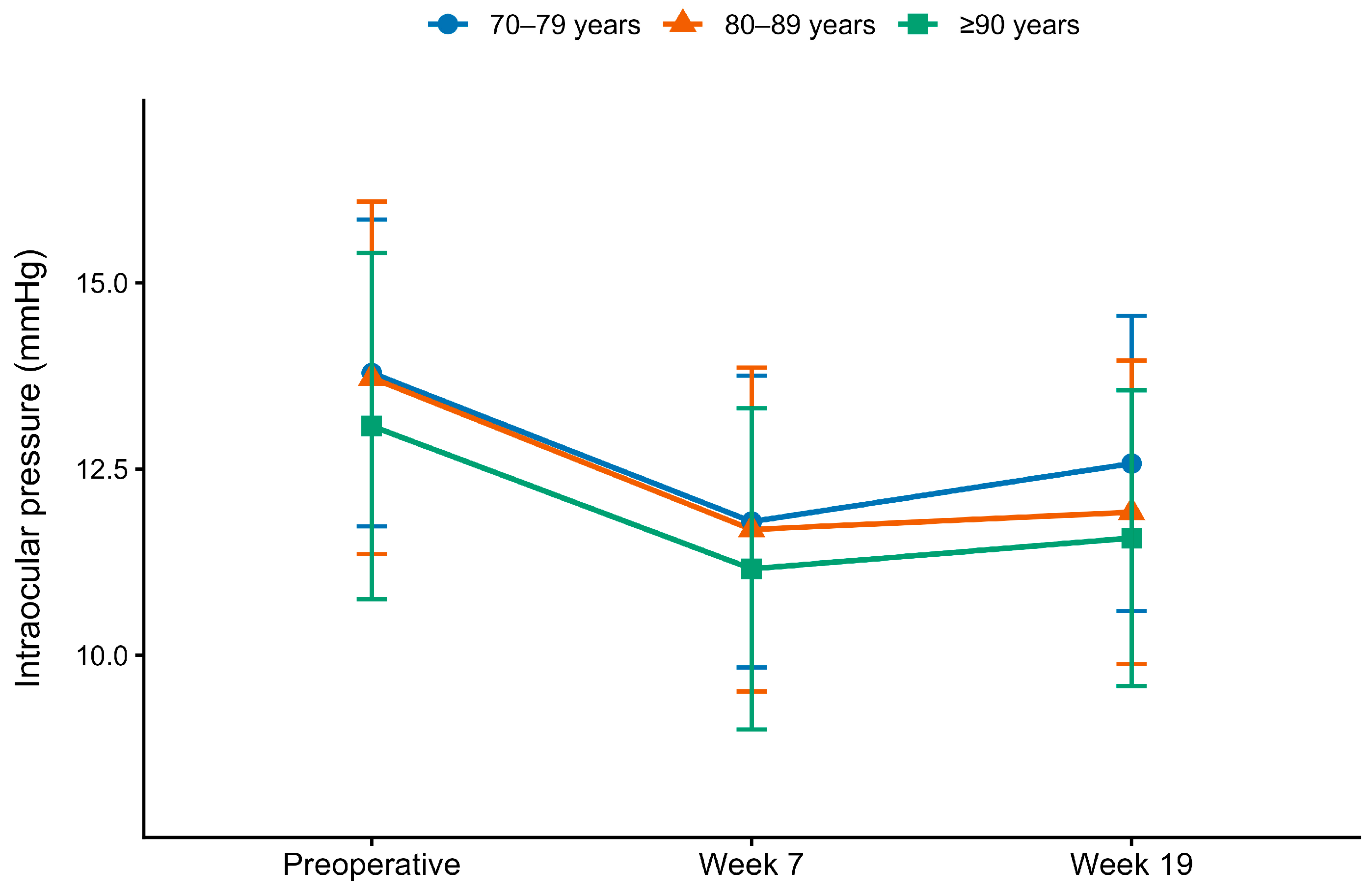
| Preoperative IOP, mmHg | 13.8 ± 2.1 | 13.7 ± 2.4 | 13.1 ± 2.3 | 0.265 |
| Preoperative CECD, cells/mm2 | 2588.1 ± 237.9 | 2566.3 ± 336.2 | 2585.3 ± 267.8 | 0.968 |
| Preoperative CV, % | 39.3 ± 5.8 | 40.9 ± 6.1 | 40.8 ± 5.6 | 0.132 |
| Preoperative PHC, % | 43.3 ± 7.4 | 44.3 ± 7.2 | 45.5 ± 6.9 | 0.335 |
| Preoperative CCT, μm | 531.1 ± 39.0 | 528.6 ± 31.7 | 522.0 ± 30.2 | 0.251 |
| Intraoperative Parameter | 70–79 Years n = 60 | 80–89 Years n = 60 | ≥90 Years n = 49 | p-Value |
|---|---|---|---|---|
| Operative time, min | 6.0 ± 2.5 | 6.7 ± 3.7 | 10.7 ± 6.1 | <0.001 |
| Phacoemulsification time, s | 17.6 ± 7.6 | 20.3 ± 8.3 | 30.2 ± 12.0 | <0.001 |
| Aspiration time, s | 76.8 ± 27.7 | 84.6 ± 35.6 | 110.2 ± 35.0 | <0.001 |
| Cumulative dissipated energy | 6.67 ± 2.51 | 7.68 ± 3.45 | 10.72 ± 3.76 | <0.001 |
| Fluid volume, mL | 30.5 ± 11.9 | 33.8 ± 15.6 | 45.2 ± 14.9 | <0.001 |
| Iris retractor hooks use | 3 (5.0%) | 7 (12%) | 13 (27%) | 0.005 |
| Age Group | Eyes Analyzed | Patients | Operative Time, min | Estimated Marginal Mean, min (95% CI) |
|---|---|---|---|---|
| 70–79 years | 57 | 57 | 5.61 ± 1.88 | 5.61 (5.02–6.21) |
| 80–89 years | 53 | 53 | 5.88 ± 2.46 | 5.88 (5.26–6.49) |
| ≥90 years | 36 | 21 | 7.65 ± 2.55 | 7.80 (6.88–8.72) |
| Mean difference, min (95% CI) | p-Value | |||
| Overall age-group effect | F = 8.29; denominator df = 116.96 | <0.001 | ||
| ≥90 vs. 70–79 years | 2.18 (0.87–3.50) | <0.001 | ||
| ≥90 vs. 80–89 years | 1.92 (0.60–3.25) | 0.002 | ||
| 70–79 vs. 80–89 years | −0.26 (−1.29 to 0.76) | 0.818 |
| Postoperative Outcome | 70–79 Years n = 60 | 80–89 Years n = 60 | ≥90 Years n = 49 | p-Value |
|---|---|---|---|---|
| BCVA at postoperative week 7, logMAR | −0.046 ± 0.064 | −0.016 ± 0.106 | 0.121 ± 0.344 | <0.001 |
| BCVA at postoperative week 19, logMAR | −0.047 ± 0.062 | −0.013 ± 0.110 | 0.096 ± 0.364 | <0.001 |
| IOP at postoperative week 7, mmHg | 11.8 ± 2.0 | 11.7 ± 2.2 | 11.2 ± 2.2 | 0.518 |
| IOP at postoperative week 19, mmHg | 12.6 ± 2.0 | 11.9 ± 2.0 | 11.6 ± 2.0 | 0.102 |
| CECD at postoperative week 7, cells/mm2 | 2615.1 ± 236.9 | 2573.7 ± 321.0 | 2474.0 ± 328.1 | 0.043 |
| CECD at postoperative week 19, cells/mm2 | 2615.9 ± 261.7 | 2594.4 ± 364.1 | 2483.4 ± 313.8 | 0.104 |
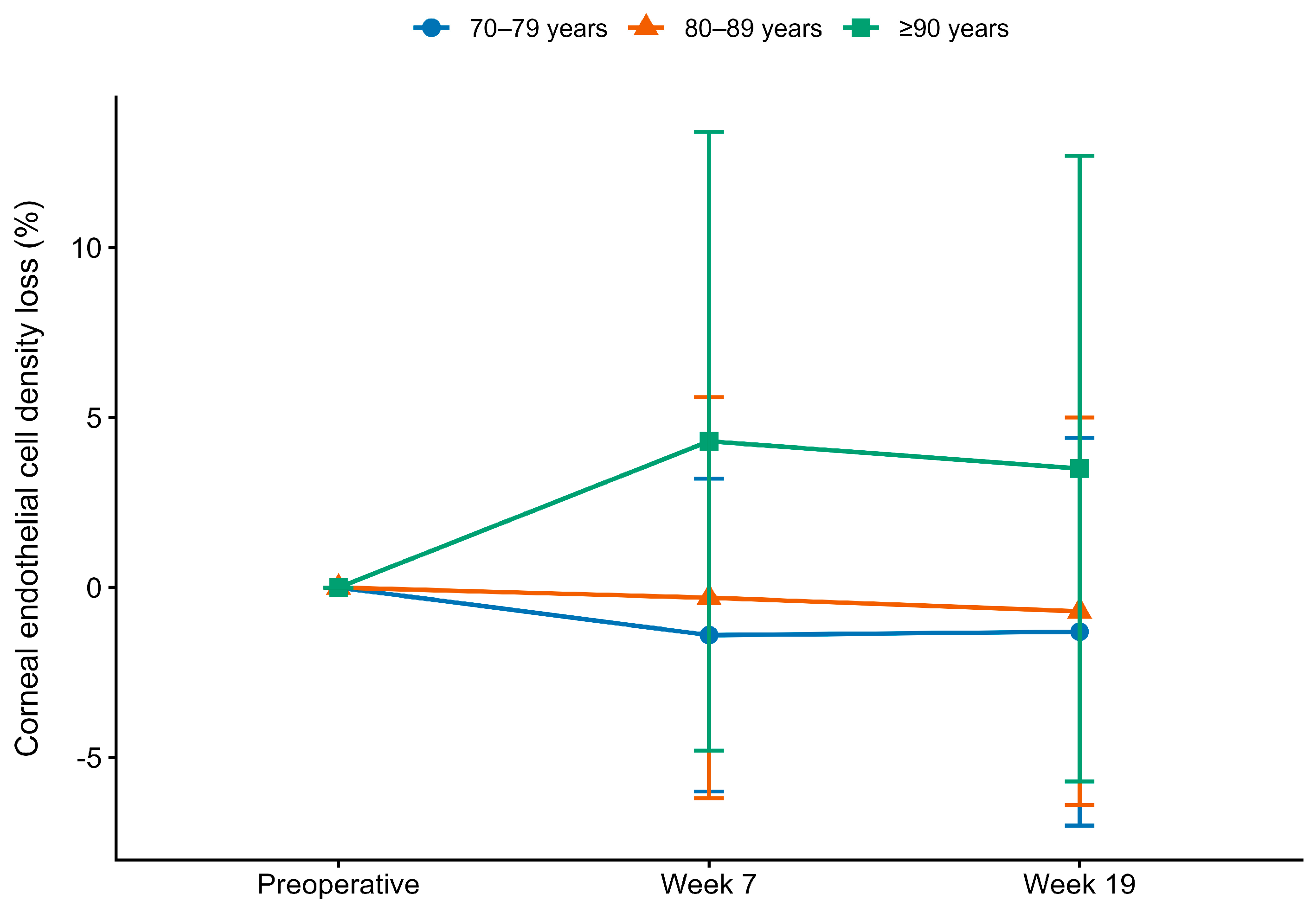
| CECD loss at postoperative week 7, % | −1.4 ± 4.6 | −0.3 ± 5.9 | 4.3 ± 9.1 | <0.001 |
| CECD loss at postoperative week 19, % | −1.3 ± 5.7 | −0.7 ± 5.7 | 3.5 ± 9.2 | 0.001 |
| CV at postoperative week 7, % | 39.3 ± 4.5 | 38.7 ± 5.4 | 39.2 ± 4.4 | 0.399 |
| CV at postoperative week 19, % | 37.3 ± 5.2 | 36.7 ± 5.7 | 38.9 ± 4.7 | 0.082 |
| PHC at postoperative week 7, % | 44.5 ± 5.7 | 45.1 ± 6.4 | 44.1 ± 6.1 | 0.771 |
| PHC at postoperative week 19, % | 46.6 ± 6.1 | 47.4 ± 7.0 | 45.1 ± 4.6 | 0.243 |
| CCT at postoperative week 7, μm | 535.0 ± 38.8 | 530.7 ± 37.1 | 522.6 ± 32.4 | 0.329 |
| CCT at postoperative week 19, μm | 536.3 ± 37.1 | 525.3 ± 34.0 | 525.9 ± 32.6 | 0.386 |
Disclaimer/Publisher’s Note: The statements, opinions and data contained in all publications are solely those of the individual author(s) and contributor(s) and not of MDPI and/or the editor(s). MDPI and/or the editor(s) disclaim responsibility for any injury to people or property resulting from any ideas, methods, instructions or products referred to in the content. |
© 2026 by the author. Licensee MDPI, Basel, Switzerland. This article is an open access article distributed under the terms and conditions of the Creative Commons Attribution (CC BY) license.
Share and Cite
Sato, T. The Eight-Chop Technique in Nonagenarian Patients: Real-World Intraoperative and Postoperative Outcomes. J. Clin. Med. 2026, 15, 6737. https://doi.org/10.3390/jcm15176737
Sato T. The Eight-Chop Technique in Nonagenarian Patients: Real-World Intraoperative and Postoperative Outcomes. Journal of Clinical Medicine. 2026; 15(17):6737. https://doi.org/10.3390/jcm15176737
Chicago/Turabian StyleSato, Tsuyoshi. 2026. "The Eight-Chop Technique in Nonagenarian Patients: Real-World Intraoperative and Postoperative Outcomes" Journal of Clinical Medicine 15, no. 17: 6737. https://doi.org/10.3390/jcm15176737
APA StyleSato, T. (2026). The Eight-Chop Technique in Nonagenarian Patients: Real-World Intraoperative and Postoperative Outcomes. Journal of Clinical Medicine, 15(17), 6737. https://doi.org/10.3390/jcm15176737