Single Coronary Artery in the Context of Vascular Disease: Anatomy, Development, Multimodality Imaging, and Clinical Interpretation


Abstract
1. Introduction
Literature Search and Review Scope
2. Developmental Basis of Single Coronary Artery
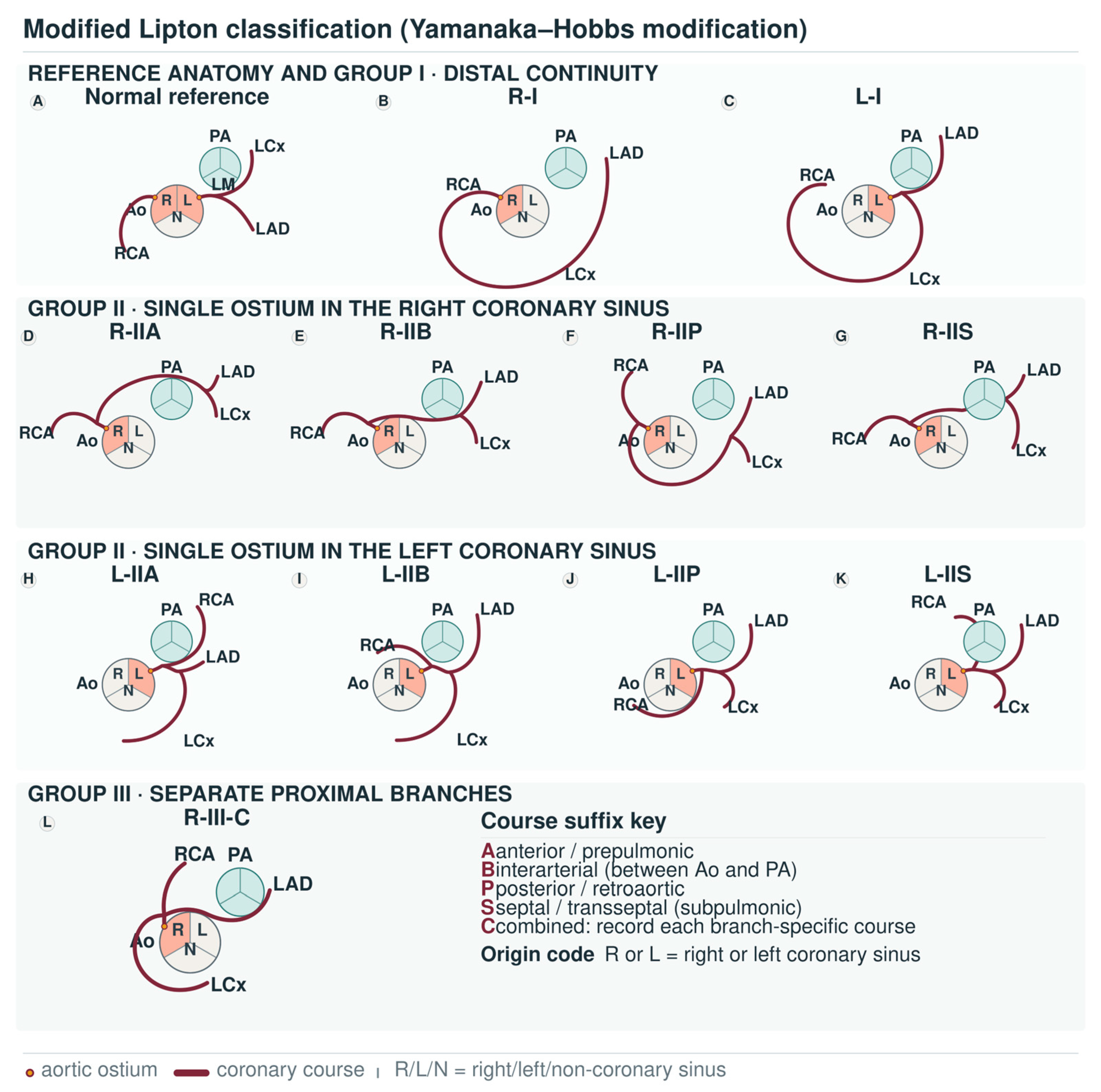
3. Anatomical Spectrum and Classification

| Framework Element | Definition and Representative Coding | CCTA-Oriented Reporting Focus | Interpretive Boundary | Evidence Category and Support |
|---|---|---|---|---|
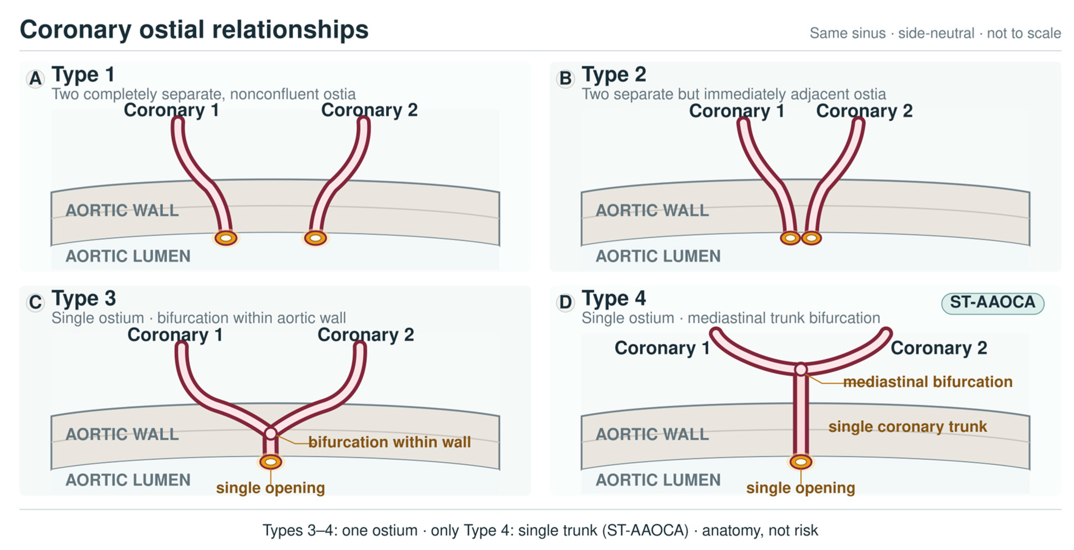
| Definition, true single ostium and common trunk | Traditional SCA usage denotes an entire epicardial coronary circulation arising from one aortic ostium. A common trunk requires a measurable shared proximal segment before major branches divide. | Confirm both aortic sinuses; exclude pulmonary-origin and fistula- or collateral-dependent mimics; and state whether branching occurs within the aortic wall, immediately beyond it, or after a measurable common trunk. | By itself, a single ostium does not prove a common trunk, define ST-AAOCA, or determine clinical significance. | Traditional SCA definition [6,7]. Left-main agenesis dilemma [31]. Pulmonary-origin and fistula differential context [32]. Component-based AAOCA terminology [30]. |
| Distribution group I | R-I or L-I: One vessel continues distally to supply the contralateral coronary territory. | Describe distal continuity, PDA origin, posterolateral branches, vessel calibre, terminal-branch duplication, supplied territory, and luminal disease. | Usually a descriptive category; interpretation depends on course, symptoms, objective ischaemia, and acquired disease. | Classical classification [6]. Direct angiographic SCA evidence [7,51,52]. |
| Distribution group II | R-II or L-II: An anomalous branch arises from the proximal main trunk and reaches the contralateral coronary distribution. A/B/P are classical course suffixes; S and C appear in the Yamanaka–Hobbs modification. | Report take-off site and angle, course relative to Ao/PA, proximal narrowing, suspected intramural segment, and branch-level anatomy. | Course and proximal morphology are more informative than the group label alone. | Classical Lipton and Yamanaka–Hobbs classification evidence [6,33]. |
| Distribution group III | R-III: LAD and LCx arise separately from the proximal RCA or a right-sided common trunk; complex variants may have branch-specific mixed courses. | Map LAD and LCx separately, including their courses, supplied territories, calibre, and procedural implications. | Complex patterns should be reported branch by branch rather than compressed into a single clinical risk label. | Classical classification [6]. Direct SCA report with dual LAD [38]. |
| A suffix | Anterior/prepulmonic course; representative coding R-IIA or L-IIA. | Confirm the anterior relationship to PA and document ostial/proximal geometry. | Anterior/prepulmonic course should be described anatomically; its clinical relevance remains dependent on patient-specific morphology and context. | Classical classification evidence [6,33]. |
| B suffix | Interarterial course between the great vessels; representative coding R-IIB or L-IIB. | Assess the Ao–PA relationship, slit-like ostium, acute take-off angle, proximal narrowing, intramural segment, and suspected dynamic compression. | Anatomy warranting heightened attention; interpretation should integrate detailed morphology, symptoms, and functional evidence when indicated. | Classical course description [6,33]. Proximal morphology interpretation is derived mainly from AAOCA evidence [3,11]. |
| P suffix | Posterior/retroaortic course; representative coding R-IIP or L-IIP. | Describe retroaortic trajectory, relationship to the aortic root/valves, and coexisting aortic or coronary disease. | The retroaortic label is descriptive and should not be used alone to infer clinical risk. | Classical classification evidence [6,33]. |
| S suffix | Septal/subpulmonic/transseptal course introduced in the Yamanaka–Hobbs modification; representative coding R-IIS or L-IIS. | Define the septal or subpulmonic pathway, any intramyocardial/intraseptal component, and relationship to PA/RV outflow. | S is an anatomical descriptor; clinical interpretation depends on patient-specific morphology, symptoms, and functional evidence. | Yamanaka–Hobbs modification and SCA descriptive evidence [20,33]. |
| C suffix | Combined or mixed course involving more than one directional component in the Yamanaka–Hobbs modification. | State which branch follows which course (for example, LAD-B with LCx-P), rather than relying on C alone. | C is a descriptive modifier, not a validated or stand-alone risk category. | Yamanaka–Hobbs modification and SCA descriptive evidence [20,33]. |
| Classification systems | Lipton combines sinus of origin, distribution group, and proximal course suffix; Yamanaka–Hobbs adds S and C; Shirani–Roberts classifies the site of the solitary ostium and route of the aberrant artery. | State the system used and provide a component-level anatomical description when one code does not capture the full pattern. | The modified Lipton system is used as the primary clinical language in this review; Shirani–Roberts terminology is complementary. | Classical primary classification sources [6,33,35]. |
| Coronary dominance and inferior supply | Dominance is conventionally defined by the artery giving rise to the PDA. | Identify the origin of the PDA and the number and distribution of posterolateral branches. | Right- or left-dominance labels have limited discriminating value when the whole myocardium is supplied from one aortic origin. | Direct SCA CCTA series [41,42,43]. Contextual inferior-branch anatomy [36,37]. |
| Terminal-branch duplication | Dual LAD or duplicated PDA patterns alter the distal perfusion map despite a common ostial origin. | Report branch number, calibre, length, course, and supplied myocardial territory. | Branch-level anatomy identifies the vessels that require protection during PCI or surgery. | Dual LAD reported with SCA [38]. Broader dual-LAD evidence [39,40] and double-PDA evidence [37]. |
| CCTA morphology beyond classification | Classic labels do not fully capture ostial morphology, take-off angle, a suspected intramural segment, proximal narrowing, or Ao–PA relationships. | Use multiplanar and three-dimensional CCTA to describe the complete proximal course and adjacent great vessel relationships. | CCTA complements classification; morphology alone should not be converted into a treatment rule. | Direct SCA CCTA series [41,42,43]. Additional SCA imaging context [5]. Risk-agnostic AAOCA terminology [30]. |
4. Pathophysiological and Haemodynamic Implications
5. Clinical Presentation and Heterogeneity of Clinical Significance
6. Imaging-Based Evaluation and Diagnostic Strategy
7. Single Coronary Artery in Adult Vascular and Procedural Context
8. Structured Reporting and Interpretive Boundaries
9. Management Implications and Decision-Making Considerations
10. Future Directions and Knowledge Gaps
Evidence Boundaries and Limitations of the Review
11. Conclusions
Supplementary Materials
Author Contributions
Funding
Institutional Review Board Statement
Informed Consent Statement
Data Availability Statement
Acknowledgments
Conflicts of Interest
References
- Angelini, P. Coronary Artery Anomalies—Current Clinical Issues: Definitions, Classification, Incidence, Clinical Relevance, and Treatment Guidelines. Tex. Heart Inst. J. 2002, 29, 271–278. [Google Scholar] [PubMed]
- Gentile, F.; Castiglione, V.; De Caterina, R. Coronary Artery Anomalies. Circulation 2021, 144, 983–996. [Google Scholar] [CrossRef] [Scilit] [PubMed]
- Gaudino, M.; Di Franco, A.; Arbustini, E.; Bacha, E.; Bates, E.R.; Cameron, D.E.; Cao, D.; David, T.E.; De Paulis, R.; El-Hamamsy, I.; et al. Management of Adults with Anomalous Aortic Origin of the Coronary Arteries. J. Am. Coll. Cardiol. 2023, 82, 2034–2053. [Google Scholar] [CrossRef] [Scilit] [PubMed]
- Lalani, K.; Rao, M.S.; Ramachandran, P.; Ashwal, A.J.; Razak, A.U.K.; Devasia, T.; Paramasivam, G.; Parikh, P. Single Coronary Artery from Right Sinus of Valsalva: A Single Center Experience of Seven Cases. Case Rep. Med. 2022, 2022, 1513474. [Google Scholar] [CrossRef] [Scilit] [PubMed]
- Gać, P.; Trejtowicz-Sutor, A.; Poręba, R. Single Coronary Artery as a Rare Developmental Variant in Cardiac Computed Tomography Angiography. Diagnostics 2023, 13, 1369. [Google Scholar] [CrossRef] [Scilit] [PubMed]
- Lipton, M.J.; Barry, W.H.; Obrez, I.; Silverman, J.F.; Wexler, L. Isolated Single Coronary Artery: Diagnosis, Angiographic Classification, and Clinical Significance. Radiology 1979, 130, 39–47. [Google Scholar] [CrossRef] [Scilit] [PubMed]
- Desmet, W.; Vanhaecke, J.; Vrolix, M.; Van De Werf, F.; Piessens, J.; Willems, J.; De Geest, H. Isolated Single Coronary Artery: A Review of 50 000 Consecutive Coronary Angiographies. Eur. Heart J. 1992, 13, 1637–1640. [Google Scholar] [CrossRef] [Scilit] [PubMed]
- Ahmed, A.I.; Han, Y.; Soliman, A.; Al-Mallah, M.H. Multimodality Imaging of a Patient with Single Coronary Artery: CTA, FFRCT, SPECT, and Beyond. J. Nucl. Cardiol. 2022, 29, 871–873. [Google Scholar] [CrossRef] [Scilit] [PubMed]
- Burnette-Phelps, K.; Sohn, L.S.; Bhardwaj, A.; Sharma, V.; Kritya, M.; Sahai, A.; Shamim, S. The Single Coronary Artery: A Rare Anatomic Variant. Methodist DeBakey Cardiovasc. J. 2025, 21, 118–120. [Google Scholar] [CrossRef] [Scilit] [PubMed]
- Bigler, M.R.; Ashraf, A.; Seiler, C.; Praz, F.; Ueki, Y.; Windecker, S.; Kadner, A.; Räber, L.; Gräni, C. Hemodynamic Relevance of Anomalous Coronary Arteries Originating from the Opposite Sinus of Valsalva—In Search of the Evidence. Front. Cardiovasc. Med. 2021, 7, 591326. [Google Scholar] [CrossRef] [Scilit] [PubMed]
- Koppel, C.J.; Verheijen, D.B.H.; Kiès, P.; Egorova, A.D.; Lamb, H.J.; Voskuil, M.; Jukema, J.W.; Koolbergen, D.R.; Hazekamp, M.G.; Schalij, M.J.; et al. A Comprehensive Analysis of the Intramural Segment in Interarterial Anomalous Coronary Arteries Using Computed Tomography Angiography. Eur. Heart J. Open 2022, 2, oeac031. [Google Scholar] [CrossRef] [Scilit] [PubMed]
- Rizzo, S.; De Gaspari, M.; Frescura, C.; Padalino, M.; Thiene, G.; Basso, C. Sudden Death and Coronary Artery Anomalies. Front. Cardiovasc. Med. 2021, 8, 636589. [Google Scholar] [CrossRef] [Scilit] [PubMed]
- Said, S.A.; de Voogt, W.G.; Bulut, S.; Han, J.; Polak, P.; Nijhuis, R.L.; Op den Akker, J.W.; Slootweg, A. Coronary Artery Disease in Congenital Single Coronary Artery in Adults: A Dutch Case Series. World J. Cardiol. 2014, 6, 196–204. [Google Scholar] [CrossRef] [Scilit] [PubMed]
- Lv, F.; Tao, Y. Single Coronary Artery with Severe Coronary Artery Disease and Aortic Valve Disease: A Case Report. Int. Heart J. 2023, 64, 955–958. [Google Scholar] [CrossRef] [Scilit] [PubMed]
- Koshy, A.N.; Kumar, K.R.; Giustino, G.; Sartori, S.; Fisher, R.; Dhulipala, V.; Iruvanti, S.; Suleman, J.; Sharma, S.K.; Kini, A.S. Procedural Success and Clinical Outcomes in Patients Undergoing Percutaneous Coronary Intervention of Anomalous Coronary Arteries. J. Soc. Cardiovasc. Angiogr. Interv. 2023, 2, 100564. [Google Scholar] [CrossRef] [Scilit] [PubMed]
- Capponi, G.; Castaldi, B.; Sirico, D.; Reffo, E.; Assanta, N.; Santoro, G.; Prati, F.; Pozza, A.; Cantinotti, M.; Salvo, G.D. Multi-Modality Imaging in Coronary Anomalies: Focus on Anomalous Aortic Origin of Coronary Arteries. Echocardiography 2025, 42, e70213. [Google Scholar] [CrossRef] [Scilit] [PubMed]
- Baz, R.O.; Refi, D.; Scheau, C.; Savulescu-Fiedler, I.; Baz, R.A.; Niscoveanu, C. Coronary Artery Anomalies: A Computed Tomography Angiography Pictorial Review. J. Clin. Med. 2024, 13, 3920. [Google Scholar] [CrossRef] [Scilit] [PubMed]
- Fuss, C.; McCallum, R.; Ghoshhajra, B.B.; Litmanovich, D.; Agarwal, P.P.; Bloom, S.; Brown, W.M.; Chelliah, A.; De Cecco, C.N.; Frommelt, P.; et al. ACR Appropriateness Criteria® Evaluation of Coronary Artery Anomalies. J. Am. Coll. Radiol. 2025, 22, S234–S242. [Google Scholar] [CrossRef] [Scilit] [PubMed]
- Schiavone, M.; Gobbi, C.; Gasperetti, A.; Zuffi, A.; Forleo, G.B. Congenital Coronary Artery Anomalies and Sudden Cardiac Death. Pediatr. Cardiol. 2021, 42, 1676–1687. [Google Scholar] [CrossRef] [Scilit] [PubMed]
- Zhang, J.; Yuan, M. Single Coronary Artery: A Comprehensive Review. Cardiol. Rev. 2026. online ahead of print. [Google Scholar] [CrossRef] [Scilit] [PubMed]
- Bigler, M.R.; Kadner, A.; Räber, L.; Ashraf, A.; Windecker, S.; Siepe, M.; Padalino, M.A.; Gräni, C. Therapeutic Management of Anomalous Coronary Arteries Originating from the Opposite Sinus of Valsalva: Current Evidence, Proposed Approach, and the Unknowing. J. Am. Heart Assoc. 2022, 11, e027098. [Google Scholar] [CrossRef] [Scilit] [PubMed]
- Adam, E.L.; Generoso, G.; Bittencourt, M.S. Anomalous Coronary Arteries: When to Follow-up, Risk Stratify, and Plan Intervention. Curr. Cardiol. Rep. 2021, 23, 102. [Google Scholar] [CrossRef] [Scilit] [PubMed]
- Pérez-Pomares, J.M.; De La Pompa, J.L.; Franco, D.; Henderson, D.; Ho, S.Y.; Houyel, L.; Kelly, R.G.; Sedmera, D.; Sheppard, M.; Sperling, S.; et al. Congenital Coronary Artery Anomalies: A Bridge from Embryology to Anatomy and Pathophysiology—A Position Statement of the Development, Anatomy, and Pathology ESC Working Group. Cardiovasc. Res. 2016, 109, 204–216. [Google Scholar] [CrossRef] [Scilit] [PubMed]
- Sharma, B.; Chang, A.; Red-Horse, K. Coronary Artery Development: Progenitor Cells and Differentiation Pathways. Annu. Rev. Physiol. 2017, 79, 1–19. [Google Scholar] [CrossRef] [Scilit] [PubMed]
- He, L.; Lui, K.O.; Zhou, B. The Formation of Coronary Vessels in Cardiac Development and Disease. Cold Spring Harb. Perspect. Biol. 2020, 12, a037168. [Google Scholar] [CrossRef] [Scilit] [PubMed]
- Chen, H.I.; Poduri, A.; Numi, H.; Kivela, R.; Saharinen, P.; McKay, A.S.; Raftrey, B.; Churko, J.; Tian, X.; Zhou, B.; et al. VEGF-C and Aortic Cardiomyocytes Guide Coronary Artery Stem Development. J. Clin. Investig. 2014, 124, 4899–4914. [Google Scholar] [CrossRef] [Scilit] [PubMed]
- Tomanek, R.J. Developmental Progression of the Coronary Vasculature in Human Embryos and Fetuses. Anat. Rec. 2016, 299, 25–41. [Google Scholar] [CrossRef] [Scilit] [PubMed]
- Ioakeimidis, N.S.; Pepis, P.; Mitrousi, K.; Valasiadis, D. One Artery to Rule Them All: A Case of Single Coronary Artery Arising from the Right Sinus of Valsalva. Radiol. Case Rep. 2025, 20, 391–394. [Google Scholar] [CrossRef] [Scilit] [PubMed]
- Han, P.; Diao, K.; Huang, S.; Gao, Y.; Guo, Y.; Yang, Z.; Yang, N. Anatomical Characteristics of Anomalous Left Coronary Artery from the Opposite Sinus (Left-ACAOS) and Its Clinical Relevance: A Serial Coronary CT Angiography Study. Int. J. Cardiol. Heart Vasc. 2020, 31, 100649. [Google Scholar] [CrossRef] [Scilit] [PubMed]
- Mery, C.M.; Nathan, M.; Gräni, C.; Krishnamurthy, R.; Basso, C.; Jacobs, M.L.; Brothers, J.A.; Padalino, M.A.; Molossi, S. Proposed Standardized Nomenclature for Anomalous Aortic Origin of a Coronary Artery. JACC Cardiovasc. Imaging 2026, 19, 983–1001. [Google Scholar] [CrossRef] [Scilit] [PubMed]
- Kök, Z.; Sökmen, E.; Bingöl, B. Classification Dilemma between Right Single Coronary Artery and Agenesis of Left Main Coronary Artery: A Rare Coronary Anomaly. Turk. J. Thorac. Cardiovasc. Surg. 2025, 33, 113–116. [Google Scholar] [CrossRef] [Scilit] [PubMed]
- Hoover, J.A.; Catakam, K.; Wittenberg, R.E.; Bloom, J.P.; Yeh, D.D.; Han, Q.J.; Stefanescu Schmidt, A.C. Coronary Artery Anomalies in Review: Anomalous Origin, Aneurysms, and Fistulae. Methodist DeBakey Cardiovasc. J. 2025, 21, 54–64. [Google Scholar] [CrossRef] [Scilit] [PubMed]
- Yamanaka, O.; Hobbs, R.E. Coronary Artery Anomalies in 126,595 Patients Undergoing Coronary Arteriography. Cathet. Cardiovasc. Diagn. 1990, 21, 28–40. [Google Scholar] [CrossRef] [Scilit] [PubMed]
- Lindsey, W.W.; Tomasulo, C.E.; Gillespie, M.J.; Harris, M.A.; Brothers, J.A. Isolated Single Right Coronary Artery in a Young Patient: A Rare Shirani-Roberts Type IIC3. JACC Case Rep. 2025, 30, 103676. [Google Scholar] [CrossRef] [Scilit] [PubMed]
- Shirani, J.; Roberts, W.C. Solitary Coronary Ostium in the Aorta in the Absence of Other Major Congenital Cardiovascular Anomalies. J. Am. Coll. Cardiol. 1993, 21, 137–143. [Google Scholar] [CrossRef] [Scilit] [PubMed]
- Nerantzis, C.E.; Lefkidis, C.A.; Smirnoff, T.B.; Agapitos, E.B.; Davaris, P.S. Variations in the Origin and Course of the Posterior Interventricular Artery in Relation to the Crux Cordis and the Posterior Interventricular Vein: An Anatomical Study. Anat. Rec. 1998, 252, 413–417. [Google Scholar] [CrossRef] [Scilit]
- Wróbel, G.; Spałek, M.; Kuder, T. Double Posterior Descending Artery Arising from a Right Coronary Artery: A Post-Mortem Examination of Two Cases. Int. Heart J. 2019, 60, 1226–1229. [Google Scholar] [CrossRef] [Scilit] [PubMed]
- Macchi, E.; Piacentino, F.; Curti, M.; Gnesutta, A.; Ossola, C.; Timb, F.; De Ponti, R.; Fontana, F.; Venturini, M. Type II Single Coronary Artery from Right Aortic Sinus, Retro-Aortic Left Coronary Artery and Dual LAD: A Rare Association of Coronary Arterial Variations. Surg. Radiol. Anat. 2023, 45, 283–287. [Google Scholar] [CrossRef] [Scilit] [PubMed]
- Bozlar, U.; Uğurel, M.Ş.; Sarı, S.; Akgün, V.; Örs, F.; Taşar, M. Prevalence of Dual Left Anterior Descending Artery Variations in CT Angiography. Diagn. Interv. Radiol. 2015, 21, 34–41. [Google Scholar] [CrossRef] [Scilit] [PubMed]
- Wróbel, G.; Spałek, M.; Spałek, J.; Kuder, T. Dual Left Anterior Descending Coronary Artery (Type III) and the Presence of Myocardial Bridges: A Post-Mortem Examination. Folia Morphol. 2020, 79, 634–639. [Google Scholar] [CrossRef] [Scilit] [PubMed]
- Michalowska, A.M.; Tyczynski, P.; Pregowski, J.; Skowronski, J.; Mintz, G.S.; Kepka, C.; Kruk, M.; Witkowski, A.; Michalowska, I. Prevalence and Anatomic Characteristics of Single Coronary Artery Diagnosed by Computed Tomography Angiography. Am. J. Cardiol. 2019, 124, 939–946. [Google Scholar] [CrossRef] [Scilit] [PubMed]
- Mandal, S.; Tadros, S.S.; Soni, S.; Madan, S. Single Coronary Artery Anomaly: Classification and Evaluation Using Multidetector Computed Tomography and Magnetic Resonance Angiography. Pediatr. Cardiol. 2014, 35, 441–449. [Google Scholar] [CrossRef] [Scilit] [PubMed]
- Al Umairi, R.; Al-Khouri, M. Prevalence, Spectrum, and Outcomes of Single Coronary Artery Detected on Coronary Computed Tomography Angiography (CCTA). Radiol. Res. Pract. 2019, 2019, 2940148. [Google Scholar] [CrossRef] [Scilit] [PubMed]
- Al-Shammari, A.; Danial Azmy Habib, S.; Gad, F.R.S. Spontaneous Coronary Artery Dissection in a Patient with a Single Coronary Artery. Cureus 2024, 16, e71647. [Google Scholar] [CrossRef] [Scilit] [PubMed]
- Sheng, C.C.; Ghobrial, J.; Cho, L. Patients with Varying Courses of Single Coronary Artery: Case Series. Eur. Heart J. Case Rep. 2021, 5, ytab314. [Google Scholar] [CrossRef] [Scilit] [PubMed]
- Gać, P.; Żórawik, A.; Poręba, R. A Single Coronary Artery Originating from the Right Coronary Sinus with a Typical Course of the Right Coronary Artery and the Interarterial Course of the Left Main, Left Anterior Descending, and Left Circumflex as an Example of a Rare Case of High-Risk Coronary Anomaly. Diagnostics 2022, 12, 167. [Google Scholar] [CrossRef] [Scilit] [PubMed]
- Pandey, N.N.; Deshpande, A.A.; Jagia, P.; Mishra, S. Single Coronary Artery with Transseptal Course of Left Main Coronary Artery. Ann. Thorac. Surg. 2022, 113, e483. [Google Scholar] [CrossRef] [Scilit] [PubMed]
- Mahmoud, A.; Saadeh, M.; Alfawara, M.S.; Maaiah, W.G.; Al-awaisheh, A. Absence of Right Coronary Ostium with Single Coronary Artery: A Rare Anomaly. Methodist DeBakey Cardiovasc. J. 2024, 20, 94–97. [Google Scholar] [CrossRef] [Scilit] [PubMed]
- Yang, T.I.; Kim, M.; Hwang, J.H.; Shin, M.-S. Images in Cardiovascular Disease. The Right Type of Single Coronary Artery. J. Cardiovasc. Imaging 2024, 32, 5. [Google Scholar] [CrossRef] [Scilit] [PubMed]
- Maller, V.V.; Cohen, H.L.; Boston, U.; Sainathan, S.; Knott-Craig, C.J. Virtual Endoluminal Aortic Root Views Determined at Coronary CT Angiography—An Important Tool for Improving Anomalous Coronary Artery Visualization and Surgical Planning. Pediatr. Radiol. 2021, 51, 296–306. [Google Scholar] [CrossRef] [Scilit] [PubMed]
- Türkmen, S.; Yolcu, M.; Sertçelik, A.; İpek, E.; Dokumacı, B.; Batyraliev, T. Single Coronary Artery Incidence in 215,140 Patients Undergoing Coronary Angiography. Folia Morphol. 2014, 73, 469–474. [Google Scholar] [CrossRef] [Scilit] [PubMed]
- Akcay, A.; Tuncer, C.; Batyraliev, T.; Gokce, M.; Eryonucu, B.; Koroglu, S.; Yilmaz, R. Isolated Single Coronary Artery: A Series of 10 Cases. Circ. J. 2008, 72, 1254–1258. [Google Scholar] [CrossRef] [Scilit] [PubMed]
- Birgün, A.; Çelik, M.C.; Yetim, M.; Kalçık, M.; Karavelioğlu, Y. Coronary Computed Tomography Angiography Identifying a Single Coronary Artery with a Critical Left-Main-Equivalent Stenosis Treated by Elective PCI. Catheter. Cardiovasc. Interv. 2026, 107, 1403–1406. [Google Scholar] [CrossRef] [Scilit] [PubMed]
- Bigler, M.R.; Stark, A.W.; Caobelli, F.; Rominger, A.; Kakizaki, R.; Biccirè, F.G.; Al-Sabri, S.M.A.; Shiri, I.; Siepe, M.; Windecker, S.; et al. Noninvasive Anatomical and Functional Imaging for Hemodynamic Relevance in Right Coronary Artery Anomalies. JAMA Cardiol. 2025, 10, 1055–1060. [Google Scholar] [CrossRef] [Scilit] [PubMed]
- Nakano, Y.; Waseda, K.; Tajima, A.; Ando, H.; Takashima, H.; Amano, T. Effect of Deep Breathing in a Patient with Single Coronary Artery Compressed by Aorta and Pulmonary Artery—Intravascular Ultrasound and Fractional Flow Reserve Assessment—. Circ. J. 2020, 84, 677. [Google Scholar] [CrossRef] [Scilit] [PubMed]
- Muszyński, P.; Święcicka, M.; Musiałowska, D.; Pura, D.; Kazberuk, M.; Kożuchowska-Eljasiewicz, A.; Sasinowski, C.; Bajda, U.; Grądzka-Matys, W.; Tomaszuk-Kazberuk, A. “Single Coronary Artery” from Right Sinus—Uncommon Causes of Ischemia with Non-Obstructive Coronary Arteries. Diagnostics 2025, 15, 1971. [Google Scholar] [CrossRef] [Scilit] [PubMed]
- Suryanarayana, P.; Kollampare, S.; Riaz, I.B.; Lee, J.; Husnain, M.; Luni, F.K.; Movahed, M.R. Presence of Anomalous Coronary Seen on Angiogram Is Not Associated with Increased Risk of Significant Coronary Artery Disease. Int. J. Angiol. 2014, 23, 243–246. [Google Scholar] [CrossRef] [Scilit] [PubMed]
- Li, K.; Hu, P.; Luo, X.; Li, F.; Chen, L.; Zhao, J.; Wang, Z.; Luo, W.; Jin, J.; Qin, Z. Anomalous Origin of the Coronary Artery: Prevalence and Coronary Artery Disease in Adults Undergoing Coronary Tomographic Angiography. BMC Cardiovasc. Disord. 2024, 24, 271. [Google Scholar] [CrossRef] [Scilit] [PubMed]
- Gentile, F.; Carapellucci, E.; Uggenti, V.; Lorenzoni, V.; Arsì, F.; Fulceri, G.; Coceani, M.; Monteleone, A.; Chiappino, D.; Passino, C.; et al. Clinical and Prognostic Significance of Anomalous Origin of a Coronary Artery in Adults. Circulation 2025, 152, 1473–1484. [Google Scholar] [CrossRef] [Scilit] [PubMed]
- Verheijen, D.B.H.; Egorova, A.D.; van der Kley, F.; Koppel, C.J.; Jongbloed, M.R.M.; Beijk, M.A.M.; de Winter, R.J.; Voskuil, M.; Dickinson, M.G.; Damman, P.; et al. Adults with Anomalous Aortic Origin of a Coronary Artery: Impact of Invasive Functional Testing on Clinical Decision Making—Insights from the MuSCAT Registry. Eur. Heart J. Imaging Methods Pract. 2025, 3, qyag010. [Google Scholar] [CrossRef] [Scilit] [PubMed]
- Jin, C.D.; Kim, M.H.; Jin, X.; Park, K. Hybrid Cardiac Imaging-Guided Optimal Management of Right Anomalous Coronary Artery Origin from the Opposite Sinus with Interarterial Course (R-ACAOS): A Case Report. Eur. Heart J. Case Rep. 2021, 5, ytab210. [Google Scholar] [CrossRef] [Scilit] [PubMed]
- Mboup, W.N.; Riviere, J.-F. Rare Coronary Artery Anomaly: Single Coronary Artery from Distal Circumflex Detected by CT. Am. J. Case Rep. 2025, 26, e946330. [Google Scholar] [CrossRef] [Scilit] [PubMed]
- Bhatia, R.T.; Forster, J.; Ackrill, M.; Chatrath, N.; Finocchiaro, G.; Fyyaz, S.; MacLachlan, H.; Malhotra, A.; Marwaha, S.; Papadakis, M.; et al. Coronary Artery Anomalies and the Role of Echocardiography in Pre-Participation Screening of Athletes: A Practical Guide. Echo Res. Pract. 2024, 11, 5. [Google Scholar] [CrossRef] [Scilit] [PubMed]
- Vijayvergiya, R.; Kasinadhuni, G.; Vemuri, K.S.; Gupta, A.; Kumar, B.; Lal, A.; Revaiah, P.C.; Rajan, P.; Uppal, L. Percutaneous Coronary Revascularization in Patients with Single Coronary Artery. Cardiovasc. Revasc. Med. 2021, 29, 32–37. [Google Scholar] [CrossRef] [Scilit] [PubMed]
- Krishnamurthy, R.; Masand, P.M.; Jadhav, S.P.; Molossi, S.; Zhang, W.; Agrawal, H.M.; Mery, C.M. Accuracy of Computed Tomography Angiography and Structured Reporting of High-Risk Morphology in Anomalous Aortic Origin of Coronary Artery: Comparison with Surgery. Pediatr. Radiol. 2021, 51, 1299–1310. [Google Scholar] [CrossRef] [Scilit] [PubMed]
- Ferraro, A.M.; Uslenghi, A.; Lu, M.; Newburger, J.W.; Nathan, M.; Quinonez, L.G.; Beroukhim, R.S. Computed Tomography Angiography (CTA) of Anomalous Aortic Origin of a Coronary Artery (AAOCA): Which Measurements Are Accurate and Reliable? J. Cardiovasc. Comput. Tomogr. 2023, 17, 130–137. [Google Scholar] [CrossRef] [Scilit] [PubMed]
- Abbara, S.; Blanke, P.; Maroules, C.D.; Cheezum, M.; Choi, A.D.; Han, B.K.; Marwan, M.; Naoum, C.; Norgaard, B.L.; Rubinshtein, R.; et al. SCCT Guidelines for the Performance and Acquisition of Coronary Computed Tomographic Angiography: A Report of the Society of Cardiovascular Computed Tomography Guidelines Committee: Endorsed by the North American Society for Cardiovascular Imaging (NASCI). J. Cardiovasc. Comput. Tomogr. 2016, 10, 435–449. [Google Scholar] [CrossRef] [Scilit] [PubMed]
- Capisizu, A.S.; Cuzino, D.; Stanciu, S.M. A Pilot Study on the Role of Computed Tomography in the Management of Patients with Coronary Artery Anomalies in Romania. J. Cardiovasc. Dev. Dis. 2023, 10, 170. [Google Scholar] [CrossRef] [Scilit] [PubMed]
- Stark, A.W.; Ferroni, A.; Mathey-de-l’Endroit, R.L.; Kakizaki, R.; Bigler, M.R.; Biccirè, F.G.; Ilic, M.; Shiri, I.; Haeberlin, A.; Siepe, M.; et al. Invasive Right Anomalous Coronary Arteries Assessment: Intravascular Ultrasound and Adenosine vs. Dobutamine Fractional Flow Reserve. JACC Adv. 2026, 5, 102526. [Google Scholar] [CrossRef] [Scilit] [PubMed]
- Farooqi, K.M.; Hsien, S.; Zhang, Y.; Donovan, D.J.; Yammine, M.; Moscicki, P.; Contento, J.; Del Mundo, R.S.; Channing, A.; Shah, A.M.; et al. Association of Lower Noninvasive Fractional Flow Reserve Values with High Risk Anatomic Characteristics in Pediatric Patients with Anomalous Coronary Arteries. J. Cardiovasc. Comput. Tomogr. 2025, 19, 755–761. [Google Scholar] [CrossRef] [Scilit] [PubMed]
- Adjedj, J.; Hyafil, F.; Halna du Fretay, X.; Dupouy, P.; Juliard, J.-M.; Ou, P.; Laissy, J.-P.; Muller, O.; Wijns, W.; Aubry, P.; et al. Physiological Evaluation of Anomalous Aortic Origin of a Coronary Artery Using Computed Tomography-Derived Fractional Flow Reserve. J. Am. Heart Assoc. 2021, 10, e018593. [Google Scholar] [CrossRef] [Scilit] [PubMed]
- Saleem, M.W.; Ullah, S.; Zaib, U.K.; Amjad, M.; Ullah, I.; Khan, M.I. Primary Percutaneous Coronary Intervention in Single Ostium Coronary Arteries: Challenges and Considerations. Cureus 2025, 17, e78332. [Google Scholar] [CrossRef] [Scilit] [PubMed]
- Brothers, J.A.; Frommelt, M.A.; Jaquiss, R.D.B.; Myerburg, R.J.; Fraser, C.D., Jr.; Tweddell, J.S. Expert Consensus Guidelines: Anomalous Aortic Origin of a Coronary Artery. J. Thorac. Cardiovasc. Surg. 2017, 153, 1440–1457. [Google Scholar] [CrossRef] [Scilit] [PubMed]
- Al-Sarraf, N.; Chandrasekar, B. Coronary Artery Bypass Graft in an Isolated Single Coronary Ostium with Triple Vessel Disease. J. Surg. Case Rep. 2021, 2021, rjab516. [Google Scholar] [CrossRef] [Scilit] [PubMed]
- Koutroulos, V.; Chalikias, G.; Stakos, D.; Lampropoulos, K.; Tziakas, D. Case Report of Mitral Valve Replacement in a Single Coronary Artery Patient: Devil Is Hidden in the Details. Eur. Heart J. Case Rep. 2026, 10, ytaf668. [Google Scholar] [CrossRef] [Scilit] [PubMed]
- Yammine, M.; Hsien, S.; Moscicki, P.; Zhang, Y.; Rosenbaum, M.; Einstein, A.J.; Kochav, J.; Channing, A.; Buratto, E.; Goldstone, A.; et al. Surgical Approach and Outcomes in Adults with Anomalous Aortic Origin of Coronary Arteries at a Reference Center: Outcomes of Proximal Coronary Surgery. JTCVS Open 2025, 27, 62–68. [Google Scholar] [CrossRef] [Scilit] [PubMed]
- Stephens, E.H.; Jegatheeswaran, A.; Brothers, J.A.; Ghobrial, J.; Karamlou, T.; Francois, C.J.; Krishnamurthy, R.; Dearani, J.A.; Binsalamah, Z.; Molossi, S.; et al. Anomalous Aortic Origin of a Coronary Artery. Ann. Thorac. Surg. 2024, 117, 1074–1086. [Google Scholar] [CrossRef] [Scilit] [PubMed]
- Mainwaring, R.D.; Ma, M.; Maskatia, S.; Petrossian, E.; Reinhartz, O.; Lee, J.; Hanley, F.L. Outcomes of 230 Patients Undergoing Surgical Repair of Anomalous Aortic Origin of a Coronary Artery. Ann. Thorac. Surg. 2025, 119, 1297–1305. [Google Scholar] [CrossRef] [Scilit] [PubMed]
- Kim, J.H.; Baggish, A.L.; Levine, B.D.; Ackerman, M.J.; Day, S.M.; Dineen, E.H.; Guseh, J.S., II; La Gerche, A.; Lampert, R.; Martinez, M.W.; et al. Clinical Considerations for Competitive Sports Participation for Athletes with Cardiovascular Abnormalities: A Scientific Statement from the American Heart Association and American College of Cardiology. J. Am. Coll. Cardiol. 2025, 85, 1059–1108. [Google Scholar] [CrossRef] [Scilit] [PubMed]
- Elias, M.D.; Meza, J.; McCrindle, B.W.; Brothers, J.A.; Paridon, S.; Cohen, M.S. Effects of Exercise Restriction on Patients with Anomalous Aortic Origin of a Coronary Artery. World J. Pediatr. Congenit. Heart Surg. 2017, 8, 18–24. [Google Scholar] [CrossRef] [Scilit] [PubMed]
- Stark, A.W.; Matthey-de-l’Endroit, R.L.; Ferroni, A.; Kakizaki, R.; Bigler, M.R.; Biccirè, F.G.; Ueki, Y.; Haeberlin, A.; Siepe, M.; Shiri, I.; et al. Coronary CT Anatomy-Based Prediction of Invasively Assessed Hemodynamic Significance in Middle-Aged Patients with Right Coronary Artery Anomaly: The NARCO Study. Circulation 2025, 151, 578–580. [Google Scholar] [CrossRef] [Scilit] [PubMed]
- Bigler, M.R.; Stark, A.W.; Shiri, I.; Illi, J.; Siepe, M.; Caobelli, F.; Giannopoulos, A.A.; Buechel, R.R.; Haeberlin, A.; Obrist, D.; et al. Noninvasive Anatomical Assessment for Ruling out Hemodynamically Relevant Coronary Artery Anomalies in Adults—A Comparison of Coronary-CT to Invasive Coronary Angiography: The NARCO Study Design. Contemp. Clin. Trials Commun. 2024, 42, 101394. [Google Scholar] [CrossRef] [Scilit] [PubMed]
- Shiri, I.; Baj, G.; Mohammadi Kazaj, P.; Bigler, M.R.; Stark, A.W.; Valenzuela, W.; Kakizaki, R.; Siepe, M.; Windecker, S.; Räber, L.; et al. AI-Based Detection and Classification of Anomalous Aortic Origin of Coronary Arteries Using Coronary CT Angiography Images. Nat. Commun. 2025, 16, 3095. [Google Scholar] [CrossRef] [Scilit] [PubMed]
- Niizeki, T.; Iwayama, T.; Kumagai, Y.; Ikeno, E.; Saito, N.; Kimura, T. Preprocedural Planning Using a Three-Dimensional Printed Model for Percutaneous Coronary Intervention in an Anomalous Coronary Artery. Am. J. Case Rep. 2020, 21, e923007. [Google Scholar] [CrossRef] [Scilit] [PubMed]
- Li, B.; Ding, L.; Sun, H. Surgical Outcomes in Adolescents and Adults with Anomalous Aortic Origin of a Coronary Artery. Front. Cardiovasc. Med. 2024, 11, 1489303. [Google Scholar] [CrossRef] [Scilit] [PubMed]

| Modality | Primary Role | Strengths | Main Limitations | Practical Role and Evidence Boundary |
|---|---|---|---|---|
| Transthoracic echocardiography | Screening and ostial suspicion | Widely available; no radiation; useful especially in children and selected young patients. | Acoustic windows and incomplete course visualisation limit reliability in many adults. | Initial or complementary test. Complete adult SCA course mapping is often limited; supporting practice comes mainly from the broader anomaly and athlete screening literature [63]. |
| Invasive coronary angiography | Lumen assessment and concomitant obstructive disease | High temporal resolution; familiar technique; useful when coronary disease or intervention is under consideration. | Two-dimensional projection can underrepresent complex three-dimensional relationships and a suspected intramural segment. | Important for luminal disease and intervention. Direct SCA PCI evidence is limited to a report and a small series [64,72]; a broader anomalous coronary PCI cohort provides indirect context [15]. Three-dimensional anatomy may remain uncertain without CCTA. |
| CCTA | Comprehensive anatomical mapping | Preferred non-invasive modality for detailed assessment of ostial origin, proximal course, spatial relationships, ostial morphology, and a suspected intramural segment. | Radiation and contrast exposure; functional significance cannot be assumed from anatomy alone. | Primary anatomical modality in many adults [18,43]. Selection depends on renal function, contrast, radiation, rhythm, heart rate, and image quality [18,67]; morphology alone does not establish functional significance [3,54]. |
| CMR | Complementary structural and functional assessment | No ionising radiation; adds ventricular function, tissue characterisation, and selected perfusion information. | Usually less detailed than CCTA for fine coronary anatomy; availability and workflow vary. | Complementary for ventricular function, scar, tissue characterisation and selected perfusion questions; usually not the primary method for fine coronary mapping [16,18]. |
| Intravascular ultrasound (IVUS) | Cross-sectional assessment of the ostium and proximal lumen | Directly demonstrates lumen shape and may show dynamic lateral compression during a defined manoeuvre. | Invasive and operator-dependent; direct SCA evidence is limited to an individual report and a small case series. | Practical invasive reference assessment of ostial and proximal lumen shape or dynamic compression. Direct SCA evidence is limited to a small case series and an individual report [45,55]; prospective cohort evidence is derived from R-AAOCA [69]. |
| CT-derived fractional flow reserve (FFRCT) | Non-invasive physiological assessment derived from CCTA | Adds lesion-specific physiological information without an additional invasive procedure. | Model- and image-quality-dependent; SCA-specific validation and outcome thresholds are lacking. | May complement CCTA. Direct SCA evidence is limited to a multimodality case report [8]; SCA-specific validation and outcome thresholds are lacking. Cohort evidence is derived from broader AAOCA populations [70,71]. |
| Exercise ECG and stress imaging | Symptom reproduction and physiological correlation | Exercise ECG, stress echocardiography, nuclear perfusion, and stress CMR provide different forms of physiological information. | Protocols and endpoints differ; fixed coronary disease and other non-obstructive mechanisms may complicate attribution in adults. | Exercise ECG and stress imaging answer different questions. Across these functional modalities, direct SCA evidence is limited to a case report and a small multimodality case series [8,45]; neither publication validates every test listed in this row. Comparative physiological evidence is derived from R-AAOCA [54]. |
| Invasive physiological assessment (FFR) | Assessment of fixed or dynamic pressure loss using a defined provocation protocol | Can interrogate a specific proximal segment using adenosine or a dobutamine–atropine–volume challenge. | Invasive, protocol-dependent, and technically demanding; adenosine and dobutamine results are not equivalent. | Direct SCA evidence is limited to an individual report [55]. Comparative protocol data come from R-AAOCA [69] and adult AAOCA [60] cohorts and require cautious extrapolation. |
| Adult Scenario | Priority Question | Priority Information | Interpretation, Competing Explanation, Procedural Concern, and Evidence Boundary |
|---|---|---|---|
| Incidental SCA without symptoms or plaque | Is there a proximal or branch-level feature that changes interpretation? | Complete CCTA anatomy and baseline clinical context | Lipton et al. reported an angiographic frequency of 0.024% [6], and Desmet et al. reported 0.066% [7]. CCTA referral cohorts reported different frequencies [41,43]. In larger angiographic datasets, Türkmen et al. reported 0.031% [51], while Akcay et al. identified 10 cases among 70,850 angiograms [52]. Detection alone does not establish a need for treatment. |
| Interarterial or suspected intramural course without demonstrable ischaemia | Did the test reproduce the suspected dynamic mechanism? | Ostium, proximal narrowing, intramurality, and the quality and protocol of functional testing | Mechanistic interpretation is derived mainly from AAOCA morphology studies [3,11]. Invasive functional evidence and documented test discordance also come from AAOCA cohorts [60,69], while recommendations are AAOCA-specific [73]. No SCA-specific outcome threshold is available. |
| Obstructive disease in a distal branch | Does the fixed stenosis explain the symptoms or ischaemia? | Lesion severity, supplied territory, and lesion-specific physiology when indicated | Acquired disease may coexist with SCA. Direct evidence includes small adult SCA and PCI series [13,64]; the anomaly itself should not be assumed to cause the plaque. |
| Sole-ostial or true common trunk stenosis | How much myocardium depends on the diseased segment? | Lesion location relative to branch points, trunk calibre and length, plaque burden, and distal targets | A lesion before the major branches separate may be left-main-equivalent [53]. PCI evidence is limited to reports and a small SCA series [64,72]; CABG evidence includes an isolated case [74]. |
| Acute coronary syndrome | How can the culprit lesion be reached without jeopardising the sole ostium or another dependent branch? | Ostial orientation, complete branch map, culprit lesion, and catheter strategy | Urgent treatment follows the acute presentation. Direct SCA reports describe catheter selection and guide support [64,72]; a broader anomalous coronary PCI cohort provides indirect procedural context [15]. |
| Symptoms without obstructive coronary disease | Is SCA causal, incidental, or one of several possible mechanisms? | Detailed anatomy, mechanism-matched stress findings, and targeted assessment of alternative cardiac causes when indicated | Symptoms should not be attributed automatically to SCA. INOCA and other cardiac causes remain competing explanations [56]. |
| SCA identified before valve or aortic surgery | Could the operation jeopardise the sole inflow or a dependent branch? | Three-dimensional relationship to the annulus and aortic root, trunk length, branch course, and planned method of coronary protection | The ostium and all dependent branches require explicit protection. Technique is individualised and supported mainly by case evidence [14,75]. |
| Concordance or discordance | Do anatomy, physiology, and symptoms indicate the same mechanism? | Test quality and timing, alternative causes, and the purpose of the assessment | Concordance strengthens a causal interpretation; discordance identifies uncertainty and may justify focused reassessment. Discordance across modalities is illustrated in AAOCA evidence [60,61]. This row is an organisational aid, not a validated score. |
Disclaimer/Publisher’s Note: The statements, opinions and data contained in all publications are solely those of the individual author(s) and contributor(s) and not of MDPI and/or the editor(s). MDPI and/or the editor(s) disclaim responsibility for any injury to people or property resulting from any ideas, methods, instructions or products referred to in the content. |
© 2026 by the authors. Licensee MDPI, Basel, Switzerland. This article is an open access article distributed under the terms and conditions of the Creative Commons Attribution (CC BY) license.
Share and Cite
Muhtaroglu, M.; Birtan, H. Single Coronary Artery in the Context of Vascular Disease: Anatomy, Development, Multimodality Imaging, and Clinical Interpretation. J. Clin. Med. 2026, 15, 6435. https://doi.org/10.3390/jcm15166435
Muhtaroglu M, Birtan H. Single Coronary Artery in the Context of Vascular Disease: Anatomy, Development, Multimodality Imaging, and Clinical Interpretation. Journal of Clinical Medicine. 2026; 15(16):6435. https://doi.org/10.3390/jcm15166435
Chicago/Turabian StyleMuhtaroglu, Musa, and Hasan Birtan. 2026. "Single Coronary Artery in the Context of Vascular Disease: Anatomy, Development, Multimodality Imaging, and Clinical Interpretation" Journal of Clinical Medicine 15, no. 16: 6435. https://doi.org/10.3390/jcm15166435
APA StyleMuhtaroglu, M., & Birtan, H. (2026). Single Coronary Artery in the Context of Vascular Disease: Anatomy, Development, Multimodality Imaging, and Clinical Interpretation. Journal of Clinical Medicine, 15(16), 6435. https://doi.org/10.3390/jcm15166435